Characterization of Ocular Fungal Infections in Egypt - The Egyptian ...
Characterization of Ocular Fungal Infections in Egypt - The Egyptian ...
Characterization of Ocular Fungal Infections in Egypt - The Egyptian ...

You also want an ePaper? Increase the reach of your titles
YUMPU automatically turns print PDFs into web optimized ePapers that Google loves.
<strong>Egypt</strong>ian Journal <strong>of</strong> Medical Microbiology, January 2006 Vol. 15, No 1came from agricultural areas with a history <strong>of</strong>trauma with vegetable or animal matter. Fullcl<strong>in</strong>ical history was obta<strong>in</strong>ed as well as fullophthalmic exam<strong>in</strong>ation and any previoustreatments were reported. Patients alreadyreceiv<strong>in</strong>g antifungal drugs were excludedfrom the study. Informed consent wasobta<strong>in</strong>ed from all subjects participat<strong>in</strong>g <strong>in</strong> thisstudy.Specimens: Three samples were taken fromeach subject: one sample for direct sta<strong>in</strong><strong>in</strong>g,the 2 nd sample for culture and the 3 rd samplewas collected <strong>in</strong> sterile phosphate bufferedsal<strong>in</strong>e (PBS, Dulbecco's GIBCO, BRL,Paisely, Scotland) and preserved at -70 o C forPCR. Specimens <strong>in</strong>cluded conjunctival swabsfrom cases <strong>of</strong> conjunctivitis (and also fromcontrols), scrapp<strong>in</strong>gs from the base, marg<strong>in</strong>and the lead<strong>in</strong>g edge <strong>of</strong> the <strong>in</strong>filtrate fromcases <strong>of</strong> corneal ulcers (by means <strong>of</strong> Kimuralikeplat<strong>in</strong>um spatula, us<strong>in</strong>g slit lampbimicroscopy) 14,15 and vitreous aspirate fromcases <strong>of</strong> endophthalmitis (us<strong>in</strong>g steriledispasable 27 gauge needle with a tubercul<strong>in</strong>syr<strong>in</strong>ge) 16,17 .Methods:I. Identification <strong>of</strong> fungi by direct smearus<strong>in</strong>g Spot Test Calc<strong>of</strong>luor White Reagent(Difco Lab., Detroit, Michigan, USA),accord<strong>in</strong>g to manufacturer's <strong>in</strong>structions.Briefly, the spot test was done by transferr<strong>in</strong>gthe specimen on a clean glass slide and gentlymix<strong>in</strong>g it with two drops <strong>of</strong> 10% KOH. Anequal volume <strong>of</strong> Calc<strong>of</strong>luor white reagent wasdispensed and mixed onto the slide thenmounted with a cover slip. <strong>The</strong> specimen wasf<strong>in</strong>ally exam<strong>in</strong>ed by a fluorescent microscope(Olympus BX40). <strong>Fungal</strong> elements display abrilliant apple-green fluorescence.II. Culture: Specimens were immediately<strong>in</strong>oculated onto Sabouraud's dextrose agar(SDA, Oxoid) 18 and Chromagar Candida(Chromagar Microbiology, France) 19 . Plateswere <strong>in</strong>cubated aerobically at 37 o C. ChromagarCandida plates were <strong>in</strong>cubated for 48 hourswhereas SDA plates were <strong>in</strong>cubated for 3weeks at least. Plates were exam<strong>in</strong>ed every 48hours for evidence <strong>of</strong> growth 14,17 . Suspectedcolonies on Chromagar Candida wereidentified accord<strong>in</strong>g to manufacturer's<strong>in</strong>structions.III. Sta<strong>in</strong><strong>in</strong>g <strong>of</strong> the suspected fungal coloniesby Lactophenol Cotton Blue (BectonDick<strong>in</strong>son Microbiology Systems, Maryland,USA) and visualization by light microscopy(Olympus BX50).IV. Identification <strong>of</strong> the different yeastcolonies by API Candida (Biomerieux, Lyon,France) us<strong>in</strong>g a 3 McFarland turbidity standardyeast suspension. <strong>The</strong> strips were <strong>in</strong>cubatedaerobically at 37 o C for 24-48 hours 20 .V. Detection <strong>of</strong> fungal DNA bysemi-nested PCR: A rapid semi-nested PCRapproach for the detection <strong>of</strong> C. albicans or A.fumigatus was applied for all specimens (fromcases and controls). A first round PCR wasachieved us<strong>in</strong>g two outer primerscorrespond<strong>in</strong>g to nucleotides 40 to 63(forward primer, UNI-F) and 637 to 654(reverse primer, UNI-R), derived from thelarge subunit ribosomal DNA (rDNA)complex <strong>of</strong> Saccharomyces cerevisiae V3region. Identification <strong>of</strong> C. albicans or A.fumigatus was then achieved by a 2 ndamplification reaction us<strong>in</strong>g species-specific<strong>in</strong>ner forward primers (Ca-F or Af-F,respectively) <strong>in</strong> comb<strong>in</strong>ation with the outerreverse primer (UNI-R) 21,22 . <strong>The</strong> follow<strong>in</strong>gselected oligonucleotide primers weresynthesized by a commercial vendor(Biosynthesis, Germany): Outer forwardprimer (UNI-F), 5'- GCA TAT CAA TAAGCG GAG GAA AAG -3', Outer reverseprimer (UNI-R), 5'- GGT CCG TGT TTCAAG ACG -3', C. albicans (<strong>in</strong>ner) forwardprimer (Ca-F), 5'- TTG GAG CGG CAGCAG GAT AAT GG -3' and A. fumigatus(<strong>in</strong>ner) forward primer (Af-F), 5'- GCA TTCGTG CCG GTG TAC TTC -3'.DNA extraction was done with aQIAamp DNA M<strong>in</strong>i Kit (Qiagen, UK). Outerprimers were <strong>in</strong>cluded <strong>in</strong> a 50 µl first PCRreaction which conta<strong>in</strong>ed 3 µl <strong>of</strong> extractedDNA, 50 mM KCl, 10 mM Tris-HCl, pH 8.3,1.0 mM MgCl 2 , 20 µM <strong>of</strong> each dATP, dCTP,dGTP and dTTP, 0.5 unit <strong>of</strong> Taq DNApolymerase (all Fermentas, Germany) and 2µM <strong>of</strong> each primer. <strong>The</strong> cycl<strong>in</strong>g parameters(us<strong>in</strong>g Perk<strong>in</strong>-Elmer 9600 thermal cycler)consisted <strong>of</strong> 30 cycles: 94 o C for 30 sec(except for 5 m<strong>in</strong>utes for the first cycle),66 o C for 90 sec and 72 o C for 15 sec (exceptfor 5 m<strong>in</strong> for the last cycle; f<strong>in</strong>al extension).Species-specific PCR (30 cycles) wasperformed <strong>in</strong> exactly the same way as the firstround PCR except that UNI-F was replacedwith either Ca-F or Af-F primer, <strong>in</strong> additionto UNI-R primer. Aliquots <strong>of</strong> 1 µl <strong>of</strong> the 1 st238
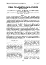
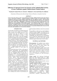
<strong>Egypt</strong>ian Journal <strong>of</strong> Medical Microbiology, January 2006 Vol. 15, No 1PCR amplification products were used astargets <strong>in</strong> the species-specific PCR.Post-PCR gel electrophoresis wasperformed on 2% agarose gel (Hispan agar,Spa<strong>in</strong>), conta<strong>in</strong><strong>in</strong>g 0.5 µg/ml ethidiumbromide (Amresco, USA) <strong>in</strong> 1% TBE buffer(GIBCO, BRL, Life Technologies, USA).<strong>The</strong> amplification products were visualizedus<strong>in</strong>g Spectrol<strong>in</strong>e Bio-Vision UV/White LightTransillum<strong>in</strong>ator and compared to acommercial molecular weight marker(ΦX174 DNA-HaeIII Digest, New EnglandBioLabs), which gives bands at 1353, 1078,872, 603, 310, 281, 271, 234, 194, 118, 72 bp(N.B., 281 and 271 bp bands appear as as<strong>in</strong>gle band). <strong>The</strong> expected PCR productlengths were 156 bp and 192 bp for C.albicans and A. fumigatus, respectively.Negative (sterile distilled water) and positivecontrols [C. albicans standard stra<strong>in</strong> (ATCC10232) and A. fumigatus standard stra<strong>in</strong>(ATCC 10827)] were <strong>in</strong>cluded <strong>in</strong> each PCRrun.Statistical Analysis: Data were analyzedus<strong>in</strong>g Statistical Package for Social Sciences(SPSS) s<strong>of</strong>tware, Version 11. P value < 0.05was considered significant.RESULTS:<strong>The</strong> present study <strong>in</strong>cluded 50 cases (30males and 20 females) <strong>of</strong> cl<strong>in</strong>ically suspectedoculomycosis [39 cases (78%) <strong>of</strong>keratoconjunctivitis and 11 cases (22%) <strong>of</strong>endophthalmitis] as the study group (cases),<strong>in</strong> addition to 20 healthy <strong>in</strong>dividuals (10males and 10 females) serv<strong>in</strong>g as the controlgroup.Table (1): Results <strong>of</strong> positive direct sta<strong>in</strong> with Calc<strong>of</strong>luor white sta<strong>in</strong> among cases and controlsFungusCases (N o = 50) Controls (N o = 20) P valueN o % N o %Yeasts 12 24 1 5 < 0.001Hyphae 20 40 1 5Total 32 64 2 101-a 1-b 1-cFigure (1): Calc<strong>of</strong>luor white sta<strong>in</strong>ed specimens show<strong>in</strong>g:1-a: Budd<strong>in</strong>g oval cells <strong>of</strong> C. albicans1-b: Germ tube <strong>of</strong> C. albicans1-c: Hyphal elements <strong>of</strong> septate filamentous fungi239
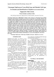
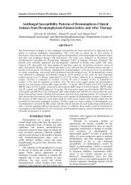
<strong>Egypt</strong>ian Journal <strong>of</strong> Medical Microbiology, January 2006 Vol. 15, No 1Table (2): Comparison between the isolation rates <strong>of</strong> different fungal species on Sabouraud'sdextrose agar (SDA) and Chromagar Candida among cases and controlsSDAChromagar CandidaCases Controls Cases ControlsN o % N o % N o % N o %No growth 25 50 16 80 42 84 19 95Candida Spp.* 8 16 1 5 8 16 1 5Aspergillus spp.** 8 16 1 5 0 0 0 0Penicillium 4 8 1 5 0 0 0 0Alternaria 2 4 1 5 0 0 0 0Curvularia 2 4 0 0 0 0 0 0Rhodotorula rubra 1 2 0 0 0 0 0 0Total 50 100 20 100 50 100 20 100N.B.: *Isolated candida species <strong>in</strong>cluded 4 C. albicans, 2 C. krusei, 1 C. parapsilosis and 1 C.tropicalis among cases, whereas only 1 C. parapsilosis was identified among controls(confirmed by API Candida).** Isolated Aspergillus species <strong>in</strong>cluded 4 A. fumigatus, 2 A. niger and 2 A. flavus amongcases, whereas only 1 A. fumigatus was isolated from controls (confirmed bylactophenol cotton blue sta<strong>in</strong>).2-a 2-b 2-c2-d 2-e 2-fFigure (2): Lactophenol cotton blue sta<strong>in</strong> <strong>of</strong>:2-a: A. fumigatus (show<strong>in</strong>g conidiophore hav<strong>in</strong>g a flask-shaped swollen end called vesicle).2-b: A. flavus (show<strong>in</strong>g conidiophore aris<strong>in</strong>g from a foot cell and term<strong>in</strong>at<strong>in</strong>g <strong>in</strong> a vesicle, whichproduces phialides <strong>in</strong> one or two series and unicellular conidia).2-c: A. niger (show<strong>in</strong>g black spore heads).2-d: Penicillium (show<strong>in</strong>g the brush appearance)2-e: Alternaria (show<strong>in</strong>g multi-celled conidium).2-f: C. albicans (show<strong>in</strong>g the characteristically asexual budd<strong>in</strong>g).240
<strong>Egypt</strong>ian Journal <strong>of</strong> Medical Microbiology, January 2006 Vol. 15, No 1Table (3): Results <strong>of</strong> culture <strong>in</strong> relation to the cl<strong>in</strong>ical diagnosis <strong>of</strong> casesCulture ResultsKeratoconjunctivitis EndophthalmitisN o % N o %No growth 22 56.4 3 27.2Aspergillus species 6 15.4 2 18.2A. fumigatus 2 5.1 2 18.2A. niger2 5.1 0 0A. flavus 2 5.1 0 0Candida species 4 10.3 4 36.4C. albicans 2 5.1 2 18.2C. krusei1 2.6 1 9.1C. tropicalis 1 2.6 0 0C. parapsilosis 0 0 1 9.1Penicillium 2 5.1 2 18.2Alternaria 2 5.1 0 0Curvularia 2 5.1 0 0Rhodotorula rubra 1 2.6 0 0Total 39 100 11 100N.B.: - Positive fungal culture was obta<strong>in</strong>ed <strong>in</strong> 17 cases (43.6%) <strong>of</strong> keratoconjunctivits and <strong>in</strong> 8 cases(72.7%) <strong>of</strong> endophthalmitis.Comparison between keratoconjunctivitis andendophthalmitis cases, regard<strong>in</strong>g the commonpredispos<strong>in</strong>g factors, revealed that out <strong>of</strong> the39 cases <strong>of</strong> keratoconjunctivitis, 22 cases(88%) had a def<strong>in</strong>ite history <strong>of</strong> trauma and 3cases (12%) were post-surgical. However, out<strong>of</strong> the 11 cases <strong>of</strong> endophthalmitis, 9 caseswere exogenous endophthalmitis [6 cases(54.5%); follow<strong>in</strong>g cataract operations and 3cases (27.2%); follow<strong>in</strong>g trauma] and 2 cases(18.2%) were endogenous endophthalmitis.Table (4): Comparison between direct smear and culture, regard<strong>in</strong>g detection <strong>of</strong> mycosis amongcasesCultureTestPositiveNegativeTotalN o % N o % N o (%)Direct Positive 24 48 8 16 32 (64%)Smear a Negative 1 2 17 34 18 (36%)Total 25 50 25 50 50 (100%)aSensitivity = 96% Specificity = 68% Accuracy = 82%Positive Predictive Value (PPV) = 75% Negative Predictive Value (NPV) = 94.4%API Candida results revealed 4isolates <strong>of</strong> C. albicans, 2 isolates <strong>of</strong> C. krusei,1 isolate <strong>of</strong> C. tropicalis and 1 isolate <strong>of</strong> C.parapsilosis (a total <strong>of</strong> 8 Candida isolates)among cases, whereas among controls, APICandida could identify only one Candidaspecies (C. parapsilosis). <strong>The</strong>re was acomplete agreement (100%) between theresults <strong>of</strong> Chromagar Candida and APICandida <strong>in</strong> the identification <strong>of</strong> the differentCandida species.Among the 50 cases <strong>of</strong> suspectedocular mycosis, PCR - performed directly oncl<strong>in</strong>ical specimens - revealed 6 positive (12%)C. albicans and 7 positive (14%) A.fumigatus. However, among controls, onlyone positive A. fumigatus (5%) was detectedby PCR. No statistically significant differencewas found - regard<strong>in</strong>g the detection rates <strong>of</strong> A.fumigatus by PCR - between cases andcontrols (P value 0.28). Compared to culture,PCR proved to be 100% sensitive <strong>in</strong> thedetection <strong>of</strong> C. albicans and A. fumigatus.241
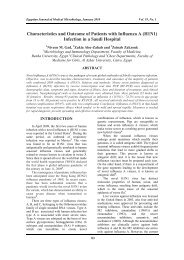
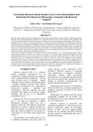
<strong>Egypt</strong>ian Journal <strong>of</strong> Medical Microbiology, January 2006 Vol. 15, No 11 2 3 4 5 6 7 8Figure (3): Gel electrophoresis show<strong>in</strong>g C. albicans and A. fumigatus - PCR productsLane 1: positive control for A. fumigatus (ATCC 10827), giv<strong>in</strong>g band at 192 bp. Lanes 2and 3: positive samples for A. fumigatus. Lane 4: PCR molecular weight marker(ΦX174 DNA-HaeIII Digest, New England BioLabs), which gives bands at 1353,1078, 872, 603, 310, 281, 271, 234, 194, 118, 72 bp (281 and 271 bp bandsappear<strong>in</strong>g as a s<strong>in</strong>gle band). Lane 5: positive control for C. albicans (ATCC10232), giv<strong>in</strong>g band at 156 bp. Lanes 6 and 7: positive samples for C. albicans.Lane 8: negative control (H2O).Table (5): Results <strong>of</strong> culture and PCR analysis for C. albicans <strong>of</strong> specimens from patients withpresumed oculomycosisSpecies-Specific PCRC. albicans CultureforPositiveNegativeTotalC. albicans a N o % N o % N o (%)Positive 4 8 2 4 6 (12%)Negative 0 0 44 88 44 (88%)Total 4 8 46 92 50 (100%)aSensitivity = 100% Specificity = 95.7% Accuracy = 97.9%Positive Predictive Value (PPV) = 66.7% Negative Predictive Value (NPV) = 100%for A. fumigatusTable (6): Results <strong>of</strong> culture and PCR analysis for A. fumigatus <strong>of</strong> specimens from patients withpresumed oculomycosisSpecies-Specific PCRA. fumigatus CulturePositiveNegativeTotalN o % N o % N o (%)Positive 4 8 3 6 7 (14%)Negative 0 0 43 86 43 (86%)Total 4 8 46 92 50 (100%)aSensitivity = 100% Specificity = 93.5% Accuracy = 96.8%Positive Predictive Value (PPV) = 57.1% Negative Predictive Value (NPV) = 100%DISCUSSION<strong>Ocular</strong> <strong>in</strong>fections due to opportunisticfungal pathogens are steadily <strong>in</strong>creas<strong>in</strong>g 6,23 .<strong>Fungal</strong> keratitis is a major ophthalmologicproblem <strong>in</strong> the tropical regions <strong>of</strong> the worldand it is one <strong>of</strong> the frequent causes <strong>of</strong> cornealdamage <strong>in</strong> develop<strong>in</strong>g countries 24-26 .242
<strong>Egypt</strong>ian Journal <strong>of</strong> Medical Microbiology, January 2006 Vol. 15, No 1Preservation <strong>of</strong> vision requires early diagnosisand early <strong>in</strong>stitution <strong>of</strong> fungal therapy 6,27 .Non-specific fluorochromatic sta<strong>in</strong>shave become popular for the detection <strong>of</strong>fungi <strong>in</strong> ocular samples. Calc<strong>of</strong>luor whiteappears to be the most widely used <strong>of</strong> thesesta<strong>in</strong>s 28 . In the present study, 50 cases <strong>of</strong>suspected oculomycosis were <strong>in</strong>vestigated.Direct microscopic exam<strong>in</strong>ation withCalc<strong>of</strong>luor white sta<strong>in</strong>, mounted with 10%KOH revealed fungal elements <strong>in</strong> 32 cases(64%), whereas positive fungal cultures; ondifferent culture media; were obta<strong>in</strong>ed <strong>in</strong> 25cases (50%). Direct smear and fungal culturematched (were both positive or negative) <strong>in</strong>41 (82%) <strong>of</strong> the 50 specimens from cases.Similar results were reported by Nag et al. 29who stated that a wet mount with 10% KOHshowed fungal elements <strong>in</strong> 68% <strong>of</strong> cornealulcers, while culture was positive <strong>in</strong> only26.4% <strong>of</strong> the cases. Moreover, other studiesreported higher sensitivity <strong>of</strong> fungal detectionus<strong>in</strong>g KOH and Calc<strong>of</strong>luor white sta<strong>in</strong> thanculture 7,24 . Calc<strong>of</strong>luor white sensitivity <strong>of</strong> 80–90% <strong>in</strong> culture-proven mycotic keratitis hasbeen reported 28 . This agrees with our results,where Calc<strong>of</strong>luor white sta<strong>in</strong> showed asensitivity <strong>of</strong> 96% <strong>in</strong> culture-proven cases <strong>of</strong>oculomycosis. Calc<strong>of</strong>luor white fluorescentsta<strong>in</strong><strong>in</strong>g proved to be a simple technique thatovercomes the difficulties and time <strong>in</strong>volved<strong>in</strong> sta<strong>in</strong><strong>in</strong>g 24,28,30 .Even with the advent <strong>of</strong> many newtechniques, culture rema<strong>in</strong>s the “goldstandard” and the cornerstone <strong>of</strong> the diagnosis<strong>of</strong> most ophthalmic mycoses, except forrh<strong>in</strong>osporidiosis, where direct microscopicexam<strong>in</strong>ation <strong>of</strong> samples yields more reliableresults 3,24,25,28,31 . Wherever possible, it is bestto use more than one culture medium, and to<strong>in</strong>cubate these at 37°C and at 25-30°C for theoptimal recovery <strong>of</strong> ocular fungi 32 .Our culture results among cases(show<strong>in</strong>g 50% positivity) are similar to those<strong>of</strong> El-Sawy et al. 33 and El-Mowafy et al. 34who detected fungal isolation from cultures <strong>in</strong>45% and 47.5% <strong>of</strong> cases <strong>of</strong> cl<strong>in</strong>icallysuspected fungal keratitis, respectively.In the present study, positive fungalculture was obta<strong>in</strong>ed <strong>in</strong> 17 cases (43.6%) <strong>of</strong>keratoconjunctivitis. <strong>The</strong> most prevalentisolates were Aspergillus species thenCandida species, followed by Penicillium,Alternaria, Curvularia and lastlyRhodotorula rubra. This agrees with theresults <strong>of</strong> Tanure et al. 35 . On the other hand,different fungal species were isolated <strong>in</strong> 8cases (72.7%) <strong>of</strong> endophthalmitis; the mostcommon were Candida species followed byAspergillus fumigatus and lastly Penicillium.Internationaly, Aspergillus species isthe most common isolate (27%-64%) <strong>in</strong> cases<strong>of</strong> fungal keratitis worldwide, followed byfusarium (6%-32%) and penicillium (2%-29%) 1 . Similar studies also concluded thatfilamentous fungi are the pr<strong>in</strong>cipal causes <strong>of</strong>mycotic keratitis <strong>in</strong> most parts <strong>of</strong> the world;either Aspergillus spp. or Fusarium spp. werethe most common isolates. Dematiaceousfungi, such as Curvularia spp. and Bipolarisspp., are the third most important cause <strong>of</strong>keratitis <strong>in</strong> a number <strong>of</strong> studies 3,24,25,36-39 . Astudy conducted <strong>in</strong> Saudi Arabia, concludedthat the higher prevalence <strong>of</strong> Aspergillusspecies; among cases <strong>of</strong> keratitis; could beexpla<strong>in</strong>ed by the fact that its spores couldsurvive the hot dry weather 37 , as is the case <strong>in</strong><strong>Egypt</strong>.Keratitis; due to yeasts and yeast-likefungi; is most frequently caused by C.albicans. Keratitis due to this organism tendsto occur more frequently <strong>in</strong> areas wheretraumatic keratitis is uncommon, but whereother predispos<strong>in</strong>g factors caus<strong>in</strong>g epithelialor stromal ulceration are important 40 , forexample, due to previous herpes simplexkeratitis, contact lens-<strong>in</strong>duced cornealabrasions 14 or <strong>in</strong> patients with dry eyes 41 . C.albicans and related fungi have been<strong>in</strong>frequent isolates <strong>in</strong> most studies performed<strong>in</strong> tropical countries, possibly due to thepredom<strong>in</strong>ance <strong>of</strong> livelihoods, such asagriculture, which carry a higher risk for theoccurrence <strong>of</strong> trauma-related keratitis causedby filamentous fungi than for keratitis due toC. albicans 42 .In the present study, <strong>of</strong> the 39 cases <strong>of</strong>keratoconjunctivitis, 22 cases (88%) had adef<strong>in</strong>ite history <strong>of</strong> trauma and 3 cases (12%)were post-surgical. Moreover, <strong>of</strong> the 11 cases<strong>of</strong> endophthalmitis, 6 cases (54.5%) werecl<strong>in</strong>ically manifested follow<strong>in</strong>g cataractoperations, 3 cases (27.2%) followed traumaand 2 cases (18.2%) occurred secondary tohematogenous dissem<strong>in</strong>ation <strong>in</strong> patientshav<strong>in</strong>g debilitat<strong>in</strong>g diseases (endogenousendophthalmitis).Many studies reported that a def<strong>in</strong>itehistory <strong>of</strong> ocular trauma (usually withvegetable matter) was the most common243
<strong>Egypt</strong>ian Journal <strong>of</strong> Medical Microbiology, January 2006 Vol. 15, No 1predispos<strong>in</strong>g factor for mycotic keratitis,(occurr<strong>in</strong>g <strong>in</strong> 44 to 55% <strong>of</strong> patients); lessfrequently reported risk factors <strong>in</strong>cludeprolonged use <strong>of</strong> topical corticosteroids oranti-bacterials, systemic diseases such asdiabetes mellitus, pre-exist<strong>in</strong>g ocular diseases,contact lens wear and previous ocularsurgery 2,24,39,42-44 . On the other hand, mycoticendophthalmitis has been reported to occurafter cataract surgery (61%), trauma (28%) oras metastatic endogenous endophthalmitis(11%) 45 . <strong>The</strong> yeast C. albicans is the mostcommon cause <strong>of</strong> endogenousendophthalmitis 46 . Moreover, Candidaspecies are particularly likely to causeexogenous endophthalmitis. In this sett<strong>in</strong>g,<strong>in</strong>fection may be due to peri-operativecontam<strong>in</strong>ation <strong>of</strong> lens prostheses orcontam<strong>in</strong>ation <strong>of</strong> fluids used for irrigation <strong>of</strong>the eye. Infection may be also enhanced bythe pre- and post-operative use <strong>of</strong> topicalcorticosteroids and anti-bacterial agents 42 .However, Aspergillus endophthalmitis is thecommonest type <strong>of</strong> vision-threaten<strong>in</strong>g fungalendophthalmitis encountered <strong>in</strong> India 47 .Our results revealed that Sabouraud'sdextrose agar allowed for the recovery <strong>of</strong>most fungal pathogens with<strong>in</strong> 2 weeks <strong>of</strong><strong>in</strong>cubation. Generally, the duration <strong>of</strong><strong>in</strong>cubation - for most yeasts - is one week,although the range <strong>of</strong> time for recovery <strong>of</strong>dimorphic fungi may extend to 4-6weeks 25,48,49 .<strong>The</strong> present study also showed acomplete agreement (100%) between theresults <strong>of</strong> Chromagar Candida and APICandida <strong>in</strong> the identification <strong>of</strong> the differentCandida species, however, the yeast colonycolor development (on Chromagar Candida)or its proper identification (by API Candida)was better reported after 48 hours <strong>of</strong><strong>in</strong>cubation, as previously noted 20,50 . Thisdelay <strong>in</strong> the identification by cultural and/orbiochemical procedures or the limited yield <strong>of</strong>vitreous cultures (<strong>in</strong> cases <strong>of</strong> endophthalmitis)has led to a search for more rapidtechniques 51 . <strong>The</strong> speed and sensitivity <strong>of</strong>PCR makes it an ideal choice for the basis <strong>of</strong>a rapid identification system 51,52 . PCR hasbeen reported to be a more sensitive and rapiddiagnostic tool, compared to the conventionalmycologic methods <strong>in</strong> the diagnosis <strong>of</strong>oculomycosis, where 55.8% <strong>of</strong> cases <strong>of</strong>fungal endophthalmitis were positive byconventional methods versus 74.4% byPCR 47 . Similarly, Hidalgo et al. 51 andLohmann et al. 53 concluded that PCR had ahigher rate <strong>of</strong> positive fungal identificationthan by microscopy or diagnostic culture.This is similar to our results.Us<strong>in</strong>g species-specific primers <strong>in</strong> thepresent study, PCR could detect 6 positive C.albicans (12%) and 7 positive A. fumigatus(14%) among cases <strong>of</strong> suspectedoculomycosis; versus only 1 positive A.fumigatus (5%) among the control group. Nostatistically significant difference was found -regard<strong>in</strong>g the detection rates <strong>of</strong> A. fumigatusby PCR - between cases and controls (P value0.28), which might be expla<strong>in</strong>ed by the smallsample size <strong>in</strong>cluded <strong>in</strong> the study. However,us<strong>in</strong>g culture, 4 C. albicans (8%) and 4 A.fumigatus (8%) isolates were recovered fromcases and only one A. fumigatus isolate wasrecovered from controls. Among the 50 cases<strong>in</strong> the present study, PCR and fungal culturefor C. albicans and A. fumigatus matched <strong>in</strong>48 (96%) and 47 (94%) <strong>of</strong> the specimens,respectively. In 2 (4%) and 3 (6%) casespecimens, PCR detected C. albicans and A.fumigatus, respectively, where no organismwas found <strong>in</strong> culture. All <strong>of</strong> these patientsappeared cl<strong>in</strong>ically to have ocular fungal<strong>in</strong>fections, and fungi were present onCalc<strong>of</strong>luor white smear for all.PCR would probably be most valuable<strong>in</strong> provid<strong>in</strong>g a positive result <strong>in</strong> a shorterperiod than that required for culture 6,12,53 and<strong>in</strong> identification <strong>of</strong> a fungal isolate whichdoes not sporulate 54 . Moreover, PCR <strong>of</strong>fersthe ability to analyze specimens far fromwhere they are collected 11,13 . Eventually, PCRmight solidly complement the current “goldstandard” diagnostic techniques for guid<strong>in</strong>gmanagement or support<strong>in</strong>g research studies <strong>of</strong>oculomycosis 13 . However, concern persistsregard<strong>in</strong>g the specificity <strong>of</strong> this technique andthe problems that may arise from theproduction <strong>of</strong> false-positive results. PCR doesnot dist<strong>in</strong>guish viable from non-viableorganisms; it may therefore be difficult toassess the relevance <strong>of</strong> a positive PCR result<strong>in</strong> a heal<strong>in</strong>g corneal ulcer, where culture isnegative 55 , or <strong>in</strong> locations such as theconjunctival sac, where fungi may be foundas transient commensals 13 . Although PCR isextremely sensitive and specific, it cannot beused to monitor the patient’s response totreatment. Moreover, A few culture mediawill suffice to detect and grow the common244
<strong>Egypt</strong>ian Journal <strong>of</strong> Medical Microbiology, January 2006 Vol. 15, No 1ocular pathogens, but PCR must bemultiplexed for each microorganism that issuspected 56 . <strong>The</strong> difficulty <strong>of</strong> DNA extraction(some filamentous fungi have a sturdy cellwall; which is resistant to standard DNAextraction procedures) and the presence <strong>of</strong>PCR <strong>in</strong>hibitors <strong>in</strong> human specimens are some<strong>of</strong> the difficulties encountered with fungaldetection <strong>in</strong> ocular samples 57 . F<strong>in</strong>ally, PCRcan detect only fungi for which the DNAsequence is known and primers areavailable 58 .To conclude, the <strong>in</strong>creased awareness<strong>of</strong> ocular fungal <strong>in</strong>fections, the betterrecognition <strong>of</strong> their cl<strong>in</strong>ical features and theimproved laboratory diagnostic techniques,will all lead to an <strong>in</strong>crease <strong>in</strong> the frequency <strong>of</strong>correct diagnosis. A rapid and accuratediagnosis <strong>of</strong> oculomycosis will improve thechances <strong>of</strong> a complete recovery, especially <strong>in</strong>the tropics, where patients may delaypresent<strong>in</strong>g to an ophthalmologist. Calc<strong>of</strong>luorwhite fluorescent sta<strong>in</strong><strong>in</strong>g proved to be asimple and reliable technique for <strong>in</strong>itiation <strong>of</strong>antifungal therapy. Culture methods rema<strong>in</strong>important diagnostic tools that add to theproper identification and characterization <strong>of</strong>oculomycosis, however, new culture medianeed to be developed. PCR is a potentiallyvaluable technique for diagnos<strong>in</strong>g ocularfungal pathogens, particularly, if panfungalprimers are used and optimized. AlthoughPCR is expensive and requires laboratoryexpertise, yet the loss <strong>of</strong> an eye, due to delay<strong>in</strong> laboratory diagnosis, is devastat<strong>in</strong>g.References:1. Alexandrakis G, Kanellopoulous J,Donald S, et al. (2002): <strong>Fungal</strong>keratitis. In: Kuhn F and Pieramici D(eds.): <strong>Ocular</strong> trauma: Pr<strong>in</strong>ciples andPractice, P. 293-301, Thieme MedicalPublishers, New York.2. Sr<strong>in</strong>ivasan R, Kanungo R and GoyalJL (1991): Spectrum <strong>of</strong> oculomycosis <strong>in</strong>South India. Acta Ophthalmologica, 66:774-779.3. Garg P, Gop<strong>in</strong>ath G, Choudhary Kand Rao GN (2000): Keratomycosis:cl<strong>in</strong>ical and microbiological experiencewith dematiaceous fungi. Ophthalmol.,107: 574-580.4. Bloom PA, Laidlaw DA, Easty DL, etal. (1992): Treatment failure <strong>in</strong> a case <strong>of</strong>fungal keratitis caused byPseudallescheria boydii. Br. J.Ophthalmol., 76 (6): 367-368.5. Thomas PA (2003): <strong>Fungal</strong> <strong>in</strong>fection <strong>of</strong>the cornea. Eye, 17: 852-862.6. Kumar M and Shukla PK (2005): Use<strong>of</strong> PCR target<strong>in</strong>g <strong>of</strong> <strong>in</strong>ternal transcribedspacer regions and s<strong>in</strong>gle-strandedconformation polymorphism analysis <strong>of</strong>sequence variation <strong>in</strong> different regions <strong>of</strong>rRNA genes <strong>in</strong> fungi for rapid diagnosis<strong>of</strong> mycotic keratitis. J. Cl<strong>in</strong>. Microbiol.,43(2): 662-668.7. Sharma S, Kunimoto DY, Gop<strong>in</strong>than Uet al. (2002): Evaluation <strong>of</strong> cornealscrap<strong>in</strong>g smear exam<strong>in</strong>ation methods <strong>in</strong>the diagnosis <strong>of</strong> bacterial and fungalkeratitis: a survey <strong>of</strong> eight years <strong>of</strong>laboratory experience. Cornea, 21(7):643-647.8. Chandler FW (1991): Histologicdiagnosis <strong>of</strong> mycotic diseases, p. 235-242. In: Gatti F, de Vroey C and Persi A(eds.): Human mycoses <strong>in</strong> tropicalcountries. Health Cooperation Papers N o13. Organizzazione per la CooperazioneSanitaria Internazionale, Bologna, Italy.9. Mendonza L, Ajello L and McG<strong>in</strong>nisMR (1996): <strong>Infections</strong> caused by theoomycetous pathogen Pythium<strong>in</strong>sidiosum. J. Mycol. Med., 6: 151-164.10. Ahmad, S, Khan Z, Mustafa AS et al.(2002): Semi-nested PCR for diagnosis<strong>of</strong> candidemia: comparison with culture,antigen detection, and biochemicalmethods for species identification. J.Cl<strong>in</strong>. Microbiol., 40: 2483-2489.11. Wu Z, Tsumura Y, Blomquist G, et al.(2003): 18S rRNA gene variation amongcommon airborne fungi anddevelopment <strong>of</strong> specific oligonucleotideprobes for the detection <strong>of</strong> fungal isolate.Appl. Environ. Microbiol., 69: 5389-5397.12. Ferrer C, Munoz G, Alio JL et al.(2002): Polymerase cha<strong>in</strong> reactiondiagnosis <strong>in</strong> fungal keratitis caused byAlternaria alternata. Am. J. Ophthalmol.,133: 398-399.13. Gaudio, PA, Gop<strong>in</strong>athan U, SangwanV, et al. (2002): Polymerase cha<strong>in</strong>reaction based detection <strong>of</strong> fungi <strong>in</strong><strong>in</strong>fected corneas. Br. J. Ophthalmol.,86(7): 755-760.14. Forster RK (1994): <strong>Fungal</strong> keratitis andconjunctivitis: Cl<strong>in</strong>ical aspects. In:245
<strong>Egypt</strong>ian Journal <strong>of</strong> Medical Microbiology, January 2006 Vol. 15, No 1Smol<strong>in</strong> G and Thr<strong>of</strong>t RA (eds.): <strong>The</strong>Cornea: Scientific foundations andCl<strong>in</strong>ical practice, 3 rd ed., P. 239-250,Little Brown & Co., Boston.15. Mounir A, Hamza I, Salah M et al.(1996): Recent trends <strong>in</strong> management <strong>of</strong>fungal keratitis. Bul. Opthalmol. Soc.<strong>Egypt</strong>, Vol 89: 809.16. Jones DB, Liesegang TJ et al. (1981):Cumitech 13, American Society forMicrobiology, Wash<strong>in</strong>gton DC.17. Goodman NL and Roberts GD (1998):Laboratory diagnosis. In: Ajello L andHay RJ (eds.): Topley & Wilson'sMicrobiology and Microbial <strong>Infections</strong>,Vol. 4, Ch. 5, P. 75-87, Arnold, NewYork.18. Liesegang TJ (1998): <strong>Fungal</strong> keratitis.In: Kaufman HE, Barron BA andMcdonald MB (eds.): <strong>The</strong> cornea, Vol. 1,2 nd ed., P. 219-245, Butterworth –He<strong>in</strong>emann.19. Odds FG and Bernaerts R (1994):Chromagar Candida, a new differentialisolation medium. Presumptiveidentification <strong>of</strong> cl<strong>in</strong>ically importantCandida species. J. Cl<strong>in</strong>. Microbiol., 32:1923-1929.20. Hidalgo HF, Vandapel O, DuchesneMA et al. (1996): Comparison <strong>of</strong> the newAPI Candida system to the ID 32 Csystem for identification <strong>of</strong> cl<strong>in</strong>icallyimportant yeast species. J. Cl<strong>in</strong>.Microbiol. , 34 (7): 1846-1848.21. Haynes KA, Westerneng TJ, Fell W etal. (1995): Rapid detection andidentification <strong>of</strong> pathogenic fungi bypolymerase cha<strong>in</strong> reaction amplification<strong>of</strong> large subunit ribosomal DNA. J. Med.Vet. Mycol., 33: 319-325.22. Haynes KA and Westerneng TJ (1996):Rapid identification <strong>of</strong> Candida albicans,Candida glabrata, Candida parapsilosisand Candida krusei by species-specificPCR <strong>of</strong> large subunit ribosomal DNA. J.Med. Microbiol., 44: 390-396.23. Feldmesser M (2001): <strong>Ocular</strong> Fungi. In:Agarwal S and Apple D (eds.): Textbook<strong>of</strong> Ophthalmology, Vol. 4, (1 st ed.), P.2783, Jaypee Brothers, New Delhi.24. Gop<strong>in</strong>athan U, Garg P, Fernandes S, etal. (2002): <strong>The</strong> epidemiological featuresand laboratory results <strong>of</strong> fungal keratitis.A 10-year review at a referral eye carecenter <strong>in</strong> South India. Cornea, 21(6): 555-559.25. Leck AK, Thomas PA, Hagan M, et al.(2002): Aetiology <strong>of</strong> suppurative cornealulcers <strong>in</strong> Ghana and South India andepidemiology <strong>of</strong> fungal keratitis. Br. J.Ophthomol., 86 (11): 1211-1215.26. Prats CH, Tello FL, Jose ABS, et al.(2004): Voriconazole <strong>in</strong> fungal keratitiscaused by Scedoporium apiospermum.Ann. Pharmacol., 38: 414-417.27. Araffa RC, Av<strong>in</strong> I, Ishibashi Y, et al.(1985): Calc<strong>of</strong>luor and <strong>in</strong>k-potassiumhydroxide preparation for identify<strong>in</strong>gfungi. Am. J. Ophthalmol., 100: 719.28. Chander J and Sharma A (1994):Prevalence <strong>of</strong> fungal corneal ulcers <strong>in</strong>Northern India. Infection, 22: 207–209.29. Nag DR, Chatterjee S, Chattersee S, etal. (2002): Role <strong>of</strong> potassium hydroxidemount <strong>in</strong> rapid diagnosis <strong>of</strong> fungalcorneal ulcers. J. Indian Med. Assoc., 100(1): 18.30. Sharma S, Silverberg, M., Mehta, P, etal. (1998): Early diagnosis <strong>of</strong> mycotickeratitis: predictive value <strong>of</strong> potassiumhydroxide preparation. Indian J.Ophthalmol., 46: 31-35.31. Richardson MD and Shankland GS(1999): Rhizopus, Rhicomucor,Absidia,and other agents <strong>of</strong> systemic andsubcutaneous zygomycoses, In: MurrayPR, Baron EJ, Pfaller MA, et al. (eds.):Manual <strong>of</strong> Cl<strong>in</strong>ical Microbiology, 7 th ed.,P. 1242–1258, ASM, Wash<strong>in</strong>gton, D.C.32. Upadhyay M P, Karmacharya PCD,Koirala S, et al. (1991): Epidemiologicalcharacteristics, predispos<strong>in</strong>g factors andaetiological diagnosis <strong>of</strong> cornealulceration <strong>in</strong> Nepal. Am. J. Ophthalmol.,111, 92–99.33. El-Saway A, Youssef W and Refai MK(1990): Prevalence and management <strong>of</strong>mycotic <strong>in</strong>fections <strong>of</strong> external eye. Bull.Ophthalmol. Soc. <strong>Egypt</strong>, 83: 661.34. El-Mowafy N, Abdalla MI and Refai M(1983): <strong>Fungal</strong> flora <strong>of</strong> the conjunctivalsac <strong>in</strong> healthy and diseased eyes. Bull.Ophthalmol. Soc. <strong>Egypt</strong>, 76: 19.35. Tanure MA, Cohen EJ, Grewal S et al.(2000): Spectrum <strong>of</strong> fungal keratitis atWills Eye Hospital, Philadelphia,Pennsylvania. Cornea, 19: 307-312.36. Sr<strong>in</strong>ivasan M, Gonzales CA, George C,et al. (1997): Epidemiology and246
<strong>Egypt</strong>ian Journal <strong>of</strong> Medical Microbiology, January 2006 Vol. 15, No 1aetiological diagnosis <strong>of</strong> cornealulceration <strong>in</strong> Madurai, South India. Br. J.Ophthalmol., 81: 965–971.37. Khairallah SH, Byrne KA andTabbara KF (1992): <strong>Fungal</strong> keratitis <strong>in</strong>Saudi Arabia. Doc. Ophthalmol., 79: 269.38. Sun X-G, Zhang Y, Ran LT, et al.(2004): Etiological analysis on ocularfungal <strong>in</strong>fection <strong>in</strong> the period 1989-2000.Ch<strong>in</strong>ese Med. J., 117(4): 598-600.39. Bharathi MJ, Ramakrishna R, Vasu S,et al. (2003): Epidemiologicalcharacteristics and laboratory diagnosis <strong>of</strong>fungal keratitis. A three-year study.Indian J. Opthalmol., 51(4): 315-321.40. Jones DB (1980): Strategy for the <strong>in</strong>itialmanagement <strong>of</strong> suspected microbialkeratitis. In: Barraquer JI, B<strong>in</strong>der PS,Buxton, JN, et al. (eds.): Symposium onmedical and surgical diseases <strong>of</strong> thecornea. Transactions <strong>of</strong> the New OrleansAcademy <strong>of</strong> Ophthalmology, 293-301, C.V. Mosby, St. Louis, Mo.41. Klotz SA, Penn CC, Negvesky GJ, et al.(2000): <strong>Fungal</strong> and parasitic <strong>in</strong>fections <strong>of</strong>the eye. Cl<strong>in</strong>. Microbiol. Rev., 13(4):662-685.42. Thomas PA (2000): Current perspectiveson ophthalmic mycoses. Cl<strong>in</strong>. Microbiol.Rev., 16(4): 730-797.43. Panda A, Sharma N, Das G et al.(1997): Mycotic keratitis <strong>in</strong> children:epidemiologic and microbiologicevaluation. Cornea, 16: 293-299.44. Wilhelmus KR and Jones DB (2001):Curvularia keratitis. Trans. Am.Ophthalmol. Soc. , 99: 111-132.45. Newell F (1982): Mycotic <strong>in</strong>fections.Ophthalmol., 5: 391-395.46. Liesegang TJ and Forster RF (1980):Spectrum <strong>of</strong> microbial keratitis <strong>in</strong> SouthFlorida. Am. J. Ophthamol., 90: 38.47. Anand AR, Madhavan HN, Sudha NV,et al. (2001): Polymerase cha<strong>in</strong> reaction<strong>in</strong> the diagnosis <strong>of</strong> Aspergillusendophthalmitis. Indian J. Med. Res.,114: 133-140.48. Telenti A and Roberts GD (1989):Laboratory detection <strong>of</strong> fungaemia. J.Cl<strong>in</strong>. Microbiol., 8: 825-831.49. Goldblum D, Frueh BE, Gian-MarcoSarra G-M, et al. (2005): Topicalcasp<strong>of</strong>ung<strong>in</strong> for treatment <strong>of</strong> keratitiscaused by Candida albicans <strong>in</strong> a rabbitmodel. Antimicrob. Agents Chemother.,49(4): 1359-1363.50. Haynes KA and Westerneng TJ (1996):Rapid identification <strong>of</strong> Candida albicans,Candida glabrata, Candida parapsilosisand Candida krusei by species-specificPCR <strong>of</strong> large subunit ribosomal DNA. J.Med. Microbiol., 44: 390.51. Hidalgo JA, Alangaden GJ, Elliot D etal. (2000): <strong>Fungal</strong> endophthalmitisdiagnosis by detection <strong>of</strong> Candidaalbicans DNA <strong>in</strong> <strong>in</strong>traocular fluid by use<strong>of</strong> a species-specific polymerase cha<strong>in</strong>reaction assay. J. Infect. Dis., 181(3):1198-1201.52. Okhravi N, Adamson P and Lighman S(2000): Use <strong>of</strong> PCR <strong>in</strong> endophthalmitis.<strong>Ocular</strong> Immunol. Inflamm., 8: 189.53. Lohmann CP, L<strong>in</strong>de H and Reische U(2000): Improved detection <strong>of</strong>microorganisms by polymerase cha<strong>in</strong>reaction <strong>in</strong> delayed endophthalmitis aftercataract. Ophthalmol., 107(6): 1047.54. Badenoch PR, Coster DJ, WetherallBI, et al. (2001): Pythium <strong>in</strong>sidiosumkeratitis confirmed by DNA sequenceanalysis. Br. J. Ophthalmol., 85: 502.55. Alexandrakis G, Jalali S and Gloor P(1998): Diagnosis <strong>of</strong> Fusarium keratitis<strong>in</strong> an animal model us<strong>in</strong>g polymerasecha<strong>in</strong> reaction. Br. J. Ophthalmol., 82:306-311.56. Reiss E, Tanaka K, Bruker G, et al.(1998): Molecular diagnosis andepidemiology <strong>of</strong> fungal <strong>in</strong>fections. Med.Mycol., 36: 249-25757. Hui M, Ip M, Chan PK, et al. (2000):Rapid identification <strong>of</strong> medicallyimportant Candida to species level bypolymerase cha<strong>in</strong> reaction and s<strong>in</strong>glestrandconformational polymorphism.Diagn. Microbiol. Infect. Dis., 38: 95-99.58. Rajeev B and Biswas J (1998):Molecular biologic techniques <strong>in</strong>ophthalmic pathology. Indian J.Ophthalmol., 46: 3–13.247
<strong>Egypt</strong>ian Journal <strong>of</strong> Medical Microbiology, January 2006 Vol. 15, No 1توصيف عدوى فطريات العين في مصر٢١١١إيمان أحمد الصعيدي ، زينب عبد الخالق إبراهيم ، شروق خميس الحدقي ، هشام عادل حسنقسم الميكروبيولوجيا الطبية٢١والمناعة وقسم الرمد – كلية الطب -جامعة القاهرةيعتبر التهاب العين الفطرى من الأمراض المتزايدة والتى تمثل تحديا ً كبيرا ً وذلك لتأخر تشخيصهاوبالتالى تأخر علاجها مما قد يؤدى إلى مضاعفات كثيرة بالعين. ويعد التهاب القرنية أكثر هذه الأمراضشيوعا"، ولكن يمكن أن تحدث مثل هذه الإصابة في أى جزء آخر من العين. وتمثل الإصابات الفطرية للقرنيةحواليلها.%٥٠من كل حالات العدوى الميكروبية للقرنية والتي تم إثباتها فعليا" بالزرع.تحدث الإصابات الفطرية بواسطة عدد كبير من أنواع وسلالات الفطريات، تبعا" للمنطقة الجغرافيةوهذه الأعداد في تزايد مستمر. ويمكن أن يساعد التشخيص السريع والدقيق لهذه الفطريات في بدء العلاج الفعالكان الهدف من هذه الدراسة هو تقييم استعمال التفاعل المتسلسل لخميرة البلمرة في تشخيص عدوىفطريات العين، بالمقارنة بالطرق التشخيصية التقليدية الآخرى، وذلك من خلال عدد ٥٠ حالة بها احتمالالتهاب فطرىبالعين تم تشخيصها إكلينيكيا" وأيضا" عدد٢٠ شخصا"وجودكمجموعة ضابطة. تم أخذ مسحات منالملتحمة أو عينات من القرح القرنية أوالسائل الزجاجى بالعين من الحالات، بينما تم أخذ مسحات من الملتحمةمن المجموعة الضابطة.تشخيص عددعن نتائجحالات إيجابيةوقد أظهرت نتائج البحث أنه تم تشخيص عدد٣٢ حالة (%٦٤)٢٥ حالة (%٥٠)عن طريق صبغة الكلكوفلور و كذلكعن طريق الزرع في المرضى. أما بالنسبة للمجموعة الضابطة، فقد تم الكشفإيجابية بنفس هذه الصبغة وكذلك بالزرع في عدد٢ و٤ أشخاص فقط، على التوالي.تم تشخيص نسبة أكبر من الحالات عن طريق التفاعل المتسلسل لخميرة البلمرة، حيث وجد عدد٦(%١٢)للفطور البيض وكذلك عدد٧ حالاتإيجابيةأما بالنسبة للمجموعة الضابطة، فقد تم تشخيص حالة واحدة فقط فيها(%١٤)(%٥)للرشاشيات الدخناء في المرضى.للرشاشية الدخناء.خلص البحث إلى أن صبغة الكلكوفلور وكذلك التفاعل المتسلسل لخميرة البلمرة من الطرق الواعدة فيتشخيص حالات عدوى فطريات العين، ولكن يبقى للزرع أهمية تشخيصية كبرى.248