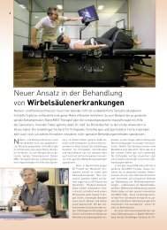
B§Iifl''f!!l'!ihld!MMW4'1•1
B§Iifl''f!!l'!ihld!MMW4'1•1
B§Iifl''f!!l'!ihld!MMW4'1•1

You also want an ePaper? Increase the reach of your titles
YUMPU automatically turns print PDFs into web optimized ePapers that Google loves.
LASER TREATMENT OF MUSCLELESIONSDr. Enzo Guaitlcri' - D<strong>l'</strong>. [·ì·onccsco Fontana·•TJ•aumatoJogy anù Sports Mcdicìne waJk ill d in ie Cesena (I~C ) 1tal;fdldiij§!lt.$Ri§i41Miii8KEY WORDS:Muscle lesions, Sport Medicine.INTRODUCTIONMuscle lesions in most cases affect subjectspracticing sports activities, and also personsduring their normal daily activities, or also dueto traumas connected with road accidents.Here, we shall purposely not include any anatomica[and microscopic descriptions of themuscular structure, instead we will concentrateon a clinica[ research based on dailypractice which will allow us to immediatelycarry out an efficacious treatment of thesubject pathology.Therefore we believe it is fundamenta l toshortly mention the classification of themuscle lesions (Tab 1-2) in order to identifythe pathologies that are susceptible to validtreatments with laser equipment.T~BLE 1.Musm LES/ONSWSSJF/CATIO.V.ACUTESUBACUTEElongationsSprains 1st degree 2nd degreeRupture or sprain - 3rd degreeContusionsTable 2 describes the characteristics of eachmuscle lesion.TABLE 2.MUSCLI LESJO.'ISOESCRJPTION.ElongationsThese occur due to strain when stretching the muscle, butwithout exceeding the muscle's maximum tension capacity.However, no lesions are produced.Sprain3rd degree Rupture or SprainContusionsIs characterised by the rupture of muscle fibres; and subdividedas follows: 1st degree, i.e. when some fibres areinvolved; 2nd degree, the number of fibres that are involvedis greater and quantifiable by means of ultrasoundmeasurement.Total or Partial; in which the continuity of most of themuscle belly is interrupted, and also the connective tissueis i nvolved.These are the effect of acute exogenous traumas that acton the muscle with a direct mechanism : a differentiationis made between muscle stupor, without any relevantlesions, muscle ecchymosis in which there are lesions inthe small Ointerstitial vessels; muscle hematoma involvingan interruption in the muscle fibres and rupture of vesselsof a considerable calibre; muscle compression with extensivelesion and tissue mortification.m
At this stage it is essential to previde a correctdiagnostic classification, also with the help ofultrasound techniques and/or MRI, that arealso useful in order to identify the location,the size and depth of the lesion.And especially ultrasound evaluation is primarilyused for muscle lesions, as it is a non-invasiveexamination, that is easy to perform andhas also been used in the subsequent checksafter the laser treatment in order to check theevolution of the lesion, and therefore theeffect of the laser radiations.Some patients were als~. subjected toTelethermography, another non invasive technique,that can be performed, which accordingto various authors, can define the "restituitoad integrum" state of the interested tissuemore precisely (a thermal map that can becompared to the contralateral healthy area toshow healing with the new formation of microcirculation).During the course of these years (from 1997 to2002), 300 patients were selected for thistrial. They were affected with different clinicalpictures of elongation, 1st and 2nd degreesprain and partial rupture, and also cases ofmuscle contusion (muscle stupor, ecchymosisand hematoma) that were very often associatedwith the previous pictures; in Table 3, thedifferent cases and the related class eachbelonged to, are described more specifically. Itmust be pointed out that in 12-13% of thecases approximately, a mixed clinical picturewas noted.Elongation 56Sprain - 1st degree 113Sprain - 2nd degree 75Rupture or sprain - 3rd degree 26Stupor and Ecchymosis 44H ematoma 23TA81f 3. 0ISTRIBUTIO~ OF THE [UNICA! OOESMATERIAlS ANO METHODSTwo types of lasers were used : the first was aLaser providing contemporaneous emission at623 nm (HeNe) with 20 mW energy and at 904nm ( GaAs ) with 60 mW energy (MectronicOpton 800) used to scan the area to be treated,and the second was a Neodymium-Yaglaser, 1064 nm (Mectronic NDS, and subsequentlyYag 145) with energy up to 50 W, broughtto the patient by means of optical fibreand a spacer, 3.5 sq. cm spot.In order to assess the symptom of pain objectively,the patients were asked to indicate theintensity of the pain they felt, using a visualanalogue scale (VAS with values from 1 to 10),on the occasion of the first visit, before eachtreatment session and at the end of the treatment.Furthermore, another scale, designed by TAstr 4.us, with values from 1 to 5 was used to identifymuscle functionality. (Table 4).Value l MeaningyScoREs INDICATINGHuscu FUNmONAUIY1 Impossible to perform a movement or muscle contractionagainst gravity, orto apply weight on the concerned limb.2 Possible to obtain mini mum muscle contraction withoutmovement and minimum application of weight in staticcondit ions.3 Possible to obtain muscle contraction equal to 50%, andarticular movement against gravity, application of weightto 50%.4 Possible to obtain muscle contraction below maximumlevels, complete articular movement against gravity, andcomplete application of weight.5 Complete functional recovery, withsports activities resumed.TREATMENTUse of Laser scan techniques was (i.e. HeNe +GaAs lasers) reserved for those cases in whichultrasound examination showed the presenceof a perilesional blood effusion which wasmore than 10 mm in size; in these cases thefirst 5 sessions were performed with thismethod in order to facilitate drainage of theeffusion and to activate micro-circulation,subsequently the Neodymium Yag laser wasused for 5-7 more sessions. In the cases inwhich subjects did not have any effusions, orif the recorded perilesional blood effusion wasLess than 10 mm, treatment was performedexclusively with Neodymium Yag , for 10/12sessi o ns.Subsequently, many of the subject patients inthe trial were subjected to functional rehabilinormaldaily and
•<strong>B§Iif<strong>l'</strong>'f</strong>!!<strong>l'</strong>!<strong>ihld</strong>!<strong>MMW4'1•1</strong>*TABlE 5.No:YA(, LASlRUT/UlArJONPAW4fTEII5)Ilo-tation in water andjor in a gym, which wasalso associated with electric stimulation andproprioceptive gym, in order to obtain completerecovery of both articulation andmuscularityThe scanning laser method was used for all thecases using maximum frequency (5000Hz) andtherefore using maximum energy for both theavailable wavelengths.The Nd:Yag laser was used in the continuousemission method (CW) and with variableenergy depending on the depth of the lesion(of th_e skin layer), from minimum 15 Watt tomaximum 25 Watt; in the latter situation, interms of energy emission (Joule) and bearingin mind a spot of approximately 3.5 sq. cm, anaverage of 500 to 800 Joule per spot per pulse,were distributed, trying not to provoke anylesions caused by skin burns, and thereforeusing the technique of a number of passagesover the same spot in each session.Table 5 summarizes the criteria used in thechoice, according to the intensity of theenergy and the amount of energy distributedper spot jpulse, when using the NeodymiumYag laser.Up to 15mm 15 500\550from 15 to 20mm 17 550from 20 to 25 mm 20 600\650from 25 to 35 mm 22 700Over 35 mm 25 800Out of the total number of treated patients, 93patients received double treatment (He\Ne +IR + Nd:Yag ), while 207 patients were treatedwith Nd:Yag alone.OUTCOME ANO COMPLICATIONSThree fundamental types of acute musclelesion outcome types were noted:1) Blood serum muscle cysts; 2) myositis ossificans;3) and muscle fibrosis.It is universally agreed that from an etiologicpoint of view, primary lesions were not treatedcorrectly or were not pointed out due to incorrector incomplete diagnoses. The final outcomefor all, was reduced muscle functionality.1) Blood serum muscle cysts can be aspiredwith the assistance of ultrasound equipmentand subsequent compression andcryotherapy in the initial phases, whenthese are still being organized, while lateronly surgery is applicable.2) Myositis ossificans is generally noted whenin the traumatic dynamics there is a periostallesion associated with a hematomafacing the bone surface. In this way, theperiostal osteogenetic elements are activatedand a heterotopic ossification processis induced, which leads to the formation ofosteoid tissue and finally to the structuredbone, in the time-interval ranging from 1to 3 months from the traumatic event.3) Myositis ossificans at times determined analteration in the muscle function, and itwas possible to treat the same with surgeryor Roentgen therapy, but the resultswere not however satisfactory.It is therefore important to determine thetype of lesion, the dynamics of the trauma,to perform an ultrasound measurement,that provides the morphological data of thelesion (size, depth, relation with the otherstructures, blood sampling if required, etc.)and to determine the parameters that aremost suited for energy laser treatmentapplications.Therefore in these cases it is extremelyimportant to newly modulate the energylaser utilization parameters, in order toprevent their biostimulating effect fromtriggering the biologica( mechanisms thatare responsible for the described outcomes.Therefore low dosages will be used,and the blood collection will be aspiredwith the assistance of ultrasound equipment.Fibrosis is the most frequent complicationin muscle lesions, and it takes placeeither because of the presence of hematomasthat determine a poor muscle stumpcoaptation, or due to excessively longperiods of immobilization which are notfollowed by suitable physiokinesitherapy,which lead to the consequent formation ofdense fibrous tissue.RESULTSIn all the cases of 1st degree elongation and
sprai n, by the end of the laser treatment, thepatients showed a practically normal functionalclinical picture, with scores of the VASscale equal to 0/1, and a score equal to 5 inthe functional scale.At the end of the treatment, the subjects witha clinical picture of 2nd degree muscle sprainhad an average score equal to 3, rated in thevisual analogue scale (VAS) and scores equal to3/ 4 in the functional scale. Obviously thesesubjects also underwent a further period ofphysiotherapy treatment (exercises in water,electric stimulation, proprioçeptive gym) inorder to obtain full fu nctional recovery.Photograph 1/2/3/4 illustrate a typical ultrasoundpicture of an intermediate lesion, assessedbetween 1st and 2nd degree at the beginning,and towards the end of the treatment.Among the cases of 3rd degree muscle sprainor rupture that were treated, subjects indicatedan average value equal to 4 in the visualanalogue scale, while they indicated an assessmentamounting to 3 points in the functionalscale. All the patients were subjected to ultrasoundchecks, and in particular those belongingto this group, where it was possible topoint out the tissue repairing process and thealmost complete reabsorption of the perilesionalblood effusion.It was interesting to note that during thetreatment of these affections, the typicalincrease in the pain that occurs in the 4/ 5 session,generally pointed out when treatingchronic, degenerative types of pathologies,was not noted here. In fact it must be underlined that a careful analysis of the data relatedto the VAS scale of the individua[ subjects,showed that the curve had a constant descendingtrend, which was more significant duringthe initial 4-5 sessions, and was more evidentin subjects who were immediately subjected to~~~PHOTOGRAPH 1151·2110 OfGRff SPRAJ/1 -BffORC lASER TREATHENT:TRA'ISVfRSAL 5CANPHOTOGRAPH 2PREVIOU5 CASE, (O.'fG/TUOINALSCAN BffORE TRWilENT.PHOTOGRAPH 3T HE SA~f PREV/0/JS CASE,(ONGITUDJNAL 5CAN AFTER lNojYAG LASER 5E55/0N5.Functional scale -baseline value~TABLE 6.5UHMARY Of THERESUlJSHematoma 1 \ 23 \ 4 4 \ 5
•ilii''iib'·IU!IIN'M~·i·*r- -..PHOTOGRAPH 4T HE 5AME PREVIOU5 CASE,TRANSVER5AL SCAN AffiR 7NojYAG LASER SESS/ONS.r···-·- ........ ~PHOTDGRAPH 5TELfTHERMOGRAPHl[EXAMINATlON OF A LEFT LATERA!MUSCULUS GEMELLUS LESION(HYPOTHERMIA - GREEN AREA).treatment witti Neodymium Yag laser. In ouropinion, this can be explained by the greatercasting off of endorphin substances and anacceleration in the repairing processes andvascular neogenesis.Many patients were also subjected to atelethermographic examination (Photos 4-5)Tab 6 summarizes the mean results.CONCLUSION:Laser treatment of muscle lesions and speciallytreatment performed with the NeodymiumYag laser, proved to be highly efficacious. Theresults, as indicated in Tab 6, are positive andencouraging, and enable us to state that outof all the types of equipment for the emissionof physical energy we have available, this isthe one that enables a precautious formationof the muscle tissue repair process, it decreasesreabsorptio n times of any blood or edemaextravasation, and t he time requi red for newformation of vessels, and it precautiouslydecreases pain, so in conclusion in enables afunctiona l recovery in a shorter amount oftime than in the past.In more severe cases, use of laser technologyhas enabled a precautious repetition of subsequentrehabilitation treatme nts req uired toreduce complications and the undesirableeffects to minimum, thus enabling thesubJect's complete functional recovery.We believe we can also state that t he newlyformed cicatrix has a good elasticity and resistance,as no relapses were noted in the samefocus of the previous lesion, specially insubjects doing sports activities, even at highlevels.•REFERENCES1) Romano Frignani - Traumatologia delloSport - Edizioni Piccin - 19912) Ferrario, Monti, Jelmoni - Traumatologia elesioni muscolari - Edi Ermes -19993) Algeri G., Conforti M.: "Laser Terapia -Trattato di Medicina Fisica e Riabi litazione"ed. UTET vol. zo - a cura di Valobra G.