08a The Skeletal System.pdf - Haspi.org
08a The Skeletal System.pdf - Haspi.org
08a The Skeletal System.pdf - Haspi.org

You also want an ePaper? Increase the reach of your titles
YUMPU automatically turns print PDFs into web optimized ePapers that Google loves.
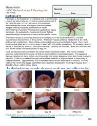
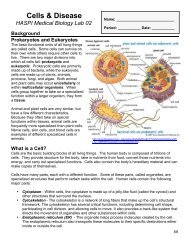
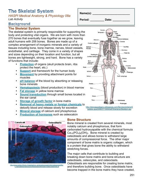
HASPI Medical Anatomy & Physiology <strong>08a</strong>Lab ActivityName(s): ________________________Period: _________ Date: ___________<strong>The</strong> <strong>Skeletal</strong> <strong>System</strong><strong>The</strong> skeletal system is primarily responsible for supporting thebody and protecting vital <strong>org</strong>ans. We are born with more than270 bones that eventually fuse together as we grow, leavingadult humans with 206 bones. Bones are made up of acomplex arrangement of in<strong>org</strong>anic minerals and a variety oftissues including bone, bone marrow, nerves, blood vessels,endothelial, and cartilage. <strong>The</strong>y come in a variety of shapesand sizes depending on their location and function, but allbones are lightweight, strong, and hard. Bone has a varietyof functions that include:• Protection of <strong>org</strong>ans (skull protects brain, ribsprotect the heart, etc.)• Support and framework for the human body• Movement by providing attachment points formuscles• pH balance of the blood by absorbing or releasingbone minerals• Hematopoiesis (blood production) in blood marrow• Fat storage in yellow bone marrow• Sound transduction through small bones located inthe ear canal• Storage of growth factor in bone matrix• Removal of heavy metals or foreign chemicals todetoxify blood and release slowly for excretion• Mineral storage of calcium and phosphorous• Production of hormones such as osteocalcinhttp://www.medes.fr/home_fr/applications_sante/osteoporose/eristo/osteoporosis/Bone_Remodeling/mainColumnParagraphs/04/image1/OsteocytesSmall.gifhttp://danceguadagno.wikispaces.com/file/view/anteriorSkeleton.jpg/248837719/640x879/anteriorSkeleton.jpgBone StructureBone mineral is created from several minerals, mostnotably calcium and phosphorous, that formcarbonated hydroxyapatite with the chemical formulaCa 10 (PO 4 ) 6 (OH) 2 . Bone mineral is created byosteoblasts and allows bones to withstand largeamounts of compressional force. <strong>The</strong> other majorcomponent of bone matrix is <strong>org</strong>anic collagen, whichis a protein that gives bone the ability to withstandstretching forces.<strong>The</strong> major cells that contribute to building andbreaking down bone matrix and bone structure areosteoblasts, osteocytes, and osteoclasts.Osteoblasts are responsible for creating bone matrix,and therefore building bone. Once osteoblasts havebecome trapped in the bone matrix they have created,251
they become osteocytes. Osteocytes function tomaintain the bone matrix and calcium homeostasis.<strong>The</strong>y are unable to move from their assigned location orspace, which is called the lacunae. Osteoclasts are largecells that are capable of reabsorbing bone minerals,and therefore remodeling bone structure.Osteoclasts also remove minerals to thebloodstream for a variety of bodily functions, suchas muscle contraction.<strong>The</strong> bone matrix can be arranged into twoclassifications of bone; compact and trabecular bone.Compact bone, also known as dense or corticalbone, is extremely hard and compact with very littlespace. Bone mineral in compact bone is arrangedinto tight circles called osteons, with nerves and bloodvessels passing through the center. Compact boneaccounts for 80% of the total bone mass.Trabecular bone, also known as spongy or cancellous bone, is porous and more like a network thatallows nerves, blood vessels, and bone marrow to easily fill trabecular bone. Stress on trabecularbone causes it to create new and stronger networks, making it extremely adaptable. Althoughtrabecular bone only accounts for 20% of the total bone mass, it has a much greater surface areathan compact bone.Bone Types<strong>The</strong>re are five main types of bone based on their shape. <strong>The</strong>se include long bones, short bones,irregular bones, sesamoid bones, and flat bones. <strong>The</strong> following table provides examples of thesebone types.Bone TypeLongBonesShortBonesDescription and ExamplesBones which are longer than they are wide andmade up primarily of compact bone. Examplesinclude arm bones, leg bones, and phalanges.Cube-shaped with a thin layer of compact bone.Examples include wrist and ankle bones.http://antranik.<strong>org</strong>/wp-content/uploads/2011/09/microscopic-structure-ofcompact-bone.pngSesamoidBonesFlatBonesIrregularBonesBones embedded in tendons. Examplesinclude the patella and pisiform.Thin and curved with parallel layers of compactbone. Examples include the sternum andbones of the skull.Bones that do not fit in any of the othercategories. Examples include the vertebra andbones of the sinus.252http://dc219.4shared.com/doc/QD-VsUDC/preview004.pngSaladin, K. 2012. <strong>The</strong> <strong>Skeletal</strong> <strong>System</strong>. Anatomy and Physiology: <strong>The</strong> Unity of Form and Function. New York,McGraw-Hill Publishing.
Station 3: <strong>Skeletal</strong> <strong>System</strong> Histology<strong>The</strong> cell and tissue structure of skeletal <strong>org</strong>ans are suited for the functions performed. Redraw andlabel Image B below. Image A on each chart is for reference!Compact BoneUsing colored pens/pencils, draw the histologyImage B from the “Compact Bone” chart in thespace below. Using Image A as a reference, labelyour drawing with the canaliculi, osteocytelacunae, and Haversian canal.Red Bone MarrowUsing colored pens/pencils, draw the histologyImage B from the “Red Bone Marrow” chart inthe space below. Using Image A as a reference,label your drawing with the compact bone,megakaryocytes, developing blood cells, andvascular sinus.Trabecular BoneUsing colored pens/pencils, draw the histologyImage B from the “Trabecular Bone” chart inthe space below. Using Image A as areference, label your drawing with the yellowbone marrow and trabeculae.PeriosteumUsing colored pens/pencils, draw the histologyImage B from the “Periosteum” chart in thespace below. Using Image A as a reference,label your drawing with the bone andperiosteum.256
Station 5: <strong>Skeletal</strong> DiseaseUsing the skeletal disease charts complete the following table. List ONLY THREE Causes or RiskFactors, Symptoms, and Treatment Options for each disease.OsteoarthritisDescription Causes or Risk Factors (3) Symptoms (3) Treatment Options (3)Approximately how many MORE people are expected to bediagnosed with osteoarthritis in 2030 than 2005? Hypothesize why.Osteogenesis ImperfectaDescription Causes or Risk Factors (3) Symptoms (3) Treatment Options (3)From Table 3-4, which type of OI is the worst? Is it dominant orrecessive?OsteosarcomaDescription Causes or Risk Factors (3) Symptoms (3) Treatment Options (3)According to the graph, what is the most common age formales to be diagnosed with osteosarcoma? Females?OsteomyelitisDescription Causes or Risk Factors (3) Symptoms (3) Treatment Options (3)What is the most common bone site of osteomyelitis?Paget’s DiseaseDescription Causes or Risk Factors (3) Symptoms (3) Treatment Options (3)What is the most common age for males to be diagnosed withPaget’s disease? Females?258
Station 6: <strong>Skeletal</strong> ProportionsHumans have used the proportions of skeleton throughout history to predict adult height or even todetermine the size of the “ideal man.” <strong>The</strong> scientific accuracy of these proportions is questionable. Inthis activity you will look at three common skeletal proportions and determine whether they have anyaccuracy in determining your actual height.DirectionsWing <strong>The</strong> wingspan measurement from fingertip to fingertip is the same as the measurement ofSpan an individual’s height.Step 1 Get a partner and a tape measure.Step 2Use the tape measure to determine the heights of you and your partner. Recordyour height in inches in Table 8.Step 3Spread your arms to the side and measure the wingspans from fingertip tofingertip of you and your partner. Record your wingspan in Table 8.Step 4Use the following formula to calculate the percent of error of your wingspanmeasurement from your measured height. Record in Table 8.SkullCircumference(Wingspan ÷ Measured Height) x 100 – 100 = Percent Error✔when complete<strong>The</strong> height of an individual should be 3x the circumference of an average-sizedhead.Step 1 Get a partner and a tape measure.Use the tape measure to measure the circumference around the foreheads ofStep 2you and your partner. Record your skull circumference in Table 8.Step 3 Multiply the skull circumference by 3 and record for calculated height in Table 8.Use the following formula to calculate the percent of error of your calculatedStep 4height from your measured height. Record in Table 8.(Calculated Height ÷ Measured Height) x 100 – 100 = Percent ErrorPerfectManStep 1 Get a partner and a tape measure.…or woman! <strong>The</strong> Greeks decided that the ideal man’s body would be seven heads tall.Only the “perfect man” proportions were used in their artwork.Use the tape measure to measure the height of the head from chin to top of theStep 2head, of you and your partner. Record your head height in Table 8.Step 3 Multiply the head height by seven and record for calculated height in Table 8.Step 4Use the following formula to calculate the percent of error of your calculatedheight from your measured height. Record in Table 8.(Calculated Height ÷ Measured Height) x 100 – 100 = Percent ErrorTable 8. Skeleton ProportionsMeasured Height Wingspan Percent of ErrorSkull Circumference Calculated Height (x3) Percent ErrorHead Height Calculated Height (x7) Percent Error259
Analysis Questions - on a separate sheet of paper complete the followingStation 11. What are the five types of bone? Give an example of each.2. What type of bone is the humerus? <strong>The</strong> vertebrae? <strong>The</strong> carpals?3. How many carpal bones are there?4. How many tarsal bones are there?5. What are the three parts of a long bone?6. Where is bone marrow located?Station 27. Explain how the structure of bone is similar to reinforced concrete?8. What types of force do collagen and bone mineral resist?9. How much weight was your paper roll (long bone) able to hold? Hypothesize how muchmore weight it would be able to hold if you taped 5 of the paper rolls together.Station 310.What passes through the Haversian canal?11.What is created in red bone marrow?12.What is found throughout trabecular bone?13.What is the function of the periosteum?Station 414.Explain why calculating height from bone length is useful to a forensic pathologist.15.Compare the heights you calculated and measured. How accurate were the calculations?16.Which bone most accurately calculated height? Hypothesize why.Station 517.What were the common causes & risk factors found between the majority of the skeletaldisorders?18.What were the common symptoms found between the majority of the skeletal disorders?Station 619.How close was your wingspan measurement to your actual height?20.How close was your height calculated from your skull circumference to your height?21.How close was your height calculated from your head height to your height?22.Explain how you could set up an experiment to determine whether the wingspanmeasurement is scientifically accurate.23.CONCLUSION: In 1-2 paragraphs summarize the procedure and results of this lab.Review Questions - on a separate sheet of paper complete the following1. How many bones are you born with?2. How many bones are in an adult skeleton?3. What types of tissues are found in bone?4. What are the functions of the skeletal system?5. What are the components of bone matrix?6. What are the functions of osteoblasts, osteocytes, and osteoclasts?7. What is the difference between compact and trabecular bone?8. What are the five many bone types?260