You also want an ePaper? Increase the reach of your titles
YUMPU automatically turns print PDFs into web optimized ePapers that Google loves.
The five colour dimensions of toothEnamel plus HFO is a rational system made by five types of bodies (dentines, generic enamels,opalescent enamels, intensive enamels, stains) that reproduce the five dimensions of naturalcolour of teeth (Fig. 14), following the technique of Dr. Lorenzo Vanini.Fig. 14.Chromaticity7 UNIVERSAL DENTINEUD1 (A1) - UD2 (A2) - UD3 (A3) UD3,5 (A3,5) - UD4 (A4) - UD5 - UD6The fluorescent dentine Enamel Plus HFO is reacting to the light excellently, in the same wayof natural toothValue3 GENERIC ENAMELSG.E.1 (low value) - G.E.2 (medium value) - G.E.3 (higher value)Translucency and brightness calibrated to the natural enamel (different value depending onthe age of the patient)Intensive2 INTENSIVE ENAMELS WHITESI.M. (Intensive Milky: warm and strong white hue) - I.W. (Intensive White: cold white hue)Intensive whites are <strong>use</strong>d <strong>for</strong> further characterisation of the surface enamel.Opalescent enamels1 UNIVERSAL OPALESCENT ENAMELO.B.N. (opalescent blue natural)3 OPALESCENT ENAMELS FOR CHARACTERIZATIONS AND SPECIAL CASESO.W. (white) – O.A. (amber) – O.G. (grey <strong>for</strong> special cases)Natural opalescent enamel OBN, OG and OA, reproduce the internal incisal opalescence.Opalescent enamels amber (OA) and white (OW) are also <strong>use</strong>d <strong>for</strong> characterizations type 1,2,3.Characterizations6 FLUORESCENT STAINSwhite, yellow, orange, blue, brown, dark brownThe fluorescent stains and OW, IW, IM, OA are <strong>use</strong>d to reproduce characterizations (internalhues and cracks)5
Colour registrationInstructions <strong>for</strong> a correct <strong>use</strong> of the Colour ChartColour chart and Enamel plus HFO shade guide, madewith original composite, are unique elements <strong>for</strong>registration of the colour. Due to the wedge-<strong>for</strong>m of theshade guide, it is possible to simulate layers with varyingthickness.The five colour dimensions of tooth have to be determinedin the order suggested in the chart.1. BC = Basic ChromaticityConsider 4 basic chromaticities (1, 2, 3, 4) that are obtained by building up 7 dentines (UD1,UD2, UD3, UD 3,5, UD4, UD5, UD6). The areas of the tooth that are most suitable <strong>for</strong> the studyof this dimension are the cervical and the middle third.2. V = ValueConsider 3 values (1, 2, 3) that refer to enamels with a low (1), middle (2) and high value (3).The three numbers are written on the chart in a colour from grey (1) to cold white (2) to milkywhite (3), in order to remind the <strong>use</strong>r the relation to the value. The corresponding compositemasses are GE1 (1), GE2 (2), GE3 (3). The area of the tooth that is most suitable <strong>for</strong> the studyof this dimension is the middle third.3. I = Intensive whitesThe numbers 1, 2, 3, 4 refer to the classification by shapes that is reproduced on the back of thechart. The <strong>use</strong>r has to follow this classification when choosing the colour.W-M indicates the shades of intensive white that can be found in natural teeth: w is a coldwhite, whereas m is a warmer and milky white. The masses of the composite system that shouldbe <strong>use</strong>d to reproduce these dimensions are IW (cold white) and IM (warm white). The Intensivewhites can affect all areas of the tooth (cervical, middle and incisal third).4. O = OpalescenceThe numbers 1, 2, 3, 4, 5, refer to the classification by shapes that are reproduced on the backof the chart. The <strong>use</strong>r has to follow this classification when choosing the colour. B-G-A indicatethe shades of the opalescences that can be found in natural teeth: B (blue), G (grey), A (amber).The masses of the composite system that should be <strong>use</strong>d to reproduce these dimensions areOBN (blue), OG (grey) and OA (amber). The opalescence’s only affect the incisal third of a tooth(interproximal and marginal area).6
5. C = CharacterizationsThe numbers 1, 2, 3, 4, 5, refer to the classification by shapes that are reproduced on the backof the chart. The <strong>use</strong>r has to follow this classification when choosing the colour.Each number is written with the same colour shades that can be found in natural teeth.W-A-Y-B indicates and reminds the <strong>use</strong>r the shades of natural teeth (white, amber, yellow,brown). The masses of the composite system that should be <strong>use</strong>d to reproduce these dimensionsare OW (soft white), IW (cold white), IM (milky white) OA (amber), SW (intense white), SY(intense yellow) and SB (intense brown). Type 1 and 3 characterizations are typical of the incisalthird of a tooth; type 2 characterizations affect both the middle and the cervical third, while type4 and 5 can be seen in all three coronal regions (cervical, middle and incisal third).Fig. 15. Taking of the colour dimensions in the three areas of the tooth(cervical, middle and incisal third).Glass ConnectorGlass Connector is a highly flowable body, with high elasticity and calibrated light diffusion thatreproduces the protein layer of the natural tooth. Glass Connector can be <strong>use</strong>d in direct andindirect restorations (inlay, onlay, veneers) and should be positioned in a thin layer (less than0,1mm) between enamel and dentine using a small brush. Glass connector increases theinternal light diffusion, strengthens the fluorescence of the dentine body, reduce the decreaseof value that is typical of the vitreous masses and create an elastic layer between enamel anddentine that reduces the internal tension due to the polymerisation.Fig. 16. 17. 18.Section of natural toothwhere the protein layerbetween enamel anddentine is very evident(on the left) comparedwith a section of toothmade with in Enamelplus HFO where GlassConnector layer ismodulating in a naturalway the internal lightdiffusion.7
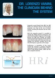
Anatomic stratification technique of Dr. L. VaniniIn order to maximise the characteristics of the Enamel plus HFO System, we suggest following theanatomic stratification technique of Dr. Lorenzo Vanini; any other stratification technique thatdoes not respect the anatomy of a natural tooth would considerably limit the aestheticper<strong>for</strong>mance of the system. To follow the anatomic stratification technique, first the lingualenamel is built up, then the internal dentine body and the vestibular enamel.The lingual enamel is applied bymeans of a silicone matrix, using aGeneric Enamel (GE1, GE2, GE3).The interproximal enamel wall isbuilt up using generic enamel.A thin layer of Glass Connector isapplied to the internal surface ofgeneric enamel.To obtain a natural chromatic composition,<strong>for</strong> the construction of the internal dentinebody three chromas are <strong>use</strong>d. Start with thebasic hue and increase by 2 shades (<strong>for</strong>example <strong>for</strong> the basic hue UD2: dentinebody shade UD2, UD3, UD4) in order tomake up <strong>for</strong> the lower chroma due to thetransition of the composite from the gelphase to the glass phase and to thedesaturation ca<strong>use</strong>d by the generic enamel.The highest chroma (UD4) is built upstarting from the margin of the cavity.8
Then this is covered by theintermediate chroma (UD3).Finally the basic hue shade (UD2)covers the underlying chromas.Using this “slice technique” it ispossible to obtain a rich andnatural chromatic composition.The completed dentine body iscovered with a thin layer of GlassConnector.The opalescent enamel (OBN) isapplied between the mamelons, inthe incisal area, in order to reproducethe internal opalescence.The intensive whites (IM, IW) arebuilt up and the characterizationsare reproduced with the opalescentmaterials (OA, OW, IW, IM) andwith stains.Finally the vestibular enamel isapplied, that should include somegeneric enamel (GE1, GE2, GE3).9
Direct techniquea) Enamel etching: orthophosphoric acid gel 37%; the application times are the same as theones <strong>use</strong>d <strong>for</strong> the dentinal substrate (30”, 60”);b) Dentine etching: orthophosphoric acid 37%; recommended application times: livingdentine 30”, sclerotic and non-vital dentine 60”. Total etching completely removes thesmear layer, produces a demineralization of the dentine and opens the dentinal tubules;c) Suction of the acid, rinsing and application of a new generation bonding system (EnaBond);following light curing;d) If also the lingual wall has to be built up, it is advisable to fabricate a silicone matrix. Afterfabricating the matrix (by means of the impression of a temporary restoration or a wax-upcarried out in the laboratory), the clinician can start with the application of the materialoutside the oral cavity; a portion of generic enamel is applied directly to the matrix in hishands and pulled in a thin layer using a small brush. When doing this it is advisable to switchoff the light from the dental unit, to avoid early polymerization of the composite (Fig.20);e) Once the generic enamel has been adapted to the matrix surface, the matrix is brought intoproper position in the mouth; the composite is pressed against the preparation margins withRESTORATION OF THE ANTERIORSFig. 19.Child Class IV fractureFig. 20.Stratification of GE 3 directly on thesilicon matrixFig. 21.Silicon matrix is positioned in mouthFig. 22.Cured lingual wallFig. 23.Build up of interproximal enamel walland application of Glass ConnectorFig. 24.Build up of the internal dentine bodyand incisal halo and application of G.C.Fig. 25.Build up of vestibular areas with GE3characterization with IMFig. 26.Build up polished with Enamel plusShiny pastesFig. 27.Palatal view of the restoration10
the flat brush. Ensure the material adheres well to the cavity and then cure. Aftercuring, remove the matrix (Fig.21-22);f) Shape the interproximal generic enamel, then light cure. The restoration is trans<strong>for</strong>medfrom a complex cavity into a simple one. After completing the enamel shell, the highdiffusion layer is reproduced with Glass Connector; do not apply this fluid mass to themargins, but only inside in a film that covers the internal enamel wall (Fig.23);g) Now you can start with the construction of the dentine body. Here you have to desaturatethe hue proceeding from cervical to incisal and from palatal to vestibular. If the basicchromaticity is UD2, you start with UD4 at the most cervical margin of the cavity; this hasto be covered with UD3 that ends a little more in an incisal position, followed by UD2 thatextends to an even more incisal area. The first dentine layer extends up to the enamelmargin of the cavity, while the other two layers go up to fill about half of the bevel thicknesson the enamel. With this technique it is possible to perfectly hide the margin. Be<strong>for</strong>e curingthe dentine at the incisal third, the grooves <strong>for</strong> the mamelons have to be cut in (Fig.24);h) The free vestibular surface of the dentine body is covered with a film of Glass Connectorthat is pulled using the flat brush and then cured;i) The opalescent enamel OBN is inserted in the grooves made between the mamelons of thedentine body. Always pull the material with the flat brush. For characterizations it is alsopossible to <strong>use</strong> the enamels OW and OA and the Stain Flows (stains);l) The following step consists in the application of the intensive white (generally IM <strong>for</strong> anteriorrestorations, IW <strong>for</strong> posterior restorations) onto the dentine body and then covers it with thevestibular generic enamel. The intensive white has to be pulled in a very thin layer, shaped asdesired and have a very limited thickness compared to the generic enamel (Fig.25);m) The last material to be built up is the vestibular generic enamel. By means of a small brushthe enamel is pulled so as to achieve an ideal aspect of the surface. After, the restoration iscured, finished and then polished (Fig.25-26).RESTORATION OF THE POSTERIORSFig. 28.Case be<strong>for</strong>eFig. 29.Preparation of the cavitiesFig. 30.Build up of the ridges with generic enamelFig. 31.Build up of the dentine bodyFig. 32.Build up of intensive enamels and genericenamelFig. 33.Completed build up11
Indirect techniqueEnamel plus HFO can be <strong>use</strong>d indirectly <strong>for</strong> anterior (laminated veneers: Fig. 34-43; onlays:Fig. 44-54) and posterior restorations (inlays: Fig. 55-57).The dental technicians <strong>use</strong> Enamel plus HFO with the same strafication technique as withmodern ceramic systems.VENEERSFig. 34.Adult central with old restorationsFig. 35.Removal of old restorations andsecondary cariesFig. 36.Filling of cavities with Enamel plus HFOand preparation <strong>for</strong> veneersFig. 37.Study model with diesFig. 38.Application of wax as spacerFig. 39.Stratification of dentineFig. 40.Finished Veneers after application ofOBN and GE3Fig. 41.Lingual view of VeneersFig. 42-43.Clinical case be<strong>for</strong>e and after application of composite veneers12
ONLAYSFig. 44. 45. 46.Complex fractures. On 1.1 also the lingual wall is fractured to the crest. After periodontal treatment, 1.1 is prepared <strong>for</strong>an overlay and 2.1 <strong>for</strong> a direct restoration.Fig. 47. 48. 49.The finished case: notice the extension on the palatal side due to the depth of the fracture.Fig. 50. 51. 52.Details under the microscope: the restorations show a natural opalescence that is pointed out by different lights, whilethe Generic Enamel is perfectly integrated with a calibrated and very delicate translucency.Fig. 53.Transilluminated sectionof a natural tooth restoredwith a composite veneer.Fig. 54.Transilluminated sectionof a natural tooth restoredwith a composite crown.13
INLAYFig. 55. Composite restoration withinfiltrationFig. 56. Cavities prepared <strong>for</strong> newrestorationsFig. 57. Cemented composite inlaysRESTORATION OF PROSTHETIC CORESGE2IWIMUD3UD4UD3UD4FLOWFig. 58. Complex restoration withcarbon fibre posts and compositeFig. 59. Scheme <strong>for</strong> the build-up ofthe restorationFig. 60. X-ray control of the restorationFig. 61. Section of a transilluminated Class 2 restoration in compositeon extracted toothCLINICAL INDICATIONSClassi I (all cavities)Classi II (small and medium cavities)Classi III (all cavities)Classi IV (all cavities)Classi V (all cavities)SealingsTotal and partials vestibular coveringCosmetic correctionsComplex restorationsOnlays Class I (all cavities)CrownsInlays Class II (all cavities)Inlays Class IV (all cavities)VeneersOnlaysRestoration of prosthetic coresMetal and fiber-glass bridgesThe Enamel plus HFO composite system has been designed and verified thanks to the researches of Dr. Lorenzo Vanini, assistedby the dental technicians Alessandro Tentardini e Franco Monti, with the R&D Department of G.D.F.14
Bibliography1. Vanini L., Toffenetti F. Nuovi concetti estetici nell’uso dei materiali compositi. Quaderni di progresso odontostomatologicoa cura degli “Amici di Brugg”. 1995;132. Dietschi D. Free-hand composite resin restorations: A key to anterior aesthetics. Pract. Periodont. Aesthet. Dent. 1995; 7(7): 15-253. Devoto V. L’intarsio in composito come soluzione di restauro estetico. Conservativa dei settori latero-posteriori.Attualità dentale 1996;02 22-314. Vanini L. Sistema composito microibrido fluorescente e opalescente. Dental Cadmos 1996; 8:36-46.5. Vanini L., Devoto W. Rifinitura e lucidatura di restauri in composito. I dossier: Materiali dentali. Supplemento a “Il dentistamoderno”, 5, 19966. C.L. Davidson, A.J. de Gee and A. Werner. Wear of 3 shades of enamel plus HFO and three other resin based fillingmaterials. Acta May-June, 19967. B. Hugo, A. Stassinakis, P. Hotz Ästhetische. Behandlungsmethoden. September 19968. Vanini L. Light and color in anterior composite restorations. Pract. Periodont. Aesthet. Dent. 1996; 8(7): 673-6829. Dietschi D. Current Developments in composite Materials And Techniques. Practical periodontics and Aestheticdentistry. September 199610 L. Portalier. Diagnostic Use of Composite in Anterior Aesthetic. Practical periodontics and Aesthetic dentistry Sept. 199611. Svanetti M., Turillazzi O. Gli intarsi in composito. Rivista di tecnologie dentali, febbraio 199712. Boschian, Gagliani, Brenna. Dentista Moderno. May 199713. Pascal Magne. Megabrasion: A Conservative Strategy <strong>for</strong> the Anterior Dentition.Practical periodontics and Aestheticdentistry May 199714. Vanini L., De Simone F., Tammaro S. Indirect composite restorations in the anterior region: a predictable technique <strong>for</strong>complex cases. Pract. Periodont. Aesthet. Dent. 1997;9(7):795-80415. G. Goracci, G. Mori. Università degli Studi di Roma “La Sapienza”. Ricostruzione estetica nei settori posteriori. DentalCadmos n. 13/199716. Vanini L. The control phases <strong>for</strong> checking the final aesthetic result in composite restoration of the anterior sector.Accademia Italiana di Conservativa 5th International Congress, Riva del Garda, 199717. Vanini L., Tasca G. Dalla <strong>for</strong>ma al colore, tecnica standardizzata per restauri in composito nei settori anteriori. Rivistadegli Amici di Brugg n. 2/199918. Hugo “Directe Veneers” Asthetische 4/9919. Mangani F., Vanini L., D. Cocchia, S. Condò “Polimerizzazione rapida delle resine composte valutazione della lampadaal plasma” Dental Cadmos 6/200020. Milnar “Recreating natural esthetics with a direct composite resin in the treatment of a complex class IV fracture–a casereport” The journal of cosmetic dentistry , Spring 200121. Dolecki “Kompozytowe rekonstrukcje podobne do porcelany–jak to si_ robi?” Compendium stomatologi 3/200122. Vanini L., Mangani F. “Determination and communication of the color using the five dimensions of teeth” PPAD Jan/Feb 200123. Mangani F., Sigalot C., Vanini L. “Intarsi in resina composita nel restauro estetico dei settori latero–posteriori” Il dentistamoderno febbraio 200124. Rollny, S. Gmünd, J. Dieterich, Winnenden “Das geheimnis eines natürlichen erscheinungsbildes: Veneers” Teamwork 4/200125. Ricciardi, M. Grande, V. Campanella, L. Cianconi “Analisi di un composito a basso modulo di elasticità” Il dentistamoderno, Gennaio 200226. Brenna, S. Porro, G. Artioli “Clinica e laboratorio nella realizzazione di restauri estetici indiretti nei settori posteriori” Ildentista moderno, Maggio 200227. Vanini L., Mangani F., Klimovskaia O., Il restauro conservativo dei denti anteriori, 2002 Promoden, Viterbo.28. Vanini L., Theunissen J.P:“Development of Esthetics in the Anterior Region” Journal of Dental Symposia, Fall 200215
MICERIUM S.p.A.Via Marconi,83 -16030 Avegno (GE) ItalyTel. +39 0185 7887 880 Fax +39 0185 7887 970www.micerium.it • e-mail:hfo@micerium.itFILE: HFO MANUALE 2004 ING