adaina primulacea meyrick, 1929 - Pterophoridae of North America
adaina primulacea meyrick, 1929 - Pterophoridae of North America
adaina primulacea meyrick, 1929 - Pterophoridae of North America

You also want an ePaper? Increase the reach of your titles
YUMPU automatically turns print PDFs into web optimized ePapers that Google loves.
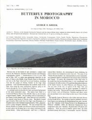
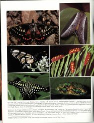
66 TROP. LEPID. RES., 19(2):64-70, 2009 MATTHEWS & MAHARAJH: Life history <strong>of</strong> Adaina plume mothLabral notch obtuse, shallow. Lateral flange <strong>of</strong> labrum with 3-4 points distad.Thorax: Prothorax with anterior setal fringe. Fringe continued alonglateral and part <strong>of</strong> posterior margin <strong>of</strong> scobinate cervical shield area. Seta D2 atleast 2× length <strong>of</strong> D1. XD and SD setae difficult to distinguish from secondaryfringe setae. About 4 L setae present, 2 apparently primary, with distinctperitremes. Three SV setae present, posterior seta 0.5× length <strong>of</strong> the other2. Setae numerous on thoracic legs. Dorsum <strong>of</strong> segments T2-T3 with slighttransverse indentation so that segment dorsum appears bilobed in lateral aspect.Dorsal primary and secondary setae present on anterior half (lobe) only. About6 minute to short setae forming anterior fringe, D1 within fringe, D2 posteriad,distinct. A group <strong>of</strong> about 6 SD setae adjacent to scobinate area. Five or 6 Lsetae present: 1 longer seta surrounded by 3-4 shorter setae and 1 isolated shortseta directly posteriad <strong>of</strong> the others. Three - 5 SV setae present, 2 distinctlylonger than the others.Abdomen: Segments A1-A7 with dorsal area subdivided as on T2-T3but with seta D2 on posterior lobe. Seta D1 about 0.67× length <strong>of</strong> D2, usuallydistinct and posteriad <strong>of</strong> anterior fringe. Setae D1 and D2 erect, anterior fringesetae projecting more anteriad. One SD seta dorsad <strong>of</strong> spiracle and about 4secondary setae anteriad <strong>of</strong> SD seta, continuing anterior fringe. Lateral setaearranged with L1 and L2 close together, L3 ventroposteriad. Several minutesecondary setae (about 6) also ventrad <strong>of</strong> L1-L2. One - 2 SV setae present.Proleg scar sometimes visible on A3-A6. Segment A8 with scobinations alonganterior margin prominent, distinctly dentate. Fringe setae minute. Spiracleenlarged, conical, positioned more posteriodorsad than on A7. Seta D1 directlydorsad <strong>of</strong> A8 spiracle, primary SD seta, 2 secondary setae, and L1 and L2directly anteriad. Two additional L setae ventrad <strong>of</strong> spiracle. Venter <strong>of</strong> A8 with 1SV seta and 1 V seta. Dorsum <strong>of</strong> A9 and A10 with scobinations and sclerotizedarea forming a contiguous anal plate. Posterior extent <strong>of</strong> A9 distinguished byrow <strong>of</strong> dentate scobinations. Anterior fringe setae lacking on A9 but D and SDsetae lateral, forming contiguous fringe with A10 anal plate setae. Venter <strong>of</strong> A9with 1 SV and 1V seta. Venter <strong>of</strong> A10 with about 15 setae surrounding vestigialproleg. Area laterad <strong>of</strong> proleg lightly sclerotized.Fig. 7–12. Female genitalia <strong>of</strong> Adaina: 7) A. <strong>primulacea</strong>, composite drawing<strong>of</strong> slides DM 661 and DM 1458; 8) antrum, ductus bursae, corpus bursae, andductus seminalis <strong>of</strong> A. microdactyla, slide DM 1460; 9) outline <strong>of</strong> dorsal anteriormargin <strong>of</strong> segment VIII showing anterior apophyses <strong>of</strong> A. bipunctata, slide DM236; 10) same, A. microdactyla, slide DM 1460; 11) same, A. simplicius, slideDM 237; 12) same, A. <strong>primulacea</strong>, slide DM 1458.Head capsule measurements indicate five larval instars (Matthews2006). Rows <strong>of</strong> closely spaced galls are seen on stems <strong>of</strong> heavilyinfested plants (Fig. 15b). Pupation occurs within the hollowed-outgall chamber formed by larval feeding on gall tissue. While larvaemove freely within the gall, the pupa (Fig. 15e,f) is positioned headup,toward the small operculum, through which the larva expels frass.These openings are also used by colonies <strong>of</strong> small ants that inhabitabandoned gall chambers. The host grows in hammocks and thickets inits native range <strong>of</strong> South Florida and the Florida Keys, the West Indies,Central and South <strong>America</strong>, and southern Texas (Cronquist 1980).Final instar larva. Figures 13a-h and 15c, d. Maximum length 9 mm.Body cream colored. Dorsum <strong>of</strong> each segment covered with band <strong>of</strong> smallferrugineous, dentate scobinations, most conspicuous on T1 and A7-A10.Cuticle bearing lightly sclerotized scobinations on T1 and A7-A10. Prolegsabsent on A3-A6 and vestigial on A10 with 0-3 crochets present (minute prolegswithout crochets are present on A3-A6 <strong>of</strong> early instars but lost before the 4thinstar). Setae minute to medium length, longest seta not exceeding head width.Localized secondary setae present, some forming short anterior fringe alongdorso-anterior margin <strong>of</strong> each segment. Spiracles, T1, A1-A7 round, slightlyexserted, with sclerotized peritremes. Spiracle on A8 (Fig. 13e) conspicuouslyenlarged, conical, directed posteriad, arising from near segment posteriormargin. Setal nomenclature for both larva and pupa descriptions follows Stehr(1987).Head: Width 0.67-0.84 mm. Light brown to brownish yellow. Adfrontalsclerite reaching anteclypeus. Mandibles reddish brown, broad, 5-toothed.Dorsal tooth very small. Distal seta replaced by pore. Labrum with 4 setae.Pupa. Figures 14a-c and 15e, f. Maximum length 7.5 mm. Dorsum withT2-A4 width nearly uniform. Lower abdominal segments tapered to a roundedtip at A10. Cephalic end with front, antenna base, pronotum, and anterior 0.33×<strong>of</strong> mesonotum flattened in the same plane to form near circular crown fringedwith setae. Setae simple. Primary setae short to medium length, longest setaabout 0.5× <strong>of</strong> maximum body width. Secondary setae present, two types. Onetype minute, without distinct peritremes, forming dense pubescence on cephaliccrown. The other type similar to primary setae, short to medium length, withdark peritremes, and grouped with primary setae. All setae on abdomen directedposteriad. Setal pinacula on cephalic crown slightly elevated. No protuberancesor dorsal ridges present on thorax and abdomen. Body color light yellow, headand cephalic crown light brown, crown moderately sclerotized. Abdomenwith ventroposterior margin <strong>of</strong> A8 fused to A9 and immobile, dorsum partlymovable. Anterior hamuli absent, posterior hamuli consisting <strong>of</strong> a uniformfringe <strong>of</strong> medium length, ventrally curved setae lacking hooked tips.Head: Front with cephalic ridge dividing dorsoanterior and ventral surfaces.Cephalic ridge continued as ridge on antenna base. Dorsum <strong>of</strong> front with thickcovering <strong>of</strong> minute setae, setae ending at cephalic ridge and forming marginalfringe. Two short primary setae on dorsum <strong>of</strong> front, anterolaterad near antenna.Setae straight, directed anteriad. Vertex present as tiny triangular posterolateralsclerite. Ventral face <strong>of</strong> front with minute sclerotized thorn-like process nearcephalic margin. A longitudinal row <strong>of</strong> about 3 minute setae between medianprocess and lateral margin with antenna, venter <strong>of</strong> front otherwise smooth.Frontoclypeal suture absent. One ventrally projecting clypeal seta present, setaabout 0.5× length <strong>of</strong> frontal setae. Clypeolabral suture lacking, lateral margins<strong>of</strong> clypeus barely indicated by a small furrow. Labrum small, with u-shapedmargin, seta lacking. Pilifers minute, touching at meson or not. Gena rounded,completely smooth and glossy, seta minute. Suture with glazed eye barelydiscernible. Sculptured eye obscure, setae lacking. Maxilla extending to 0.46×<strong>of</strong> T2 leg length. Distal tip <strong>of</strong> maxilla not exposed. Antenna extending to about0.65× <strong>of</strong> T2 leg length. Antenna base mesad <strong>of</strong> foreleg with fringe <strong>of</strong> about 15minute setae. No setae or ridge laterad <strong>of</strong> T1 leg.Thorax: Pronotum indented at anterior margin near midline. Setae D1and D2 in transverse alignment at about 0.67× from anterior segment margin.Setae short, about 0.5× length <strong>of</strong> frontal setae. Lateral margin forming roundedflange with 10-15 variable length setae, 1-2 setae nearly as long as frontalsetae. Midline suture visible. Foreleg extending to about 0.95× length <strong>of</strong> T2leg. Anterior half <strong>of</strong> foreleg noticeably broad, swollen at position <strong>of</strong> adultepiphysis but lacking longitudinal ridge. Coxal sclerite exposed. Anteriormargin <strong>of</strong> mesonotum straight (not convex). Spiracle along anterior margin
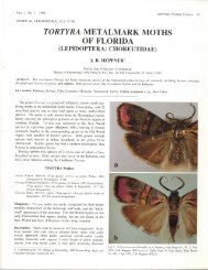
MATTHEWS & MAHARAJH: Life history <strong>of</strong> Adaina plume mothTROP. LEPID. RES., 19(2):64-70, 2009 67Immature material examined. L = larva, LS = larval skin/exuvium,P = pupa, PC = pupa case/exuvium. USA: FLORIDA: Broward Co.: Davie,University Drive, south <strong>of</strong> Rolling Hills Golf Course 11 Sep 1993 D. Matthews,T.A. Lott, & B. Maharajh, in stem gall on Chromolaena odorata (199 L, 19LS, 8 P, 14 PC) [DMC]; same, preserved 24 Sep 1993 (29 L, 5 LS, 4 P, 5 PC);same, preserved 1 Nov 1993 reared on artificial diet (1 P); Davie, University <strong>of</strong>Florida, FLREC 23 Aug 1993 B. Maharajh, in stem gall on C. odorata (1 L); 11Sep 1993 D. Matthews, T.A. Lott, & B. Maharajh, in stem gall on C. odorata(1 L); Davie, Treetops County Park 11 Sep 1993 D. Matthews, T.A. Lott, & B.Maharajh, in stem galls on C. odorata (8 L, 1 LS, 1 1st instar head capsule);Davie, Nob Hill Road 21 Nov 2001 D. Matthews & T.A. Lott, in stem gallson C. odorata (38 L, 1 LS, 1 P). All specimens currently held in DMC withvouchers to be deposited at MGCL and USNM.Fig. 13. Larval morphology <strong>of</strong> final instar A. <strong>primulacea</strong>: a) head, frontalview; b) labrum epipharyngeal surface on right; c) right mandible; d) seta D2,SD1, and A7 spiracle; e) seta D2, SD1, and A8 spiracle; f) chaetotaxic map <strong>of</strong>segment T1; g) segment T2; h) segment A3.minute, peritreme sclerotized. Anterior 0.67× <strong>of</strong> mesonotum covered withminute secondary setae. A transverse band <strong>of</strong> about 15 short to medium lengthprimary and secondary setae at 0.33× from anterior margin. Setae <strong>of</strong> band allwith dark peritremes. Setae D1 and D2 difficult to distinguish from secondarysetae <strong>of</strong> band, SD1 and SD2 usually distinct, with SD1 longest. Forewingnearly extending to tip <strong>of</strong> midleg, smooth (veins unmarked), a small cluster <strong>of</strong>about 10 minute setae near base. Midlegs not touching at meson, separated byfore and hindlegs. Midleg reaching middle <strong>of</strong> A5. Midline suture apparent onmesonotum but spliting only to about 0.67× at emergence. Metanotum withv-shaped mark at midline instead <strong>of</strong> suture line. Metanotum with 3-4 short setaeanteriad in D position and 3-4 setae in SD position within anterolateral angle.Hindwing extending to about 0.33× <strong>of</strong> A2, even with spiracle. Tips <strong>of</strong> hindlegsexposed posteriad <strong>of</strong> forelegs, between midleg tips, slightly exceeding T2 leg.Abdomen: Setae lengths variable, very short to medium length, all directedposteriad, with dark pertremes. Primary setae usually longer but difficult todistinguish from secondary setae (especially D setae). Dorsal setae shorter onlower abdominal segments. Segment A1-A2 with 4 D setae and 4 SD setae,A2 also with L1-L2 setae near forewing margin. Segment A3 with 5 D, 3-4SD setae present, and L1 and L2 between spiracle and forewing margin; A4with 5-6 D, 4 SD setae, and L1 and L2 present (no L3 or SV setae); A5 with6-7 D, 3 SD, 3 L (close together), and 1-2 SV setae; A6 with 7-9 D, 5 SD, 2-3L, 1-2 SV setae; A7 with 5 D, 5-8 SD, 2 L, and 1 SV seta. Spiracles on A2-A7 round with dark peritremes, very slightly exserted and with dark traces <strong>of</strong>tracheae visible from beneath cuticle. Segment A8 fused to posterior margin <strong>of</strong>A7 in both males and females except for a slightly movable part on dorsum.Segments A8-A10 completely fused, segment margins obscure. Segment A8with 3-4 short D setae, 3-4 short SD setae, L1 short, L2 medium length andno SV setae. Genital slit conspicuous on females. Spiracular scar present onA8. Segment A9 with about 8 setae on dorsum (groups indistinct), no setae onventer. Segment A10 with 2 setae slightly anteriad <strong>of</strong> hamuli fringe but difficultto distinguish. Hamuli consisting <strong>of</strong> dense fringe along tip and sides <strong>of</strong> A10,hamuli medium length, curving ventrad but lacking hooked tips. Tip <strong>of</strong> A10rounded. Anterior hamuli absent but a small, flat circular mark present. Venter<strong>of</strong> A8-A10 rounded, no lateral ridges or ventral plate present.Distribution and phenology. This species was originallydescribed from one male from Toboga Island, Gulf <strong>of</strong> Panama, (500ft.) collected in September (Meyrick <strong>1929</strong>). Gielis (1992) also listedone male from Costa Rica (Punt. Monteverde) collected in December1987. The Florida specimens are all from Broward County. As thehost is widespread thoughout the Neotropics, including the WestIndies, and Central and South <strong>America</strong>, it is likely this species will befound in these areas and possibly in Old World tropical regions wherethe host is an invasive weed. The Florida specimens were reared fromlarvae collected from stems <strong>of</strong> C. odorata in August and Septemberbefore the plants were in bloom. The seasonal habits and number<strong>of</strong> generations per year are unknown. It is possible that flowers arealso used as reported for A. microdactyla, which has two broods, onewhich bores in stems and produces galls, the other feeding in flowers,although Gielis (pers. comm.) indicates flower feeding has not beenrecently confirmed. In the northern hemisphere, flowering <strong>of</strong> SiamWeed occurs in late December and is brought on by short day lengths(Walton & Waterhouse 1998).DISCUSSIONMiller (2005) reported 179 identified species <strong>of</strong> lepidopterangallers worldwide representing 20 families. Of these, the family<strong>Pterophoridae</strong> included two palearctic species, A. microdactylaand Platyptilia nemoralis Zeller, the later on Senecio cacliasterLam. A third palearctic species, Gillmeria ochrodactyla(Denis & Schiffermüller) is known to feed within stem gallson Tanacetum vulgare L. (Cees Gielis, pers. comm.) but pupateexternally on the stems. Adaina <strong>primulacea</strong> adds anotheridentified species to the list <strong>of</strong> cecidophylous Lepidoptera,a fourth species to the known cecidogenous pterophorids,and is the first cecidogenous pterophorid identified from theNeotropics. As hosts and life histories are known for only 8<strong>of</strong> the 28 species <strong>of</strong> Adaina, and descriptions <strong>of</strong> several newneotropical species are anticipated, the discovery <strong>of</strong> additionalgallicolous species is likely. A recent experimental study byDiamond et al. (2008) demonstrated that larvae <strong>of</strong> the stemboring pterophorid Hellinsia glenni (Cashatt) attained greatermass when transferred and reared on Solidago galls inducedby a tephritid fly compared to larvae reared in stems, thussupporting the nutrition hypothesis for the adaptive nature <strong>of</strong>insect galls with an empirical test using a non-adapted species.While it is not known whether or not Adaina <strong>primulacea</strong> hascontinuous broods or also feeds on flowers and shoots asreported for A. microdactyla, both <strong>of</strong> these species presentunique opportunities for further studies on the evolutionarysignificance <strong>of</strong> gall induction.Although Gielis (1992) revised the genus Adaina, as thelife history <strong>of</strong> more species <strong>of</strong> these moths becomes known,
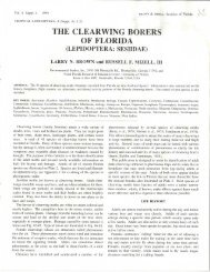
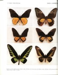
68 TROP. LEPID. RES., 19(2):64-70, 2009 MATTHEWS & MAHARAJH: Life history <strong>of</strong> Adaina plume mothadditional insight into relationships within the genus as wellas the family may be gained from morphological studies <strong>of</strong> theimmatures and from reared series <strong>of</strong> adults with definitivelyassociated males and females. The genus is presently definedby characters <strong>of</strong> the wing venation and <strong>of</strong> male and femalegenitalia. Adaina and Hellinsia Tutt are similar in some generalfeatures <strong>of</strong> the genitalia such as asymmetry in the male valvaeand lateral placement <strong>of</strong> the antrum in females. The forewingvenation <strong>of</strong> Adaina, however, is distinguished from that <strong>of</strong>Hellinsia having veins R 3and R 4stalked vs. free (Fig. 2).Larval morphology <strong>of</strong> internally feeding Hellinsia speciessuch as the type species H. osteodactylus (Zeller) vs. internalfeeders in Adaina is very similar. In both genera, setae <strong>of</strong> theinternal feeders are unmodified and arise individually instead<strong>of</strong> on tubercles or verrucae and the dorsum <strong>of</strong> the thoracic andabdominal segments is covered with numerous tiny sclerites.External feeders <strong>of</strong> both genera tend to have modified andclustered setae and have one or more extra teeth added to thebasic 5-toothed mandible, and in many species, the adfrontalsclerite on the larval head does extend all the way to theanteclypeus (Matthews 2006). The A4-A6 prolegs <strong>of</strong> Adainaflower and stem/gall borers examined are reduced, withoutcrochets, and with either the length not exceeding the width, orabsent as in A. <strong>primulacea</strong>. In Hellinisa flower and stem borers,the prolegs are shorter than in the external feeders but crochetsare present. In Adaina, the A8 spiracle is noticeably enlargedin certain species, such as A. <strong>primulacea</strong> (Fig. 13e) and externalfeeders, such as A. ambrosiae (Murtfeldt) (Matthews 2006),although not in the flower borer A. simplicius (Grossbeck). Thepupal chaetotaxy in these genera is generally a reduced version<strong>of</strong> the larva. In both Hellinsia and Adaina internal feeders,the anterior hamuli <strong>of</strong> segment A10 in pupae are absent orreduced in number. Pupae <strong>of</strong> Adaina borers (A. microdactyla,Fig. 14. Pupa <strong>of</strong> A. <strong>primulacea</strong>: a) ventral view; b) dorsal view; c) lateralview.<strong>primulacea</strong>, and simplicius) are unique however, in having aminute hooked process near the anterior margin <strong>of</strong> the head(Mellini 1954, Matthews 2006).While general characters and wing venation distinctlyplace A. <strong>primulacea</strong> within the genus, it is not necessarily moreclosely related to A. microdactyla than other Adaina based onthe common pattern <strong>of</strong> gall induction in the host. The left valvesaccular process <strong>of</strong> male genitalia <strong>of</strong> A. microdactyla (Fig.4), and two other species occuring in Florida, A. bipunctata(Möshler) (Fig. 5), and A. simplicius (Fig. 6) are similar inhaving a curved thin sclerotized sulcus extending basad <strong>of</strong> themain process which is absent in A. <strong>primulacea</strong>. The saccularprocess <strong>of</strong> A. <strong>primulacea</strong> also differs from the previous speciesin having the spine portion extending laterad from the basebefore curving posteriad, as opposed to extending directlyanteriad from the thickened base. The anterior apophysis in thefemale genitalia <strong>of</strong> A. <strong>primulacea</strong> (Fig. 12) also differs from A.bipunctata (Fig. 9), A microdactyla (Fig. 10), and A. simplicius(Fig. 11) in that it is a simple thorn-like process as opposed toan ornate bifurcate structure. The antrum in females also lacksthe paired sclerites found in A. microdactyla (Fig. 8), and theothers. Another interesting but variable character is the ductusseminalis, which is straight in A. simplicius, bent and distallyswollen in A. <strong>primulacea</strong>, and spiraled in both A. microdactyla(Fig. 8) and A. bipunctata. These last two species seem closestoverall in genital characters but are easily distinguished bywing maculation, the first being yellowish with scattered darkscaling, the later white with localized dark spots.In addition to differences in the morphology <strong>of</strong> A.microdactyla and A. <strong>primulacea</strong>, these two galler speciesdiffer somewhat in the reported larval feeding habits. Mellini(1954) illustrates multiple openings in a single large gall forA. microdactyla. Larvae <strong>of</strong> A. <strong>primulacea</strong> inhabit individualgalls with only a single opening. Gielis (pers. comm.) indicatesidentical habits for A. microdactyla. Larvae <strong>of</strong> A. <strong>primulacea</strong>bore within the stems beyond gall tissue, but not to the extentdescribed by Mellini (1954) for A. microdactyla. Althoughseasonal habits <strong>of</strong> A. <strong>primulacea</strong> are not completely known,A. microdactyla is limited to two distinct broods. Whilephytochemistry, linked with closely related hostplant genera,in this case Eupatorium and Chromolaena, is the most probabledeterminant in host selection, host physiology is most likely thesignificant factor in the evolution <strong>of</strong> larval habits in this group.Galler species have probably evolved in separate lineageswithin the genus Adaina as they have throughout the orderLepidoptera (Miller 2005).Crutwell (1974) listed an Adaina sp. in hollow stems <strong>of</strong> E.odoratum from Veracruz, Mexico which most likely refers toAdaina <strong>primulacea</strong>. Cruttwell (1968, 1974) also reported A.bipunctata from flowers <strong>of</strong> E. odoratum and E. iresinoides fromTrinidad. These records have not been confirmed with museumspecimens and could refer to A. <strong>primulacea</strong>, A. simplicius, or A.bipunctata. The latter two species are easily distinguished fromA. <strong>primulacea</strong> by the white as opposed to yellowish ground color<strong>of</strong> the wings. The true identity <strong>of</strong> A. bipunctata is problematic,however, as the holotype is not available and was reported asprobably lost from the Zoological Museum <strong>of</strong> Berlin (Gielis1992). In Florida, A. simplicius is more common than what
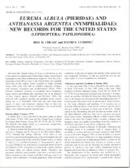
MATTHEWS & MAHARAJH: Life history <strong>of</strong> Adaina plume mothTROP. LEPID. RES., 19(2):64-70, 2009 69Fig. 15. Life history <strong>of</strong> Adaina <strong>primulacea</strong>: a) terminal shoot on hostplant, Chromolaena odorata, showing frass extruding from developing stem galls;b) mature stem galls; c) cross-section <strong>of</strong> stem exposing dorsal view <strong>of</strong> larva feeding within the developing gall; d) lateral view <strong>of</strong> final instar larva; e)lateral view <strong>of</strong> pupa within gall chamber; f) ventral view <strong>of</strong> pupa.is generally known as A. bipunctata. Larvae <strong>of</strong> A. simpliciusare flower borers on several species <strong>of</strong> composites, especiallyCarphephorus odoratissimus (J.F.Gmel.) Herb. (Matthews2006). Adaina simplicius, or a close relative has also beencollected in Argentina and exported to South Africa for study asa biological control agent for Pompom Weed, Campulocliniummacrocephalum (Less.) DC. (ARC/LNR 2007).The status <strong>of</strong> populations <strong>of</strong> A. <strong>primulacea</strong> in South Floridashould be monitored as this area is under heavy pressure fromdevelopment. As <strong>of</strong> 2001 the stand <strong>of</strong> hostplants supporting thepopulation along University Drive (County Road 817) in Daviewas completely eliminated by development. Although the hostis a weedy species, growing in dense tangled stands reaching 3meters, it is native to the area. While A. <strong>primulacea</strong> is considereda species <strong>of</strong> low priority for the biological control <strong>of</strong> SiamWeed because <strong>of</strong> difficulties in inducing sufficient ovipositionin captivity and in handling these small moths (Zacharideset al. 1998), as the spread and severity <strong>of</strong> this invasive weed
70 TROP. LEPID. RES., 19(2):64-70, 2009 MATTHEWS & MAHARAJH: Life history <strong>of</strong> Adaina plume mothcontinues in tropical and subtropical regions <strong>of</strong> the world,the candidate status <strong>of</strong> this moth may need to be reassessed.Despite the fact that Chromolaena odorata is widespread inthe Neotropics, along with preserving species diversity, it isimportant to maintain local reservoirs <strong>of</strong> the associated insectfauna for future biological control studies since these isolatedpopulations may include unique genotypes, variably suited fordifferent host strains and environments.ACKNOWLEDGMENTSWe thank Cees Gielis for his critical review <strong>of</strong> themanuscript, for providing images <strong>of</strong> type material for studyand comparison with our specimens, and for sharing personalobservations. Ernst Arenberger supplied comparative material<strong>of</strong> Adaina microdactyla. Line illustrations <strong>of</strong> the pupa are byMargo Duncan. Terry A. Lott assisted in fieldwork. Dale H.Habeck provided logistic support to the first author for travelto collecting sites and funding during graduate sudies. We alsogratefully acknowledge the support <strong>of</strong> staff and colleagues atthe Fort Lauderdale Research and Education Center. Thanksare also due to Charles V. Covell, Jr., Vitor O. Becker, ReedA. Watkins and anonymous peers for their careful reviews andhelpful comments.REFERENCES CITEDARC/LNR2007. Management and control. Pompom weed Campuloclinummacrocephalum. http://www.arc.agric.za/home.asp?pid=4538.Cronquist, A.1980. Vascular Flora <strong>of</strong> the Southeastern United States. Volume 1.Asteraceae. University <strong>of</strong> <strong>North</strong> Carolina Press, Chapel Hill. 261pp.Crutwell, R. E.1968. Preliminary survey <strong>of</strong> potential biological control agents <strong>of</strong>Eupatorium odoratum in Trinidad. Proceedings <strong>of</strong> the 9th BritishWeed Control Conference 1968: 836-841.1974. Insects and mites attacting Eupatorium odoratum in the neotropics. 4.An annotated list <strong>of</strong> the insects and mites recorded from Eupatoriumodoratum L., with a key to the types <strong>of</strong> damage found in Trinidad.Commonwealth Institute <strong>of</strong> Biological Control Technical Bulletin17: 87-125.Diamond, S. E., C. P. Blair and W. G. Abrahamson.2008. Testing the nutrition hypothesis for the adaptive nature <strong>of</strong> insectgalls: does a non-adapted herbivore perform better in galls?Ecological Entomology 33: 385-393.Gielis, C.1992. Neotropical <strong>Pterophoridae</strong> 8: The genus Adaina Tutt, 1905(Lepidoptera: <strong>Pterophoridae</strong>). Shilap Revista de Lepidopterologia20: 373-404.2003. <strong>Pterophoridae</strong> & Alucitoidea - In: World Catalogue <strong>of</strong> Insects 4:1-198.Kimball, C. P.1965. Kimball, C.P. 1965. Arthropods <strong>of</strong> Florida and neighboring landareas. Volume 1. The Lepidoptera <strong>of</strong> Florida an annotated checklist.Division <strong>of</strong> Plant Industry, Florida Department <strong>of</strong> Agriculture,Gainesville. 363 pp.Matthews, D. L.1989. The Plume Moths <strong>of</strong> Florida (Lepidoptera: <strong>Pterophoridae</strong>). MSThesis, University <strong>of</strong> Florida, Gainesville. 347 pp.2006. Larvae and Pupae <strong>of</strong> Nearctic <strong>Pterophoridae</strong>: A Synopsis<strong>of</strong> Life Histories, Morphology, and Taxonomy (Lepidoptera:Pterophoroidea). PhD Thesis, University <strong>of</strong> Florida, Gainesville.959 pp.Matthews, D. L., D. H. Habeck and D. W. Hall1990. Annotated checklist <strong>of</strong> the <strong>Pterophoridae</strong> (Lepidoptera) <strong>of</strong> Floridaincluding larval food plant records. Florida Entomologist 73: 613-621.Matthews, D. L. and T. A. Lott2005. Larval Hostplants <strong>of</strong> the <strong>Pterophoridae</strong> (Lepidoptera:Pterophoroidea). Memoirs <strong>of</strong> the <strong>America</strong>n Entomological Institute76: 1-324.Mellini, E.1954. “Pterophorus microdactylus” Hbn. (Lepidoptera <strong>Pterophoridae</strong>)nella biocenosi di “Eupatorium cannabinum”. Bollettino dell’Instituto di Entomologia della Università Degli Studi di Bologna20: 275-307.Meyrick, E.<strong>1929</strong>. The micro-lepidoptera <strong>of</strong> the “St. George” expedition. Transactions<strong>of</strong> the Entomological Society <strong>of</strong> London 76: 489-521.Miller, W.E.2005. Gall-inducing Lepidoptera. Pp. 431-465 in: Biology, Ecology, andEvolution <strong>of</strong> Gall-inducing Arthropods. Vol. 2. Raman, A., C. W.Schaefer, and T. M. Withers (Eds.) Science Publishers, Inc., Enfield,NH.Muniappan, R. and Bamba, J.2000. Biological Control <strong>of</strong> Chromolaena odorata: Successes andFailures. Pp. 81-85 in: Neal R. Spencer [ed.], Proceedings <strong>of</strong> the XInternational Symposium on Biological Control <strong>of</strong> Weeds 4-14 July1999, Montana State University, Bozeman, Montana, USA.Stehr, F. W.1987. Immature Insects. Kendall/Hunt, Dubuque, Iowa, 754 pp.Walton, C. and B. Waterhouse1998. Siam Weed. Australian Quarantine and Inspection Service,Department <strong>of</strong> Primary Industries and Energy http://www.dpie.gov.au/aquis/homepage/public/industry/siamweed.html.Zacharides, C., R. L. Kluge, S. Neser and L.W. Strathie1998. Promising New Candidates for the Biocontrol <strong>of</strong> Chromolaenaodorata. http://www.ehs.cdu.edu.au/chromolaena/proceedings/fourth/zach.htm.