
You also want an ePaper? Increase the reach of your titles
YUMPU automatically turns print PDFs into web optimized ePapers that Google loves.
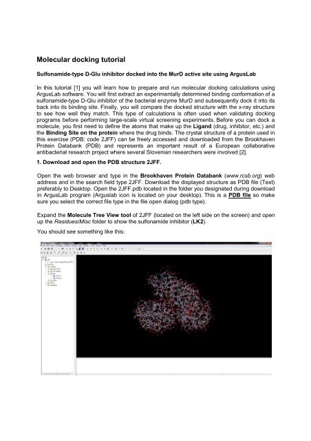
2. Create "Ligand and Binding Site groups".Left-click on "1440 LK2" in the Tree View to select the sulfonamide-type D-Glu inhibitor. Itshould appear yellow.Select the Edit/Hide Unselected menu option to hide all atoms that are not selected. The onlyatoms showing on the screen should be the D-Glu sulfonamide inhibitor.Center the sulfonamide inhibitor in the window by selecting the View/Center Molecule inWindow menu or pressing thebutton on the toolbar.While the inhibitor is selected, add hydrogens by and pressing theYou should see something like this:button on the toolbar.Right-click on "1440 LK2" in the Tree View and select the "Make a Ligand Group from thisResidue" option. ArgusLab will construct a group underneath the Groups folder with thesame name "1 LK2" that is of type = Ligand.Left-click on either the "1440 LK2" residue in the Residues/Misc folder. This action will againselect the atoms of the Ligand on the screen.Copy and Paste the selected residue. Look in the Residues/Misc folder in the Tree View tooland you will see a new highlighted residue with a name "2480 LK2". (different number is alsopossible). This is an extracted conformation of a ligand that you will dock into its binding site.
Make a Ligand group of this new residue in the same manner you did previously. Right-clickon "2480 LK2" and select "Make a Ligand Group from this Residue". You should now havetwo Ligands in the Groups folder named "1 LK2" and "2 LK2".Rename the two Ligand groups: Right-click on "1 LK2" in the Groups folder and select"Modify Group..." option. In the Modify Group Dialog box, type in a new name. Let's call it"ligand-xray". Make sure you don't change the Groups type. It should be a Ligand. Do thesame for "2 LK2", but name it "ligand".To more easily distinguish the two ligand in the graphics window, you can do a number ofdifferent things. One would be to to change the rendering style of one of the ligands Rightclickon the Group named "ligand-xray" and select "Set Render Mode" and choose the"Cylinder med" option.Make the binding site for the ligand-xray group. Right-click on the ligand-xray group in theGroups folder and select the "Make a BindingSite Group for this Group" menu option. Thiswill generate a BindingSite that consists of all residues that have at least one atom within 3.5Angstroms from any atom in the ligand-xray groupYou should see something like this:
3. Dock the ligand into the defined biding siteBring up the Dock Settings dialog box by selecting the Calculation/Dock a Ligand menuoption or clicking on the button on the toolbar.Here is what the Dock Settings dialog box looks like:Select the ligand to dock in the "Ligand" drop-box. Make sure to select the group named"ligand" group and NOT the "ligand-xray" group.Click on the "Calculate Size" button and a <strong>docking</strong> box tailored to the binding site will bemade and shown on the screen.Make sure "ArgusDock" is theselected <strong>docking</strong> engine, the Calculation type = Dock, andthe Ligand is Flexible.Click the "Start" button and the <strong>docking</strong> calculation will begin.Notice the messages that appear in the status line at the bottom of the molecule window. Youshould see ArgusLab first generate the scoring grids used during the <strong>docking</strong>, then thevarious search phases will occur, and finally the candidate poses should be processed untilthe calculation is done. Only the coordinates of the group named "ligand" should have beenmodified.
4. Analyze and save the resultsThere are two sources of information about the performed <strong>docking</strong> calculation: The separatelog file and information located in the Tree View tool. Expand the ArgusDock calculation inthe Calculations folder in the Tree View. You will see a listing of the <strong>docking</strong> settings andseveral "poses" starting with the most stable as Pose 1 according to the calculated energy inkcal/mol. (A term "pose" is usually used to designate the specific set of coordinates of adocked structure). Right click on pose 2 and select the "Display" option. The coordinates ofligand will change to this pose and its score will be displayed on the screen.The screen should look like this:Now you can examine how close the docked structure is to the x-ray structure. You willcalculate the RMSD difference between the x-ray and docked ligand - Pose 1 wich you mustselect. Then in the Tree View tool, select both the ligand and ligand-xray groups by holdingdown the "Ctrl" key and left-clicking on both groups in the Groups folder. Both groups shouldbe highlighted on the screen. Now, right-click on the "Groups" folder tab in the Tree Viewand select option "Calc RMSD position between two similar Groups". An information dialogwill appear on the screen giving the root mean square deviation (RMSD) difference in thecoordinates of the two ligands. In order to be able to conclude that the experimental andcalculated conformation are comparable the RMSD value should be less than approximately2.5 Angstroms. Write the calculated RMSD value between the Pose1 and experimentalligand-xray structure on your seminar answer sheet.Finally, to save the work you have done, you will need to save this molecule to an ArgusLab.agl file. Select the File/Save As... menu option and choose the ArgusLab file type and saveit. The file name should be: Naloga4.agl.
Literature:1. Original Arguslab <strong>docking</strong> <strong>tutorial</strong> available on-line (www.arguslab.com) and in the helpsection of the program.2. Article describing the X-ray structure: Kotnik M, Humljan J, Contreras-Martel C, Oblak M,Kristan K, Hervé M, Blanot D, Urleb U, Gobec S, Dessen A, Solmajer T Structural andfunctional characterization of enantiomeric glutamic acid derivatives as potentialtransition state analogue inhibitors of MurD ligase. J Mol Biol 2007, 370:107–115.