Immediate Implant Placement and Provisionalization - Zimmer Dental
Immediate Implant Placement and Provisionalization - Zimmer Dental
Immediate Implant Placement and Provisionalization - Zimmer Dental

You also want an ePaper? Increase the reach of your titles
YUMPU automatically turns print PDFs into web optimized ePapers that Google loves.
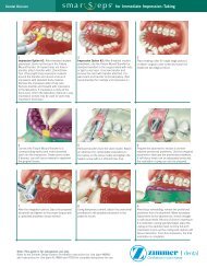
CE 1Figure 1—Diagnostic wax-up.Figure 2—TempStent surgical stent/provisionalization system.Figure 3—Analogs set into maxillary study cast.tapered, which provides the best chance forobliterations of the coronal portion of thesocket. It also mimics the natural convergence,which occurs in the natural tooth system, tothe apical portion of the socket. The implantsurface should be roughened with surfaceenhancements to promote a rapid integration<strong>and</strong> to enhance the initial stabilization of thefixture. The polished implant collar should be≤ 1 mm in height, which allows the final positionof the implant collar to be at a positionthat is superior to the crest of the bone. Thisposition allows soft tissue attachment in theregion of the collar. The roughened implantsurface at the first thread, or at its smoothtransfer from the polished collar to the roughenedsurface, allows bone attachment. Placingthe implant in this fashion eliminates the“dieback” phenomenon.Figure 4—Preparation of stock abutments on theTempStent model.Grafting MaterialAccording to the author’s clinical experience,autogenous bone is the best grafting materialavailable. It is osetoconductive, osteoinductive,<strong>and</strong> osteogenic. 16-19 However, one of thedrawbacks of autogenous bone is that a secondsurgical site is necessary to harvest the bonetissue. Many of these techniques require complicatedsurgical procurement <strong>and</strong> add significantsurgical time <strong>and</strong> morbidity. Other optionsinclude the use of allogenic, alloplastic, <strong>and</strong>xenographic 20,21 grafting materials, either with orwithout the use of various regenerative barriers.All have been well documented. 22 The nonautogenousgrafting material should be biocompatiblewith the host tissue, osteoconductive, 23-27osteoinductive, 28,29 <strong>and</strong> osteotrophic. 30,31 Not allallogenic or alloplastic grafting materials possessthese important qualities. Some must rely on avehicle to reconstitute their granular form. Theauthor 32-34 <strong>and</strong> others 35,36 have observed thatplatelet-rich plasma (PRP) has enhanced theosteoconductive, <strong>and</strong> possibly the osteoinductive,properties of various allogenic, alloplastic,<strong>and</strong> xenographic materials.Platelet-Rich PlasmaThe ideal autogenous vehicle for reconstitutingthe alloplastic, allogenic, or xenographic substrateis PRP (autologous platelet gel), 37,38 whichis developed from autologous blood with a cellseparator. The cell separator used herein followsa dual spin cycle <strong>and</strong> is complete in 12minutes (Smart PreP ,c ). Centrification of 55mL of whole blood results in 10 mL of PRPcHarvest Technologies Corporation, Plymouth, MA 02360;(508) 732-75004 Compendium / February 2003 Vol. 24, No. 2
Table 5—Substances Released byDegranulation of PlateletsCE 1Platelet-derivedgrowth factorTransforming growthfactor-betaPlatelet-derived endothelialcell growth factorInsulin-like growth factorVascular endothelialcell growth factor(PDGF)(TGF-ß)(PD-ECCF)(IGF)(VEGF)Figure 5A—Pretreatment view of edentulous site at tooth No. 8.Figure 5B—Preoperativeperiapicalradiograph.which, when simultaneously mixed withthrombin <strong>and</strong> calcium chloride, results in thedegranulation of the platelets <strong>and</strong> subsequentrelease of growth factors. This stimulates bothhard <strong>and</strong> soft tissue maturation <strong>and</strong> promoteshealing. For this reason, PRP is the ideal vehiclefor reconstituting the substrate because itenhances the osteoconductive qualities <strong>and</strong>,perhaps, osteoinductive properties of the allogenic,alloplastic, or xenographic graft materials.In addition, some have observed that PRPaccelerates soft tissue maturation, which allowsthe gingival tissues to rapidly heal with minimalpostsurgical contour alterations. Table 5 lists substancesreleased by the degranulated platelets.Abutment SelectionAbutment selection is achieved by using thecarrier mechanism of the implant system (a temporaryabutment) or a stock abutment (whichthe restorative dentist could use as a final abutment).Both of these require preparation. Theincorporation of the temporary/surgical guide system,which the author uses, allows advancepreparation of the stock abutment in the dentallaboratory. This eliminates the repeated removalof carrier mechanisms, intraoral preparation ofthe abutment (which produces heat <strong>and</strong> micromovementvia vibration), <strong>and</strong> impression copingsfor implant indexing at the time of placement.It is important to remember that manytimes, with immediate tooth removal/immediateimplant placement (especially in sinus elevations),the quality of bone is compromised.Whatever the surgeon can do to minimizeexternal forces to the fixture is beneficial. Astock-angled abutment that is easily preparedsuffices when the stock abutment provideddoes not allow an appropriate path of insertionfor the restoration because of the angle ofimplant placement.Temporary Restorative Material/TemporaryConstructionThe next step is the ideal temporaryconstruction method, as discussed previously.The TempStent method allows the efficienttransfer of a surgical guide into anesthetic provisional restoration that closelymimics that of the planned final restoration,in regard to emergence profile formation,interdental contours, contact points, <strong>and</strong>gingival contour at the facial marginalaspect. After completion of the temporary,the clinician must carefully evaluate thepatient’s occlusion. The provisional shouldnot have occlusal contact in the centric relationposition, or lateral excursive or protrusivecontacts. After 3 months (4 or 5 monthsin sinus grafted cases) the prosthodontist orreconstructive dentist can fabricate the finalimplant-supported restoration. The temporaryVol. 24, No. 2 Compendium / February 20035
CE 1Figure 5C—Creation of the sculpted implant receptacle site withPRP inserted into the osteotomy site.Figure 5D—Prepared, polished stock abutment seated.Figure 5E—Provisional restoration cemented.Figure 5F—Emergence profile formation at 3 monthspostoperatively.Figure 5H—Case complete periapicalradiograph.Figure 5G—Final implant-supported restoration.restoration can be cemented with a temporarycement, <strong>and</strong> once again, confirmation of nonloadcan be obtained.Suture MaterialA 5.0 monofilament (Monocryl d ), or4.0 Vicryl Rapide suture is recommended.The suture is small enough in diameter <strong>and</strong>resorbs over 30 to 45 days (Vicryl Rapide in10 to 14 days), allowing proper stabilization<strong>and</strong> maturation of the soft tissues.Postoperative CourseAs stated earlier, the prescribed healingphase is 3 months in immediate extraction ordEthicon, Inc, Somerville, NJ 08876; (800) 255-2500edentulous ridge cases, <strong>and</strong> 4 to 5 months insinus grafted cases. After 3 to 5 months, if therestorative clinician uses the stock abutmentseated at the initial visit, the abutment istorqued to 30 Ncm, <strong>and</strong> the temporary is recemented.Routine restorative procedures arefollowed from that point.If the restorative dentist requests acustom abutment, the patient is referred back tothe restorative clinician for routine impression6 Compendium / February 2003 Vol. 24, No. 2
CE 1Figure 6A—Preoperative view of the m<strong>and</strong>ibular left posteriorsextant at teeth Nos. 19 <strong>and</strong> 20.Figure 6B—TempStent seated clinically after creation of thesculpted implant receptacle site.Figure 6C—Seating the prepared, polished stock abutments.Figure 6D—Closure <strong>and</strong> the left lateral clinical view.techniques at fixture level with a transfer coping.When fabrication of the custom abutment iscompleted, the abutment is seated <strong>and</strong> torquedat 30 Ncm, <strong>and</strong> completion of the implant supportedrestoration proceeds as normal.The case reports that follow describe theimmediate implant placement <strong>and</strong> provisionalizationprocedure outlined in an edentuloussite, an immediate extraction site,<strong>and</strong> a sinus grafted site.Case 1—Single Tooth <strong>Implant</strong>A 33-year-old woman (nonsmoker) presentedfor implant reconstruction at the edentuloussite at tooth No. 8 (Figure 5A). The preoperativeperiapical radiograph is shown in Figure 5B.After completing a diagnostic waxing of boththe hard <strong>and</strong> soft tissues that need to bereplaced, the clinician can evaluate the necessaryregeneration procedures that need to be performed,in conjunction with implant placement;ascertain the width <strong>and</strong> length of the plannedfinal restoration; plan soft tissue contours; <strong>and</strong>fabricate a surgical stent/provisional restoration(TempStent ).After administering an appropriate localanesthetic <strong>and</strong> full-thickness flap elevation,the clinician prepares a sculpted implantreceptacle site in the alveolar crest (Figure 5C)by mimicking the flow of the facial bone marginsfrom tooth No. 8 to tooth No. 10. Thissculpted implant receptacle site also allows thecreation of interdental bone contours (Figure5C) <strong>and</strong> osseous foundation, which sculpts theemergence profile of the final restoration.After atraumatic site preparation, the clinicianapplies PRP to the osteotomy site (Figure 5C)<strong>and</strong> coats the implant surface with PRP beforeplacement. A 4.7-mm Tapered Screw Vent e ,HA-coated, 13-mm length implant is placed inthe osteotomy site. After implant placement<strong>and</strong> removal of the implant carrier, an HLA4/5 stock abutment e is prepared <strong>and</strong> polishedextraorally, <strong>and</strong> securely seated in the implant(Figure 5D).Retrofitting of the TempStent surgicalguide/provisional restoration completes thetransitional restoration, while marginal adaptationis completed with the abutment that hasbeen prepared extraorally. After the abutment/provisionalmargins have been adapted,the entire abutment/temporary restorationcomplex is reinserted <strong>and</strong> cemented, respectively,at the implant site (Figure 5E).Cementation is completed with a strong temporarycement. Veneering of the buccal plateVol. 24, No. 2 Compendium / February 20037
CE 1Figure 6E—Case complete clinical appearance of three immediaterestored implants at 4 months after surgery.Figure 6F—Case complete panoramic radiograph.Figure 7A—Preoperative clinical view, maxillary left posteriorsextant.<strong>and</strong> correction of the fenestration at the facialof tooth No. 9, with a PRP/PepGen P-15 graft complex f , provide the foundation forregeneration of the insufficient alveolar buccalcrest. Creation of a bioactive membrane by applicationof PRP, followed by platelet-poor plasma(PPP) over the entire graft complex <strong>and</strong> surgicalsite, aids in the local delivery of growth factors tothe bone surface, implant surface, <strong>and</strong> inferiorsurface of the flap. Closure is accomplished with5.0 Monocryl suture <strong>and</strong> a bioactive wounddressing created by additional application of PRP<strong>and</strong> PPP over the surgical site before dismissal.After 3 months, the removal of the temporaryallows not only for the final torquing of the stockabutment to 30 Ncm, but also for inspection ofthe sulcular environment <strong>and</strong> emergence profilecreated (Figure 5F).Figure 5G illustrates the final implantsupportedrestoration <strong>and</strong> AGC ® (AestheticGalvanotechnik Crown) g , which was seated4 months after the initial surgical appointment.Note the excellent soft tissue maturitylevel, interproximal contours, <strong>and</strong> emergenceprofile that have been formed <strong>and</strong> maintainedthroughout the healing phase. The caseeCenterpulse, Carlsbad, CA 92008-7308; (760) 929-4300fCeraMed, Lakewood, CO 80228; (800) 426-7836Figure 7B—Preoperative panoramic view, maxillary leftposterior sextant.complete final periapical radiograph is shown inFigure 5H. Note how the bone contours createdby the sculpted implant receptacle site have beenmaintained in the final radiograph <strong>and</strong> a stablesulcular/biologic width environment bony interfacehas been established.Case 2—Multiple Tooth ReplacementA 56-year-old man (nonsmoker) presentedfor implant reconstruction of the m<strong>and</strong>ibularleft posterior sextant at teeth Nos. 19 <strong>and</strong>20 (Figure 6A). After maxillary <strong>and</strong> m<strong>and</strong>ibularstudy models are obtained, <strong>and</strong> mountedon a KaVo Protar articulator h , the fabricationof the TempStent surgical guide/provisionalsystem is completed, <strong>and</strong> the appropriate sitesfor implant position cored. After administeringan appropriate local anesthetic <strong>and</strong> crestalincision, followed by full-thickness mucoperiostealflap elevation, the surgeon creates thesculpted implant receptacle site, precedingreinsertion of the surgical guide (Figure 6B).Observe how creation of the proposed osseouscontour (sculpted implant receptacle site) forthe final restoration follows the contours ofthe surgical guide.gWiel<strong>and</strong> <strong>Dental</strong> Systems, Milford, CT 06460; (866) 876-0885hKaVo USA, Lake Zurich, IL 60047; (800) 323-80298 Compendium / February 2003 Vol. 24, No. 2
CE 1Figure 7C—<strong>Implant</strong> placement in conjunction with sinuselevation surgery.After atraumatic coring procedures, <strong>and</strong>application of PRP in the osteotomy sites,placement of the implants is accomplished.Two Paragon Tapered Screw Vents e areplaced: a 5.7-mm diameter, 13-mm lengthimplant at the No. 19 position, <strong>and</strong> a 4.7-mmdiameter, 13-mm length implant at the No.20 position. The creation of the sculptedimplant receptacle site eliminates the countersinkingprocedure, <strong>and</strong> allows the implantcollar to remain above the crest of the bone,but within the environment of the interdentalbone height (Figure 6C). After preparing<strong>and</strong> polishing two HLA abutments e , a 4/5<strong>and</strong> 5/6 at the appropriate implant sites,placement of the abutments (Figure 6C) precedesretrofitting <strong>and</strong> marginal adaptation ofthe provisional restorations. Closure isaccomplished with 4.0 Vicryl Rapide suturein a continuous sling/horizontal mattresssuturing technique (Figure 6D). Evaluationof the patient’s occlusion confirms noocclusal contact in centric relation or lateralexcursive movements.After observation for 2 months, a fractureis detected at tooth No. 18, <strong>and</strong> a thirdimmediate restored implant is placed in them<strong>and</strong>ibular left posterior sextant. Figure 6Eshows the case complete clinical appearanceof the three immediate restored implants at4 months after surgery. Figure 6F shows thecase complete panoramic radiograph of them<strong>and</strong>ibular left posterior sextant.Case 3—Multiple Tooth Replacementwith Sinus ElevationA 63-year-old woman (nonsmoker) presentedfor treatment of a vertical fracture attooth No. 13 (Figure 7A). The preoperativepanoramic radiograph is shown in Figure 7B.Figure 7D—Abutments seated <strong>and</strong> conversion of theTempStent into the provisional restoration.After the pretreatment diagnostic <strong>and</strong> planningphase previously mentioned is completed,TempStent is fabricated not only to providesurgical guidance, but also to serve as the provisionalrestoration. Because of the pneumatizedmaxillary left sinus, sinus elevationsurgery is added to the treatment plan, in additionto placement of the three implants.After administering an appropriate localanesthetic, a sulcular incision is made at thebuccal <strong>and</strong> palatal of teeth Nos. 13 <strong>and</strong> 15,crestally at No. 14, with two vertical releasingincisions at the distal of No. 12 <strong>and</strong> distal lineangle of No. 15. After full-thickness mucoperiostealflap elevation, the creation of anosteotomy in the lateral wall of the maxillae,just inferior to the zygomatic arch, allows foraccess to, <strong>and</strong> elevation of, the lateral wall ofthe maxillae. Attached to the Schneiderianmembrane, the lateral wall <strong>and</strong> the inferiorportion of the sinus membrane itself are rotatedmedially <strong>and</strong> superiorly to create the receptaclefor the sinus graft. Insertion of the surgicalguide allows for preparation of the implantsites at the Nos. 13, 14, <strong>and</strong> 15 areas.Before implant placement, PRP isapplied to the sinus cavity via the accessopening, <strong>and</strong> the graft complex (PRP <strong>and</strong>PepGen P-15 ) is placed anteriorly, medially,<strong>and</strong> distally in the sinus. This techniqueensures that grafting material is present atthe aforementioned areas of the sinus beforeplacement of the implants. Additional applicationof PRP in the sinus precedes implantplacement. Three Paragon Tapered ScrewVent implants are placed: a 5.7-mm diameter,13-mm length at the No. 15 site, followed bytwo 4.7-mm diameter 13-mm in lengthimplants at the Nos. 13 <strong>and</strong> 14 sites, respectively(Figure 7C). After removal of theVol. 24, No. 2 Compendium / February 20039
CE 1Figure 7E—Closure <strong>and</strong> left lateral centric occlusion view.Figure 7F—Case complete clinical view of the immediaterestored implants placed in conjunction with sinus elevationsurgery at 5½ months.Figure 7G—Case complete periapical radiograph.implant carriers, the preprepared HLA stockabutments are seated, <strong>and</strong> the provisionalrestoration is inserted to confirm implant placement,before retrofitting with a composite material(Figure 7D). After the conversion of the surgicalguide to the provisional restoration, cementationof the restoration is achieved with a strongtemporary cement. Application of PRP over thelateral aspect of the maxillae precedes additionalgrafting with the PRP/PepGen graft complex atthe osteotomy site in the lateral wall of the maxillae,<strong>and</strong> the peri-implant defect sites. Closureis accomplished with 4.0 Vicryl Rapide sutures(Figure 7E). Note that in centric occlusion,disclusion is present from Nos. 13 to 15. In thenormal chewing cycle, small amounts of loadwill undoubtedly be present, <strong>and</strong> the authorobserves that enhancement of the maturationphase of the graft occurs, based on earlierdefinitive load times.After careful observation for 4½ months, thepatient is referred for completion of implantprosthetics. The final implant-supported restorations(Figure 7F) are seated 5½ months after theinitial, <strong>and</strong> only, surgical procedure. The casecomplete periapical radiograph shows the sinusgraft well incorporated at the level of theimplants (Figure 7G).Conclusion<strong>Implant</strong> treatment has previously beenseparated into the surgical <strong>and</strong> restorativephases. Depending on the complexity of theplanned site, the surgical phase may requiretwo to four procedures in conventionalimplant treatment. The restorative phase,especially in the esthetic zone, may requiretissue sculpting <strong>and</strong> provisionalization toachieve the foundation not only for esthetics,but also for long-term restorative success. Overthe years, conventional implant treatment hasproven to be a highly successful treatmentoption for replacing the natural tooth system.Advancements in surgical techniques,bone grafting materials, <strong>and</strong> bioengineering ofthe surgical site, have allowed the implantsurgeon to decrease treatment times <strong>and</strong> possiblysurgical visits. However, without propercommunication among the surgeon, restorativedentist, <strong>and</strong> laboratory technician, poor orimproper treatment planning leads to complex,<strong>and</strong>/or compromised prosthetic procedures.Continued advancements in surgical stentdesigns <strong>and</strong> the incorporation of provisionalrestorations during implant placement haveallowed the implant team (ie, surgeon, reconstructivedentist, <strong>and</strong> laboratory technician) tobetter communicate the parameters for thefunctional, biological, <strong>and</strong> esthetic success inimplant restorations.The immediate restoration of dentalimplants is an exciting option the implantteam can offer patients seeking implant treatment.The incorporation of a provisionalrestoration during implant placement providesthe patient with a stable, esthetic temporaryrestoration. From a periodontal perspective,the author observes that the preservation of10 Compendium / February 2003 Vol. 24, No. 2
interdental bone, creation or maintenance of thesoft tissue, <strong>and</strong> formation of a sound biologicwidth environment are all benefits of immediaterestoration—in addition to decreasing patienttreatment time. The author recommends continuedresearch <strong>and</strong> clinical studies on this procedureto supplement initial clinical reports presentedin recent literature.DisclosureThe author of this article works asa consultant for Centerpulse, DentsplyFriadent CeraMed, <strong>and</strong> Harvest TechnologiesCorporation.References1. Wohrle PS. Single-tooth replacement in the aesthetic zonewith immediate provisionalization: fourteen consecutive casereports. Pract Periodontics Aesthet Dent. 1998;10:1107-1114.2. Salama H, Salama MA, Garber D, et al. The interproximalheight of bone: the guidepost to predictable aesthetic strategies<strong>and</strong> soft tissue contours in anterior tooth replacement.Pract Periodontics Aesthet Dent. 1998;10:1131-1141.3. Saadoun AP, Le Gall MG. Periodontal implications inimplant treatment planning for aesthetic results. PractPeriodontics Aesthet Dent. 1998;10:655-664.4. Kois JC. Altering gingival levels: the restorative connections.Part 1: biological variables. J Esthet Dent. 1994;6:3-9.5. Tarnow DP, Magne AW, Fletcher P. The effect from the distancefrom the contact point to the crest of one on the presenceor absence of interproximal dental papillae.J Periodontol. 1992;63:995-996.6. Tarnow DP, Cho SC, Wallace SS. The effect of inter-implantdistance on the height of the inter-implant bone crest.J Periodontol. 2000;71:546-549.7. Salama H, Salama M. The role of orthodontic extrusiveremodeling in the enhancement of soft <strong>and</strong> hard tissueprofiles prior to implant placement: a systematic approachto the management of extraction site defects. IntJ Periodontics Restorative Dent. 1993;13:312-333.8. Kan JY, Rungcharassaeng K. <strong>Immediate</strong> placement <strong>and</strong>provisionalization of maxillary anterior single implants: asurgical <strong>and</strong> prosthetic rationale. Pract Periodontics AesthetDent. 2000;12:817-824.9. Saadoun AP. <strong>Immediate</strong> implant placement <strong>and</strong> temporizationin extraction <strong>and</strong> healing sites. Compend ContinEduc Dent. 2002;23:309-323.10. Marx RE, Carlson ER, Eichstaedt RM, et al. Platelet richplasma. Growth factor enhancement for bone grafts. OralSurg Oral Med Oral Pathol Radiol Endod. 1998;85:638-646.11. Lynch SE, Genco RJ, Marx RE, (eds). Tissue Engineering:Applications in Maxillofacial Surgery <strong>and</strong> Periodontics. CarolStream, IL: Quintessence Publishing Co, 71-77, 1999.12. Petrungaro PS. <strong>Immediate</strong> restoration of dental implantsafter tooth removal: A new technique to provide estheticreplacement of the natural tooth system. InternationalMagazine of Oral <strong>Implant</strong>ol. 2001;2:6-16.13. Petrungaro PS. <strong>Immediate</strong> restoration of multiple toothimplants for aesthetic implant restorations. <strong>Implant</strong> Dent.2002;11:118-126.14. Petrungaro PS, Maragos C, Matheson O. Using the MasterDiagnostic Model ® to enhance restorative success inimplant treatment. Compend Contin Educ Dent.2000;21:33-42.15. Petrungaro PS. Using the TempStent technique to simplifysurgical stent <strong>and</strong> esthetic temporary fabrication in immediatelyrestored implants in the esthetic zone. ContemporaryEsthetics <strong>and</strong> Restorative Practice. 2002;6(5):84-90.16. Jensen J, Sindet-Pedersen S. Autogenous m<strong>and</strong>ibular bonegrafts <strong>and</strong> osseointegrated implants for reconstruction of theseverely atrophied maxilla: a preliminary report. J OralMaxillofac Surg. 1991;49:1277-1287.17. Misch CM, Misch CE, Resnik RR, et al. Reconstruction ofmaxillary alveolar defects with m<strong>and</strong>ibular symphysis graftsfor dental implants: a preliminary procedural report. Int JOral Maxillofac <strong>Implant</strong>s. 1992;7:360-366.18. Jensen J, Sinder-Pedersen S, Oliver AJ. Varying treatmentstrategies for reconstruction of maxillary atrophy withimplants: results in 98 patients. J Oral Maxillofac Surg.1994;52:210-216.19. Misch CM, Misch CE. The repair of localized severe ridgedefects for implant placement using m<strong>and</strong>ibular bone grafts.<strong>Implant</strong> Dent. 1995;4:261-267.20. Krauser JT, Rohrer MD, Wallace SS. Human histologic <strong>and</strong>histomorphometric analysis comparing OsteoGraf/N withPep-Gen P-15 in the maxillary sinus elevation procedure: Acase report. <strong>Implant</strong> Dent. 2000;9:298-302.21. Smiler DG. Comparison of anorganic bovine material with<strong>and</strong> without synthetic peptide in a sinus elevation: A casestudy. <strong>Implant</strong> Dent. 2001;10(2)139-142.22. Mellonig JT, Bowers GM, Bailey RC. Comparison of bonegraft materials Part 1. New bone formation with autografts<strong>and</strong> allografts determined by Strontium–85. J Periodontol.1981;52:291-296.23. Jarcho M. Calcium phosphates as hard tissue prosthetics. ClinOrthop. 1981;157:259-278.24. Froum SJ. Human histologic evaluation of HTR polymer<strong>and</strong> freeze-dried bone allograft. J Clin Periodontol.1996;23:615-620.25. Shapoff CA, Alex<strong>and</strong>er DC, Clark AE. Clinical use of bioactiveglass particulate in the treatment of human osseousdefects. Compend Contin Educ Dent. 1997;18:352-358.26. Sottasanti JS. Calcium sulfate: a biodegradable <strong>and</strong> biocompatiblebarrier for guided tissue regeneration. CompendContin Educ Dent. 1992;13:226-234.27. Sottasanti JS. Aesthetic extractions with calcium sulfate <strong>and</strong>the principles of guided tissue regeneration. Pract PeriodonticsAesthet Dent. 1993;5:61-69.28. Urist MR, Strates BS. Bone morphogenic protein. J Dent Res.1971;50:1392-1406.29. Urist MR, Silverman BF, Buring K, et al. The bone inductionprinciple. Clin Orthop. 1967;53:243-283.30. Burwell RG. The function of bone marrow in the incorporationof a bone graft. Clin Orthop. 1985;200:125-141.31. Callan DP, Rohrer MD. Use of bovine-derived hydroxylapatitein the treatment of edentulous ridge defects: ahuman clinical <strong>and</strong> histologic case report. J Periodontol.1993;64:575-582.32. Petrungaro PS. Platelet-rich plasma for dental implants <strong>and</strong>soft-tissue grafting. Dent <strong>Implant</strong>ol Update. 2001;12:41-46.33. Petrungaro PS. Using platelet-rich plasma to accelerate softtissue maturation in esthetic periodontal surgery. CompendContin Educ Dent. 2001;22:729-746.34. Petrungaro PS. <strong>Immediate</strong> restoration of dental implants inthe aesthetic zone. Dent <strong>Implant</strong>ol Update. 2001;12:89-95.35. Marx RE, Garg AK. Bone graft physiology with use of PRP<strong>and</strong> hyperbaric oxygen. In: Jensen OT. The Sinus Bone Grafts,Chicago, IL, Quintessence Publishing Co, 1999.36. Anitua E. Plasma rich in growth factors: preliminary resultsof use in the preparation of future sites for implants. Int J OralMaxillofac <strong>Implant</strong>s. 1999;14:529-535.CE 1Vol. 24, No. 2 Compendium / February 200311
Quiz11. Administration of which antibioticis recommended the daybefore surgery?a. tetracyclineb. Augmentin ®c. erythromycind. cephalosporin2. In sinus elevation cases,whether using the lateral wallosteotomy approach, or theosteotome technique, thesurgeon must also be sure topreserve the:a. buccal plate.b. lingual plate.c. Schneiderian membrane.d. Tetralogy of Fallot.3. Removal of what is absolutelyessential so as to provide theexistence of a bone/implantinterface?a. gingival epitheliumb. gingival sulcusc. buccal epitheliumd. granulomatous tissue4. The implant of choice for thistechnique should be:a. one with a thread designthat is self-tapping in nature.b. one that is tapered.c. one with a roughenedsurface.d. all of the above5. The polished implant collarshould be:a. ≤ 1 mm in height.b. ≤ 2 mm in height.c. ≤ 3 mm in height.d. ≤ 4 mm in height.6. According to the author’sclinical experience, which boneis the best grafting materialavailable?a. allogenicb. autogenousc. alloplasticd. xenographic7. The ideal autogenous vehicle toreconstitute the alloplastic,allogenic, or xenographicsubstrate is:a. PRP.b. saline.c. concentrated saliva.d. reconstituted saliva.8. The incorporation of the temporary/surgicalguide system,which the author uses, allowsfor the preparation of the stockabutment to be accomplished:a. during implant insertion.b. after implant insertion.c. in advance.d. implant preparation is neverrequired.9. The prescribed healing phase ishow long in sinus grafted cases?a. 1 monthb. 2 monthsc. 3 monthsd. 4 to 5 months10. If the restorative clinician willuse the stock abutment seatedat the initial visit, the abutmentis torqued to:a. 10 Ncm.b. 20 Ncm.c. 30 Ncm.d. 40 Ncm.This article provides 1 hour of CE credit from <strong>Dental</strong> Learning Systems, in association with the University of SouthernCalifornia School of Dentistry <strong>and</strong> the University of Pennsylvania School of <strong>Dental</strong> Medicine, representatives of which havereviewed the articles in this issue for acceptance. Record your answers on the enclosed answer sheet or submit them on a separatesheet of paper. You may also phone your answers in to (888) 596-4605 or fax them to (703) 404-1801. Be sure to include your name,address, telephone number, <strong>and</strong> social security number.12 Compendium / February 2003 Vol. 24, No. 2