Working with children from a lymphatic and fluid motion perspective
Working with children from a lymphatic and fluid motion perspective
Working with children from a lymphatic and fluid motion perspective

Create successful ePaper yourself
Turn your PDF publications into a flip-book with our unique Google optimized e-Paper software.
Los archivos en la nube…
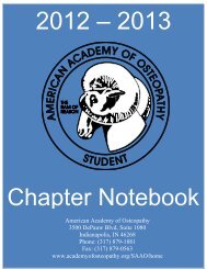
<strong>Working</strong> <strong>with</strong> <strong>children</strong> <strong>from</strong> a <strong>lymphatic</strong> <strong>and</strong> <strong>fluid</strong> <strong>motion</strong> <strong>perspective</strong>SEATED OR STANDING RIB BALANCEDLIGAMENTOUS TENSION1. The child is seated or st<strong>and</strong>ing <strong>with</strong> thephysician behind them.2. One of the physician’s h<strong>and</strong>s is placed on theanterior <strong>and</strong> posterior aspect of the rib(s) to betreated, noting that <strong>with</strong> young <strong>children</strong> that therib runs in a more horizontal plane than in older<strong>children</strong> <strong>and</strong> adults. The other h<strong>and</strong> stabilizes thevertebra attached to the rib being treated.3. Using gentle pressure, the rib <strong>and</strong> itssurrounding tissue is engaged by using a pincergrasp. Then the physician brings the area intobalance by bringing the entire rib up or down,internal or external rotation, <strong>and</strong> inferior orsuperior inclination.4. Once balanced tension is achieved, the physician maintains that position until a correction of the mechanicalstrain or improvement in the tissue <strong>motion</strong> is noted.SEATED ABDOMINAL DIAPHRAGM RELEASE1. The child is seated <strong>with</strong> the physician behind them <strong>and</strong>supporting their back <strong>with</strong> their hip or leg.2. The physicians fingers anteriorly contact the inferiorborder of the rib cage <strong>and</strong> gently hook posteriorly <strong>and</strong>superiorly to engage the abdominal diaphragmatic fascia.Posteriorly (see bottom picture) the thumbs are engaging thethoracolumbar junction, including the 11-12 ribs <strong>and</strong> T12-L1.3. The child is gently encouraged to slump into the fingerswhile the entire diaphragm is brought into ease or bind,whichever feels more conducive to treatment, until thepassive breathing of the child is felt easily. This is balance.4. This position is held until a release of the mechanical strain orimprovement in tissue <strong>motion</strong> is noted.Heather Ferrill DO March 2013 Convocation Page 4
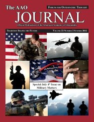
<strong>Working</strong> <strong>with</strong> <strong>children</strong> <strong>from</strong> a <strong>lymphatic</strong> <strong>and</strong> <strong>fluid</strong> <strong>motion</strong> <strong>perspective</strong>SEATED OR STANDING THORACIC INLET MYOFASCIAL RELEASE1. The child is seated or st<strong>and</strong>ing <strong>with</strong> thephysician behind them.2. The physician contacts the first <strong>and</strong>second ribs <strong>and</strong> possibly the manubriumanteriorly, <strong>and</strong> the costotransverse junctionof T1 posteriorly. The focus of treatment ison the fascial connections of the thoracicinlet.3. The area is engaged by gently liftingsuperiorly. Balance is sought by bringingthe area into ease or bind through engagingflexion/extension, sidebending <strong>and</strong>rotational barriers.4. When the breath is easily felt coming through the tissues, the position is held until a correction of themechanical strain occurs or improvement in tissue <strong>motion</strong> is noted.SEATED OR STANDING CLAVICLE BALANCEDLIGAMENTOUS TENSION1. The child is seated or st<strong>and</strong>ing <strong>with</strong> the physicianbehind them. The clavicle is contacted at both thesternoclavicular <strong>and</strong> acromioclavicular ends <strong>and</strong> thephysician evaluates passive <strong>motion</strong> <strong>with</strong> respiration.2. First to be addressed should be any interosseous strains.To do this, a gentle compression or distraction force alongthe long axis of the clavicle is applied. The force vectorhere can be changed to accommodate tissue response <strong>and</strong>torsional restrictions of the clavicle.3. After the interosseous balancing is achieved, then the clavicle is balanced in relationship to the supportingstructures <strong>and</strong> attachments such as the clavipectoral fascial, <strong>and</strong> the sternoclavicular <strong>and</strong> acromioclavicularligaments. Both of these are then balanced <strong>with</strong> the respiratory <strong>motion</strong> of the first rib.4. This balanced tension system is then maintained until correction of the mechanical strain or improvement inthe tissue <strong>motion</strong> is noted.Heather Ferrill DO March 2013 Convocation Page 5
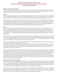
<strong>Working</strong> <strong>with</strong> <strong>children</strong> <strong>from</strong> a <strong>lymphatic</strong> <strong>and</strong> <strong>fluid</strong> <strong>motion</strong> <strong>perspective</strong>SEATED OR STANDING CERVICALFACILITATED POSTITIONAL RELEASE:Lower cervical, Occipito-Atlantal (OA) <strong>and</strong>Atlanto-Axial (AA) joints1. The child is seated <strong>with</strong> the physician to theside for best control of the head.2. One h<strong>and</strong> is used to monitor tissue response totreatment at the level of the dysfunctionalsegment(s). The other h<strong>and</strong> is placed on the head.3. The child’s head is gently placed in relativeflexion until the cervical spine is in a posturalneutral position.4. A gradual <strong>and</strong> gentle axial compression isapplied until there is a softening of the tissuesjust under the monitoring h<strong>and</strong>. Force used should be no more than 2.5kg.5. While maintaining the axial compression, the segment monitored is then brought into is position of ease, orinto the position of diagnosis. For example, if the diagnosis was C4FRSr, than C4 would be gently brought intoa flexion, sidebending <strong>and</strong> rotation to the right using the head as well as translational <strong>motion</strong> of the monitoringh<strong>and</strong>.6. This position is held for 3-5 seconds <strong>and</strong> then released <strong>and</strong> the area is re-assessed. This procedure can bereapplied as many times <strong>and</strong> the child allows.Heather Ferrill DO March 2013 Convocation Page 6
<strong>Working</strong> <strong>with</strong> <strong>children</strong> <strong>from</strong> a <strong>lymphatic</strong> <strong>and</strong> <strong>fluid</strong> <strong>motion</strong> <strong>perspective</strong>Some of my favorite resources:All techniques described above are adaptations of things I have learned <strong>from</strong> theseoutst<strong>and</strong>ing physicians though direct contact or through their writing.‣ Carreiro, J. Pediatric Manual Medicine, An Osteopathic Approach. Churchill, Livingstone,Elsevier 2009.‣ Carreiro, J. An Osteopathic Approach to Children, 2 nd ed. Chruchill Livingston Elsevier, 2009.‣ Frymann, V. The Collected Papers of Viola M. Frymann, DO. Legacy of Osteopathy to Children.American Academy of Osteopathy, 1998.‣ Sergueef, N. Cranial Osteopathy for Infants, Children <strong>and</strong> Adolescents: A Practical H<strong>and</strong>book.Churchill, Livingstone, 2007.‣ Arbuckle, B. The Selected Writings of Beryl Arbuckle DO. American Academy of Osteopathy.Heather Ferrill DO March 2013 Convocation Page 7