OPD-Scan II ARK-10000 - Amedeolucente.it
OPD-Scan II ARK-10000 - Amedeolucente.it
OPD-Scan II ARK-10000 - Amedeolucente.it
Create successful ePaper yourself
Turn your PDF publications into a flip-book with our unique Google optimized e-Paper software.
Averaged Corneal Pupil Power (APP)By utilizing the IOL-Station's Averaged Corneal Pupil Power (APP), calculation for selection of an IOL is nowsafer and more accurate.Post-Op Vision SimulationBy selecting an IOL, Post-Op vision is simulated on screen w<strong>it</strong>h chart.The IOL-Station's Post-OpVision Simulation is a useful tool for selection of an Aspheric IOL, as well as informedconsent.Flexible selection of IOL formulaPopular IOL formulas such as BINKHORST, HOFFER Q or HOLLADAY formula are available.Post LASIK calculation availableCamellin-Calossi IOL formula is applicable for a patient who underwent refractive surgery (RK, AR, CK, PRK,LASIK, PTK, LK, etc.). Also, Non-History method IOL formula is selectable for a patient w<strong>it</strong>h unknown history ofrefractive surgery.Formula Comparison ScreenUp to six results of IOL calculation w<strong>it</strong>h different formulas can be compared or be averaged w<strong>it</strong>h FormulaComparison Screen. For a patient w<strong>it</strong>h unknown history of refractive surgery, comparing calculation result w<strong>it</strong>hCamellin-Calossi formulas and Non-History method is beneficial.One-Click data importConnecting the IOL-Station PC w<strong>it</strong>h the <strong>OPD</strong>-<strong>Scan</strong> <strong>II</strong> Optical Path Difference <strong>Scan</strong>ning System and the US-4000 /US-500 Echoscan by network enables one-click import of such data like K-Value, Aberration, CornealTopography, Axial Length, Anterior Chamber Depth, Lens Thickness, Corneal Thickness.Accommodation Measurement FunctionAccommodation Measurement Function is available as anadd-on the to built-in function of the <strong>OPD</strong>-<strong>Scan</strong> <strong>II</strong>.The distribution of refractive power is measured on far visionand near vision w<strong>it</strong>h specific accommodation. The functionbenef<strong>it</strong>s in confirming correct function of accommodative IOL.Accommodation amount is variable w<strong>it</strong>hin the range of -10Dto 0.0D.far visionnear vision
Optional SoftwareCorneal NavigatorCorneal Navigator can be used as a integrated function of the<strong>OPD</strong>-Station or can be integrated separately into the <strong>OPD</strong>-<strong>Scan</strong> <strong>II</strong>.Utilizing various corneal parameters from topography, theCorneal Navigator automatically determines corneal featuresand shows by percentage the possibil<strong>it</strong>y of having a cond<strong>it</strong>ion of normal (NRM), astigmatic (AST), keratoconussuspects (KCS), keratoconus (KC), pellucid marginal degeneration (PMD), myopic refractive surgery (MRS),hyperopic refractive surgery (HRS), and penetrating keratoplasty (PKP).Instant analysis by the CornealNavigator helps improve the qual<strong>it</strong>y of examination / diagnosis.*The Corneal Navigator is developed in collaboration w<strong>it</strong>h Stephen D. Klyce, PhD & Michael K. Smolek, PhD.The <strong>OPD</strong>-Station Corneal / Refractive Analysis SoftwareThe <strong>OPD</strong>-Station software makes a variety of corneal / refractiveanalysis possible using advanced, unique and intelligentfunctions including the Holladay Summary and CornealNavigator (optional).The <strong>OPD</strong>-Station provides various maps such as the <strong>OPD</strong> HOMap, PSF, MTF, MTF Graph and Visual Acu<strong>it</strong>y Chart in add<strong>it</strong>ionto the <strong>OPD</strong>-<strong>Scan</strong> <strong>II</strong> maps. For wavefront maps such as the PSFand MTF, clinicians can select the target (<strong>OPD</strong>, Cornea, Internal)and also the type (Total, HO, Group) according to their needs.Holladay Summary*<strong>OPD</strong>-Station ScreenThe "Holladay Summary" shows the patient where the aberrations are located and how they affect thequal<strong>it</strong>y of vision using the Wavefront, MTF, PSF and VA-chart simulations.*Developed in cooperation w<strong>it</strong>h Jack T. Holladay, MD.PSF SimulationCalculates the Point Spread Function (PSF) based on the <strong>OPD</strong> data, and displays in simulation the distributionof the point spread. The Strehl Ratio serving as a metric of the visual qual<strong>it</strong>y of the eye is also displayed.Retinal Image SimulationCalculates the distortion of incoming light based on the results of PSF analysis, and displays the simulatedretinal image of the projected chart. This simulation can be used in explanations to patients for informedconsent.<strong>OPD</strong> HO MapDisplays in diopters the high order aberrations and shows the refractive errors which cannot be correctedw<strong>it</strong>h glasses.Averaging Multiple ExamsThe <strong>OPD</strong>-Station creates an exam data average from multiple exams. Noise components such as tear film andfixation dispar<strong>it</strong>y are excluded, providing more stable and reliable data.
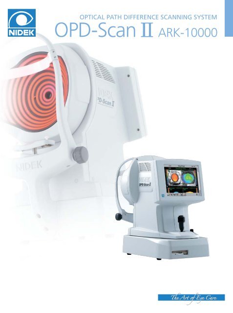
Features of the <strong>OPD</strong>-<strong>Scan</strong> <strong>II</strong>Measurement Selection forImproved Reliabil<strong>it</strong>yThe <strong>OPD</strong>-<strong>Scan</strong> <strong>II</strong> offers increased reliabil<strong>it</strong>y of examinationby automatically selecting the best measurement frommultiple measurements, allowing a more reliable clinicaldecisionFast Processing SpeedThe <strong>OPD</strong>-<strong>Scan</strong> <strong>II</strong> offers fast processing speed, minimizingstress in daily clinical use.Measurement Selection ScreenImproved Accessibil<strong>it</strong>y to a Patient EyeW<strong>it</strong>h the improved forehead rest, <strong>it</strong> is easier to reach andkeep the patient's eyelids open.Wide Measurement RangeHas the abil<strong>it</strong>y to measure high power Cylinder providingaccuracy in irregular aberration measurements.(Sphere -20.0 to +22.0D and Cylinder 0.0 to ±12.0D)Easy Data Maintenance w<strong>it</strong>h aDetachable Hard DrivePatient data is saved to a detachable Hard Drive, allowingquick and easy data transfer.Improved Accessibil<strong>it</strong>yNetwork Capabil<strong>it</strong>iesData from the <strong>OPD</strong>-<strong>Scan</strong> <strong>II</strong> may also be analyzed at a remote location using the <strong>OPD</strong>-Station."The <strong>OPD</strong>-<strong>Scan</strong> <strong>II</strong> is the only instrument that couples Wavefront, Topography and Refractioninto one un<strong>it</strong>. This allows the isolation of any optical problem to cornea or crystalline lensmaking <strong>it</strong> easy to decide if lensectomy or corneal surgery is the procedure of choice. It alsoprovides the best data for Customized Corneal Refractive Surgery."Jack T. Holladay, M.D., M.S.E.E., F.A.C.S."I see for the future the coupling of wavefront sensors w<strong>it</strong>h corneal topography devices for theoptimal correction of aberrations in a patient's eye. "Stephen D. Klyce, Ph.D.Professor of Ophthalmology and Anatomy, Louisiana State Univers<strong>it</strong>yRecipient of ASCRS 2000 Innovator's Award for contributions to the field of corneal topography and refractive surgery
<strong>OPD</strong>-<strong>Scan</strong> <strong>II</strong> (<strong>ARK</strong>-<strong>10000</strong>) SpecificationsPower mappingSpherical power rangeCylindrical powerAxisMeasuring areaMeasuring pointsMeasuring timeMeasuring methodMapping methodsCorneal topographyMeasuring ringsMeasuring areaDioptric rangeAxis rangeMeasuring pointsMapping methodsWorking distanceAuto trackingObservation areaOperating systemDisplayPrinterPower supplyPower consumptionDimensions / Weight- 20.00 to + 22.00 D0.00 to ± 12.00 D0 to 180˚2.0 to 6.0 mm diameter (4 zone measurement)1,440 points (4 x 360)< 0.4 secondsAutomated objective refraction(dynamic skiascopy)<strong>OPD</strong>, Internal <strong>OPD</strong>, Wavefront maps,Zernike graph, PSF19 vertical, 23 horizontal0.5 to 11.0 mm dia. (r=7.9)10 to 100 D0 to 359˚More than 6,800Axial, Instantaneous, "Refractive", Elevation75 mmX-Y-Z directions14 x 8 mmWindows XP embedded*10.4-inch color LCD touch panelBuilt-in thermal type line printer for data printExternal color printer (optional) for map print100 / 120, 220 / 240 Vac50 / 60 Hz170 VA290 (W) x 499 (D) x 520 (H) mm / 25 kg11.4 (W) x 19.6 (D) x 20.4 (H) " / 55 lbs.<strong>OPD</strong>-Station SpecificationsAnalysis and map displayCorneal topography<strong>OPD</strong>WavefrontOthersCorneal navigator (optional)PupillometryComputer requirementsCPUFree disk spaceMemoryGraphicLAN port (RJ-45)CD-ROM driveUSB portOSComputer requirementsCPUHard disk spaceMemoryGraphicMon<strong>it</strong>orLAN portUSB portOSAxial, Instantaneous, "Refractive",Elevation, Topoclassifier**W<strong>it</strong>h corneal navigator only<strong>OPD</strong>, <strong>OPD</strong> HO, Zonal refractionWavefront, Zernike graph, PSF,MTF, MTF graph, Visual acu<strong>it</strong>yInternal <strong>OPD</strong>, Target refractive, Differential,Eye image, Aspheric<strong>it</strong>y index (Q, e, S)8 kinds of corneal classification, StatisticsDiameters, Distances, Contours(photopic / mesopic cond<strong>it</strong>ion)Pentium <strong>II</strong>I 1200 MHz or higher30 GB or more256 MB or more (above 512 MB recommended)1024 x 768 pixels, 32 b<strong>it</strong> true color or moreWindows XP*IOL-Station SpecificationsPentium 4 1.3GHz or higher1GB or more512MB or more1024 x 768 pixels, 32 b<strong>it</strong> true color or moreCompatible w<strong>it</strong>h graphic mode as specified above1 or more1 or moreWindows XP SP2 32b<strong>it</strong> English** Windows is a trademark of Microsoft Corporation U.S.A.*Specifications and design are subject to change w<strong>it</strong>hout notice for improvement.HEAD OFFICE34-14 Maehama, HiroishiGamagori, Aichi 443-0038, JapanTelephone : 81-533-67-6611Facsimile : 81-533-67-6610URL : http://www.nidek.co.jpTOKYO OFFICE(International Div.)3F Sum<strong>it</strong>omo Fudosan Hongo Bldg.,3-22-5 Hongo, Bunkyo-ku, Tokyo,113-0033 JapanTelephone : 81-3-5844-2641Facsimile : 81-3-5844-2642URL : http://www.nidek.comNIDEK INC.47651 Westinghouse DriveFremont, CA 94539, U.S.A.Telephone : 1-510-226-5700: 1-800-223-9044 (US only)Facsimile : 1-510-226-5750URL : http://www.usa.nidek.comNIDEK SOCIETE ANONYMEEuroparc13, rue Auguste Perret94042 Creteil, FranceTelephone : 33-1-49 80 97 97Facsimile : 33-1-49 80 32 08URL : http://www.nidek.frNIDEK TECHNOLOGIES SRL.Via dell'Artigianato, 6 / A35020 Albignasego (Padova), ItalyTelephone : 39 049 8629200 / 8626399Facsimile : 39 049 8626824URL : http://www.nidektechnologies.<strong>it</strong>CNIDEK 2008 Printed in Japan <strong>OPD</strong>-SCAN <strong>II</strong> NQDNM6








![scarica questo file [PDF, 524 kB] - Gerlos - Altervista](https://img.yumpu.com/48083579/1/190x143/scarica-questo-file-pdf-524-kb-gerlos-altervista.jpg?quality=85)
