UROLITHIASIS IN A BUFFALO CALF - A CASE REPORT
UROLITHIASIS IN A BUFFALO CALF - A CASE REPORT
UROLITHIASIS IN A BUFFALO CALF - A CASE REPORT

You also want an ePaper? Increase the reach of your titles
YUMPU automatically turns print PDFs into web optimized ePapers that Google loves.
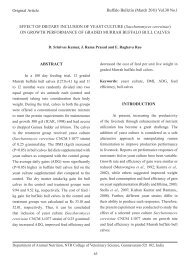
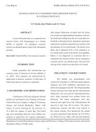
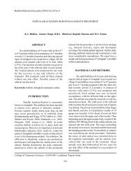
Case ReportBuffalo Bulletin (December 2011) Vol.30 No.4<strong>UROLITHIASIS</strong> <strong>IN</strong> A <strong>BUFFALO</strong> <strong>CALF</strong> - A <strong>CASE</strong> <strong>REPORT</strong>R.V. Suresh Kumar, P. Veena, P. Sankar, N. Dhana Lakshm and S. Kokila<strong>IN</strong>TRODUCTIONUrolithiasis is defined as the formation ofurolith anywhere in the urinary system. The diseaseis reported worldwide and occurs in all species ofthe animals but has most frequently been recordedin feeder steer and lambs (Radostitis et al., 2000).In India, urolithiasis has mostly been reported inbullocks, goat, sheep and buffaloes from differentcorners of the country (Tyagi and Singh, 1993).Sporadic cases of urolithiasis have also beenreported in buffalo and cow calves. Predisposingfactors like age, types of feed and water, season,castration, etc. have been identified as playingimportant roles in pathogenesis of disease. Amongbovines, buffalo calves had a significantly higheroccurrence of obstructive urolithiasis than cowcalves and buffaloes. The present paper describesa case of obstructive urolithiasis with cystorrhexsisin a buffalo calf and its surgical management.Keywords: urolithiasis, buffao calf, urinarysystem<strong>CASE</strong> HISTORY AND OBSERVATIONSA 7-month-old buffalo calf presented at theDepartment of Veterinary Surgery and Radiology,College of Veterinary Science, Tirupati, with ahistory of not having passed urine for the previous 15days and being treated by a local veterinarian. Therewas complete cessation of urination, abdominaldistension and unusual posture. The other clinicalsigns were kicking, posture to urinate, pumpingmotion of the tail. Ultrasonographic examinationof urinary bladder revealed presence of hypoechoicfluid in the abdomen with intestinal loops (Figure1) and also the presence of hyperechoic calculifloating in the fluid (Figure 2). Based on the history,clinical symptoms and ultrasonogram, the case wasdiagnosed as a case of cystic calculi.TREATMENT AND DISCUSSIONThe animal was prepared for asepticsurgery under local infiltration with 2% lignocaine.Laparotomy was done through left para medianincision. Urine present in the abdomen was siphonedout as much as possible without any damage to theany other abdominal organs. The urinary bladderwas identified and the tear in the urinary bladderwas located (Figure 3). Then the urinary bladderwas incised, and sandy, white colored calculi wereremoved. There was thickening of wall of theurinary bladder with severe congestion indicativeof a chronic case of cystic calculi (Figure 4). Theurinary bladder was flushed with normal saline.Normograde catherization of urethra was done toDepartment of Veterinary Surgery and Radiology, College of Veterinary Science, Sri Venkateswara VeterinaryUniversity, Tirupati-517 502 (A.P), India222
Buffalo Bulletin (December 2011) Vol.30 No.4Figure 1. Anechoic fluid in the abdomen with intestinal loops.Figure 2. Presence of hyperechoic calculi floating in the abdominal fluid.223
Buffalo Bulletin (December 2011) Vol.30 No.4Figure 3. Photograph showing thickened urinary bladder wall with leakage of urine.Figure 4. Photograph showing sand like cystic calculi with congested wall and petechial haemorrhage.224
Buffalo Bulletin (December 2011) Vol.30 No.4ensure the patency.The urinary bladder was sutured with adouble layer of inversion sutures using 3/0 chromiccatgut. The laparotomy wound was closed withsimple interrupted pattern using No.1 chromiccatgut and the skin incision was apposed withhorizontal mattress suture pattern using No. 1 silk.Post-operatively animal was given fluid therapyand antibiotic and anti inflammatory treatment for5 days. Animal recovered uneventfully on the 10 thpost operative day.Clinical signs associated with urolithiasisdepend upon the severity of blockage and thereaction of surrounding tissue (Van Saun, 1997).Complete blockage results in various stages ofstranguria, exaggerated and prolonged urinationposture, urine dribbling and hematuria. Affectedanimals may be depressed and lethargic, grindtheir teeth, and show abdominal distention andsigns of pain (Van Saun, 1977). Rupture of theurinary bladder is the most common sequel toobstructive urolithiasis especially in buffalocalves. While a discrete dorsal tear may sometimesheal spontaneously, ventral tear requires surgicalintervention (Tyagi and Singh, 1993).REFERENCESRadostitis, O.M., D.C. Blood, CC. Gay and K.W.Hinchcliff. 2000. Veterinary Medicine: A textBook of the Diseases of Cattle, Sheep, Pigs,Goats and Horses, 9 th ed. Bailliere Tiondal,London. 1877p.Tyagi, R.P.S. and Jit Singh. 1993. RuminantSurgery. CBS Publishers and Distributors,New Delhi. 484p.Van Saun, R.J. 2007. Urinary blockage in llamasand alpacas. Lamalink.com, 3(8): 30-31.225