CLINICAL REPORTBILATERAL INGUINAL HERNIA WITH DISTINCT HYSTEROCELEAND OMENTOCELE IN A DACHSHUND BITCH1 2 3John Martin K. D. , Susannah Bijee Philip , Sherin B. Sarangom4and Ashay P. KankonkarCollege of Veterinary and Animal Sciences, Mannuthy, Thrissur, Kerala 680651INTRODUCTIONInguinal hernias may be congenital or acquiredof which the former in dogs occur as a result of adefect in the inguinal ring through which theabdominal contents protrudes into the subcutaneousspace (Pratschke, 2002). Inguinal hernias may becongenital or acquired. The former are rare in dogsand may co-exist with the umbilical hernias (Fossum,2007), while the latter are often seen in middle agedintact bitches (Waters et al., 1993). The potentialfactors involved in the development of inguinalhernias might be anatomical, hormonal andmetabolic in nature. However, the exact etiopathogenesisis still unknown (Smeak, 2003).Polygenic inheritance of inguinal hernia had beendescribed in Cocker Spaniels and Dachshunds byRoberts (1986). The usual contents of inguinal herniamay include omentum, fat, ovary, uterus, smallintestine, colon, bladder and spleen, with omentumbeing the commonest. (Bellenger, 1996). Herniationof gravid uterus and pyometra uterus through theinguinal ring are also report (Munro and Stead, 1993;Byers, 2007). The present paper reports bilateralinguinal hernia in a dachshund bitch and its surgicalmanagement.CASE HISTORY AND OBSERVATIONSA five year old nulliparous dachshund bitch waspresented to the Kerala Veterinary and AnimalSciences University Hospital, Kokkalai with abilateral swelling in the inguinal region. The swellingwas noticed since one month, which increased in sizeover the last two weeks. The animal was in estrus onemonth back and had no previous history of any1 2,3&4Associate Professor, M.V.Sc. ScholarDepartment of Veterinary Surgery and Radiologytrauma. The animal was bright, active and alert. Allthe physiological parameters were within the normalrange. On palpation, the swellings were non-painful,soft and doughy in consistency. The left inguinalswelling was bigger in size compared to the rightone. The contents of both the swelling were nonreducible,even on application of moderate pressure.The bladder was catheterized, urine was relieved andthus the possibility of vesicocele was ruled out.Ultrasonographic examination of the left inguinalmass could revealed strands of hypoechoic regionwith areas of normal echogenicity and the rightinguinal mass with moderate echogenicity. Based onhistory, physical inspection and ultrasonographicexamination, the condition was diagnosed as anacquired bilateral inguinal hernia. The reduction ofthe hernial contents and herniorrhaphy under generalanaesthesia were resorted to.SURGICAL INTERVENTIONAND TREATMENTThe dog was premedicated with atropineasulphate at the rate of 0.045 mg/kg body weightbfollowed by xylazine hydrochloride at the rate of 1.5mg/kg body weight, both given intramuscularly. Thethe surgical site ware shaved, scrubbed and paintedwith Tr Iodine for aseptic surgery. Generalanaesthesia was induced with ketaminechydrochloride at the rate of 5 mg/kg body weightintramuscularly and was maintained by incrementalintravenous injection of a combination of xylazinehydrochloride and ketamine hydrochloride, equaldquantity by volume and diazepam given 'to effect'.The dog was positioned on dorsal recumbency. Thesite was painted with Tincture iodine and the patientJIVA Vol. 10 Issue 1 <strong>April</strong> <strong>2012</strong>45
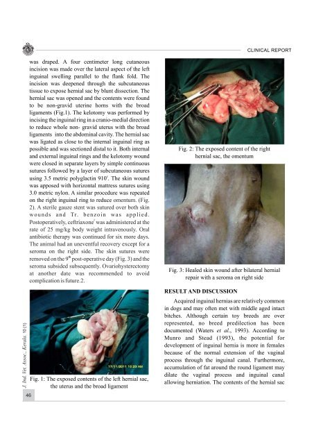
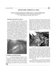
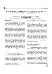
CLINICAL REPORTJ. Ind. Vet. Assoc., Kerala. 10 (1)was draped. A four centimeter long cutaneousincision was made over the lateral aspect of the leftinguinal swelling parallel to the flank fold. Theincision was deepened through the subcutaneoustissue to expose hernial sac by blunt dissection. Thehernial sac was opened and the contents were foundto be non-gravid uterine horns with the broadligaments (Fig.1). The kelotomy was performed byincising the inguinal ring in a cranio-medial directionto reduce whole non- gravid uterus with the broadligaments into the abdominal cavity. The hernial sacwas ligated as close to the internal inguinal ring aspossible and was sectioned distal to it. Both internaland external inguinal rings and the kelotomy woundwere closed in separate layers by simple continuoussutures followed by a layer of subcutaneous sutureseusing 3.5 metric polyglactin 910 . The skin woundwas apposed with horizontal mattress sutures using3.0 metric nylon. A similar procedure was repeatedon the right inguinal ring to reduce omentum. (Fig.2). A sterile gauze stent was sutured over both skinwounds and Tr. benzoin was applied.fPostoperatively, ceftriaxone was administered at therate of 25 mg/kg body weight intravenously. Oralantibiotic therapy was continued for six more days.The animal had an uneventful recovery except for aseroma on the right side. The skin sutures werethremoved on the 9 post-operative day (Fig. 3) and theseroma subsided subsequently. Ovariohysterectomyat another date was recommended to avoidcomplication is future.2.46Fig. 1: The exposed contents of the left hernial sac,the uterus and the broad ligamentFig. 2: The exposed content of the righthernial sac, the omentumFig. 3: Healed skin wound after bilateral hernialrepair with a seroma on right sideRESULT AND DISCUSSIONAcquired inguinal hernias are relatively commonin dogs and may often met with middle aged intactbitches. Although certain toy breeds are overrepresented, no breed predilection has beendocumented (Waters et al., 1993). According toMunro and Stead (1993), the potential fordevelopment of inguinal hernia is more in femalesbecause of the normal extension of the vaginalprocess through the inguinal canal. Furthermore,accumulation of fat around the round ligament maydilate the vaginal process and inguinal canalallowing herniation. The contents of the hernial sac