2012 Jiva April Cover 220 GSM Art Card Glossy ... - Jivaonline.net
2012 Jiva April Cover 220 GSM Art Card Glossy ... - Jivaonline.net
2012 Jiva April Cover 220 GSM Art Card Glossy ... - Jivaonline.net

Create successful ePaper yourself
Turn your PDF publications into a flip-book with our unique Google optimized e-Paper software.
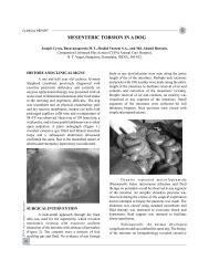
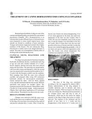
RESEARCH ARTICLEJ. Ind. Vet. Assoc., Kerala. 10 (1)Schmithz, 1977). So recently molecular techniqueslike polymerase chain reaction using 16S rRNA(LaFontaine et al., 1993; Zakaria et al., 1998,Waniet al., 2004; Moore et al., 2005) or fimA genesequences (Dhungyel et. al., 2002) for D. nodosusand lktA gene sequences (Zhou et. al., 2009;Hickford et. al., 2010) for F. necrophorum weretried and observed to yield rapid, confirmatory andsensitive diagnosis of these bacteria in clinicalsamples. The present study was conducted toscreen the samples from animals of Centre for PigProduction and Research, Mannuthy, having severefoot lesions and lameness.MATERIALS AND METHODSCollection of samplesSevere lameness and foot lesions werereported in 26 pigs of Centre for Pig Productionand Research, Mannuthy. Lesions werecharacterized by hot, painful, swollen limbs,separation of hard horny wall from the heel andabscessation. Animals were unable to stand or takefeed. Representative samples were taken from 10pigs using sterile cotton swabs (Hi-Media,Mumbai) from the deeper portion of the lesion andwere transported quickly to the laboratory.Extraction of bacterial DNAThe samples were processed to isolate theDNA of the pathogens by crude method. Thesamples were suspended in 200µl of sterilephosphate buffered saline (PBS), boiled for 10min,immediately chilled on ice for 5 min andcentrifuged at 15000 rpm for 10min. Thesupernatant was collected and stored in 1.5mlmicrocentrifuge tubes, which were later used astemplates for PCR reaction.PCR detection of D. nodosusD. nodosus was detected by PCRamplification of 16S rRNA using primers5'CGGGGTTATGTAGCTTGC3' (forward)and5'TCGGTACCGAGTATTTCTACCCAACACCT3' (reverse)(La Fontaine et al., 1993; Moore etal., 2005; Wani et. al., 2007). The PCRamplifications were performed in 25µl volumes.The final concentration contained 2µl of template,2.5µl of 10X Taq buffer (10mM Tris-HCl (pH 9.0),50mM KCl, 15mM MgCl 2), 25pM each of forwardand reverse primer, 200µM of eachdeoxyribonucleotide triphosphate and 1IUTaqDNA polymerase. The amplification wascarried out in a thermal cycler (Eppendorf) with aninitial denaturing step of 94°C for 10min, followedby 35 cycles of 94°C for 1min, 58°C for 30s and72°C for 30s, with a final extension step at 72°Cfor 5min.PCR detection of F. necrophorumF. necrophorum was detected by PCRamplification of leukotoxin (lktA) gene usingprimers 5'AATCGGAGTAGTAGGTTCTG-3'(Forward) and 5'CTTTGGTAACTG CCACTGC3'(Reverse) (Zhou et al., 2009). The PCRamplifications were performed in 25µl volumes.The final concentration contained 2µl of templateDNA, 2.5µl of 10X Taq buffer (10mM Tris-HCl(pH 9.0), 50mM KCl, 15mM MgCl 2), 25pM of eachprimer,200µM of each deoxyribonucleotidetriphosphate and 1IU TaqDNA polymerase.Thermal profile for amplification of lktA geneconsisted of denaturation at 94°C for 2minfollowed by 35 cycles of 94°C for 30s, 58°C for 40sand 72°C for 30s, with a final extension step at72°C for 10min.Analysis of PCR productsThe PCR products were subjected toelectrophoresis in 1 percent agarose (Genei,Bangalore) gels, using 1x Tris Borate EDTA buffer,containing 200ng/ml of ethidium bromide. Thegels were visualized under ultraviolet illuminationand photographed using gel- documentationsystem.RESULTS AND DISCUSSIONOut of the ten hoof swabs examined, a total14