Diagnosis and Management of Hemochromatosis - AASLD
Diagnosis and Management of Hemochromatosis - AASLD
Diagnosis and Management of Hemochromatosis - AASLD

Create successful ePaper yourself
Turn your PDF publications into a flip-book with our unique Google optimized e-Paper software.
<strong>AASLD</strong> Practice Guidelines<strong>Diagnosis</strong> <strong>and</strong> <strong>Management</strong> <strong>of</strong> <strong>Hemochromatosis</strong>ANTHONY S. TAVILLPREAMBLEPractice guidelines, intended for use by physicians, suggestpreferable approaches to the diagnostic, therapeutic, <strong>and</strong> preventativeaspects <strong>of</strong> care. These guidelines are intended to beflexible, in contrast with “st<strong>and</strong>ards <strong>of</strong> care,” which are inflexiblepolicies to be followed in almost every case. 1 They aredeveloped in a manner consistent with the American GastroenterologicalAssociations’ Policy Statement on Development<strong>and</strong> Use <strong>of</strong> Practice Guidelines. 2Specific recommendations are based on relevant publishedinformation. In an attempt to st<strong>and</strong>ardize recommendations,the Practice Guidelines Committee <strong>of</strong> the American Associationfor the Study <strong>of</strong> Liver Diseases modified the categories <strong>of</strong>the Infectious Diseases Society <strong>of</strong> America’s Quality St<strong>and</strong>ards.3 These categories are reported with each recommendation,using the letters A through E to determine the strength <strong>of</strong>recommendation (Table 1) <strong>and</strong> Roman numerals I through IVto determine quality <strong>of</strong> evidence upon which recommendationsare based (Table 2).These guidelines provide data-supported peer-reviewedrecommendations for the care <strong>of</strong> patients with hemochromatosis.They are based on the following: (1) a formal review <strong>and</strong>analysis <strong>of</strong> the recent published literature on hemochromatosis(Medline Search from 1990-2000); (2) the American College<strong>of</strong> Physicians’ Manual for Assessing Health Practices <strong>and</strong>Designing Practice Guidelines; (3) several published guidelines,including the American Association for the Study <strong>of</strong>Liver Diseases’ Policy Statement on Development <strong>and</strong> Use <strong>of</strong> PracticeGuidelines <strong>and</strong> the American Gastroenterological Association’sPolicy Statement on Guidelines 2 ; <strong>and</strong> (4) the experience<strong>of</strong> the author in the clinical care <strong>of</strong> patients with hemochromatosis.Abbreviations: HH, hereditary hemochromatosis; HIC, hepatic iron concentration;HII, hepatic iron index; HCC, hepatocellular carcinoma; TIBC, total iron-binding capacity;UIBC, unsaturated iron-binding capacity.From the MetroHealth Medical Center, <strong>and</strong> Case Western Reserve University, Clevel<strong>and</strong>,OH.Received February 2, 2001; accepted March 16, 2001.This Guideline has been commissioned <strong>and</strong> approved by the American Association forthe Study <strong>of</strong> Liver Diseases <strong>and</strong> has received the endorsement <strong>of</strong> the American College <strong>of</strong>Gastroenterology <strong>and</strong> the American Gastroenterological Association.Address correspondence to: Anthony S. Tavill, M.D., Gastroenterology Division,MetroHealth Medical Center, 2500 MetroHealth Dr., Clevel<strong>and</strong>, OH 44109. E-mail:ast2@po.cwru.edu; fax: 216-778-4873.Individual copies <strong>of</strong> these guidelines can be obtained from the <strong>AASLD</strong> website atwww.aasld.org. For multiple reprints (100 copies or more) contact W.B. Saunders Company,The Curtis Center, Independence Square West, Philadelphia, PA 19106-3399, <strong>and</strong>obtain permission from Anthony S. Tavill.Copyright © 2001 by the American Association for the Study <strong>of</strong> Liver Diseases.0270-9139/01/3305-0038$35.00/0doi:10.1053/jhep.2001.247831321BACKGROUNDHereditary hemochromatosis (HH) is the most common,identified, genetic disorder in the Caucasian population. Althoughits geographic distribution is worldwide, it is concentratedin individuals <strong>of</strong> northern European origin, particularly<strong>of</strong> Nordic or Celtic ancestry, in whom it occurs with a prevalenceclose to 1 per 200 <strong>of</strong> the population. 4-6 The pathophysiologicpredisposition to increased <strong>and</strong> inappropriate absorption<strong>of</strong> dietary iron may lead to the progressive development<strong>of</strong> life-threatening complications <strong>of</strong> cirrhosis, hepatocellularcancer, diabetes, <strong>and</strong> heart disease. The gene defect describedin 1996 7 isaGtoAmissense mutation (C282Y) leading to thesubstitution <strong>of</strong> tyrosine for cysteine at the 282 amino acidposition <strong>of</strong> the protein product <strong>of</strong> the newly discovered HFEgene located on the short arm <strong>of</strong> chromosome 6 (6p). Anothermutation (H63D) in which aspartic acid is substituted forhistidine at position 63 has also been associated as a c<strong>of</strong>actorin some cases <strong>of</strong> hemochromatosis. The homozygous state inwhich both alleles <strong>of</strong> chromosome 6 possess the C282Y mutationor the compound heterozygous state with C282Y onone chromosome <strong>and</strong> H63D on the other, are the predominantgenetic abnormalities associated with phenotypic HH. Inmost studies to date, C282Y/C282Y homozygosity has beenfound in more than 90% <strong>of</strong> patients with hemochromatosis,while compound heterozygosity (C282Y/H63D) accounts for3% to 5% <strong>of</strong> such cases in published series. Possession <strong>of</strong> theC282Y mutation on both alleles <strong>of</strong> the chromosome pair has ahigh positive predictive accuracy for phenotypic HH. In theonly large population study published to date in which penetrance<strong>of</strong> the HFE gene mutation has been studied comprehensively,all C282Y homozygotes had elevated transferrinsaturation (100% positive predictive accuracy). However, fullexpression as defined by progressive tissue iron overload occurredin only 58% <strong>of</strong> these homozygotes. 6Although the vast majority <strong>of</strong> familial cases <strong>of</strong> hemochromatosisin the Anglo-Celtic population are associated with thedescribed pathogenic mutations <strong>of</strong> the HFE gene, it is highlyprobable that genes other than HFE play a role in familial ironoverload in other populations. In particular, there are welldocumented families in Italy with iron overload comparablewith HFE-related hemochromatosis, 8-10 in whom neither theC282Y nor H63D mutation existed, <strong>and</strong> in whom the geneticabnormality could not be located to chromosome 6p.The clinical condition <strong>of</strong> hereditary hemochromatosisevolves in a series <strong>of</strong> stages beginning with clinically insignificantiron accumulation (0-20 years <strong>of</strong> age, 0-5 g parenchymaliron storage). This evolves to a stage <strong>of</strong> iron overload withoutdisease (approximately 20-40 years <strong>of</strong> age, 10-20 g parenchymaliron storage), which if left untreated, may progress to astage <strong>of</strong> iron overload with organ damage (usually more than40 years <strong>of</strong> age <strong>and</strong> 20 g parenchymal iron storage). 11,12Ideally, any strategy for diagnosis should identify cases before
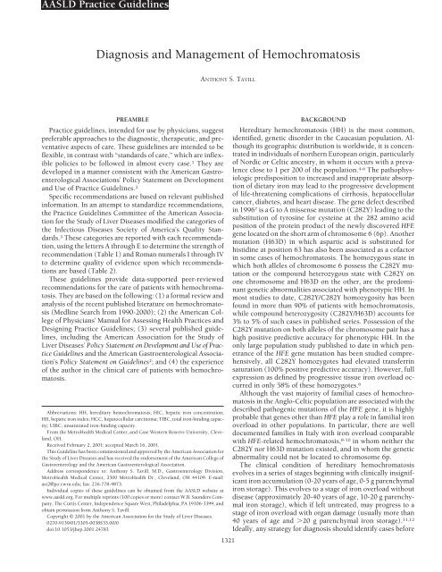
1322 TAVILL HEPATOLOGY May 2001TABLE 1. Categories Reflecting the Evidence to Support the Use <strong>of</strong> aGuideline RecommendationCategoryABCDEAdapted <strong>and</strong> modified from Gross et al. 3DefinitionSurvival benefitImproved diagnosisImprovement in quality <strong>of</strong> lifeRelevant pathophysiologic parameters improvedImpacts cost <strong>of</strong> health carethe third stage <strong>of</strong> disease has developed so that therapy toremove iron can prevent progression to irreversible tissuedamage. Fortunately, biochemical serum testing with indirectiron markers is capable <strong>of</strong> identifying most cases <strong>of</strong> iron overloadwell before tissue damage has become irreversible.Therefore, these guidelines will emphasize the fundamentalobjective <strong>of</strong> detection <strong>of</strong> HH before organ damage has occurred(Table 3).Current clinical practice in diagnosis <strong>and</strong> management <strong>of</strong>HH has evolved from experience in screening healthy blooddonors or selected populations <strong>and</strong> managing patients <strong>and</strong>their discovered relatives with phenotypic HH. 6,12-14 Early institution<strong>of</strong> phlebotomy has proven to be a highly effectivetherapy for HH, which prevents morbidity <strong>and</strong> promotes normallongevity. 15 As a result, r<strong>and</strong>omized, controlled trials <strong>of</strong>other therapies or observation have been regarded as unethical<strong>and</strong> have not been done.The development <strong>of</strong> liver injury in those with HH is relatedto the progressive accumulation <strong>of</strong> hepatic iron. 15-18 Hepaticiron concentration increases with age in most homozygotes.In HH patients over the age <strong>of</strong> 40 years, hepatic iron concentrationis likely to exceed 10,000 g/g dry weight <strong>and</strong> liverbiopsy results are more likely to show fibrosis or cirrhosis. 16-18It was the observation that the hepatic iron concentration(HIC) increased with age that led to the concept <strong>of</strong> hepaticiron index (HIC in micromoles per gram dry weight dividedby age in years). A hepatic iron index (HII) in excess <strong>of</strong> 1.9mol/g per year <strong>of</strong> life was found to effectively distinguishhomozygous hemochromatosis from heterozygotes <strong>and</strong> patientswith alcohol-induced liver disease. However, it is nowclear that the rate <strong>of</strong> iron accumulation is variable <strong>and</strong> exceptionsmay occur in between 8% <strong>and</strong> 50% <strong>of</strong> individuals withHH. 6,12,14,19 Therefore, while an HII less than 1.9 does notentirely exclude HH, a value greater than this certainly documentssignificant iron overload in the C282Y homozygote <strong>and</strong>in individuals with certain forms <strong>of</strong> secondary iron overload.TABLE 2. Quality <strong>of</strong> Evidence on Which Recommendation Is BasedGradeDefinitionIIIIIIIVEvidence from multiple well-designed r<strong>and</strong>omized controlledtrials each involving a number <strong>of</strong> participants to be <strong>of</strong>sufficient statistical powerEvidence from at least one large well-designed clinical trialwith or without r<strong>and</strong>omization, from cohort or case-controlanalytic studies, or well-designed meta-analysisEvidence based on clinical experience, descriptive studies, orreports <strong>of</strong> expert committeesNot ratedAdapted <strong>and</strong> modified from Gross et al. 3TABLE 3. <strong>Management</strong> Objectives for HH<strong>Management</strong> ObjectivesEarly diagnosis to prevent organ damage <strong>and</strong> dysfunction due to tissue irontoxicityScreening <strong>and</strong> early detection <strong>of</strong> asymptomatic HH cases to reducemortalityRecognition <strong>and</strong> diagnosis <strong>of</strong> symptomatic cases <strong>of</strong> HH, to minimizeprogression <strong>and</strong> complications <strong>of</strong> the diseaseAdequate treatment <strong>of</strong> HH to promote rapid, safe, <strong>and</strong> effective removal <strong>of</strong>ironVigilant follow-up <strong>and</strong> maintenance treatment <strong>of</strong> all cases <strong>of</strong> HHThe degree <strong>of</strong> iron overload has a direct impact on lifeexpectancy <strong>of</strong> the individual with HH. The major causes <strong>of</strong>death are decompensated cirrhosis, hepatocellular carcinoma(HCC), diabetes mellitus, <strong>and</strong> cardiomyopathy. 15 These occurredwith a frequency 10- to 119-fold higher than expectedin an age- <strong>and</strong> sex-matched population without HH. Survivalwas normal in HH patients in whom treatment was initiatedbefore the development <strong>of</strong> cirrhosis or diabetes, confirmingthe importance <strong>of</strong> early diagnosis <strong>and</strong> treatment.DIAGNOSIS OF HEREDITARY HEMOCHROMATOSISTarget PopulationsTarget populations are shown in Table 4. The diagnosis <strong>of</strong>hemochromatosis is based on documentation <strong>of</strong> increasediron stores, namely increased hepatic iron concentrations associatedwith elevated serum ferritin levels. HH can be furtherdefined genotypically by the familial occurrence <strong>of</strong> iron overloadassociated with C282Y homozygosity or C282Y/H63Dcompound heterozygosity. 20 As serologic iron markers havebecome more widely available over the last several years, themajority <strong>of</strong> patients with HH are now identified while stillasymptomatic <strong>and</strong> without evidence <strong>of</strong> hepatic fibrosis or cirrhosis.15 Diagnostic screening strategies should target highriskgroups such as those with suspicious organ involvement,a familial history <strong>of</strong> HH, <strong>and</strong> those with chance detection <strong>of</strong>biochemical or radiologic abnormalities suggestive <strong>of</strong> the possibility<strong>of</strong> iron overload.TABLE 4. Target Populations for Screening for HHTarget Populations for <strong>Hemochromatosis</strong> EvaluationSymptomatic patientsUnexplained manifestations <strong>of</strong> liver disease or a presumably knowncause <strong>of</strong> liver disease with abnormality <strong>of</strong> one or more indirect serumiron markersType 2 diabetes mellitus, particularly with hepatomegaly, elevated liverenzymes, atypical cardiac disease or early-onset sexual dysfunctionEarly-onset atypical arthropathy, cardiac disease, <strong>and</strong> male sexualdysfunctionAsymptomatic patientsPriority groupsFirst-degree relatives <strong>of</strong> a confirmed case <strong>of</strong> hemochromatosisIndividuals with abnormal serum iron markers discovered duringroutine testingIndividuals with unexplained elevation <strong>of</strong> liver enzymes or theserendipitous finding <strong>of</strong> asymptomatic hepatomegaly or radiologicdetection <strong>of</strong> enhanced computed tomography attenuation <strong>of</strong> theliverGeneral populationSee Fig. 1.
HEPATOLOGY Vol. 33, No. 5, 2001 TAVILL 1323FIG. 1.Proposed algorithm for management <strong>of</strong> HH.Evidence is accumulating to support the cost effectiveness <strong>of</strong>serologic strategies for screening the general population for ironoverload. 13,21-25 Most <strong>of</strong> these reports have only assessed the usefulness<strong>of</strong> st<strong>and</strong>ard serologic tests such as serum iron, transferrinsaturation, or serum ferritin; only one study has included the recentlydiscovered HFE gene mutation. 25 This latter study comparedscreening <strong>of</strong> blood donors by phenotypic or genotypic methods.It was concluded that the most cost-effective strategy foridentifying cases in the general population was phenotypic screening(st<strong>and</strong>ard iron markers) with genotypic confirmation <strong>of</strong> homozygosityin those with indirect markers <strong>of</strong> iron overload($2,700 per case). This strategy had a high predictive value for thedetection <strong>of</strong> homozygotes with iron overload <strong>and</strong> remained costeffective even when it was assumed that as few as 20% <strong>of</strong> caseswould ever develop life-threatening complications <strong>of</strong> the disease.25,26 In contrast, genotypic screening (by mutation analysis)<strong>of</strong> the general population would be prohibitively expensive($110,000 to detect one case) <strong>and</strong> the strategy would have specificitylimitations in the light <strong>of</strong> accumulating evidence for the incompletepenetrance <strong>of</strong> the gene mutations. 6 These limitationsmay become less important as newer <strong>and</strong> less expensive techniques<strong>of</strong> mutation analysis are developed. At this time we aresupportive <strong>of</strong> a low-cost phenotypic approach for screening thegeneral population.Data on sensitivity, specificity, <strong>and</strong> predictive value <strong>of</strong> phenotypicscreening tests have been provided by studies both inasymptomatic populations (e.g., healthy blood donors <strong>and</strong>large-scale screening <strong>of</strong> a healthy population) <strong>and</strong> in families<strong>of</strong> detected homozygotes. 6,27-29 More recent studies are availablefor sensitivity <strong>and</strong> specificity <strong>of</strong> genotyping studies. 6,12,20The following diagnostic algorithm proceeds in 3 steps, beginningwith phenotypic evaluation followed by genotyping <strong>of</strong> thosewith elevated iron markers (Fig. 1). The proposed algorithm isconstructed to detect iron overload caused by HH with a highdegree <strong>of</strong> accuracy, while providing a pathway for those cases <strong>of</strong>iron overload unassociated with the HFE mutation.ALGORITHM STEP 1Indirect Serologic Markers <strong>of</strong> Iron StoresThe initial approach to diagnosis <strong>of</strong> HH is by indirect serologicmarkers <strong>of</strong> iron stores. Transferrin saturation (TS) isderived by dividing the serum iron by the total iron bindingcapacity. When the fasting value exceeds 50% for women <strong>and</strong>60% for men, TS has a sensitivity <strong>of</strong> 0.92, a specificity <strong>of</strong> 0.93,<strong>and</strong> a positive predictive value <strong>of</strong> 86% for the diagnosis <strong>of</strong>HH. 27-29 Overnight fasting avoids circadian or postpr<strong>and</strong>ialvariations <strong>and</strong> eliminates 80% <strong>of</strong> false-positive TS results. 27Lowering the cut<strong>of</strong>f TS value to 45% increases sensitivity, butreduces specificity <strong>and</strong> positive predictive value. In a recentstudy, values exceeding 45% correctly identified 97.9% <strong>of</strong> homozygoteswith no false positives among the normal population.30 However, this cut<strong>of</strong>f did include 22.2% <strong>of</strong> the heterozygotepopulation, a group recognized to occasionally havephenotypic markers <strong>of</strong> iron overload. In another report, thiscut<strong>of</strong>f was 100% sensitive for the detection <strong>of</strong> C282Y homozygotes;however, only 44% <strong>of</strong> those with TS more than 45%were genetic homozygotes. 6 Thus, lowering the threshold forTS to 45% will also identify other groups with relatively minordegrees <strong>of</strong> secondary iron overload (e.g., alcohol-inducedliver disease, steatohepatitis, chronic hepatitis C, previoussurgical portacaval shunt, etc.) <strong>and</strong> these cases will requirefurther evaluation by the clinician.In most clinical laboratories TS was customarily measuredby determining two serum iron measurements; the first a measurement<strong>of</strong> the subjects’ fasting serum iron, the second arepeat measurement after adding exogenous iron to saturatethe serum transferrin followed by removal <strong>of</strong> the nontransferrin-boundiron. The latter determines the total iron bindingcapacity (TIBC). The ratio serum iron to TIBC gives the transferrinsaturation (TS in percent). Although the serum iron isan automated test, the TIBC is not, making the traditionalmethod for deriving TS relatively expensive. Alternatively, ithas been proposed that costs could be reduced by using unsaturatediron binding capacity (UIBC). 31 Values for UIBCless than 28 mol/L are indicative <strong>of</strong> iron overload. In fact,many laboratories now determine TIBC by summing serumiron <strong>and</strong> UIBC (both automated methods). TS is then expressedas the ratio <strong>of</strong> serum iron to the calculated TIBC:Fe/(Fe UIBC). This allows the clinician to judge the significance<strong>of</strong> a raised TS, by noting those that might be spuriouslyelevated by a low TIBC (low serum transferrin concentration).It is recommended that TS be calculated in this way to reducecosts, particularly for large-scale screening.Other indirect markers <strong>of</strong> iron stores such as serum iron orferritin lack specificity when used alone. The serum iron haspositive <strong>and</strong> negative predictive values for HH <strong>of</strong> 61% <strong>and</strong>87%, respectively, compared with 74% <strong>and</strong> 93% for TS. 29 Serumferritin is also nonspecific particularly in the face <strong>of</strong> inflammatoryconditions, chronic hepatitis C, alcohol-inducedliver disease, <strong>and</strong> neoplastic diseases. However, a serum ferritinlevel in combination with TS has a negative predictivevalue <strong>of</strong> 97% <strong>and</strong> exceeds the accuracy <strong>of</strong> any <strong>of</strong> the indirecttests used in isolation. 29 In confirmed HH, a level <strong>of</strong> serumferritin 1,000 ng/mL is an accurate predictor <strong>of</strong> the degree <strong>of</strong>hepatic fibrosis (cirrhosis). 32Recommendation 1. Initial screening <strong>of</strong> individuals with suspectediron overload <strong>and</strong> those over the age <strong>of</strong> 20 years whoare first-degree relatives <strong>of</strong> known cases <strong>of</strong> HH should be doneby measurement <strong>of</strong> transferrin saturation after an overnightfast. Simultaneous serum ferritin determination increases thepredictive accuracy for diagnosis <strong>of</strong> iron overload. TS is alsothe test <strong>of</strong> choice for screening the general adult populationfor iron overload states (Fig. 1) (rating: II A, B, C, D, <strong>and</strong> E).
1324 TAVILL HEPATOLOGY May 2001ALGORITHM STEP 2Genotypic Testing: Mutation AnalysisFasting transferrin saturation less than 45% <strong>and</strong> a normalserum ferritin would require no further evaluation. Elevation<strong>of</strong> TS <strong>and</strong> serum ferritin would require genotypic testing asindicated in step 2 <strong>of</strong> the diagnostic algorithm in Fig. 1. Thepresence <strong>of</strong> the HFE mutations C282Y <strong>and</strong> H63D can now bedetected by polymerase chain reaction using whole bloodsamples. 7 Individuals with serum indicators <strong>of</strong> iron overloadwho are homozygous for the C282Y mutation require phlebotomytherapy. Those who are unlikely to have significanthepatic injury may be <strong>of</strong>fered therapeutic phlebotomy withoutthe necessity for a liver biopsy. This includes individualsless than 40 years <strong>of</strong> age who have no clinical evidence <strong>of</strong> liverdisease (raised alanine transaminase, hepatomegaly, etc.) <strong>and</strong>whose serum ferritin is less than 1,000 ng/mL. Higher values<strong>of</strong> serum ferritin are associated with an increased likelihood <strong>of</strong>significant hepatic fibrosis or cirrhosis. 20,32 On the otherh<strong>and</strong>, liver biopsy should be <strong>of</strong>fered to document the degree<strong>of</strong> fibrosis in all homozygotes who are over the age <strong>of</strong> 40 yearsor those who have an elevated serum alanine transaminaselevel, have clinical evidence <strong>of</strong> liver disease, or have a serumferritin greater than 1,000 ng/mL. Since these are likely to beindividuals over the age <strong>of</strong> 40 years, discretion is appropriatein recommending liver biopsy on the basis <strong>of</strong> age alone. In theabsence <strong>of</strong> the above indicators <strong>of</strong> cirrhosis, other risk factors(e.g., alcohol abuse, or coexisting clinical features <strong>of</strong> HH, suchas diabetes, impotence, etc.) may play a role in making thisrecommendation. Liver biopsy <strong>and</strong> hepatic iron evaluationare also recommended in compound heterozygotes (C282Y/H63D), C282Y heterozygotes, or non-HFE mutated individualswho have indirect markers <strong>of</strong> iron overload, particularly ifthey also have abnormal liver enzymes or clinical evidence <strong>of</strong>liver disease. Although these individuals account for a smallproportion <strong>of</strong> phenotypic hemochromatosis, they have a lowlikelihood <strong>of</strong> significant iron overload, <strong>and</strong> elevated iron testsare <strong>of</strong>ten due to other causes <strong>of</strong> liver disease. 33Although recognizing that the penetrance <strong>of</strong> the C282Ymutation is variable, the option is provided in step 1 <strong>of</strong> thediagnostic algorithm to proceed to gene mutation analysisregardless <strong>of</strong> the TS or serum ferritin in first-degree relatives<strong>of</strong> a known HH individual. In the case <strong>of</strong> the children <strong>of</strong> an HHpatient, mutation analysis in the spouse allows for assessment<strong>of</strong> the genotypic status <strong>of</strong> the children. 14 If the spouse possessesno C282Y mutation, the <strong>of</strong>fspring can only be heterozygous.An HH patient with a spouse who is a heterozygote forC282Y has a 50% chance <strong>of</strong> having homozygous <strong>of</strong>fspring.Because organ damage is virtually unknown in HH beforeadult life, evaluation <strong>of</strong> first-degree relatives can be postponeduntil about 20 years <strong>of</strong> age.Recommendation 2. Genotyping to detect HFE mutationsshould be performed for all individuals who have abnormaliron studies <strong>and</strong> on those who are first-degree relatives <strong>of</strong>identified homozygotes as detailed in step 1 <strong>of</strong> the diagnosticalgorithm. In the absence <strong>of</strong> indicators suggestive <strong>of</strong> significantliver disease, C282Y homozygotes under the age <strong>of</strong> 40years may be treated by therapeutic phlebotomy without theneed for liver biopsy. Liver biopsy is recommended in allhomozygotes with clinical evidence <strong>of</strong> liver disease, serumferritin greater than 1,000 ng/mL, <strong>and</strong> particularly in thosegreater than 40 years <strong>of</strong> age with other risk factors for liverdisease. Liver biopsy should also be considered in compoundor C282Y heterozygotes with elevated TS, particularly thosewho have had abnormal liver enzyme levels or clinical evidence<strong>of</strong> liver disease (rating: II A, B, C, D, <strong>and</strong> E).ALGORITHM STEP 3Liver Biopsy for HICLiver biopsy is useful to document the presence <strong>of</strong> cirrhosis(if not evident from radiologic studies) to rule out significantiron overload when iron markers are equivocal, or to investigateother possible causes <strong>of</strong> liver disease. Histopathology <strong>and</strong>staging <strong>of</strong> fibrosis is best determined with hematoxylin-eosin<strong>and</strong> Masson trichrome staining, respectively. The liver is themost easily accessible tissue for accurately assessing ironstores. The degree <strong>and</strong> cellular distribution <strong>of</strong> iron stores isbest assessed using a Perls’ Prussian blue stain. Before 1985,the extent <strong>of</strong> iron deposition was judged exclusively by thismethod <strong>and</strong>, in fact, a qualitative assessment <strong>of</strong> iron stores wasderived based on stainable iron. 34 Two qualitative scales havebeen proposed. 35,36 The most commonly used <strong>of</strong> these, theLudwig-Batts system, estimates the proportion <strong>of</strong> hepatocytesthat stain for iron, recognizing the progressive nature <strong>of</strong> ironaccretion through the hepatic acinus from Rappaport zone 1(periportal) to zone 3 (pericentral). 36 Although grade 4 irondeposition (panacinar) usually indicates HH range quantitativeiron levels, grades 2 <strong>and</strong> 3 correlate poorly with quantitativeiron content. For this reason the quantitative, biochemicalHIC has become the preferred method for evaluating thehepatic iron stores. Quantitative iron determinations fromfresh frozen <strong>and</strong> formalin-fixed, paraffin-embedded samplesare comparable. 36,37 Accordingly, a biopsy core at least 2.5 to3.0 cm in total length should be obtained. A 0.5- to 1.0-cmpiece <strong>of</strong> the tissue core should be removed <strong>and</strong> placed in a drytube or in 10% formalin (not in saline, which may leach outiron). The remainder <strong>of</strong> the fixed tissue is processed for routinehistopathologic evaluation <strong>and</strong> a Perls’ Prussian bluestain. If tissue was not separated <strong>and</strong> saved before fixation <strong>and</strong>embedding, the remaining tissue can be removed from theparaffin block <strong>and</strong> sent for quantitative iron.The normal HIC is less than 1,800 g/g dry weight (equivalentto 32 mol/g). It is now clear from several studies thatmost patients with homozygous HH steadily <strong>and</strong> inexorablyaccumulate iron at least through early adult <strong>and</strong> middle life,unless they have had blood loss or have been blood donors, incontrast to patients with secondary iron overload caused byother chronic liver diseases. The concept <strong>of</strong> the HII as a measure<strong>of</strong> the iron accretion rate was developed to distinguishHH from these other potentially confounding clinical situations,particularly alcohol-induced liver disease. 16-18 A rate inexcess <strong>of</strong> 1.9 mol/g/y is strong evidence for homozygoushemochromatosis. However, it has recently been shown thatup to 15% <strong>of</strong> genotypic homozygotes for HH do not meet thepreviously defined rate <strong>of</strong> at least 1.9 mol/g/y. Thus, an elevatedHII is no longer considered essential for diagnosis. 20Yet, even these individuals with partial expression <strong>of</strong> homozygousHH have HIC at least 3 times the upper limit <strong>of</strong> normal ifthey are more than 20 years old. Finally, it should be emphasizedthat secondary iron overload due to dyserythropoietic orhemolytic anemia may have HIC comparable with that seen inHH, particularly in those who require repeated blood transfusions.These causes should be easily distinguishable by otherclinical criteria.
HEPATOLOGY Vol. 33, No. 5, 2001 TAVILL 1325Although the rate <strong>of</strong> hepatic iron accumulation (HII) haslost some <strong>of</strong> its importance in the diagnosis <strong>of</strong> HH, it is neverthelessthe correlation between HIC <strong>and</strong> age that determinesthe age at which fibrosis will develop. Sallie et al. 18 found nopatient who developed hepatic fibrosis before the HIC levelsexceeded 14,000 g/g dry weight. Hepatic fibrosis was notpresent in any patient less than 40 years <strong>of</strong> age in the seriesreported by Bacon et al. 20 <strong>and</strong> Guyader et al. 32 <strong>and</strong> occurredonly at a younger age or lower levels <strong>of</strong> HIC in individuals whoalso abuse alcohol. 11 The latter is the basis for recommendation2 (Fig. 1) regarding the lack <strong>of</strong> need for liver biopsy insome patients. Indeed, Bacon et al. retrospectively applied thisalgorithm to 66 patients who were C282Y homozygotes <strong>and</strong>found that 19 <strong>of</strong> the 66 would not have required the liverbiopsy, which otherwise would have been necessary to determineHIC. 20The value <strong>of</strong> liver biopsy is not limited to determination <strong>of</strong>HIC. Documentation <strong>of</strong> extensive bridging fibrosis or cirrhosisby liver biopsy has a pr<strong>of</strong>ound impact on the prognosis inHH patients. Serum aminotransferase levels may be helpful inidentifying chronic liver disease but lack negative predictiveaccuracy since half <strong>of</strong> cirrhotic HH patients have normal alaninetransaminase or aspartate transaminase values. 20 Survivalin noncirrhotic HH patients is similar to the normalcontrol population, while those with cirrhosis have significantlyincreased mortality. Cirrhosis or its complications, particularlyhepatocellular cancer, account for three quarters <strong>of</strong>HH-related deaths. 15 Thus, close surveillance for HCC hasbeen proposed for cirrhotic individuals, although there arecurrently no data to guide the optimal method or interval forsuch screening in HH. Further studies are needed.Recommendation 3. Liver biopsy is helpful in suspected HHwhen documentation <strong>of</strong> HIC <strong>and</strong> the stage <strong>of</strong> fibrosis is necessary(see recommendation 2) or to rule out other causes <strong>of</strong>liver disease. In addition to routine histologic assessment,qualitative hepatic iron determination should be performedby Perls’ staining. If this suggests increased iron stores, thisshould be confirmed by a quantitative iron measurement instored tissue (rating: II A, B, C, D, <strong>and</strong> E).TREATMENT OF HEMOCHROMATOSISHereditary <strong>Hemochromatosis</strong>There is overwhelming evidence that institution <strong>of</strong> phlebotomytherapy before cirrhosis <strong>and</strong>/or diabetes develop will significantlyreduce the morbidity <strong>and</strong> mortality <strong>of</strong> HH. 15 Therefore,early identification (step 1 in algorithm, Fig. 1) <strong>and</strong>preemptive treatment <strong>of</strong> those at risk is required. This includestreatment <strong>of</strong> asymptomatic individuals with homozygousHH <strong>and</strong> markers <strong>of</strong> iron overload, as well as others withevidence <strong>of</strong> potentially toxic levels <strong>of</strong> hepatic iron. In symptomaticpatients treatment is also advocated to mitigate asmuch <strong>of</strong> the organ damage as possible. Certain clinical featuresmay be ameliorated by phlebotomy (malaise, fatigue,skin pigmentation, insulin requirements in diabetes, abdominalpain), whereas other features are either less responsive toiron removal or do not respond at all (arthropathy, hypogonadism,cirrhosis). The life-threatening complications <strong>of</strong> cirrhosis,particularly HCC, continue to be a threat to survivaleven after adequate phlebotomy. HCC accounts for about 30%<strong>of</strong> all deaths in HH, whereas other complications <strong>of</strong> cirrhosisaccount for an additional 20%. 11,15 The observation that HCCis exceedingly rare in noncirrhotic HH provides an additionalTABLE 5. Treatment <strong>of</strong> Iron OverloadTreatment <strong>of</strong> <strong>Hemochromatosis</strong>Hereditary hemochromatosisOne phlebotomy (removal <strong>of</strong> 500 mL <strong>of</strong> blood) weekly or biweeklyCheck hematocrit prior to each phlebotomy; allow hematocrit to fall byno more than 20% <strong>of</strong> prior levelCheck serum ferritin level every 10-12 phlebotomiesStop frequent phlebotomy when serum ferritin falls below 50 ng/mLContinue phlebotomy at intervals to keep serum ferritin to between 25<strong>and</strong> 50 ng/mLAvoid vitamin C supplementsSecondary iron overload due to dyserythropoiesisDeferoxamine (Desferal) at a dose <strong>of</strong> 20-40 mg/kg body weight per dayConsider follow-up liver biopsy to ascertain adequacy <strong>of</strong> iron removalAvoid vitamin C supplements<strong>and</strong> powerful argument for preventive therapy prior to thedevelopment <strong>of</strong> cirrhosis. 38The mainstay <strong>of</strong> treatment for HH remains phlebotomy(Table 5). One unit <strong>of</strong> blood (equal to about 250 mg <strong>of</strong> iron,depending on the hematocrit) should be removed once ortwice per week as tolerated. In HH patients who may havetotal body iron stores greater than 30 g, this phlebotomy regimenmay take up to 2 to 3 years to adequately reduce ironstores to the desired end point just short <strong>of</strong> iron deficiency.Each venesection should be preceded by measurement <strong>of</strong> thehematocrit. The hematocrit should have returned to within 10points <strong>of</strong> or no lower than 20% below its starting value. Transferrinsaturation usually remains elevated until iron stores aredepleted. Serum ferritin may initially fluctuate, but eventuallybegin to fall progressively with iron mobilization. Serum ferritinshould only be done after every 10 to 12 phlebotomies inthe initial stages <strong>of</strong> treatment. It can be confidently assumedthat excess iron stores have been mobilized when the serumferritin falls below 50 ng/mL. As the target figure <strong>of</strong> 50 ng/mLis approached, it may be repeated more frequently to preemptthe development <strong>of</strong> overt iron deficiency. Levels less than 25ng/mL indicate iron deficiency <strong>and</strong> require a temporary holdon further phlebotomies. Iron deficiency anemia should beavoided. At the point at which the above-mentioned criteriaindicate incipient iron deficiency, frequent phlebotomy canbe stopped <strong>and</strong> a maintenance schedule started. The frequency<strong>of</strong> maintenance phlebotomies varies among individuals,as might be expected given the variable rate <strong>of</strong> iron accumulationin HH. Certain persons (either male or female)require phlebotomy every month, whereas others who presumablyreaccumulate iron at a slower rate may need only 3 to4 units <strong>of</strong> blood removed per year. Currently, in the UnitedStates, blood acquired by therapeutic phlebotomy cannot beused for blood donation, a ruling that is under scrutiny.Therefore, phlebotomy remains a therapeutic procedure witha coding recognized by the Health Care Finance Administration<strong>and</strong> third-party insurers.Cardiac dysrhythmias <strong>and</strong> cardiomyopathy are the mostcommon causes <strong>of</strong> sudden death in iron overload states. Sincethe risk <strong>of</strong> these complications may increase during rapidmobilization <strong>of</strong> iron, certain additional precautions <strong>and</strong> therapymay be required. Pharmacologic doses <strong>of</strong> vitamin C acceleratemobilization <strong>of</strong> iron to a level that may saturate circulatingtransferrin, which results potentially in an increase inpro-oxidant <strong>and</strong> free-radical activity. Therefore, supplementalvitamin C should be avoided by patients undergoing phlebot-
1326 TAVILL HEPATOLOGY May 2001omy. Patients receiving iron chelators should not exceed 200mg <strong>of</strong> vitamin C intake daily. 39Cirrhosis does not reverse with iron removal <strong>and</strong> the development<strong>of</strong> decompensated liver disease is an indication toconsider orthotopic liver transplantation. However, survival<strong>of</strong> transplanted HH patients is lower than in those transplantedfor other causes <strong>of</strong> liver disease. 40,41 Most posttransplantationdeaths in HH patients occur in the perioperativeperiod from cardiac or infection-related complications.Whether these problems are related to inadequate removal <strong>of</strong>excess iron stores before orthotopic liver transplantation isnot known. However, this is certainly the case in some patientsin whom HH is not or cannot be diagnosed before orthotopicliver transplantation. Persistent tissue iron toxicitymay lead to posttransplantation end-organ problems in thesecases despite removal <strong>of</strong> the bulk <strong>of</strong> parenchymal iron storeswith the explanted liver.Early studies reported that HCC accounted for 30% <strong>of</strong> alldeaths in HH patients <strong>and</strong> that the risk did not decrease afteradequate iron removal by phlebotomy. 41 However, these datawere obtained before institution <strong>of</strong> screening programs thatpromote earlier identification <strong>and</strong> treatment <strong>of</strong> cases. Nonetheless,close life-long observation <strong>of</strong> HH patients with significantfibrosis or cirrhosis would seem prudent. However,there are currently no data on the optimal method or frequencyfor such surveillance for the development <strong>of</strong> HCC, norare there data supporting the cost effectiveness <strong>of</strong> such a practicein HH. Until there is information specific to HH on thislife-threatening complication, the recommendation for surveillance<strong>of</strong> HCC is based on analogy to other causes <strong>of</strong> cirrhosis,particularly chronic viral hepatitis.Recommendation 4. All patients with HH who have evidence<strong>of</strong> iron overload should be strongly encouraged to undergoregular phlebotomies until iron stores are depleted. Theseshould be continued for life, gauging the frequency <strong>of</strong> maintenancetherapy on the serum ferritin level. Vitamin C supplementsshould be avoided. HH patients with cirrhosisshould undergo regular screening for HCC (rating: II A, B, C,D, <strong>and</strong> E).Secondary Iron OverloadAlthough these guidelines have concentrated on the management<strong>of</strong> HH it is pertinent to review the treatment <strong>of</strong> nonhereditaryforms <strong>of</strong> secondary iron overload. The causes <strong>of</strong>secondary iron overload are listed in Table 6.Phlebotomy is useful only in certain forms <strong>of</strong> secondaryiron overload (Table 5). It has been used in African iron overload<strong>and</strong> porphyria cutanea tarda with reduction in morbidity<strong>and</strong> mortality. No systematic, r<strong>and</strong>omized controlled studieshave been done in secondary iron overload states associatedwith chronic liver diseases to evaluate long-term outcomes <strong>of</strong>iron removal therapy. In secondary iron overload associatedwith ineffective erythropoiesis, iron chelation therapy withparenteral deferoxamine is the treatment <strong>of</strong> choice. Multiplestudies have documented the efficacy <strong>of</strong> deferoxamine in preventingthe complications <strong>of</strong> iron overload in -thalassemia. 42Deferoxamine mesylate (Desferal; Novartis PharmaceuticalsCorporation, East Hanover, NJ) is the only approved ironchelation agent that is widely available. Usually it is administeredsubcutaneously, using an implanted minipump, by continuousinfusion over a 24-hour cycle at a dose <strong>of</strong> 20 to 40mg/kg/d. A total dose <strong>of</strong> about 2 g per 24 hours usuallyachieves maximum urinary iron excretion. Chelation therapyTABLE 6. Iron Overload StatesClassificationHereditary hemochromatosisHH: HFE relatedC282Y homozygosityC282Y/H63D compound heterozygosityOther mutations <strong>of</strong> HFEHH: non-HFE related; other gene mutationsJuvenile hemochromatosisAutosomal dominant hemochromatosis (Solomon Isl<strong>and</strong>s)Secondary iron overloadIron-loading anemias transfusionThalassemia majorSideroblastic anemiaChronic hemolytic anemiasDietary iron overloadChronic liver diseasesHepatitis C <strong>and</strong> BAlcohol-induced liver diseasePorphyria cutanea tardaFatty liver diseaseMiscellaneous causes <strong>of</strong> iron overloadAfrican iron overloadNeonatal iron overloadAceruloplasminemiaCongenital atransferrinemiato reduce hepatic iron concentrations below 15,000 g/g dryweight significantly reduces the risk <strong>of</strong> clinical disease. 43However, the aim <strong>of</strong> chelation therapy should be more ambitious<strong>and</strong> should attempt to achieve <strong>and</strong> maintain near normalhepatic iron concentrations. The application <strong>of</strong> deferoxaminetherapy is limited by cost (particularly in developing countries),the need for a parenteral route <strong>of</strong> therapy, discomfort<strong>and</strong> inconvenience (a challenging prospect in young patients),<strong>and</strong> neurotoxicity. In addition, a variety <strong>of</strong> opportunisticbacterial infections have been described after prolongedchelation therapy. 44Monitoring iron reduction in patients with secondary ironoverload is a challenge. In contrast to HH, where serum ferritinreliably reflects iron burden during therapy, ferritin levelsare misleading in secondary overload cases. In many patients,it may be necessary to repeat the liver biopsy to assessthe progress <strong>of</strong> therapy <strong>and</strong> ensure adequate chelation. 45 Superquantummagnetic susceptibility determinations are capable<strong>of</strong> measuring hepatic iron concentrations over a widerange, but it is expensive <strong>and</strong> available only at two centersworldwide. The recent advent <strong>of</strong> high Tesla magnetic resonanceimaging instruments showed some initial promise as anoninvasive way to estimate hepatic iron levels, but it hasproven to be inaccurate when there is marked iron overload orhepatic fibrosis/cirrhosis. 46 In selected patients with thalassemiamajor, allogeneic bone marrow transplantation <strong>of</strong>fers thepossibility <strong>of</strong> permanent cure <strong>of</strong> the hematologic disorder. 47However, the preexisting iron overload persists after thetransplantation <strong>and</strong> such patients may be recommended forphlebotomy with the expectation that the bone marrow iscapable <strong>of</strong> enhanced erythropoiesis. As in patients treated bychelation, the liver iron concentration provides an accurate,quantitative means for monitoring iron balance. 48Recommendation 5. Treatment <strong>of</strong> secondary iron overloadshould be tailored to the underlying cause. Phlebotomy usinga regimen similar to HH (see recommendation 4) may be
HEPATOLOGY Vol. 33, No. 5, 2001 TAVILL 1327tolerated in some forms <strong>of</strong> secondary iron overload withoutpreexisting anemia. Parenteral chelation therapy with deferoxamineis currently the treatment <strong>of</strong> choice in patients withchronic dyserythropoietic syndromes or chronic hemolyticanemia. Monitoring <strong>of</strong> the efficacy <strong>of</strong> therapy during chelationmay require repeat liver biopsies to confirm adequate reduction<strong>of</strong> HIC (rating: II A, B, C, D, <strong>and</strong> E).SUMMARYHH is one <strong>of</strong> the few genetic disorders in which phenotypicmanifestations (organ damage) are delayed to adult life. However,sensitive <strong>and</strong> specific phenotypic <strong>and</strong> genotypic testingnow allows diagnosis <strong>of</strong> HH while it is still a disorder <strong>of</strong> ironmetabolism <strong>and</strong> before it results in end-organ damage. 49 Thepractical clinical guidelines provided here <strong>of</strong>fer a cost-effective,preemptive diagnostic approach whereby early identification<strong>of</strong> individuals at risk <strong>and</strong> initiation <strong>of</strong> life-saving phlebotomytherapy in the presymptomatic stage <strong>of</strong> the disorderare facilitated.ADDENDUMSince the development <strong>of</strong> these practice guidelines was initiatedby the <strong>AASLD</strong>, an International Consensus Conferenceon Haemochromatosis was conducted by the European Associationfor the Study <strong>of</strong> Liver (EASL), cosponsored by theWHO, NIH, <strong>and</strong> CDC, <strong>and</strong> was recently published as a conferencereport. 50 Although many <strong>of</strong> the conclusions <strong>and</strong> recommendations<strong>of</strong> this conference accord with the <strong>AASLD</strong>Practice Guidelines on hemochromatosis, others remain tentativefor the international group, particularly those relatingto screening for iron overload. However, we share with theinternational group an awareness <strong>of</strong> the need for informationderived from well-designed screening programs that incorporatecareful follow-up <strong>of</strong> identified HH patients <strong>and</strong> matchedcontrols.Acknowledgment: This guideline was produced in collaborationwith the Practice Guideline Committee <strong>of</strong> the AmericanAssociation for the Study <strong>of</strong> Liver Diseases. This committeein concert with additional external consultants, suppliedextensive peer review <strong>of</strong> the document. Members <strong>of</strong> the PracticeGuidelines Committee included Henry C. Bodenheimer,Jr. (Chair), Thomas D. Boyer (Governing Board Liaison),David E. Bernstein, Gary L. Davis, Stuart C. Gordon, F. BlaineHollinger, Donald M. Jensen, Jacob Korula, Jan M. Novak,Melissa Palmer, Eve A. Roberts, Thomas Shaw-Stiffel, <strong>and</strong>James R. Spivey.REFERENCES1. Eddy DM. A Manual for Assessing Health Practices <strong>and</strong> Designing PracticeGuidelines. Philadelphia: American College <strong>of</strong> Physicians 1996:1-126.2. Position <strong>and</strong> policy statement. American Gastroenterological Associationpolicy statement on the use <strong>of</strong> medical practice guidelines by managedcare organizations <strong>and</strong> insurance carriers. Gastroenterology 1995;108:925-926.3. Gross PA, Barrett TL, Dellinger EP, Krause PJ, Martone WJ, McGowan JE,Sweet RL, et al. Infectious Diseases Society <strong>of</strong> America quality st<strong>and</strong>ardsfor infectious diseases: purpose <strong>of</strong> quality st<strong>and</strong>ards for infectious diseases.Clin Infect Dis 1994;18:421.4. Merryweather-Clarke AT, Pointon JJ, Shearman JD, Robson KJ. Globalprevalence <strong>of</strong> putative haemochromatosis mutations. J Med Genet 1997;34:275-278.5. Lucotte G. Celtic origin <strong>of</strong> the C282Y mutation <strong>of</strong> hemochromatosis.Blood Cells Mol Dis 1998;24:433-438.6. Olynyk JK, Cullen DJ, Aquila S, Rossi E, Summerville L, Powell LW. Apopulation based study <strong>of</strong> the clinical expression <strong>of</strong> the hemochromatosisgene. N Engl J Med 1999;341:718-724.7. Feder JN, Gnirke A, Thomas W, Tsuchihashi Z, Ruddy DA, Basava A,Dormishian F, et al. A novel MHC class 1-like gene is mutated in patientswith hereditary haemochromatosis. Nat Genet 1996;13:399-408.8. Carella M, D’Ambrosio L, Totaro A, Grifa A, Valentino MA, Piperno A,Girelli D, et al. Mutation analysis <strong>of</strong> the HLA-H gene in Italian hemochromatosispatients. Am J Hum Genet 1997;60:828-832.9. Cameschella C, Fargion S, Sampietro M, Roetto A, Bosio S, Garozzo G,Arosio C, et al. Inherited HFE-unrelated hemochromatosis in Italian families.HEPATOLOGY 1999;29:1563-1564.10. Pietrangelo A, Montosi G, Totaro A, Garuti C, Conte D, Cassanelli S,Fraquelli M, et al. Hereditary hemochromatosis in adults without pathogenicmutations in the hemochromatosis gene. N Engl J Med 1999;341:725-732.11. Adams PC, Deugnier Y, Moir<strong>and</strong> R, Brissot P. The relationship betweeniron overload, clinical symptoms, <strong>and</strong> age in 410 patients with genetichemochromatosis. HEPATOLOGY 1997;25:162-166.12. Crawford DHG, Jazwinska EC, Cullen LM, Powell LW. Expression <strong>of</strong>HLA-linked hemochromatosis in subjects homozygous for the C282Ymutation. Gastroenterology 1998;114:1003-1008.13. Adams PC, Gregor JC, Kertesz AE, Valberg LS. Screening blood donorsfor hereditary hemochromatosis. Decision analysis model based on a 30year database. Gastroenterology 1995;109:177-188.14. Adams PC, Chakrabarti S. Genotypic/phenotypic correlations in genetichemochromatosis: evolution <strong>of</strong> diagnostic criteria. Gastroenterology1998;114:319-323.15. Niederau C, Fischer R, Purschel A, Stremmel W, Haussinger D, StrohmeyerG. Long-term survival in patients with hereditary hemochromatosis.Gastroenterology 1996;110:1107-1119.16. Bassett ML, Halliday JW, Powell LW. Value <strong>of</strong> hepatic iron measurementin early hemochromatosis <strong>and</strong> determination <strong>of</strong> the critical iron levelassociated with fibrosis. HEPATOLOGY 1986;6:24-29.17. Summers KM, Halliday JW, Powell LW. Identification <strong>of</strong> homozygoushemochromatosis subjects by measurement <strong>of</strong> hepatic iron index. HEPA-TOLOGY 1990;12:20-25.18. Sallie RW, Reed WD, Shilkin KB. Confirmation <strong>of</strong> the efficacy <strong>of</strong> hepatictissue iron index in differentiating genetic hemochromatosis from alcoholicliver disease complicated by alcoholic haemosiderosis. Gut 1991;32:207-210.19. Kowdley KV, Trainer TD, Saltzman JR, Pedrosa M, Krawitt EL, Knox TA,Susskind K, et al. Utility <strong>of</strong> hepatic iron index in American patients withhereditary hemochromatosis: a multicenter study. Gastroenterology1997;113:1270-1277.20. Bacon BR, Olynyk JK, Brunt EM, Britton RS, Wolff RK. HFE genotype inpatients with hemochromatosis <strong>and</strong> other liver diseases. Ann Int Med1999;130:953-962.21. Balan V, Baldus W, Fairbanks V, Michels V, Burritt M, Klee G. Screeningfor hemochromatosis: a cost-effectiveness study based on 12,258 patients.Gastroenterology 1994;107:453-459.22. Phatak PD, Guzman G, Woll JE, Robeson A, Phelps CE. Cost-effectiveness<strong>of</strong> screening for hereditary hemochromatosis. Arch Int Med 1994;154:769-776.23. Baer DM, Simons JL, Staples RL, Rumore GJ, Morton CJ. <strong>Hemochromatosis</strong>screening in asymptomatic ambulatory men 30 years <strong>of</strong> age <strong>and</strong>older. Am J Med 1995;98:464-468.24. Bassett ML, Leggett BA, Halliday JW, Webb S, Powell LW. Analysis <strong>of</strong> thecost <strong>of</strong> population screening for hemochromatosis using biochemical<strong>and</strong> genetic markers. J Hepatol 1997;27:517-524.25. Adams PC, Valberg LS. Screening blood donors for hereditary hemochromatosis:Decision analysis model comparing genotyping to phenotyping.Am J Gastroenterol 1999;94:1593-1600.26. Tavill AS. Screening for hemochromatosis: phenotyping or genotyping orboth? Am J Gastroenterol 1999;94:1430-1433.27. Edwards CQ, Griffen LM, Goldgar D, Drummond C, Skolnick MH, KushnerJP. Prevalence <strong>of</strong> hemochromatosis among 11,065 presumablyhealthy blood donors. N Engl J Med 1988;318:1355-1362.28. Borwein S, Ghent CN, Valberg LS. Diagnostic efficacy <strong>of</strong> screening testsfor hereditary hemochromatosis. Cen Med Assoc 1984;131:895-901.29. Bassett ML, Halliday JW, Ferris RA, Powell LW. <strong>Diagnosis</strong> <strong>of</strong> hemochromatosisin young subjects: Predictive accuracy <strong>of</strong> biochemical screeningtests. Gastroenterology 1984;87:628-633.30. McLaren CE, McLachlan GJ, Halliday JW, Webb SI, Leggett BA, JazwinskaEC, Crawford DH, et al. Distribution <strong>of</strong> transferrin saturation in an
1328 TAVILL HEPATOLOGY May 2001Australian population: relevance to the early diagnosis <strong>of</strong> hemochromatosis.Gastroenterology 1998;114:543-549.31. Adams PC, Kertesz AE, McLaren CE, Barr R, Bamford A, Chakrabarti S.Population screening for hemochromatosis: a comparison <strong>of</strong> unboundiron binding capacity transferrin saturation, <strong>and</strong> C282Y genotyping in5,211 voluntary blood donors. HEPATOLOGY 2000;31:1160-1164.32. Guyader D, Jacquelinet C, Moir<strong>and</strong> R, Turlin B, Mendler MH, ChaperonJ, David V, et al. Non-invasive prediction <strong>of</strong> fibrosis in C282Y homozygoushemochromatosis. Gastroenterology 1998;115:929-936.33. Bacon BR, Powell LW, Adams PC, Kresina T, Ho<strong>of</strong>nagle JH. Molecularmedicine <strong>and</strong> hemochromatosis: at the crossroads. Gastroenterology1999;116:193-207.34. Deugnier YM, Turlin B, Powell LW, Summers KM, Moir<strong>and</strong> R, FletcherL, Loreal O, et al. Differentiation between heterozygotes <strong>and</strong> homozygotesin genetic hemochromatosis by means <strong>of</strong> a histological hepatic ironindex: a study <strong>of</strong> 192 cases. HEPATOLOGY 1993;17:30-34.35. Scheuer PJ, Williams R, Muir AR. Hepatic pathology in relatives <strong>of</strong> patientswith hemochromatosis. J Pathol 1962;84:53-64.36. Ludwig J, Batts K, Moyer T, Baldus W, Fairbanks V. Liver biopsy diagnosis<strong>of</strong> homozygous hemochromatosis: a diagnostic algorithm. MayoClin Proc 1993;68:263-267.37. Olynyk JK, O’Neill R, Britton RS, Bacon BR. Determination <strong>of</strong> hepaticiron concentration in fresh <strong>and</strong> paraffin-embedded tissue: diagnostic implications.Gastroenterology 1994;106:674-677.38. Deugnier YM, Gruyader D, Crantock L, Lopez J-M, Turlin B, Yaouang J,Jouanolle H, et al. Primary liver cancer in genetic hemochromatosis: aclinical pathological <strong>and</strong> pathogenic study <strong>of</strong> 54 cases. Gastroenterology1993;104:228-234.39. Cao A, Gabutti V, Galanello R, et al. <strong>Management</strong> Protocol for the Treatment<strong>of</strong> Thalassaemia Patients. Nicosia, Cyprus: Thalassaemia InternationalFederation, 1997.40. Farrell FJ, Nguyen KM, Woodley S, Imperial JC, Garcia-Kennedy R, ManK, Esquivel CO, et al. Outcome <strong>of</strong> liver transplantation in patients withhemochromatosis. HEPATOLOGY 1994;20:404-410.41. Kowdley KV, Hassanein T, Kaur S, Farrell FJ, Van Thiel DH, Keefe EB,Sorrell MF, et al. Primary liver cancer <strong>and</strong> survival in hereditary hemochromatosispatients undergoing orthotopic liver transplantation. LiverTransplant Surg 1995;1:237-241.42. Oliveri NF. The -thalassemias. N Engl J Med 1999;341:99-109.43. Brittenham GM, Griffith PM, Nienhuis AW, McLaren CE, Young NS,Tucker EE, Allen CJ, et al. Efficacy <strong>of</strong> deferoxamine in preventing complications<strong>of</strong> iron overload in patients with thalassemia major. N EnglJ Med 1994;331:567-573.44. Porter JB. A risk-benefit assessment <strong>of</strong> iron chelation therapy. Drug Saf1997;17:407-421.45. Brittenham GM, Cohen AR, McLaren CE, Martin MB, Griffith PM, NienhuisAW, Young NS, et al. Hepatic iron stores <strong>and</strong> plasma ferritin concentrationin patients with sickle cell anemia <strong>and</strong> thalassemia major. Am JHematol 1993;42:81-85.46. Angelucci E, Giovagnoni A, Valeri, Paci E, Ripalti M, Muretto P, McLarenC, et al. Limitations <strong>of</strong> magnetic resonance imaging in measurement <strong>of</strong>hepatic iron. Blood 1997;90:4736-4742.47. Angelucci E, Muretto P, Lucarelli G, Ripalti M, Barconciani D, Erer B,Galimberti M, et al. Phlebotomy to reduce iron overload in patients cured<strong>of</strong> thalassemia by some marrow transplantation. Blood 1997;90:994-998.48. Angelucci E, Brittenham GM, McLaren CE, Ripalti M, Baronciani D,Giardini C, Galimberti M, et al. Hepatic iron concentration <strong>and</strong> totalbody iron stores in thalassemia major. N Engl J Med 2000;343:327-331.49. Tavill AS. Clinical implications <strong>of</strong> the hemochromatosis gene. N EnglJ Med 1999;341:755-757.50. EASL International Consensus Conference on Haemochromatosis.J Hepatol 2000;33:485-504.