DM6000 CFS.qxd:DM6000 CFS - Duke University Light Microscopy ...
DM6000 CFS.qxd:DM6000 CFS - Duke University Light Microscopy ...
DM6000 CFS.qxd:DM6000 CFS - Duke University Light Microscopy ...

You also want an ePaper? Increase the reach of your titles
YUMPU automatically turns print PDFs into web optimized ePapers that Google loves.
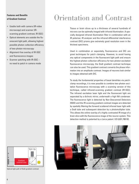
Features and Benefitsof Gradient Contrast• Usable both with camera (IR-videomicroscopy) and scanner (IRscanninggradient contrast, IR-SGC)• Optical elements are outside the fluorescentlight path, allowing highestpossible photon collection efficiencyof two-photon microscopy• Alignment-free overlay of IR-SGCand fluorescence images• Scanner patching with IR-SGC –no need to patch in camera modeOrientation and ContrastTissue or brain slices up to a thickness of several hundreds ofmicrons can be optimally imaged with infrared illumination. A speciallydesigned infrared illumination filter in combination with anIR polarizer, IR analyzer and the infrared differential interferencecontrast (DIC) prisms give extremely good resolution even in thethickest specimens.Used in combination or separately, fluorescence and DIC aregreat techniques for patch clamping. However, to avoid havingany optical components in the fluorescent light path and ensurethe highest photon collection efficiency for two-photon excitationfluorescence microscopy, the Dodt gradient contrast techniquecan also be used. This gradient contrast converts the phase informationinto an amplitude contrast. Images of neurons look similarto images obtained with DIC.To study the fundamental properties of basal dendrites via patchclamprecordings, it is now possible to combine two-photon excitationfluorescence microscopy with a scanning version of thistechnique, called infrared-scanning gradient contrast (IR-SGC).The infrared excitation laser light and the fluorescent light areseparated by a dichroic mirror, underneath a high NA condenser.The fluorescence light is detected by Non-Descanned Detectors(NDD) and the IR-scanning gradient contrast images are detectedby spatially filtering the forward scattered infrared laser light witha Dodt tube and subsequent detection by a photomultiplier tube.This allows the online-overlay of a highly contrasted IR image of abrain slice with the fluorescence image of the neuron system. Thisdetection method is patented by a Leica patent: US 6,831,780 B2.Optical light path of Dodt gradiant contrast6