DM6000 CFS.qxd:DM6000 CFS - Duke University Light Microscopy ...
DM6000 CFS.qxd:DM6000 CFS - Duke University Light Microscopy ...
DM6000 CFS.qxd:DM6000 CFS - Duke University Light Microscopy ...
You also want an ePaper? Increase the reach of your titles
YUMPU automatically turns print PDFs into web optimized ePapers that Google loves.
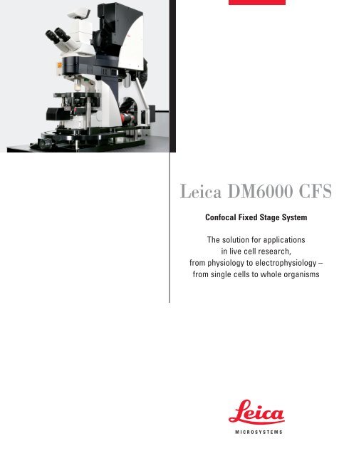
Leica <strong>DM6000</strong> <strong>CFS</strong>Confocal Fixed Stage SystemThe solution for applicationsin live cell research,from physiology to electrophysiology –from single cells to whole organisms
Excellent ResultsUnder All ConditionsLeica Microsystems sets a new standard with the integration of theLeica <strong>DM6000</strong> <strong>CFS</strong> fixed stage microscope into the Leica TCS SP5confocal platform.Leica TCS SP5 with integrated fixed stage microscopeLeica <strong>DM6000</strong> <strong>CFS</strong>Features and Benefits• Maximum workspace for mani -pulators and attached pipettes• High collection efficiency due toshort collection path beforedetectors• Integrated change of magnification(0.35x, 1x, 4x) of camera port• Remote control of all microscopefunctions via touch panel• Special booster optic to fill theentrance pupil of the 20x1.0objective• “Dip-in” feature for immersing theobjective• Patented condenser drainagesystem• High microscope and samplestabilityNeurobiological research has a long history not only in measurementsof single cells, but also of thick brain slices. Here morerealistic results can be achieved due to the intact network. Ultimately,accessing the brain directly in a whole animal providesthe most intact environment for conducting measurements onindividual cells. So for optimal results, space for whole animalsamples is an absolute must.Electrophysiological research employs micropipette systems forrecording electrical signals (patch clamp, whole cell recordings),electrical stimulation (intracellular stimulation, synaptic stimulation,soma-stimulation), dye injection and intracellular perfusion. To makesuch measurements effective, the microscope needs to have asmuch space as possible for micropipettes and other manipulators.Searching for the perfect spot in the sample requires a large fieldof view, whilst the precise positioning of the micropipettes requireshigh magnification. Furthermore, high quality confocal image scanningrequires a large numerical aperture for best performance. Tofulfill these experimental prerequisites, a camera with anadjustable magnification and specially adapted objectives areneeded. Any direct manipulation of the system potentially disturbsthe delicate positioning of the micropipettes within the sample.This emphasizes the importance of a remote control for the imagingsetup, providing convenient access to all relevant functions.Finally, recording electrophysiological and imaging data with perfectsynchrony is paramount for correct interpretation. Triggeringimage recording by external events and synchronizing the applicationof stimuli with image scanning down to the single line levelhelps realize sophisticated experimental setups. Online analysisof the resulting data allows on-the-fly adjustment of externalparameters, helping the researcher to get everything just right.The Leica TCS SP5 with the integrated fixed stage microscopeLeica <strong>DM6000</strong> <strong>CFS</strong> gives excellent experimental results under allconditions.2
Variable Fieldat High ResolutionWith the highest aperture objective 20x 1.0 NA, with the innovative,ergonomic and stable microscope stand Leica <strong>DM6000</strong> <strong>CFS</strong>,with short coupled non-descanned detectors and with the fastest,most sensitive scanning system SP5, we enter a new dimensionin imaging, from single cells to whole organisms.Leica objective HCX APO L 20x1.0The objectiveThe new Leica HCX APO L 20x 1.0 water immersion objectiveoffers both a large field and a high resolution with one singleobjective lens, making it unnecessary to change objectivesbetween overview imaging for sample preparation and detailimaging for data recording.Transmission HCX Apo L 20x/1.00 WIn addition to optimizing the optical parameters, a number ofphysical, chemical and mechanical improvements have beenintegrated into the new HCX APO L 20x1.0 application objective: Itis corrosion-free, chemically neutral and avoids diffusion of metalions. Wettability is outstanding, thermal conductivity is minimaland magnetic fields at the front of the objective have been eliminated.These new objective characteristics are possible by employing aspecial, extremely hard ceramic material for the whole front lensarea. This material is resistant to mechanical damage and muchbetter suited to the requirements of electrophysiology than typicalobjective metal sleeves.A further advantage is the access angle of the objective, which isa measure of how easily manipulators can be fitted. This has beenwidened to 39 degrees, which is almost the limit of what is theoreticallypossible. In combination with a free working distance of2 mm, the 20x objective is ideal for many applications.Features and Benefits• 20x water immersion objective• High numerical aperture (NA) of 1.0• Free working distance (FWD) of2 mm• Optimized access angle of 39°• Large objective focusing range of13 mm• Insulated ceramic tip• Minimum conductivity• High transmission in VIS and IR(see graph)• Best DIC and Dodt contrast3
The microscope standBut an objective alone doesn’t make an electrophysiology setup.The Leica <strong>DM6000</strong> <strong>CFS</strong> combines the specific needs of electrophysiologicalexperiments with the optimized imaging performanceof the DM series. With its focusing objective or nosepiece,it offers a highly stable and convenient optical platform for allfixed stage applications.To cover the demand for large scale sample screening and fineneedle approaches for electrophysiology, the <strong>DM6000</strong> <strong>CFS</strong> has aspecial observation tube with 3 magnification positions (0.35x; 1x;4x). This parfocal magnification changer allows a total change inmagnification up to a factor of 12 without changing the objective.Leica DFC350 FX Camera with parfocal magnificationchangerThe specimen is completely decoupled from the microscope, bothmechanically and electrically. The microscope provides a highdegree of free space for application-specific sample holders,bath chambers and several manipulators at a time. In this way,the sample and the patch electrodes can be moved below thefixed optical axis of the microscope. This allows the researcher toscan the entire dendritic tree and follow the axons of fluorescentlylabeled cells. The <strong>DM6000</strong> <strong>CFS</strong> is also designed for usewith a separate third-party fixed platform for holding micromanipulatorsand other devices, as well as an x,y-translation stage.To avoid the need for direct manipulation of the microscope standwhich could cause accidental vibrations, the external control panelLeica STP6000 (Smart Touch Panel) controls all the microscopefunctions. A touch screen and freely programmable function keysallow quick and easy operation without adversely affecting themeasurements.Features and Benefitsof the Leica STP6000• Intuitive user interface• Control of all microscope functions• Z-focusing wheel for coarse andfine z-movements• Freely programmable function keys• Helps to avoid vibrations caused bymanual operation4
Large range of applicationsEven though the new 20x 1.0 objective makes electrophysiologywith one single objective possible, sometimes this isn’t enough. Indevelopmental biology, there is a clear trend towards imagingeven larger organs and organisms, calling for different specialapplication objectives. The Leica <strong>DM6000</strong> <strong>CFS</strong> offers maximumflexibility in a multi-user environment to cater to everyone’sneeds. A high precision adapter is available, allowing to replacethe single objective with an electronic 6x objective nosepiece.The patented change of objectives works vibration-free, withautomatic power switch-off to avoid disturbing measurements.For each objective, the focus position can be programmed. Thus,by simply pushing a button, a quick change between the magnificationscan be achieved even in the near infrared without losingthe area of interest, implementing automatic parfocality.Exchangeable nosepieceFeatures and BenefitsTop: Zebrafish eye (courtesy of: Carl Neumann, EMBL).Right (from top to bottom): Platynereis larva (courtesy of:Raju Tomer, EMBL, Heidelberg, Germany). Neurons inbrain slice (courtesy of: Thomas Nevian, Institute ofPhysiology, Bern, Switzerland). Mouse embryo, detail ofthe heart (courtesy of: Dr. Elisabeth Ehler, King’s College,London, UK).• High precision adapter for switchingbetween single objective andobjective revolver• 6 objective nosepiece positions• Patented electronic nosepieceturret• Automatic power switch-off afterobjective change• Automatic electronic parfocality5
Features and Benefitsof Gradient Contrast• Usable both with camera (IR-videomicroscopy) and scanner (IRscanninggradient contrast, IR-SGC)• Optical elements are outside the fluorescentlight path, allowing highestpossible photon collection efficiencyof two-photon microscopy• Alignment-free overlay of IR-SGCand fluorescence images• Scanner patching with IR-SGC –no need to patch in camera modeOrientation and ContrastTissue or brain slices up to a thickness of several hundreds ofmicrons can be optimally imaged with infrared illumination. A speciallydesigned infrared illumination filter in combination with anIR polarizer, IR analyzer and the infrared differential interferencecontrast (DIC) prisms give extremely good resolution even in thethickest specimens.Used in combination or separately, fluorescence and DIC aregreat techniques for patch clamping. However, to avoid havingany optical components in the fluorescent light path and ensurethe highest photon collection efficiency for two-photon excitationfluorescence microscopy, the Dodt gradient contrast techniquecan also be used. This gradient contrast converts the phase informationinto an amplitude contrast. Images of neurons look similarto images obtained with DIC.To study the fundamental properties of basal dendrites via patchclamprecordings, it is now possible to combine two-photon excitationfluorescence microscopy with a scanning version of thistechnique, called infrared-scanning gradient contrast (IR-SGC).The infrared excitation laser light and the fluorescent light areseparated by a dichroic mirror, underneath a high NA condenser.The fluorescence light is detected by Non-Descanned Detectors(NDD) and the IR-scanning gradient contrast images are detectedby spatially filtering the forward scattered infrared laser light witha Dodt tube and subsequent detection by a photomultiplier tube.This allows the online-overlay of a highly contrasted IR image of abrain slice with the fluorescence image of the neuron system. Thisdetection method is patented by a Leica patent: US 6,831,780 B2.Optical light path of Dodt gradiant contrast6
Detection efficiencyAs with multi-photon excitation the fluorescence is only generatedin the diffraction limited focal volume, the detectors can beplaced directly behind the objective (reflected light detectors,RLD) as well as directly behind the condenser (transmitted lightdetectors, TLD) without losing spatial resolution. This close-couplingdetection scheme results in the highest possible photon collectionefficiency, as scattered fluorescent photons can also becollected over a large detection angle due to the high numericalaperture of the objective and the condenser. Two-channel detectorson both sides add a maximum of detection flexibility.Apart from its high NA, the new <strong>DM6000</strong> <strong>CFS</strong> patented turret condenserfor brightfield and interference contrast provides a numberof other advantages. The system allows the exchangebetween dry and oil condensers. The condenser base with condenserhead 1.4 NA oil S1 stands for highest collection efficiency,while the patented condenser base provides a watertight sealwith an outlet pipe for liquid leaking from the sample.ABFeatures and BenefitsDCGradient contrast with camera,magnification 0.35x (A), 1x (B), 4x (C).Online overlay of IR-SGC andfluorescence image (D).• Close-coupled two-channel NDDsboth in reflected and transmittedlight paths• Electrophysiology condenser systemwith outlet pipe for draining• Condenser base with condenserhead 0.9 NA S1• Condenser base with condenserhead 1.4 NA oil S1 for highestcollection efficiency7
Two worlds in oneTwo completely different experimental requirements can be satisfiedwith one single system. The Leica TCS SP5 with the TandemScanner provides classical morphology on large samples, wherehigh spatial resolution is required, e.g. research on structures ofcytoskeleton, organelles or tissues, as well as physiology and biophysicsimaging, where temporal resolution becomes very important.Double labeled neurons of brain slices.Courtesy of Thomas Nevian, Institute ofPhysiology, Bern, SwitzerlandCalcium is an important second messenger to trigger many signallingcascades in neurons. Functional imaging of calcium influxis possible with calcium ion sensitive indicator dyes, but calciumtransients in neurons are very fast. Therefore, they are typicallyrecorded in line scan mode, also known as xt-scan mode. Thelaser beam is continuously scanned back and forth along thesame line and fluorescence over time is recorded. The resultingimage consists of one spatial and one time axis. The resonantscanning system of the Leica TCS SP5 oscillates at 8000 Hz,enabling a line rate of 16,000 lines per second. Fast dynamics ofinitial calcium inflow can now be investigated.Even in the xy-scan mode, the scan rate can be as high as 180frames per second in the frame size 512 x 64. This frame rateenables imaging of extended regions of the dendritic tree andmultiple spines at the same time.Confocal interface8
New XYTZ-Scan Mode for 3D visualization of calcium transientsThe dendritic tree has a complex three dimensional structure.Repeatedly acquiring complete 3D stacks gives a temporal resolutionmuch too low for imaging the fast calcium transients in dendrites.To circumvent this problem, optical sectioning, the fastscanning of the resonant system and the triggering capability arecombined for the new XYTZ-Scan Mode.Individual time series are taken at different focal depths and combinedinto a 4D image stack. A stimulus is always delivered beforethe same frame of each time series, synchronized by a trigger outevent. After a complete focal series, the image data is projectedinto a 3D data stack over time. For each structure in the sampled3D volume the fluorescence transients can be analyzed. The timecourse of fluorescence in all parts of the dendritic tree can beseen clearly.XYTZ-Scan Module:Time series at different focalpositions, stimulus (t 0 ) is always delivered beforethe same frame of each time seriesScanner patching and optimized workflowIn combination with the IR-SGC, which makes electrophysiologyneedles visible in the scanning mode, the extreme speed of theresonant scanner allows electrode patching while imaging atvideo rates, without ever switching to the camera mode. Togetherwith the one-for-all electrophysiology 20x 1.0 objective, scannerpatching minimizes the number of steps between experimentsetup and data collection, optimizing your workflow.So even though you might never need it, the workflow orienteduser interface of the Leica TCS SP5 with Leica <strong>DM6000</strong> <strong>CFS</strong>includes the camera as well. Whether you are doing CCD-cameraor confocal / multiphoton imaging, the other option is always justone fast click away. Integrating both operation modes in the samesoftware allows you to concentrate on what’s in your images, noton where they came from.Features and BenefitsCamera interface• Software-integration of CCDcameraand confocal scanner• Single click switching betweencamera and confocal• Workflow oriented user interface9
Data Correlation (ψ/F)Recording of electrical (e.g. patch-clamp) data is typically relatedto stimulation (electrical or chemical sensory stimulation of theanimal). Current or voltage is recorded briefly before, during andafter stimulation. The time frame for recording data after stimulationdepends on effect-relaxation.To synchronize the image acquisition and the electrophysiologicalrecordings, precise triggers are necessary. The Leica TCS SP5hardware provides different types of outbound triggers, such asframe, line and pixel triggers. They can be used to synchronize theapplication of stimuli or external recording devices. On the otherhand, input triggers can be used to start or continue image scanningin response to arbitrary external events, thus increasing theflexibility in data acquisition. For example, the synchronization ofheart beat and image acquisition could be performed by a specialinput trigger to minimize the influence of heart activity on theimage data.With the Leica software LAS AF, data evaluation of the electrophysiologicalsignals can be performed online providing precisecorrelation with imaging data. For the analysis of electrical andoptical data, the relevant basic functions are implemented. Onlinedata evaluation enables the validation of the recorded data and isimportant in helping the researcher quickly decide how the externalparameters should be modified for the best results.Options for data correlation• Correlation of optical and electricaldata• Input triggering to start imageacquisition• Output triggering to control stimuli• Synchronization of scanning withexternal devices• Control of environmental conditionsby online data analysis10
System ComponentsLeica Confocal Fixed Stage System <strong>DM6000</strong>0 <strong>CFS</strong>11
Leica Microsystems –the brand for outstanding productsLeica Microsystems’ mission is to be the world’s first-choice provider of innovativesolutions to our customers’ needs for vision, measurement and analysis of microstructures.Leica, the leading brand for microscopes and scientific instruments, developed fromfive brand names, all with a long tradition: Wild, Leitz, Reichert, Jung and CambridgeInstruments. Yet Leica symbolizes innovation as well as tradition.Leica Microsystems – an international companywith a strong network of customer servicesAustralia: North Ryde Tel. +61 2 8870 3500 Fax +61 2 9878 1055Austria: Vienna Tel. +43 1 486 80 50 0 Fax +43 1 486 80 50 30Canada: Richmond Hill/Ontario Tel. +1 905 762 2000 Fax +1 905 762 8937Denmark: Herlev Tel. +45 4454 0101 Fax +45 4454 0111France: Rueil-Malmaison Tel. +33 1 47 32 85 85 Fax +33 1 47 32 85 86Germany: Bensheim Tel. +49 6251 136 0 Fax +49 6251 136 155Italy: Milan Tel. +39 0257 486.1 Fax +39 0257 40 3475Japan: Tokyo Tel. + 81 3 5421 2800 Fax +81 3 5421 2896Korea: Seoul Tel. +82 2 514 65 43 Fax +82 2 514 65 48Netherlands: Rijswijk Tel. +31 70 4132 100 Fax +31 70 4132 109People’s Rep. of China: Hong Kong Tel. +852 2564 6699 Fax +852 2564 4163Portugal: Lisbon Tel. +351 21 388 9112 Fax +351 21 385 4668Singapore Tel. +65 6779 7823 Fax +65 6773 0628Spain: Barcelona Tel. +34 93 494 95 30 Fax +34 93 494 95 32Sweden: Kista Tel. +46 8 625 45 45 Fax +46 8 625 45 10Switzerland: Heerbrugg Tel. +41 71 726 34 34 Fax +41 71 726 34 44United Kingdom: Milton Keynes Tel. +44 1908 246 246 Fax +44 1908 609 992USA: Bannockburn/lllinois Tel. +1 847 405 0123 Fax +1 847 405 0164and representatives of Leica Microsystemsin more than 100 countries.The companies of the Leica MicrosystemsGroup operate internationallyin three business segments, where werank with the market leaders.• <strong>Microscopy</strong> SystemsOur expertise in microscopy is the basisfor all our solutions for visualization, measurementand analysis of micro-structuresin life sciences and industry. Withconfocal laser technology and imageanalysis systems, we provide threedimensionalviewing facilities and offernew solutions for cytogenetics, pathologyand materials sciences.• Specimen PreparationWe provide comprehensive systemsand services for clinical histo- andcytopathology applications, biomedicalresearch and industrial quality assurance.Our product range includesinstruments, systems and consumablesfor tissue infiltration and embedding,microtomes and cryostats as well asautomated stainers and coverslippers.• Medical EquipmentInnovative technologies in our surgicalmicroscopes offer new therapeuticapproaches in microsurgery.Copyright © Leica Microsystems CMS GmbH • Am Friedensplatz 3 • 68165 Mannheim • Germany 2007 • Tel. +49 (0)621-7028 0 • Fax +49 (0)621-7028 1028 LEICA and the Leica Logo are registered trademarks of Leica IR GmbH.Order no.: English 1593102113 Printed on chlorine-free bleached paper. ??/07/???/???www.leica-microsystems.com


