02 Cells & Disease - Student.pdf - haspi
02 Cells & Disease - Student.pdf - haspi
02 Cells & Disease - Student.pdf - haspi

Create successful ePaper yourself
Turn your PDF publications into a flip-book with our unique Google optimized e-Paper software.
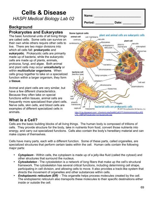
<strong>Cells</strong> & <strong>Disease</strong>HASPI Medical Biology Lab <strong>02</strong>BackgroundProkaryotes and EukaryotesThe basic functional units of all living thingsare called cells. Some cells can survive ontheir own while others require other cells tolive. There are two major divisions intowhich all cells fall: prokaryotic andeukaryotic. Prokaryotic cells are primarilymade up of bacteria, while the eukaryoticcells are made up of plants, animals,protozoa, fungi, and algae. Both animaland plant cells may occur unicellularly orwithin multicellular organisms. Whencells group together to take on a specializedfunction within a larger organism, they forma tissue.Name: ____________________________Period: ___________ Date: ___________Animal and plant cells are very similar, buthave a few different characteristics.Because they often take on specialfunctions within tissues, animal cells arefrequently more specialized than plant cells.Nerve cells, skin cells, and blood cells areexamples of different specialized cells inanimals.http://3.bp.blogspot.com/_LGy4ytnLmtg/SqM0Cz8CPTI/AAAAAAAAAEs/uRSLTp_mBPg/Procaryotic+vs+eucaryotic.jpgWhat is a Cell?<strong>Cells</strong> are the basic building blocks of all living things. The human body is composed of trillions ofcells. They provide structure for the body, take in nutrients from food, convert those nutrients intoenergy, and carry out specialized functions. <strong>Cells</strong> also contain the body’s hereditary material and canmake copies of themselves.<strong>Cells</strong> have many parts, each with a different function. Some of these parts, called organelles, arespecialized structures that perform certain tasks within the cell. Human cells contain the followingmajor parts:• Cytoplasm - Within cells, the cytoplasm is made up of a jelly-like fluid (called the cytosol) andother structures that surround the nucleus.• Cytoskeleton - The cytoskeleton is a network of long fibers that make up the cell’s structuralframework. The cytoskeleton has several critical functions, including determining cell shape,participating in cell division, and allowing cells to move. It also provides a track-like system thatdirects the movement of organelles and other substances within cells.• Endoplasmic reticulum (ER) - This organelle helps process molecules created by the cell.The endoplasmic reticulum also transports these molecules to their specific destinations eitherinside or outside the cell.69
• Golgi apparatus - The Golgi apparatus packages molecules processed by the endoplasmicreticulum in order to be transported out of the cell.• Lysosomes and peroxisomes - These organelles are the recycling center of the cell. Theydigest foreign bacteria that invade the cell, rid the cell of toxic substances, and recycle wornoutcell components.• Mitochondria - Mitochondria are complex organelles that convert energy from food into a formthat the cell can use. They have their own genetic material, separate from the DNA in thenucleus, and can make copies of themselves.• Nucleus - The nucleus serves as the cell’s command center, sending directions to the cell togrow, mature, divide, or die. It also houses DNA (deoxyribonucleic acid), the cell’s hereditarymaterial. A membrane called the nuclear envelope, which protects the DNA and separates thenucleus from the rest of the cell, surrounds the nucleus.• Cell membrane - The plasma membrane is the outer lining of the cell. It separates the cellfrom its environment and allows materials to enter and leave the cell.• Ribosomes - Ribosomes are organelles that process the cell’s genetic instructions to createproteins. These organelles can float freely in the cytoplasm or be connected to theendoplasmic reticulum.Organelles and DisordersWith some human diseases, dysfunction of the organelles is the cause. This may occur from agenetic defect that deforms the organelle, or damage that results from exposure to harmfulsubstances. Some examples include cystic fibrosis, congenital disorders of glycosylation (CDG),progeria, lysosomal storage diseases (LSD), type 1 diabetes, and familial hypercholesterolemia.Disorder Organelle/Cause SymptomsCystic FibrosisCDGProgeriaLSDType 1 DiabetesFamilialHypercholesterolemiaEndoplasmic Reticulum – defectivelyformed, which causes deformedproteins that stay trapped in the ERGolgi Apparatus – defective bondingof carbohydrates to proteinsNuclear Envelope (surrounds thenucleus) – cytoskeleton portion ofenvelope is defectiveLysosomes – do not break downundigested molecules that then buildup in the cell causing damageCell Membrane – defectivemembrane protein does not removeglucose from the blood and move itinto the cellsCell Membrane - missing membraneprotein that doesn’t allow the cells toremove cholesterol from the bloodAccumulation of thick mucus inorgans, mainly lungs and pancreasMental retardation, seizures, liverdisease, and death by the age of 2Premature aging of musculoskeletaland cardiovascular system leadingto early deathMental deterioration, heart disease,respiratory failure, leads to deathIncreased thirst, frequent urination,hunger, abdominal pain,nausea/vomiting, fatigue, poorcirculation, coma, deathHigh amount of cholesterol in theblood that begins at birth and can’tbe controlled, fatty deposits on skin,chest pain, signs of coronary arterydisease at early ageNIH. 2011. What is a cell? Genetics Home Reference, National Institutes of Health, U.S. NationalLibrary of Medicine. http://ghr.nlm.nih.gov/handbook/basics/cell70
MaterialsCompound microscope Concaved slide ToothpickWater Slide Methylene bluePlastic pipette Coverslip OnionPond mixYogurtProcedurePurpose: The goal of this lab will be to compare and contrast prokaryotic and eukaryotic cells.Scenario:You have just started working in the infectious disease lab of HASPI Hope Hospital. The following isthe medical background information for your first patient.Patient ID: 105839Your patient was admitted to the Emergency Room this morning at 6:45 am. He has just returnedfrom a backpacking trip about three days ago. He began having some abdominal pain and cramping,intermittent constipation, and well as fatigue upon his return, but awoke this morning to severediarrhea with blood and a swollen abdomen. Upon interviewing the patient he has admitted todrinking from a pond on the backpacking trip when they ran out of water. A sample from the pondhas been collected for your review.You are excited to begin the analysis for your first patient, but need to review the difference betweensome of the possible causes before you begin to make sure you can make the correct diagnosis.The possible causes have already been narrowed down to a bacterial infection (prokaryotic) from thepond water, a plant-like protist (unicellular plant cell) in the pond water, or an animal-like protist(unicellular animal cell) in the pond water. Follow the procedure below to isolate and compare aprokaryote, plant cell, and animal cell so you know what to look for in the pond water that has beencollected.Part A – Identification of a prokaryote, eukaryotic plant cell, andeukaryotic animal cellProkaryote: Bacteria in yogurtYour instructor has placed yogurt in a dark and warm area for the past 24 hours in order toallow bacteria to grow.1. Using a toothpick, collect a small amount of the yogurt culture onto a slide.2. Place a drop of water on the slide as well to dilute the yogurt, and add a coverslip over theyogurt and water to create a wet mount.3. Place the slide on the stage of your compound microscope.4. Bacteria are very small and difficult to see, so start your microscope objective at 100xmagnification. Draw what you see in the analysis section.5. Switch your microscope objective to the 400x magnification, or higher if it is available on yourmicroscope. Draw what you see in the analysis section.6. At this level, you should be able to identify some of the characteristics of the bacteria in theyogurt. Using Chart A below, draw an example of and classify the type of bacteria in theyogurt culture.7. Thoroughly rinse off your slide and coverslip with soap and water.71
Chart A: How to Classify Bacteria (Prokaryotes)CommonTermScientificTermImageBacteriaArrangementpairedchaineddiploestreptoseclusteredstaphyleBacteriaShaperoundrodcoccusbacillusspiralspirillumExamples:Streptococcushttp://2.bp.blogspot.com/_BghQ0YzD2Ns/S9t8hE2scSI/AAAAAAAAArs/jhKBFz_nIp0/s1600/11-18_streptococcus.jpgDiplobaccilushttp://www.jayzine.com/wpcontent/uploads/2011/<strong>02</strong>/bacillus.jpgStaphylospirillumhttp://staff.tuhsd.k12.az.us/gfoster/standard/spirilla.jpg72
Eukaryote: Plant cell from onionAn onion is made up of tissue of which a single cell is able to create the entire plant. Thereare curved pieces that can be pulled apart from an onion called scales. Each scale has a thinmembrane coating that can be pulled off, which is called the epidermis.1. Obtain a scale from the onion and carefully pull off the thin epidermis.2. Cut or tear a small piece--approximately 5 mm x 5 mm--of the epidermis and place it on yourclean slide. Make sure that the epidermis is flat and not folded over on itself.3. Add one drop of water and one drop of methylene blue to the slide.4. Wait approximately 30 seconds and then add the coverslip. If there is extra liquid on the slidetouch the corner of a paper towel to the extra liquid to wick it off.5. Place the slide on your microscope and the objective at 40x magnification. Draw what you seein the analysis section. The onion cells will be stained blue or purple.6. View the onion cells at 100x and 400x magnification. Draw what you see in the analysissection.7. On your 400x drawing, label the cell membrane, cell wall, chloroplasts, and nucleus.8. Thoroughly rinse off your slide and coverslip with soap and water.Eukaryote: Animal cell from cheekThe cells from your cheek can be gently scraped off and viewed. The cells you will beobserving are actually part of the human epidermis.1. Using a toothpick, scrape the inside of your cheek 3 or 4 times. DO NOT scrape hard enoughto cause bleeding!2. Gently smear the end of the toothpick on the center of a clean slide.3. Add one drop of water and one drop of methylene blue to the slide.4. Wait approximately 30 seconds and then add the coverslip. If there is extra liquid on the slidetouch the corner of a paper towel to the extra liquid to wick it off.5. Place the slide on your microscope and the objective at 40x magnification. Draw what you seein the analysis section. The cheek cells will be stained blue or purple.6. View the cheek cells at 100x and 400x magnification. Draw what you see in the analysissection.7. On your 400x drawing, label the cell membrane and nucleus for one of the cheek cells.8. Thoroughly rinse off your slide and coverslip with soap and water.Part B – Identification of Cause of InfectionYou now have reviewed the differences between bacteria (prokaryotes), plant cells(eukaryote), and animal cells (eukaryote). Follow these directions to determine what organismin the pond water may be the cause of illness in your patient.1. Place 1-2 drops of the pond water in the well of the depression slide. It is not necessary to adda coverslip to the depression slide.2. Place the slide on your microscope and view under the 40x magnification objective to start.3. Most of the living organisms in the pond water will be moving. Draw and identify any livingorganisms that you find in the analysis section. Attempt to find some of the organisms thatcould be causing the patient’s illness. A list of the possibilities can be found in Chart B.4. It may be necessary to move to a higher magnification to observe some of the organisms.Make sure you observe the pond water under 40x, 100x, and 400x magnification.5. When you are done, attempt to pour the pond water from the slide back into the originalcontainer, rinse out the depression slide, and repeat Steps 1-4 (you are looking for organismsyou might have missed in your first observation).73
Possible Bacterial Cause:Bacteria <strong>Disease</strong> Information ImageCauses Campylobacteriosis. Transmission is fecaloral,ingestion of contaminated food or water, and theCampylobacter eating of raw meat. Symptoms are inflammatory,sometimes bloody, diarrhea, periodontitis or dysenterysyndrome, mostly including cramps, fever and pain.Symptoms typically last for 5–7 days.http://www.safetables.org/img/fbi/campylobacter2.pngPossible Plant-like Protist Causes:Plant-like Protist <strong>Disease</strong> Information ImageThese protists are found in stagnant water. They do notnormally cause disease in humans, but a few caseswith similar ingestion/symptoms have been identifiedwithin the last month at Hope Hospital. YourEuglena graclilisdepartment is wondering if there may be a correlationand a possible change in the Euglena DNA that hascaused it to become infectious.Dinophysis algaeCauses Diarrhetic Shellfish Poisoning (DSP).Transmission is from ingesting shellfish containing thisalgae, but in rare cases can be contracted by drinkingcontaminated water. Symptoms of DSP includediarrhea, nausea, vomiting, abdominal pain, andcramps. DSP is generally not lethal. Onset ofsymptoms is normally 30 minutes to a few hours afteringestion.Possible Animal-like Protist Causes:http://www.fcps.edu/islandcreekes/ecology/Miscellaneous/Euglena/15L.jpghttp://algeinfo.imr.no/algebilder/50.jpgAnimal-like Protist <strong>Disease</strong> Information ImageCauses Giardiasis. Transmission occurs throughingestion, usually by drinking contaminated water.Symptoms usually appear two weeks after exposureand include: diarrhea, dehydration, stomach cramps,Giardia lambliagas, and weight loss. Onset of symptoms is normally 1-2 weeks after ingestion. The disease is diagnosed byfinding cysts in fecal smears.Entamoebahystolitica(amoeba)CryptosporidiumparvumCauses Amoebic Dysentery. Transmission by fecalcysts in contaminated food or water. Once ingested,the organism secretes enzymes that digest in theintestinal lining. Symptoms develop slowly, withintermittent diarrhea, cramps, vomiting, and a generalbad feeling. In severe cases, dehydration and anemiaresult from the loss of fluids and blood.Causes Cryptosporidiosis. This protist is present inmost surface water, and infection is through ingestion ofinfected organisms in drinking water. Symptomsinclude watery diarrhea, cramps, and headache. Thelungs and trachea can be affected as well, producinglow-grade fever and coughing. It is most dangerous tochildren and immunocompromised patients, such asAIDS patients, and people undergoing cancerchemotherapy. Healthy individuals normally havehttp://parasitecleanse.com/images/giardia_lambia.jpghttp://home.tiscali.nl/~t936927/folio/images/amoebahttp://www.diark.org/img/species_pict/Cryptosporidium_parvum_str__Iowa_IIshorter duration of symptoms.CDC. 2009. A-Z Index of Foodborne, Bacterial, and Mycotic <strong>Disease</strong>s. Center for <strong>Disease</strong> Controland Prevention; Division of Foodborne, Bacterial, and Mycotic <strong>Disease</strong>. www.cdc.gov74
AnalysisPart A - Bacteria ObservationsRecord what you see in your microscopic observations at the following magnifications:100x magnification400x magnificationBacteria in Yogurt (complete the following chart with as many as you see)BacteriaArrangementBacteriaShapeDraw aPicture ExamplePart A - Plant Cell ObservationsRecord what you see in your microscopic observation at the following magnifications:40x magnification 100x magnification 400x magnification75
Part A – Animal Cell ObservationsRecord what you see in your microscopic observation at the following magnifications:40x magnification 100x magnification 400x magnificationPart B – Pond Water ObservationsDraw and identify any organisms found in the pond water in this area. Identify at what magnificationthey were identified, what the organism may be, and whether it is a prokaryote, eukaryote (plant), oreukaryote (animal) next to your drawing.76
Part A Analysis Question - on a separate sheet of paper complete the following1. What are the names of the bacteria you found in the yogurt culture? (HINT: combine thearrangement and shape for the name)2. Were you able to observe a nucleus in the bacteria? Explain your answer.3. Are bacteria prokaryotes or eukaryotes? What is the difference?4. Identify the cell wall, plasma membrane, capsule, pili, cytoplasm, ribosome, and flagella on thefollowing bacterial cell.Bacteria CellABDEFGC5. What type of onion cells did you observe?6. Why do you think it was difficult to see any organelles besides the cell membrane, cell wall,nucleus, and chloroplasts in the cells you observed?7. Is the onion cell a prokaryote or eukaryote?8. What was the point of using methylene blue? What parts of the cells did it stain?9. List at least two differences you observed between the onion cell and the cheek cell.11. Label the cell wall, cell membrane,chloroplast, ribosomes, vacuole,cytoplasm, endoplasmic reticulum,Golgi complex, mitochondria, andnucleus on the following plant cell.HPlant CellIJABC12. Label the cell membrane, centriole,ribosomes, vacuole, mitochondria,endoplasmic reticulum, Golgi complex,cytoplasm, and nucleus on thefollowing animal cell.ABAnimal CellCA DEGFFGEDIH77
Part B Analysis Questions - on a separate sheet of paper complete the following13. What are the key symptoms for this patient that would lead you to believe the disease has beencaused by something that was drunk?14. At which magnification were the most organisms visible?15. Why do you think it was important to perform the microscope observation of the pond watertwice?16. Did you find any organisms that were on the list of possible causes? If yes, explain what theorganism is and give information on the disease. If no, give a review of the organisms that youdid find.17. CONCLUSION: In 1-2 paragraphs summarize the procedure and results of this lab.Review Questions - on a separate sheet of paper complete the following1. What is the difference between a prokaryote and a eukaryote?2. What does it mean for an organism to be unicellular or multicellular? Give an example of anorganism that is unicellular, and an organism that is multicellular.3. What is a tissue?4. What is the difference between an animal and plant cell?5. What is the function of a cell?6. List the function of the following cell parts: cytoskeleton, endoplasmic reticulum, lysosomes,mitochondria, and ribosomes.7. How does a disorder in the organelles occur?8. Choose two organelle disorders and give the name, the organelle that causes it, and thesymptoms.9. Identify the arrangement and shape of the following bacteria:ABhttp://www2.estrellamountain.edu/faculty/farabee/biobk/84150f.jpghttp://www2.estrellamountain.edu/faculty/farabee/biobk/96444c.jpgCDhttp://www.poolcenter.com/bacteria-campylobacter.bmphttp://www.steripen.com/stuff/contentmgr/files/1/78