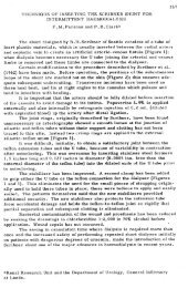
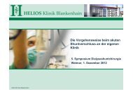
2630 K. KonnerFig. 1. Animal experiments with <strong>vascular</strong> anastomoses by Jaboulay and Briau, published 1896 [1].Fig. 2. Three-point-end-to-end anastomosis by Carrel, published1912 [2].after the first patient, Clyde Shields, had been takenon maintenance <strong>haemodialysis</strong>, Scribner published a‘Preliminary report on the treatment <strong>of</strong> chronic uremiaby means <strong>of</strong> intermittent hemodialysis’, which describedwith clairvoyance some medical problems plaguingrenal replacement therapy still today: malnutrition,hypertension, anaemia and others [5]. Clyde Shields,a Boeing machinist, survived <strong>for</strong> 11 years after theinsertion <strong>of</strong> his first AV shunt on 9 March 1960.Two thin-walled Teflon cannulas with tapered endswere inserted near the wrist in the <strong>for</strong>earm, one into theradial artery and the other into the adjacent cephalicvein. The external ends were connected by a curvedteflon bypass tube. Later, the Teflon tube was replacedby flexible silicon rubber tubing.Scribner wrote in 1990: ‘Successful treatment <strong>of</strong>Clyde Shields represents one <strong>of</strong> the few instances inmedicine where a single success was required to validatea new therapy’ [6]. The development <strong>of</strong> a permanent<strong>vascular</strong> <strong>access</strong> by the Seattle group was the decisivebreakthrough, which made maintenance dialysispossible. It is rightly considered a landmark in thehistory <strong>of</strong> dialysis: maintenance <strong>haemodialysis</strong> therapybegan on 9 March 1960. Many variants <strong>of</strong> the AV shuntcame into use during the following years when themajority <strong>of</strong> AV-shunt insertions concerned temporary<strong>vascular</strong> <strong>access</strong> at the start <strong>of</strong> chronic dialysis therapyto bridge the time when an AV fistula was absentor maturing.During early 1970s, TJ Buselmeier andco-workers (Minneapolis, USA) developed a compactU-shaped silastic prosthetic AV shunt with either oneor two Teflon plugged outlets which communicated tothe outside <strong>of</strong> the body. The U-shaped portion couldbe totally or partially implanted subcutaneously [7].This modification <strong>of</strong> the Scribner AV shunt gainedsome acceptance during the following years, especially<strong>for</strong> paediatric <strong>haemodialysis</strong> patients.1961At that time, Stanley Shaldon (London, UK) facedthe problem <strong>of</strong> finding a surgeon willing to operate onthe radial artery and cephalic vein to introducecannulae <strong>for</strong> circulatory <strong>access</strong>. To become independent,Shaldon introduced hand-made catheters intothe femoral artery and vein by the percutaneousDownloaded from http://ndt.ox<strong>for</strong>djournals.org/ by guest on February 20, 2012
<strong>History</strong> <strong>of</strong> <strong>vascular</strong> <strong>access</strong> <strong>for</strong> <strong>haemodialysis</strong> 2631Seldinger technique <strong>for</strong> immediate <strong>vascular</strong> <strong>access</strong> [8,9].Over time, vessels in different sites were used, includingthe subclavian vein. Shaldon concluded: ‘Eventually,veno-venous catheterization was preferred because thebleeding from the femoral vein was less than from thefemoral artery when the catheter was removed’ [10].Regional heparinization was practised with hexadimethrinebromide (‘Polybrene’) to neutralize the anticoagulanteffect <strong>of</strong> heparin. Obviously, these cathetersare not identical with the widely used type <strong>of</strong> catheters<strong>for</strong> temporary <strong>access</strong>, which today are usually called‘Shaldon catheters’.1962James E. Cimino and Michael J. Brescia (New York,USA) described a ‘simple venipuncture <strong>for</strong> hemodialysis’based on the experience <strong>of</strong> Dr Cimino whenhe worked part-time as a student at the BellevueTransfusion Center in New York [11]. After priorinfiltration <strong>of</strong> the overlying skin with 1% procaine, themost <strong>access</strong>ible <strong>for</strong>earm vein was punctured with aneedle. Needle sizes varied from No. 16 to No. 12gauge. Patency <strong>of</strong> the vein and adequate blood supplywere assured by application <strong>of</strong> tourniquet pressurewith a sphygmomanometer. A blood flow in the range<strong>of</strong> 150 and 410 ml/min was obtained in these mainlyfluid overloaded patients.1963Thomas J. Fogarty from Cincinnati, USA, inventedan intra<strong>vascular</strong> catheter with an inflatable balloonat its distal tip designed <strong>for</strong> embolectomy andthrombectomy – an essential device even today [12].1966The legendary paper ‘Chronic hemodialysis usingvenipuncture and a surgically created arteriovenousfistula’ was published by Brescia, Cimino, Appell andHurwich [13]. Dr Appell was the surgeon in the team.The first surgically created fistula <strong>for</strong> the purpose<strong>of</strong> <strong>haemodialysis</strong> was placed on 19 February 1965,followed by further 14 operations as <strong>of</strong> 21 June 1966.Twelve out <strong>of</strong> these 14 AV fistulae resumed primaryfunction without complications, two never functioned(in the first patient, the anastomosis ‘was made toosmall’). This represents an early failure rate whichwould be admirably low even in 2005. Dr Scribnerfrom Seattle was the first nephrologist to refer one<strong>of</strong> his patients to New York <strong>for</strong> the creation <strong>of</strong> anAV fistula.Dr Appel had per<strong>for</strong>med a side-to-side-anastomosisbetween the radial artery and the cephalic antebrachialvein at the wrist after a 3–5 mm incision had been madein the corresponding lateral surfaces <strong>of</strong> the artery andthe vein. The suture was achieved using arterial silk incontinuous fashion.Many years later, in 1994, Dr Cimino stated that‘the decision to connect an artery and vein subcutaneously,thus creating an internal shunt, appeared notonly logical but was the classic example <strong>of</strong> necessityas the mother <strong>of</strong> invention’ and ‘that arteriovenousfistulas could lead to heart failure, and this would beparticularly hazardous in patients whose cardio<strong>vascular</strong>systems were already compromised’ [14].Drs Cimino and Appell left the VeteransAdministration Hospital, Bronx, New York, a fewyears later: Dr Cimino is still busy practising palliativemedicine at the Calvary Hospital, Bronx, New York;Dr Appell has retired after a successful careeras a ‘countryside’ general surgeon in the greaterNew York area [Personal communication, April 2005].1967One year after the article <strong>of</strong> Brescia and Cimino,M. Sperling (Wu¨rzburg, Germany) reported thesuccessful creation <strong>of</strong> an end-to-end-anastomosisbetween the radial artery and the cephalic antebrachialvein in the <strong>for</strong>earm <strong>of</strong> 15 patients using a stapler [15].This type <strong>of</strong> AV anastomosis gained widespreadacceptance during the next decade, mainly based onthe rationale to restrict the inflow <strong>of</strong> blood into the AVfistulae to the flow provided by the feeding radialartery. The creation <strong>of</strong> an end-to-end anastomosis wastechnically challenging; an additional problem arosebecause the diameters <strong>of</strong> artery and vein were different;various patch techniques were tried to solve thisproblem. Because <strong>of</strong> the increasing numbers <strong>of</strong> elderly,hypertensive and diabetic patients with difficultvessels and high risk <strong>of</strong> a steal syndrome, this type<strong>of</strong> AV fistula was abandoned as the first <strong>vascular</strong><strong>access</strong> <strong>of</strong> choice. End-to-end-anastomoses are still awell established technique in revision procedures. Thestapler, however, never was accepted as a routinetechnical tool.For the transluminal recanalization <strong>of</strong> arteriesobstructed by atherosclerotic plaques Charles T.Dotter and co-workers (Portland, USA) introduceda type <strong>of</strong> balloon catheter. The first angioplastyrepresented an essential contribution to resolve one<strong>of</strong> the great problems in <strong>vascular</strong> surgery and <strong>vascular</strong><strong>access</strong> surgery [16].1968Lars Ro¨hl from Heidelberg, Germany, publishedhis results in 30 radial-artery-side-to-vein-endanastomoses[17]. After completion <strong>of</strong> the anastomosis,the radial artery was ligated distal to theanastomosis, thus resulting in a functional end-toend-anastomosis.With this technique, an antebrachialcephalic vein located at a more lateral position whichwould not have been suitable <strong>for</strong> a side-to-sideanastomosis,could be used successfully. Later on,the ligation <strong>of</strong> the peripheral arterial limb only waspractised in patients with impending signs <strong>of</strong> peripheralischaemia.Today, the artery-side-to-vein-end-anastomosishas become a standard procedure. The handling <strong>of</strong>Downloaded from http://ndt.ox<strong>for</strong>djournals.org/ by guest on February 20, 2012