syndromic notification and laboratory investigation manual - Jabatan ...
syndromic notification and laboratory investigation manual - Jabatan ...
syndromic notification and laboratory investigation manual - Jabatan ...

You also want an ePaper? Increase the reach of your titles
YUMPU automatically turns print PDFs into web optimized ePapers that Google loves.
MOH / K / EPI / 38.04 (HB)SYNDROMIC NOTIFICATIONAND LABORATORYINVESTIGATIONMANUALCoordinated by:Communicable Disease Surveillance Section,Disease Control DivisionMINISTRY OF HEALTH MALAYSIA2nd EditionOctober 2004
EDITORIAL BOARDADVISOR:MEMBERS:Dr. Hj. Ramlee bin Hj. RahmatDirector of Disease ControlDisease Control Division, MOH.Dr. Lee Cheow PhengPublic Health Specialist,Disease Control Division, MOH.Dr. Zainudin Abdul WahabDeputy Director of Disease Control (Surveillance),Disease Control Division, MOH.Dr. Rohani JahisPrinciple Assistant Director (Surveillance),Disease Central Division, MOHDr. Husnina IbrahimAssistant Director (Surveillance),Communicable Disease Surveillance Section, MOH.2
CONTENTPAGEForeword 4Abbreviation 51. Introduction: <strong>syndromic</strong> approach1.1 What is a syndrome ?1.2 What is <strong>syndromic</strong> <strong>notification</strong> ?1.3 Objectives of <strong>syndromic</strong> <strong>notification</strong>1.4 Advantages of <strong>syndromic</strong> <strong>notification</strong>1.5 Criteria for infection that require <strong>syndromic</strong> <strong>notification</strong>1.6 Laboratory <strong>investigation</strong>2. Notification procedure.2.1 Notification flow666677893. Syndrome definition3.1 Acute neurological syndrome 103.2 Acute respiratory syndrome 113.3 Acute dermatological syndrome 123.4 Acute haemorrhagic syndrome 133.5 Acute jaundice syndrome 143.6 Acute diarrhoeal syndrome 144. Algorithm for <strong>syndromic</strong> approach. 155. Collection <strong>and</strong> h<strong>and</strong>ling protocol for specific specimen.5.1 Cerebrospinal fluid specimen collection 215.2 Respiratory tract specimen collection 215.3 Skin lesions specimen collection 235.4 Blood specimen collection 255.5 Faecal specimen collection 295.6 Urine specimen collection 306. Protocol for collection, storage, package <strong>and</strong> dispatch of specimen toIMR from patients with suspected unknown virus infection.6.1 Packing for samples from patient with suspected exotic virusinfections.32347. Appendix7.1 Summary tables for specimen collection. 367.2 Directory of <strong>laboratory</strong> services 417.3 Notification formats. 487.4 Glossary 507.5 Investigation format 518. Acknowledgement 62
FOREWORDCommunicable disease surveillance undergirds communicable diseasecontrol. A functional <strong>and</strong> integrated surveillance provides information foraction on communicable disease.Communicable disease surveillance is the ongoing systematic collection,analysis <strong>and</strong> interpretation of outcome specific data for use in theplanning, implementation <strong>and</strong> evaluation of communicable disease control<strong>and</strong> prevention.The current m<strong>and</strong>atory <strong>notification</strong> of certain priority communicablediseases has been useful in communicable disease surveillance but isinadequate in the event of new imerging infectious diseases. The difficultyof identifying unknown aetiological agents is part of the reason for delaysbetween the occurrence <strong>and</strong> recognition of new infectious diseases. Amore systematic approach for the early detection of unknown infectiousagents <strong>and</strong> <strong>notification</strong> is needed.Hence, a workshop was organised on 27 to 29 October 2002 in Penang tocustomise the WHO Guidelines for collection of clinical specimens duringfield <strong>investigation</strong> of outbreaks to establish protocols for use as<strong>investigation</strong> tools for unexplained deaths, critical illnesses or outbreaksdue to possible infectious diseases in Malaysia. The workshop participantscomprised clinicians, microbiologists, epidemiologists <strong>and</strong> public healthspecialists.This <strong>manual</strong> details the <strong>syndromic</strong> approach to infectious diseases<strong>notification</strong> <strong>and</strong> <strong>laboratory</strong> <strong>investigation</strong> which complements otherexisting specific disease <strong>notification</strong> <strong>and</strong> is useful for rapid response tonewly emerging <strong>and</strong> reemerging diseases <strong>and</strong> bioterrorist attacks.TAN SRI DATU DR MOHAMAD TAHA BIN ARIFDirector-General Of Health
ABBREVIATIONSA & EASAPBALCSFEDEDTAELISAHCWAccident <strong>and</strong> EmergencyAs soon as possibleBrochio-alveolar lavageCerebrospinal fluidEmergency DepartmentEthylene Di-amine Tetra AcetateEnzyme-link Immunosorbent AssayHealth Care WorkerHFMD H<strong>and</strong>-Foot <strong>and</strong> Mouth DiseaseHPEIDIDRCIFIMRMOHNPHLPCRPHLPVAVTMWHOHistopathological examinationInfectious DiseaseInfectious Diseases Research CentreImmunofluorescent testInstitute for Medical ResearchMinistry of HealthNational Public Health LaboratoryPolymerase chain reactionPublic Health LaboratoryPolyvinyl isopropyl alcoholViral transport mediaWorld Health Organisation5
1.0 SYNDROMIC APPROACH1.1 What is a syndrome?A syndrome is a symptom complex in which the symptoms <strong>and</strong> / or signs co-exist morefrequently than would be expected by chance.1.2 What is <strong>syndromic</strong> <strong>notification</strong>?Syndromic <strong>notification</strong> is the <strong>notification</strong> of a “health event” under surveillance in whichthe case definition is based on a syndrome, not on a specific disease. For example: acutehaemorrhagic fever syndrome <strong>and</strong> acute respiratory syndrome.Syndromic <strong>notification</strong> is already being practiced in Malaysia e.g.• National Acute Flaccid Paralysis Surveillance.• National Acute Gastroenteritis Surveillance.• National Conjunctivitis Surveillance.• Sentinel H<strong>and</strong>-Foot <strong>and</strong> Mouth Disease ( HFMD) Surveillance.• National Acute Respiratory Infection Surveillance.1.3 Objectives of <strong>syndromic</strong> <strong>notification</strong>1. To facilitate <strong>and</strong> expedite <strong>notification</strong> <strong>and</strong> response using clinical syndromes todefine <strong>and</strong> capture all diseases that could potentially cause outbreaks2. To alert attention to a problem at the earliest possible time <strong>and</strong> to promote rapid<strong>investigation</strong> <strong>and</strong> containment of the outbreak (when the causal organism isidentified, the specific disease should be reported)3. To complement other existing specific disease <strong>notification</strong> <strong>and</strong> is especially usefulfor rapid response to newly emerging <strong>and</strong> reemerging diseases <strong>and</strong> the deliberaterelease of biological agents.1.4 Advantages of <strong>syndromic</strong> <strong>notification</strong>1. The physician reports what he sees.2. Stable definitions of clinical syndromes are available.3. It facilitates timely <strong>notification</strong>.4. It enables rapid response to the disease outbreak without being delayed by<strong>laboratory</strong> confirmation.5. It fills in gaps in existing surveillance systems e.g. provide reporting of diseaseoutbreaks of unknown origin.6
1.5 Criteria for infections that require <strong>syndromic</strong> <strong>notification</strong>.1. High potential for spread <strong>and</strong> rapid transmission in the community.2. Unexpectedly high case fatality rates.3. Newly recognised syndromes.4. High political or media profile.5. Potential for imposition of trade <strong>and</strong> travel restrictions by other countries.6. Proximity to international borders, airports <strong>and</strong> ports.7. Unusual <strong>and</strong> unexpected events.8. Occurring in high density <strong>and</strong> urban areas.9. Significant possibility of vector <strong>and</strong> zoonotic transmission.1.6 Laboratory <strong>investigation</strong>s1. The attending doctor in the hospital should take the appropriate specimens toestablish the aetiological agent causing the syndrome following the protocols inthis <strong>manual</strong>. The nature of the illness will dictate the specimen of choice.2. When in doubt consult the Infectious Disease (ID) Specialist / Physician /Pathologist (Microbiology) appointed for this purpose in the hospital concerned.3. Where a field <strong>investigation</strong> of an outbreak is deemed necessary the RapidResponse Team should follow the protocols in this <strong>manual</strong> with regards to theappropriate specimens to be collected. If necessary they should consult the IDSpecialist / Physician / Pathologist (Microbiology).4. For further information regarding the collection <strong>and</strong> transport of specimenscontacti. For bacteriological <strong>and</strong> mycological specimens:Bacteriology Unit, Infectious 03-40402361Diseases Research Centre (IDRC)National Public Health Laboratory 03-61565109 / 03-61402209(NPHL)Ipoh Public Health Laboratory 05-5287833(Ipoh PHL)Johor Bahru Public Health 07-2387179Laboratory (JB PHL)ii.iii.For virological specimens:Virology Unit, IDRC 03-40402346NPHL 03-61565109 / 03-61402209For parasitological specimens:Parasitology Unit, IDRC 03-404024377
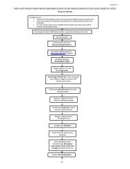
2.0 NOTIFICATION PROCEDURE1. When a doctor encounters a patient that satisfies the definition of any of the sixsyndromes in this <strong>manual</strong>, the doctor should complete the Syndromic NotificationForm (KKM-syndsurv/2003.2).2. The completed form should be sent to the nearest District Health Office (DHO)within 24 hours.3. DHO then should sent the <strong>notification</strong> form toi. State Health Officeii. Surveillance Section of Disease Control Division, Ministry of Health(MOH)e-mail: survelan@dph.gov.myfax no. : 03-888862714. DHO should conduct case <strong>investigation</strong>. The <strong>investigation</strong> format as in theappendix 5. Investigation should be done if:i. there are two or more cases of similar syndrome <strong>and</strong> they areepidemiologically link, whether by place of living or working; ORii. the case died or in bad condition <strong>and</strong> highly suspected of infectiousorigin; <strong>and</strong> / or has a direct link with any infectious disease event.5. In cases where a definitive diagnosis is confirmed (e.g. diagnosis already made bythe referring hospital or where rapid confirmatory test is available) the specificdisease should be notified under the m<strong>and</strong>atory <strong>notification</strong> system if it warranted<strong>notification</strong>.8
2.1 Notification FlowE D/A & E/WardNOTIFY SYNDROME(KKM-syndsurv/2003.2)FEEDBACKDISTRICTHEALTH OFFICEINVESTIGATESURVEILLANCE MOHAnalysisInterpretationResponseED – Emergency DepartmentE/Ward – Emergency WardREPORTFEEDBACKSTATEAnalysisInterpretationResponseA & E – Accident & Emergency DepartmentMOH – Ministry of Health9
3.0 SYNDROME DEFINITION3.1 Acute neurological syndromeA. Definition:Acute neurological dysfunction with one or more of the following:• deterioration of mental function• stupor/coma• acute paralysis• convulsion• signs of meningeal irritation e.g. neck stiffness, positive Kernig’ssign/Brudzinski’s sign• involuntary movements e.g. myoclonus, tremors• other neurological symptoms e.g. headache, visual disturbances,vomitingANDsevere illness (see glossary for definition)ANDabsence of predisposing factors (see glossary for definition).B. Possible diseases / pathogens:Poliomyelitis, Guillain Barre syndrome, enteroviral meningitis (e.g. EV71, Echovirus), Dengue, Nipah encephalitis, Japanese encephalitis, WestNile encephalitis, tick-borne virus encephalitis, meningococcal meningitis,mycoplasma, cerebral malaria, toxoplasma encephalitis, rabies, rickettsia,chemical agent / toxin.C. Specimens required include: faeces, blood / serum, CSF, throat swabD. Laboratory tests:1. Faeces : viral culture.2. Blood /serum - blood film for malaria, blood culture <strong>and</strong>sensitivity, serology <strong>and</strong> toxicology studies3. CSF: microscopic examination, cytology, biochemistry studies,culture <strong>and</strong> sensitivity, polymerase chain reaction (PCR), antigendetection <strong>and</strong> serology.4. Throat swab / washing / gargle : viral culture, bacterial culture <strong>and</strong>sensitivity.5. Post-mortem specimen ( tissue <strong>and</strong> body fluids): culture, PCR,antigen detection, serology, HPE.10
3.2 Acute respiratory syndromeA. Definition:Acute onset of cough or respiratory distress (e.g. tachypnoea, chestrecession, dyspnea, cyanosis)ANDsevere illness (see glossary for definition).ANDabsence of known predisposing factors (see glossary for definition).B. Possible diseases/pathogens:Respiratory syncytial virus, influenza, parainfluenza, adenovirus,cytomegalovirus, hantavirus pulmonary syndrome, diphtheria, pertussis,streptococcus, mycoplasma, chlamydia, legionella, respiratory anthrax,pneumonic plague, tularemia, chemical agent / toxin, SARS coronavirus,melioidosis, leptospirosis.C. Specimens required to include:1. Throat swab / gargle.2. Nasopharyngeal swab.3. Sputum.4. Bronchoalveolar lavage/tracheal aspirate.5. Pleural fluid.6. Blood /serum.7. Urine.D. Laboratory tests may include:1. Throat swab/ gargle: microscopy, culture <strong>and</strong> antigen detection.2. Nasopharyngeal swab / aspirate: microscopy, culture <strong>and</strong> antigendetection.3. Sputum: culture <strong>and</strong> antigen detection.4. Bronchoalveolar lavage/ tracheal aspirate: culture <strong>and</strong> antigendetection.5. Pleural fluid: culture <strong>and</strong> antigen detection.6. Blood / serum: culture & sensitivity / serology <strong>and</strong> toxicology.7. Urine: antigen detection ( for legionella).11
3.3 Acute dermatological syndromeA. Definition:Acute febrile illness with rash (rash can be erythematous, macular /papular <strong>and</strong> vesicular / pustular) OR other skin manifestations e.g.pruritus, desquamation, pigmentationANDabsence of known predisposing factors.B. Possible diseases / pathogens:Chickenpox, smallpox,enterovirus HFMD, measles, rubella, herpessimplex virus, monkey pox, chikungunya parvovirus B19, typhus,cutaneous anthrax, chemical agents/ toxin.C. Specimens required may include:1. Vesicular fluid2. Crust / swab at base of ulcer3. Pus4. Skin scraping / biopsy5. Blood / SerumD. Laboratory tests include:1. Vesicular fluid: viral culture, antigen detection, electron microscopy.2. Crust / swab at base of ulcer: microscopic examination , viral <strong>and</strong>bacterial culture, antigen detection3. Pus: microscopic examination, bacterial culture <strong>and</strong> sensitivity,antigen detection4. Skin scraping / biopsy: microscopic examination, culture, antigendetection, HPE5. Blood/serum – culture / serology, toxicology studyNote:Photographs of the skin lesion (preferably serial) are very useful <strong>and</strong>should be taken where possible.12
3.4 Acute haemorrhagic syndromeA. Definition:Acute onset of fever of less than 3 weeks durationANDany two of the following:• Haemorrhagic or purpuric rash• Epistaxis• Haematemesis• Haemoptysis• Blood in stool• Other haemorrhagic symptomsANDabsence of known predisposing factorsB. Possible diseases / pathogens:Dengue haemorrhagic fever <strong>and</strong> shock syndrome, haemorrhagic fever withrenal syndrome (Hantavirus), malaria, relapsing fever (Borreliosis), yellowfever, other viral haemorrhagic fevers ( Ebola, Marburg, Lassa fever, RiftValley, Tick-borne flaviviruses etc.)C. Specimens required include:Blood, blood smear: thin <strong>and</strong> thick smear, serum, post-mortem tissuespecimens: biopsies of liver <strong>and</strong> spleen <strong>and</strong> cerebrospinal fluid.D. Laboratory tests:• Blood smear: Thick <strong>and</strong> thin blood smear for demonstration ofparasites• Blood for viral culture• Serum: Bacterial <strong>and</strong> viral antigen detection, antibody levels <strong>and</strong>viral genome detection.• Cerebrospinal fluid: viral culture, viral antigen detection, antibodylevels <strong>and</strong> viral genome detection.• Post-mortem tissue specimens (biopsies of liver <strong>and</strong> spleen):Antigen detection, genome detection <strong>and</strong> histopathologicalexamination.13
3.5 Acute jaundice syndromeA. Definition:Acute onset of jaundice AND severe illness AND absence of knownpredisposing factors e.g. drugsB. Possible diseases / pathogens:Hepatitis A-E, malaria, yellow fever, leptospirosis <strong>and</strong> other spirochaetaldiseases.C. Specimens required include:Serum, blood smear, blood culture, urine, post-mortem liver biopsy.D. Laboratory tests:1. Serum: antigen detection, antibody levels, serotyping, genomedetection.2. Blood smear: thick <strong>and</strong> thin smear for demonstration of parasites.3. Blood culture: leptospiral <strong>and</strong> virus isolation <strong>and</strong> identification4. Urine: leptospiral antigen detection5. Post-mortem liver biopsy: culture, antigen detection, HPE, genomeanalysis.3.6 Acute diarrhoeal syndromeA. Definition:Acute onset of diarrhoea AND severe illness AND absence of knownpredisposing factors e.g. drugsB. Possible diseases / pathogens:Patient can present with watery diarrhoea or dysentery. The possiblecauses of watery diarrhoea are viral-gastroenteritis, cholera,enterotoxigenic E. coli, giardiasis <strong>and</strong> CyptosporidiumThe possible causes of dysentery are shigellosis, salmonellosis,campylobacteriosis, amoebic dysentery, enterohaemorrhagic E. coli,Clostridium difficile, Ebola <strong>and</strong> other haemorrhagic feversC. Specimens required include: FaecesD. Laboratory tests:1. Macro- <strong>and</strong> micro-scopic examination for parasites2. Faecal leukocytes count, culture <strong>and</strong> antimicrobial, susceptibilitytest for bacteria. Proceed to serotyping whenever applicable.3. Toxin detection in faeces for certain bacteria e.g. Bacillus cereus.4. Antigen detection <strong>and</strong> culture for virus; <strong>and</strong> proceed to genomeidentification.14
4.0 ALGORITHM FOR SYNDROMIC APPROACHSuspected outbreakACUTE NEUROLOGICAL SYNDROMEDefinitionAcute neurological dysfunction with one or more of thefollowing:• Deterioration of mental function• Stupor / coma• Acute paralysis• Convulsions• Signs of meningeal irritation e.g. neck stiffness• Involuntary movements e.g. tremor• Other neurological symptomsANDsevere illnessANDabsence of predisposing factors..Possiblediseases/pathogens• Poliomyelitis or• Guillain BarrésyndromeViral, bacterial,fungal or parasiticmeningoencephalitisRabiesSpecimensrequiredFaeces• CSF• Blood culture• Blood smear• Serum• Throat Swab• Serum• PostmortemLaboratorystudiesHPE – Histopathological ExaminationCSF - Cerebro-spinal fluidViral cultureBacterial (includingLeptospiral):• Gram stain <strong>and</strong> othermicroscopic techniques• Culture• Antimicrobialsusceptibility• Antigen detection• Toxicology• CSF Biochemistry• Viral culture• Antigendetection• Serology• HPE15
Suspected outbreakACUTE RESPIRATORY SYNDROMEDefinitionAcute onset of cough OR respiratory distressANDsevere illnessANDabsence of known predisposing factors.Possiblediseases/pathogens• Influenza• Respiratorysyncytial virus(RSV)• Diphtheria• Streptococcalpharyngitis•HantaviruspulmonarysyndromePertussisBacterial pneumoniaincluding:• Pneumococcal• Legionellosis• Mycoplasma• Respiratory anthrax• Pneumonic plague• Melioidosis• LeptospirosisLaboratorystudiesThroat swab Serum Nasopharyngealswab• Blood culture• Serum• Sputum• Urine (forLegionella)Bacterial/viral:Culture• Microscopy• Bacterial/Viral Culture• Antimicrobial susceptibility(for bacteria)• Antigen detection• Serology• Toxicology16
Suspected outbreakACUTE DERMATOLOGICAL SYNDROMEDefinitionAcute febrile illness with rash OR other skin manifestationsANDabsence of known predisposing factors.Possiblediseases/pathogens• Chickenpox• Smallpox• Measles• Rubella• TyphusCultaneousanthraxSpecimens • Vesicularfluid• CrustSerumLesion swab(vesicularexudate)LaboratorystudiesBacterial/Viral• Culture• Microscopic examination• Bacterial/Viral culture• Antigen detection• Serology• Electron microscopy on vesicularfluid ( smallpox)17
Suspected outbreakACUTE HAEMORRHAGIC FEVER SYNDROMEDefinitionAcute onset of fever of less than 3 weeks durationANDany two of the following:• Haemorrhagic or purpuric rash• Epistaxis• Haematemesis• Haemoptysis• Blood in stool• Other haemorrhagic symptomANDabsence of known predisposing factorsPossiblediseases/pathogens• Dengue haemorrhagic fever <strong>and</strong> shock syndrome• Haemorrhagic fever with renal syndrome (Hantavirus)• Malaria• Relapsing fever (Borreliosis)• Yellow fever• Other viral haemorrhagic fevers ( Ebola, Marburg, Lassafever, Rift valley, Tick-borne flaviviruses etc.)Specimens• Blood• Blood smear: Thin <strong>and</strong> thick smear• Serum• Post-mortem tissue specimens:-Biopsies of liver <strong>and</strong> spleen-Cerebrospinal fluidLaboratorystudiesParasitic:• Thick <strong>and</strong> thinblood smear fordemonstrationof parasitesBacterial:• Antigendetection• AntibodylevelsViral:• Culture• Antigen detection• Antibody levels• Genome detection18
Suspected outbreakACUTE JAUNDICE SYNDROMEDefinitionAcute onset of jaundice AND severe illness AND absence of knownpredisposing factors e.g. drugsPossiblediseases/pathogensMalaria• Leptospirosis<strong>and</strong> otherspirochaetaldiseasesHepatitisA-EYellowfeverSpecimensBloodsmear• Blood culture• UrineSerumPostmortemliver biopsyLaboratorystudiesParasitic:• Thick <strong>and</strong> thinblood smear fordemonstrationof parasitesLeptospiral:• Culture• Antibody levels• Antigen detection(urine)• Serotyping (MAT)Viral:• Culture• Antigen detection• Antibody level• Genome analysis19
Suspected outbreakACUTE DIARHOEAL SYNDROMEDefinitionAcute onset of diarrhoeaANDsevere illnessANDabsence of known predisposing factorsPossiblediseases/pathogensSpecimensWatery• Viralgastroenteritis• Cholera• EnterotoxigenicE. coli• Giardiasis• CyptosporidiumDysentry• Shigellosis• Salmonellosis• Campylobactreriosis• Amoebic dysentery• EnterohaemorrhagicE. coli• Clostridium difficile• Ebola <strong>and</strong> otherhaemorrhagis fevers.FaecesLaboratorystudiesParasitic:Macro- <strong>and</strong>micro-scopicexaminationBacterial:• Faecalleukocytes• Culture• Antimicrobial;susceptibility• Serotyping• ToxinViral:• Culture• Antigen detection• Genome detectionNote: Ebola <strong>and</strong> other haemorrhagic fever may initially present as bloody diarrhoea. If such anaetiology is suspected, refer to “Acute Haemorrhagic Fever Syndrome” for appropriate specimencollection guidelines.20
5.0 COLLECTION AND HANDLING PROTOCOL FOR SPECIFICSPECIMENS5.1 CEREBROSPINAL FLUID (CSF) SPECIMEN COLLECTIONThe specimen must be taken by a medical doctor experienced in the procedure. CSF isused in the diagnosis of viral, bacterial, parasitic <strong>and</strong> fungal meningitis/encephalitis.Materials for collectionSt<strong>and</strong>ard lumbar puncture tray is needed.Method of collectionAs only experienced personnel should be involved in the collection of CSF samples, themethod is not described in this document. CSF is collected directly into separate screwcaptubes. If the samples cannot be promptly transported, separate tubes should becollected for bacterial <strong>and</strong> viral processing (refer summary table).H<strong>and</strong>ling <strong>and</strong> transportIn general, specimens should be delivered to the <strong>laboratory</strong> <strong>and</strong> processed as soon aspossible.• CSF specimens for bacteriology are transported at ambient temperature,generally without transport medium. They must never be refrigerated as manyof the relevant pathogens do not survive well at low temperatures.• CSF specimens for virology do not need transport medium. They may betransported at 4 to 8 o C for up to 48 hours or at –70 o C / dry ice for longerperiods.5.2 RESPIRATORY TRACT SPECIMEN COLLECTIONSpecimens are collected from the upper or lower respiratory tract, depending on the siteof infection. Upper respiratory tract pathogens (viral <strong>and</strong> bacterial) are found in throat<strong>and</strong> nasopharyngeal secretions. Lower respiratory tract pathogens are found in sputumspecimens. For organisms such as Legionella, culture is difficult, <strong>and</strong> diagnosis is bestbased on the detection of antigen excreted in the urine.In patients with stridor or when acute epiglottitis is suspected, no attempt should be madeto take throat or pharyngeal specimens <strong>and</strong> neck X rays, since these procedures mayprecipitate respiratory obstruction. However the aetiological agent may be isolated fromblood culture21
5.2.1 Protection for Health Care WorkersWhen taking respiratory specimen, HCW should exercise droplet <strong>and</strong> contact precaution.HCW should wear a surgical or N95 mask as appropriate. If the procedure involves ahigh risk of splashing of contamination by the clinical specimens, the HCW should wearappropriate eye protection.Materials for collection• Transport media – bacterial <strong>and</strong> viral• Cotton swabs• Tongue depressor• Flexible wire calcium alginate tipped swab (for suspected pertussis)• Nasal speculum (for suspected pertussis – not essential)• Suction apparatus or 20-50 ml syringe• Sterile screw-cap tubes, <strong>and</strong> wide-mouthed clean sterile jars (minimum volume25 ml).5.2.2 Upper respiratory tract specimensMethod of collecting a throat swab1. Hold the tongue down with the depressor. Use a strong light source to locate areasof inflammation <strong>and</strong> exudate in the posterior pharynx <strong>and</strong> the tonsillar region ofthe throat behind the uvula.2. Rub the area back <strong>and</strong> forth with a cotton swab. Withdraw the swab withouttouching cheeks, teeth or gums <strong>and</strong> insert into a screw-cap tube containingtransport medium.3. Break off the top part of the stick without touching the tube <strong>and</strong> tighten the screwcap firmly.4. Label the specimen containers.5. Complete the <strong>laboratory</strong> request form.Method of collecting pernasal <strong>and</strong> post-nasal swabs (for suspected pertussis)1. Seat the patient comfortably, tilt the head back <strong>and</strong> insert the nasal speculum.2. Insert a flexible calcium alginate swab through the speculum parallel to the floorof nose without pointing upwards. Alternately, bend the wire <strong>and</strong> insert it into thethroat <strong>and</strong> move the swab upwards into the nasopharyngeal space.22
3. Rotate the swab on the nasopharyngeal membrane a few times, remove itcarefully <strong>and</strong> insert it into a screw-cap tube containing transport medium.4. Label the specimen tube.5.2.3 Lower respiratory tract specimensMethod of collecting sputum1. Instruct patient to take a deep breath <strong>and</strong> cough up sputum directly into a widemouthedsterile container. Avoid saliva or post-nasal discharge. Minimum volumeshould be about 1 ml.2. If no sputum, induce cough by hypertonic saline nebulisation.3. Label the specimen containers.4. Complete the <strong>laboratory</strong> request form.5.2.4 Bronchoalveolar / tracheal lavageMethod is not documented here as these procedures are only performed byexperienced personnel.H<strong>and</strong>ling <strong>and</strong> transport• All respiratory specimens except sputum are transported in appropriate bacterial /viral media.• Transport as quickly as possible to the <strong>laboratory</strong> to reduce overgrowth bycommensal oral flora.• For transit periods up to 24 hours, transport bacterial specimens at ambienttemperature <strong>and</strong> viruses at 4 to 8 °C in appropriate media.5.3 COLLECTING SPECIMENS OF SKIN LESIONSFor most dermatological conditions, diagnosis may be established on the basis of clinicalhistory <strong>and</strong> physical examination. Important characteristics to be noted on physicalexamination include the nature of the skin lesions (erythematous, macular, papular,maculopapular, vesicular, bullous, petechial, purpuric, etc.) <strong>and</strong> the anatomic distributionof spread (central, peripheral, diffuse, etc.).23
In cases of indeterminate diagnoses, unusual presentations, <strong>and</strong> some rare conditions,collection of specimens from rashes <strong>and</strong>/or skin lesions may be necessary. In the case ofvesicular rashes, specimens for microscopy <strong>and</strong> culture are taken directly from vesicles.In other exanthemata (macular <strong>and</strong>/or papular), the diagnosis may be more readilyestablished from alternative specimens (e.g. blood cultures, serology). In suspectedcutaneous anthrax or bubonic plague, specimens from the skin lesions (eschars <strong>and</strong>buboes respectively) <strong>and</strong> blood cultures should be taken.Materials for collection• Sterile saline• Sterile swabs <strong>and</strong> appropriate transport media• Sterile screw-cap vials• Sterile lancets or needles (for piercing of vesicles)• Syringe with wide-bore needle (for aspiration of abscesses/buboes)• Wide-mouthed screw-cap containers (for biopsy specimens)• Glass slides <strong>and</strong> slide boxes.Method of collectionVesicular or vesiculo-pustular rash (for diagnosis of viral infections)1. Pierce roof of fluid-containing vesicle with sterile lancet.2. Swab fluid with sterile swab. Try to get a good amount of fluid onto the swab.3. Take a clean labeled microscope slide <strong>and</strong> make a smear with the swab in thecentral area of the slide of approximately the size of a 5 sen coin.. Make 2 slides ifpossible. The slides should be left to dry in air.4. Place swab directly into virus transport medium.5. Label the bottles or tubes containing swabs in transport media.6. When glass slides have dried, place carefully into a plastic slide box.7. Do not refrigerate or freeze the slides during storage or transport. Keep in theclosed container at room temperature.Crusting stage1. Gently ease off crust with a lancet, scalpel or a pair of disposable forceps.2. Take 5-10 crusts; place them in a plastic screw-cap vial. Make sure the lid istightly closed.24
3. Label the specimen containers.4. Discard forceps, lancets, <strong>and</strong> scalpels into sharps disposal container. Do not re-useforceps on specimens from another patient.NOTE: If cutaneous anthrax is suspected, the vesicular fluid under the eschar is abetter diagnostic specimen than a piece of the crust.Aspiration of abscesses• Disinfect the skin overlying the abscess / bubo with 70% isopropyl alcohol.• Aspirate the fluid from the abscess with a sterile needle <strong>and</strong> syringe.• Transfer the aspirate aseptically into a sterile tube.Skin biopsySkin biopsies are generally not appropriate specimens for field outbreak <strong>investigation</strong>s.When necessary, skin biopsy should be done by trained personnel.H<strong>and</strong>ling <strong>and</strong> transportSpecimens for bacteriological analysis should be transported in Stuart’s or Amiestransport medium. Swabs for suspected viral pathogens should be transported in virustransport medium. Other specimens should be h<strong>and</strong>led as described in the relevantsection.If processing takes longer than 2 hours, bacteriology specimens can be maintained atambient temperature for 24 hours. Specimens for virus isolation may be refrigerated at 4to 8°C, <strong>and</strong> transported to the <strong>laboratory</strong> as rapidly as possible. In some instances, theoutbreak <strong>investigation</strong> team may bring liquid nitrogen or carbon dioxide ice for specimenpreservation.5.4 BLOOD SPECIMEN COLLECTION• Blood can be used for isolation of pathogens when inoculated into culturemedium or separated into serum for the detection of genetic material (e.g. usingPCR), specific antibodies, antigens, toxins (e.g. by ELISA).• Blood specimens for culture should be taken before antimicrobial agents areadministered to the patient.• Serum is preferable to whole blood for diagnosis of viral infections. However formolecular diagnosis, blood collected in an EDTA bottle may be required whileclotted blood may be needed for viral haemorrhagic fever diagnosis.• When specific antibodies are being assayed, it is often helpful to collect pairedsera, i.e. an acute sample at the onset of illness <strong>and</strong> a convalescent sample, one tofour weeks later.25
• Blood can also be collected by finger prick for the preparation of slides formicroscopy or for absorption onto special filter paper discs for analysis.5.4.1 Venous blood sampleContainers1) Blood culture bottles2) EDTA bottleMethod of collection1) If withdrawing with conventional disposable syringes, withdraw whole blood- 5-10 ml from adults- 2-5 ml from children- 0.5 – 2 ml from infants2) If withdrawing with vacuum systems, withdraw the desired amount of blooddirectly into each EDTA <strong>and</strong> culture bottle.3) Using aseptic technique, transfer the specimen to the relevant containers. Securecap tightly.H<strong>and</strong>ling <strong>and</strong> transport• Blood culture bottles <strong>and</strong> blood collected in EDTA tubes should be transportedupright <strong>and</strong> secured in a screw cap container or in a rack in a transport box.• Cushion or suspend bottles during transport over rough terrain to prevent lysis ofred cells. They should have enough absorbent paper around them to soak up allthe liquid in case of a spill.• If the specimen reaches the <strong>laboratory</strong> within 24 hours, most bacterial pathogenscan be recovered from blood cultures transported at ambient temperature.5.4.2 SerumMaterials1) Sterile disposable transfer pipettes2) Sterile screw-cap 5 ml tubes – 2 per sampleMethod of separation of serum from blood1) Draw 10 ml of venous blood <strong>and</strong> transfer to a screw cap tube without anticoagulant.Alternatively, blood may collected directly into a proprietarycollection <strong>and</strong> transport tube (e.g., vacutainer, etc.)2) Let the specimen clot for 30 minutes at ambient temperature, then place in a coolbox to retract at 4 to 8 o C for a minimum of 1-2 hours (it may be stored at this26
temperature for 48-72 hours). If a centrifuge is not available, allow 4-6 hours forclot retraction to occur.3) Separate the serum aseptically from the clot using the sterile disposable transferpipette. Carefully remove the clear yellow serum whilst taking care to keep the tipas far as possible from the clot. Avoid agitating the blood tube during the removalprocess. Transfer equally to 2 plastic screw cap tubes. Secure the caps tightly.4) If a centrifuge is available, spin the specimen at low speed (1000g for 10 minutes)to remove residual blood cells. If viral haemorrhagic fever is strongly suspected,samples should only be processed in properly equipped, specialized laboratories.Discuss with the <strong>laboratory</strong> whether a separation gel tube would be acceptable<strong>and</strong> whether clotted blood is required.H<strong>and</strong>ling <strong>and</strong> transport• If serum will be required for testing, separation from blood should take place assoon as possible, preferably within 24 hours at ambient temperature.• If a specimen will not reach a <strong>laboratory</strong> for processing within 24 hours, serumshould, if at all possible, be separated from blood prior to transportation.• Serum may be stored at 4 to 8 o C for up to 10 days.• If testing is delayed for a longer period, serum samples should be frozen at –20 o C.• If separation on site is not possible, or is inadvisable for safety reasons, the bloodsample should be stored at 4 to 8 o C <strong>and</strong> be sent to a nearest center as soon aspossible. Protect such unseparated samples from excessive vibration whiletransporting. WHOLE BLOOD SAMPLES SHOULD NOT BE FROZEN.5.4.3 Capillary blood sampleMaterials for collection1) Disposable sterile lancets2) Glass slides, cover slips <strong>and</strong> slide box3) Filter paper4) Fixatives such as methanolMethod of collection1) Disinfect finger or thumb for adults, thumb for children, or side of heel for infantsby swabbing with 70 % isopropyl alcohol <strong>and</strong> prick with a sterile lancet. Wipeaway the first drop of blood.2) Discard used lancets directly into the sharps disposal containers.3) Collect blood directly onto labeled glass slides <strong>and</strong>/or filter paper.27
Method of preparation of blood filmsBlood films should be made by trained personnel. If this is not possible, they can bespread from heparinised or EDTA blood specimens sent to the <strong>laboratory</strong>.Thick films for microscopy1. Label the slide with patient identification number <strong>and</strong> name.2. Disinfect <strong>and</strong> prick site with lancet as described above.3. Touch one or more large drops of blood onto the center of the slide making surethat the slide does not touch the skin.4. Spread the blood in a circle about 1 cm in diameter using the corner of anotherglass slide.5. Air dry the film in a horizontal position. Do not dry the film by heating over aflame or other heat source.Thin films for microscopy1. Label the slide with patient identification number <strong>and</strong> name.2. Touch another drop of blood to the glass slide about 2 cm from one end makingsure that the slide does not touch the skin.3. Place the slide horizontally on a flat surface.4. Hold the side of a second clean glass slide (the spreader) on to the center of thespecimen slide <strong>and</strong> move it back until it touches the drop <strong>and</strong> the blood spreadsalong its base.5. At an angle of about 45°, move the spreader firmly <strong>and</strong> steadily across thespecimen slide <strong>and</strong> air dry the film quickly. Do not dry over a flame or other heatsource.6. Fix the dried film by dipping the glass slide in methanol or other fixative for a fewseconds <strong>and</strong> air dry.28
H<strong>and</strong>ling <strong>and</strong> transport• Air dried <strong>and</strong>/or fixed films are transported at ambient temperature preferablywithin 24 hours of specimen collection. They must not be refrigerated. Thick <strong>and</strong>thin films are usually kept in separate slide boxes.5.5 FAECAL SPECIMEN COLLECTION• Stool specimens are most useful for microbiological diagnosis if collected soonafter diarrhoea (for viruses < 48 hours, for bacteria < 4 days), <strong>and</strong> preferablybefore the initiation of antimicrobial agent therapy.• If required, two or three specimens may be collected on separate days. Stool isthe preferred specimen for culture of bacterial, viral <strong>and</strong> parasitic diarrhoealpathogens.• Rectal swabs showing faeces may also be used from infants. In general, rectalswabs are not recommended for the diagnosis of viruses.Materials for collection1) Clean, dry leak-proof screw cap containers <strong>and</strong> tape.2) Appropriate bacterial transport media for transport of rectal swab from infants.3) Parasitology transport pack: 10 % formalin in water, polyvinyl isopropyl alcohol(PVA)Method of stool collection1. Collect freshly passed stool, 5 ml liquid (a teaspoonful) or 5 gm solid (peanutsized), in a container.2. Label the container.Method of rectal swab collection from infants1) Moisten a swab in sterile saline.2) Insert the swab tip just pass the anal sphincter <strong>and</strong> rotate gently.3) Withdraw the swab <strong>and</strong> examine to ensure that the cotton tip is stained withfaeces.4) Place the swab in a sterile tube/container with the appropriate bacterial <strong>and</strong>/orviral transport medium.29
5) Break off the top part of the stick without touching the tube <strong>and</strong> tighten the screwcap firmly.6) Label the specimen tube.H<strong>and</strong>ling <strong>and</strong> transport• Stool specimens should be transported at 4 to 8 o C. Bacterial yields may fallsignificantly if specimens are not processed within 1 to 2 days of collection.Shigella are particularly sensitive to elevated temperatures.• Specimens to be examined for parasites should be mixed with 10% formalin orPVA, 3 parts stool to 1 part preservative. Transport at ambient temperature incontainers <strong>and</strong> sealed in plastic bags.5.6 URINE SPECIMEN COLLECTIONMaterials for collection1) Sterile plastic cup with lid (50 ml or more)2) Clean, screw-top specimen transport containers (“universal” containers are oftenused)3) Gauze pads.4) Soap <strong>and</strong> clean water (or normal saline) if possible.Method of collection1) To reduce the contamination, it may be necessary to wash the external genitaliawith soap <strong>and</strong> clear water. If soap <strong>and</strong> clean water are not available, the area maybe rinsed with normal saline. Dry the area thoroughly with gauze pads beforecollecting the urine.2) Give the patient clear instructions to pass urine for a few seconds, <strong>and</strong> then tohold the cup in the urine stream for a few seconds to catch a mid-stream urinesample. This should decrease the risk of contamination from organisms present inthe urethra.3) To decrease the risk of contamination from skin organisms, the patient should bedirected to avoid touching the inside or rim of the plastic cup with the skin of theh<strong>and</strong>s, legs or external genitalia. Tighten the cap firmly when finished.4) Urine collection bags may be necessary for infants. For boys, clean-catchspecimens may be also collected. If used, transfer urine from the urine bag to30
specimen containers as soon as possible to prevent contamination with skinbacteria. Use a disposable transfer pipette to transfer the urine.5) Label the specimen container.H<strong>and</strong>ling <strong>and</strong> transport• Transport to the <strong>laboratory</strong> within 2 to 3 hours of collection. If this is not possibledo not freeze but keep the specimen refrigerated at 4 to 8 o C. Keeping thespecimen refrigerated will decrease the risk of contaminating organisms.• Ensure that the containers are leak-proof <strong>and</strong> tightly sealed.31
6.0 PROTOCOL FOR COLLECTION, STORAGE, PACKAGE ANDDISPATCH OF SPECIMENS WITH SUSPECTED UNKNOWNVIRUS INFECTIONSamples required- Blood in EDTA (5 mls) - Plain sterile plastic tube(paired samples)- Do not add VTM, Do not freeze, keep at 4 0 C- Throat swab place in sterile plastic tube containing 1/2 inch(from tonsillar area)of VTM (viral transport medium)[refer to figure below]- Vesicle swab*- Stool (at least 10 gm) - place in sterile container } Do not add VTMORDo not freeze, keepat 4 0 CRectal swab (must see stool - place in sterile plastic tube containing 1/2 inchon swab)of VTMPlastic tubeSwab1/2 inch of VTM- CSF (at least 1ml)*- plain sterile plastic tube*when clinically indicatedSample required from patients with lung involvement:Broncho-Alvedar Lavage (BAL) / Tracheal aspirate- place in sterile plastic tube container(keep cold in ice <strong>and</strong> ship as soon as possible)Samples required from fatal cases:Collect all specimens mentioned above, in addition collect:Cardiac tissueLiver tissueLung tissueBrain stem tissueplace in sterile plastic tube container(keep cold <strong>and</strong> ship as soon as possible)32
All samples must be sent in ice. Please provide full clinical details including name ofthe doctor filling the form <strong>and</strong> patient’s I/C in IMR 135 or hospital form of PathologyRequest Form.SAMPLES AND FORM TO BESENT TO:Bahagian Virologi (Makmal Virus Kultur)Institut Penyelidikan PerubatanJalan Pahang, 50588 Kuala LumpurTel: 03-40402345 / 346 / 347Fax: 03-26936323ORNational Public Health LaboratorySungai Buloh, Selangor.Tel: 03-61565109 / 61402209 / 61402213Fax: 03-6140224933
6.1 PACKING FOR SAMPLES FROM PATIENTS WITH SUSPECTEDEXOTIC VIRUS INFECTIONContainers <strong>and</strong> packing:Samples should be packaged in three layers (see to diagram):(1) a primary watertight non breakable container containing the sample• it must be firmly capped <strong>and</strong> the cap should then be sealed with parafilm,adhesive cloth or zinc oxide tape (not cellulose tape)• the container must then be cocooned in absorbent material• several primary containers may be packed in one secondary container(2) a secondary watertight non breakable container enclosing enough absorptive materialbetween it <strong>and</strong> the primary container to absorb all of the fluid in the specimen in caseof leakage• it must be firmly capped <strong>and</strong> sealed in the same way as the primary container• the secondary container must then be packed firmly with absorbent material intothe outer container• several secondary containers may be packed in one outer container(3) an outer container which is intended to protect the secondary package from outsideinfluence, such as physical damage <strong>and</strong> water, during transportation• absorbent, shockproof packing between the secondary <strong>and</strong> outer containers• the lid is again sealed with tape34
PACKING INFECTIOUS SUBSTANCES FOR THE POSTFigure 1: Packing infectious substance for the post35
APPENDIX 1SUMMARY FOR COLLECTING AND HANDLING SPECIMENSSPECIMEN CONTAINER AMOUNT STORAGE & TRANSPORT PRECAUTIONCSF Plain sterile bottle Minimum – 0.5ml each in 3different bottlesTransport in sealed container as soon as possible(ASAP)Virus – 4 to 8 0 CIf >48 hr, -70 0 C.Bacteria/fungus – Ambient temperature.If >24 hr, keep at 37 0 C (incubator).Parasite- Ambient temperature.If >24 hrs, keep in polyvinyl alcohol preservativeBiochemical examination – Ambient temperatureFor bacterial culture - Donot refrigerate CSF sampleThroat swabCotton bud in transportmedium.Virus- VTMBacteria- Stuart/AmiesSwab must befully immerse inthe transportmediumTransport in sealed container as soon as possible(ASAP)Virus - 4 to 8 0 CPatient with stridor orsuspect acute epiglotitis -do not do throat swabBacteria - Ambient temperature36
SPECIMEN CONTAINER AMOUNT STORAGE & TRANSPORT PRECAUTIONPernasal /nasopharyngealswabCotton bud in transportmedium.Calcium alginate swab intransport medium (forpertussis).Swab must be fully immersein the transport mediumVirus - 4 to 8 0 CBacteria - Ambient temperatureNasopharyngealaspiratePlain sterile bottleSufficient amountdepending on number oftests requestedVirus - 4 to 8 0 CBacteria – Ambient temperature.If >24 hrs - 4 to 8 0 CThroat gargle Plain sterile bottle Sufficient amountdepending on number oftests requestedVirus - 4 to 8 0 CBacteria – Ambient temperatureIf >24 hrs - 4 to 8 0 CSputumPlain sterile containerSufficient amountdepending on number oftests requestedTransport in sealed container as soon aspossible (ASAP)Virus - 4 to 8 0 CBacteria – Ambient temperatureIf >24 hrs - 4 to 8 0 C37
SPECIMEN CONTAINER AMOUNT STORAGE & TRANSPORT PRECAUTIONBronchial –aveolar lavage(BAL)Plain sterile bottleSufficient amountdepending onnumber of testsrequestedVirus - 4 to 8 0 CBacteria – Ambient temperatureIf >24 hrs - 4 to 8 0 CTrachealaspiratePlain sterile bottleSufficient amountdepending onnumber of testsrequestedVirus - 4 to 8 0 CBacteria - Ambient temperatureIf >24 hrs - 4 to 8 0 CPleural fluid Plain sterile bottle Sufficient amountdepending onnumber of testsrequestedVirus - 4 to 8 0 CBacteria - Ambient temperatureIf >24 hrs - 4 to 8 0 CVesicle fluidCotton swab - VTMfor viral cultureSwab must be fullyimmerse in thetransport mediumVirus - 4 to 8 0 CCrust / swabbase of escharVirus – in VTMBacteria- inStuart/Amiestransport mediumSwab must be fullyimmerse in thetransport mediumVirus - 4 to 8 0 CBacteria – Ambient temperature38
SPECIMEN CONTAINER AMOUNT STORAGE & TRANSPORT PRECAUTIONAbscess:- Needleaspirate- Drained abscess- SwabSterile leak-proofcontainerSwab in Amies transportmediaTransport ASAP at ambient temperatureIf more than 24 hrs, refrigerated at 4 to8 0 CSkin scraping/biopsyFor culture - sterile leakproofcontainerHPE- 10% FormalinVirus - 4 to 8 0 CBacteria –Ambient temperature.If >24 hr, store in refrigerator (4 to 8 0 C)Skin scraping : Transport to the<strong>laboratory</strong> in a cardboard mailer.Whole bloodBlood culture bottle5 to 10 ml from adults2 to 5 ml from children0.5 to 2 ml from infantsTransport upright in a rack in a transportbox.Ambient temperature if able to reach thelab within 24 hours.Blood collected in EDTAbottleDo not exceed the specimenlevel mark on the container4 to 8 o C. DO NOT FREEZEBlood smear 2 clean glass slides Thick <strong>and</strong> thin blood smear Transport in plastic slide box39
SPECIMEN CONTAINER AMOUNT STORAGE & TRANSPORT PRECAUTIONSerumFaecesSterile screw-cap 5 mltubesClean, dry leak-proofscrew cap containersAppropriate bacteriologytransport mediaVirus transport mediaAdults : 5 to 10 mlChildren : 3 to 5 mlInfant : 1 to 2 ml5 ml liquid (a teaspoonful) or5 g solid (peanut sized)4 to 8 o C for up to 10 days–20 o C up to 2 years- 70 o C for more than 2 yearsat 4 to 8 o CFor bacterial isolation,need to process within1 to 2 days ofcollectionParasitology transportpack: 10 % formalin inwater, polyvinylisopropyl alcohol (PVA)5 to 10 ml3 part stool to 1 part preservative.Transport at ambient temperature incontainers sealed in plastic bags.UrineSterile plastic cup withlid (50 ml or more)Clean, screw-topspecimen transportcontainersTransport to the <strong>laboratory</strong> within 2 to 3hours of collection or store.at 4 to 8 o C40
DIRECTORY OF LABORATORY SERVICESAPPENDIX 2DISEASE/PATHOGEN SERVICE SPECIMEN REQUIRED TESTING LABORATORYMicroscopic examination Blood smear: Thick <strong>and</strong> thin State hospitals <strong>and</strong> district hospital withmicrobiologists.Virus isolation Blood, CSF IMR (Virology Division)Acute haemorrhagic syndromeAcute jaundice syndromeAcute neurological syndromeBacterial <strong>and</strong> viral antigen Serum, tissue, CSF State hospitals <strong>and</strong> district hospital withdetectionmicrobiologists.Viral genome detection Serum, tissue, CSF IMR (Virology Division), NPHL.Antibody levels Serum, CSF State hospitalsAntigen detection, antibody Serum, tissueState hospitalslevels,Microscopic examination Blood smear: Thick <strong>and</strong> thinsmearState hospitals <strong>and</strong> district hospital withmicrobiologists.Bacterial isolation Blood, tissue State hospitalsViral isolation Blood, tissue IMR (Virology Division)NPHLVirus serotyping, genome Serum, tissueIMR (Virology Division)detectionBacterial isolation,identification <strong>and</strong>susceptibility testingThroat swab/washing/gargle,tissue <strong>and</strong> other body fluidsState hospitals <strong>and</strong> district hospital withmicrobiologists <strong>and</strong> Public Health Laboratories41
DISEASE/PATHOGEN SERVICE SPECIMEN REQUIRED TESTING LABORATORYAcute neurological syndromeAcute respiratory syndromeAcute respiratory syndromeVirus isolationFaeces, throat swab / washing IMR (Virology Division)/ gargle, tissue <strong>and</strong> other body NPHLfluidsSerology Serum State hospitals <strong>and</strong> district hospital withmicrobiologists, IMR (Virology division).Bacterial isolation,identification <strong>and</strong>susceptibility testingVirus isolation, antigentestingBlood culture, throat swab/gargle, sputum,nasopharyngeal swab /aspirate, bronchoalveolarlavage / tracheal aspirate,pleural fluidThroat swab / gargle,nasopharyngeal swab /aspirate, bronchoalveolarlavage / tracheal aspirate,pleural fluidState hospitals <strong>and</strong> district hospital withmicrobiologists.IMR (Virology Division)NPHLAdenovirus Isolation Throat swab, stool Virology division(IMR)Amoebic dysenteryMicroscopyAntigen detectionStoolState hospitals <strong>and</strong> district hospital withmicrobiologistsState hospital, Parasitology division (IMR)AnthraxMicroscopic examinationof clinical specimenBlood, sputum, skin or ulcertissueState hospitals <strong>and</strong> district hospital withmicrobiologists42
DISEASE/PATHOGEN SERVICE SPECIMEN REQUIRED TESTING LABORATORYAnthraxBacterial culture identificationBacterial typing, Pulsed FieldGel ElectrophoresisBordetella pertussis <strong>and</strong> otherbordetellaeIsolation <strong>and</strong> identification Blood, sputum, skin or ulcertissueIdentification of pure Pure, actively growingisolates determined to be of culture on suitable agar slantclinical significanceTo determine if isolates Pure isolates on agar slantsfrom different sources arethe sameIsolationSputum, nasopharyngealaspirateBSL 3 laboratories (VRI, IMR)IMR (Bacteriology division)IMR (Bacteriology division)State hospitals <strong>and</strong> district hospital withmicrobiologistsBordetella pertussis <strong>and</strong> otherbordetellaeBrucellosisIdentification susceptibility Pure cultureState hospitalstesting of BordetellaisolatesAntigen detection (IF) Nasopharyngeal swab State hospitalsBordetella pertussisserologyCharacterisation ofBordetella isolatesCulture <strong>and</strong> susceptibilitytestingSerumPure cultureBlood, bone marrow, abscess,liver or spleen biopsyState hospitals, Public Health LaboratoriesIMR (bacteriology division),Public HealthLaboratoriesState hospitals <strong>and</strong> district hospital withmicrobiologists. Primary specimens for isolation <strong>and</strong>identification are acceptable with prior consultation43
DISEASE/PATHOGEN SERVICE SPECIMEN REQUIRED TESTING LABORATORYChlamydia pneumoniaeCorynebacterium diptheriaeAntigen detection Brocho- alveolar lavage State hospitalsBracho-alvedarSerology Serum State hospitalsIsolation <strong>and</strong> identification Swab from inflamed areas of State hospitals <strong>and</strong> district hospital withthe membranes in throat <strong>and</strong> microbiologistsnasopharynx, skin lesion <strong>and</strong>materials removed fromwounds by swab or aspirationIn-vitro toxin testing Pure culture National PHLClostridium perfringesEnteric pathogens (Salmonella,Shigella, Yersinia, E. coli0157:H7, Campylobacter,Vibrio, Aeromonas,PleisomonasEnterovirusinfections(Coxsackieviruses,echoviruses, poliovirus)Culture <strong>and</strong> susceptibilitytestingCulture, identification <strong>and</strong>susceptibility testingAntigen detection (IF)Isolation <strong>and</strong> strain typingStoolStool, rectal swabStool, throat swab, CSF,vesicle fluid <strong>and</strong> tissueState hospitalsState hospitals <strong>and</strong> district hospital withmicrobiologistsState hospitalsIMR (Virology Division)44
DISEASE/PATHOGEN SERVICE SPECIMEN REQUIRED TESTING LABORATORYEnterovirusinfections(Coxsackieviruses,echoviruses, poliovirus)Genome detectionStool, throat swab, CSF,vesicle fluid <strong>and</strong> tissueIMR(Virology Division)EIA for mumps, measles SerumState hospitals <strong>and</strong> Public Health Laboratories<strong>and</strong> rubellaExanthematous viral infections IFA Vesicular fluid, lesion swab State hospitals <strong>and</strong> Public Health LaboratoriesHaemophilus ducreyi Isolation, identification <strong>and</strong> Genital ulcer swab, aspirated State hospitals <strong>and</strong> district hospital withsusceptibility testing pusmicrobiologists, Public Health LaboratoriesHIV Screening <strong>and</strong> confirmation Serum State hospitals <strong>and</strong> district hospital withmicrobiologists, Public Health LaboratoriesAntigen testState hospitals, Public Health Laboratories.Throat swab, nasopharyngeal Note: Need to confirm positives with conventionalswab, bronchial wash or other virus isolation <strong>and</strong> subtyping by Virology Division,respiratory specimen IMR. Specimens tested negative are not reported untilInfluenzae virus/ parainfluenzavirusInfluenzae virus/ parainfluenzavirusIsolation <strong>and</strong> typing ofinfluenza virus by shellvialsIsolation <strong>and</strong> typing byconventional cultureconventional culture results are rState hospitals.Throat swab, nasopharyngealNote: Specimens tested negative are not reported untilswab, bronchial wash or otherconventional culture results are reported.respiratory specimenVirology Division, IMRThroat swab, nasopharyngealswab, bronchial wash or otherrespiratory specimen45
DISEASE/PATHOGEN SERVICE SPECIMEN REQUIRED TESTING LABORATORYIsolation <strong>and</strong> identificationof Legionella pneumophilaserogroup 1Lung tissue, pleural fluid,transtracheal aspirate <strong>and</strong>lower respiratory secretionsState hospitals, Public Health LaboratoriesLegionellosisLeptospirosisMeningococcal diseaseNeisseria gonorhoeaSpeciation <strong>and</strong>serogrouping of otherlegionellaIsolation, identification <strong>and</strong>susceptibility testingPure cultureBlood <strong>and</strong> CSF (1st 10 daysof illness), urine (2nd week -30 days)Bacteriology Division, IMRState hospitals, Public Health LaboratoriesAntigen detection Urine State hospitals, Public Health LaboratoriesEIA - screeningSerumState hospitals, Public Health LaboratoriesMAT- confirmatoryBacteriology Division, IMRBacterial culture, CSF, bloodState hospitals <strong>and</strong> district hospital withidentification, susceptibilitymicrobiologists.testing <strong>and</strong> antigen testingCulture <strong>and</strong> susceptibilitytestingEndocervical swab, urethralswab, eye swab in Amiestransport media. Preferablyswab from suspected site ofinfection is streaked on toselective media e.g. ThayerMartinState hospitals <strong>and</strong> district hospital withmicrobiologists <strong>and</strong> Public Health Laboratories46
DISEASE/PATHOGEN SERVICE SPECIMEN REQUIRED TESTING LABORATORYNeisseria gonorhoea Strain typing Pure culture IMR (Bacteriology Division)Isolation onlySputum, blood, CSF, gastriclavage, skin lesion material,tissue, stool, urineState hospitals <strong>and</strong> district hospital withmicrobiologistsTuberculosisIsolation, genusidentification <strong>and</strong>susceptibility testingSputum, blood, CSF, gastriclavage, skin lesion material,tissue, stool, urineState hospitals with automated Tb culture system <strong>and</strong>Public Health LaboratoriesSpecies identification <strong>and</strong>susceptibility testing (goldst<strong>and</strong>ard method of testing)Molecular strain typing(RFLP)Pure, actively growingculture on LJ agar slantPure cultureInstitute Of Respiratory Medicine, HKL <strong>and</strong> NationalPublic Health LaboratoryNational PHLRickettsial diseases Indirect Immunoperoxidase Serum State hospitalsIsolation, identification <strong>and</strong> Blood, pus, throat swab State hospitals <strong>and</strong> district hospital withStreptococcal infections susceptibility testingmicrobiologists <strong>and</strong> Public Health LaboratoriesStrain typing Pure culture IMR (Bacteriology Division)Syphilis Serology Serum, CSF State hospitals <strong>and</strong> district hospital withmicrobiologists.Serology (latexStoolState hospitalsViral gastroenteritisagglutination, EIA)Electron microscopic StoolIMR(Virology Division)examination47
SYNDROMIC NOTIFICATON FORMDISEASE CONTROL DIVISIONMINISTRY OF HEALTH MALAYSIATEL: 03-88834327 FAX: 03-88836271Appendix 3KKM-syndssurv /2003.2Reporting A & E / Hospital: ………………………………………………………………Tel. No.: …………………… Fax No: ……………….. E-mail : ……………………Patient’s Name : …………………………………………Patient’s Address: ………………………………………………………………………..………………………………………………………………………..IC No.: ……………………………. R/N No: …………………………..(if applicable)Age: ………………… Sex: Male / Female Ethnicity: M / C / I / OtherPlease state:………………………Admission: ICU / Ward …………/ Mortuary. Date of adm: …… / ….. / …………Please tick the relevant box for the syndrome reported:CLINICAL SYNDROMESAcute dermatological syndromeAcute neurological syndromeAcute respiratory syndromeAcute haemorrhagic syndromeAcute jaundice syndromeAcute diarrhoeal syndromeDATE OF ONSET[dd/mm/yr]Working diagnosis: ……………………………………………………………….Has the patient been in a foreign country in the last three weeks?YesIf yes please state the country (s): …………………………………………………………NoName of Reporting Officer: ………………………….Designation: …………………………………………..Signature: ………………….Date: ………………………Note: Please fax this form within 24 hours to District Health Office. Thank you.(see overleaf for instructions for completing this form <strong>and</strong>the definitions of the syndromes)48
OverleafSYNDROMES DEFINITION:Acute haemorrhagic syndromeAcute onset of fever of less than 3 weeks duration ANDany two of the following:• Haemorrhagic or purpuric rash• Epistaxis• Haematemesis• Haemoptysis• Blood in stool• Other haemorrhagic symptomANDabsence of known predisposing factorsAcute Respiratory SyndromeAcute onset of cough or respiratory distress (e.g. tachypnoea, chest recession, dyspnea, cyanosis)ANDsevere illness (see glossary for definition).ANDabsence of known predisposing factors (see glossary for definition).Acute neurological syndromeAcute neurological dysfunction with one or more of the following :• deterioration of mental function• stupor/coma• acute paralysis• convulsion• signs of meningeal irritation e.g. neck stiffness, positive Kernig’s sign/Brudzinski’s sign• involuntary movements e.g myoclonus, tremors• other neurological symptoms eg headache, visual disturbances, vomitingANDsevere illness (see glossary for definition)ANDabsence of predisposing factors (see glossary for definition).Acute dermatological syndromeAcute febrile illness with rash (rash can be erythematous, macular/papular <strong>and</strong> vesicular/pustular) ORother skin manifestations e.g. pruritus, desquamation, pigmentationANDabsence of known predisposing factors.Acute jaundice syndromeAcute onset of jaundice AND severe illness AND absence of known predisposing factors e.g. drugsAcute diarrhoeal syndromeAcute onset of diarrhoeal AND severe illness AND absence of known predisposing factors.State Health Department Fax number:STATE HEALTH DEPT FAX NUMBER STATE HEALTH DEPT FAX NUMBERPerlis 04-9760764 Negeri Sembilan 06-7638543Kedah 04-7306421 Melaka 06-2839233Pulau Pinang 04-2613508 Johor 07-2232603Perak 05-2557646 Pahang 09-5135528Selangor 03-55186004 / 5 / 6 Kelantan 09-7441333Wilayah Persekutuan KL 03-26940702 Terengganu 09-6235001Sabah 088-217716 Sarawak 082-42495949
Appendix 4GLOSSARY1. ACUTE.Acute is defined as a period of 3 weeks or less.2. SEVERE ILLNESS.Severe illness are illnesses characterised by at least one of the following:• hospital admission,• major organ failure,• altered state of conciousness,• circulatory collapse• death.3. ABSENCE OF KNOWN PREDISPOSING FACTORS.Absence of known predisposing factors is the absence of known underlyingdiseases or other factors eg. drugs which can explain the occurrence of thesyndrome.50
Appendix 5INVESTIGATION FORMATFORMAT PENYIASATAN BAGI KES SINDROMIK AKUTMaklumat PesakitNama:No. K/P atau Passport:No. Telefon Untuk Dihubungi : __________________Warganegara: Malaysia / bukan Malaysia (nyatakan): ______________________Jika bukan Malaysia, lama tempoh telah berada di Malaysiahari/ bulan / tahunAlamat semasa:Tarikh Lahir:HariBulan TahunPekerjaan:Alamat Tempat Kerja:Status Pesakit Diwad ( t<strong>and</strong>a yang berkaitan )Adakah pesakit dimasukan kewadi. Tidak ( dirawat sebagai pesakit luar )ii. Masuk wad:Tarikh masuk: _______RN : _______Wad umumWad pengasinganICUDibawa kehospital, telah matiMati ( tarikh ): __________________Perkembangan diwadDiagnosa _______________________________51
Gejala berkaitan ( T<strong>and</strong>a, jika ada )Tarikh mulaDemamChills/rigorRuamTarikh mulaGejala pernafasanBatuk tak berkahak Batuk berkahak pleuritic chest painHidung tersumbatSelsemaLain-lain, sila nyatakanGejala neurologiDeterioration of stupor convulsion / sawanmental functionLemah anggota acute paralysis deria sentuhanberkurang (sensation)Lain-lain, sila nyatakanGejala Kardiovaskularsakit dadasebelah kiriLain-lain, sila nyatakanSukar bernafasPendarahanHidung berdarahGusi berdarahMuntah darahPetechiae (Ruam)Lain-lain, sila nyatakanGejala gastroentrelogiMuntahCirit biritSakit perutSembelitLain-lain, sila nyatakanGejala genitourinariKerap kencingKencing sakitTak tahan (urgency)Kencing berdarahLain-lain, sila nyatakanGejala musculoskeletalSakit sendiSakit ototLain-lain, sila nyatakanLain-lain gejala, sila nyatakan52
( T<strong>and</strong>akan yang perlu )Sejarah perubatan laluDiabetisTuberculosisHIVKegagalan buahpinggangMenghidap kanser, rawatankemoterapiRawatan aspirin (jangka panjang)Rawatan steroid (jangka panjang)Pemindahan darah baru-baru ininyatakan tarikh_________________Mengambil sebarang rawatan, sila nyatakan:Sejarah sosialPernah merokok?Pernah menyalahgunakan dadah (atau yang seumpamanya)?Ya Tidak Ya TidakSejarah perjalanan keluar negeri atau dalam negeri( dalam tempoh sebulan lalu ).Tempat Tarikh perjalanan Tarikh kembaliHobi, jika ada, nyatakan :Adakah pernah terlibat dalam aktiviti luar seperti berkhemah, berenang, laluan hut<strong>and</strong>an sebagainya dalam tempoh sebualn sebelum sakit.YaTidakJika ya, di mana ________________________ dan bila _________________ (tarikh)Sejarah sentuhan dengan binatangJika "Ya", nyatakan jenis :YaTidak53
Status PelalianLengkap mengikut umurTidak lengkapPemeriksaan fizikal semasa kemasukan wad;Tekanan darahKadar nadiPenemuan fizikal yangberkaitanPenyiasatanMakmal ( Isi yang berkaitan )SpesimenSuhuJaundice(Kuning)KeputusanNajisDarahSerumCSFCalitat tekakKahakUrinBiopsi tisunyatakan:Lain-lainnyatakan:Radiologi ( Isi yang berkaitan )A. X RayJenisKeputusanDadaLain-lainnyatakan:B. CT Scan(with/out contrast)Otak54
Paru-paruLain-lainnyatakan:C. MRIOtakParu-paruLain-lainnyatakan:Status semasa keluar wadSihatMatiKomplikasiNyatakan:Jika pesakit mati, adakan otopsi dibuatYaKeputusanTidakMenungguNama dan t<strong>and</strong>atangan Pegawai Penyiasat:Jawatan :Tarikh :Hospital :55
LAWATAN KERUMAHAlamat SemasaTarikh: _____________________Minggu EPID: _________DaerahNegeriSenarai ahlirumah atau konteks:No Nama T<strong>and</strong>a Pengenalan Umur Status Immunisasi( jika berkaitan )Bekalan Air JBA Telaga Gravity FeedLain-lain, sila nyatakanKebersihan Sekeliling BaikMemuaskanTidakmemuaskanJenis RumahHaiwan ternakan berhampiran rumahHaiwan peliharaan (pet/s) dirumahAda Tiada Ada TiadaJika ada, nyatakanJika ada, nyatakanTempat ternakan dikawasan berhampiran.AdaTiadaJika ada, jarak dari rumahJika ada, jenis binatang yang diternak,UnggasLain-Lain, nyatakanKhinzirmeter / kmLembu56
Pernahkan sesiapa ahlirumah bekerja atau melawat tempat ternakan?YaTidakAdakah terdapat rumah sembelih berhampiran rumah?AdaTiadaStatus kesihatan ahli keluarga atau konteks.Adakah sesiapa mempunyai t<strong>and</strong>a berikut: ( T<strong>and</strong>a yang berkaitan )DemamYaTidakTak pastiSakit tekak Ya Tidak Tak pastiBatukYaTidakTak pastiHidung tersumbatYaTidakTak pastiSawan (convulsion)YaTidakTak pastiSakit ototYaTidakTak pastiAcute paralysisYaTidakTak pastiKuning (jaundice) Ya Tidak Tak pastiSakit sendi Ya Tidak Tak pastiChills/rigorYaTidakTak pastiMuntah-Muntah Ya Tidak Tak pastiCirit-biritYaTidakTak pastiRuamYaTidakTak pastiHidung berdarahYaTidakTak pastiMuntah darah Ya Tidak Tak pastiNajis berdarahYaTidakTak pastiRuam maculopapularYaTidakTak pastiVesicular lesion Ya Tidak Tak pastiKelenjar limf besar Ya Tidak Tak pastiT<strong>and</strong>an lain, nyatakan57
Adakah sesiapa di kalangan jiran pernah dimasukkan ke hospital dalam tempoh seminggu lepas?YaTidakJika ya, apakah diagnosa penyakitnyaKeadaan pesakitSesiapa ahli keluarga atau konteks pernah terlibat dalam aktiviti luar seperti berkhemah,berenang, laluan hutan dan sebagainya.YaTidakPernahkah sesiapa ahlirumah atau konteks melawat daerah / negeri atau negarayang dil<strong>and</strong>a wabak sesuatu penyakit dalam tempoh 1 bulan sebelum jatuh sakit?YaNamakan daerah / negeri / negara berkaitanTidakAdakah berlaku kematian tanpa sebab diketahui di kawasan berhampiran?YaTidakKesimpulan dari lawatan kerumahDisemak oleh Peg. Kesihatan Daerah / PPKP KananNama dan t<strong>and</strong>atanganJawatan:Tarikh:Daerah:Negeri:58
ACKNOWLEDGEMENTWe would like to acknowledgethe invaluable contribution of the participants of theSyndromic Approach Workshop held in Pulau Pinang from the 27 -29 October 2002.1 Dr. Abdul Karim Tajudin2 Dr. Abdul Rahman Yusof3 Dr. Abu Hassan Asaari4 Dr. Amal Nasir Mustafa5 Dr. Amreeta Dhanoa6 Dr. Arumugam Lingam7 Dr. Bhupinder Singh8 Dr. Cheng Hoei Hon9 Dr. Chua Siew Kee10 Pn. Fauziah Md Kassim11 Dr. Goh Pik Pin12 Dr. Halimah Yahaya13 Dr. Kadir Mohamad14 Dr. Kamarul Azhar Mohamad Razali15 Dr. Lee Cheow Pheng16 Dr. Mahiran Mustafa17 Dr. Mohd Shah Mahmood18 Dr. Norain Karim19 Pn. Normah Untong20 Dr. Revathy21 Dr. Rohani Jahis22 Dr. Rohani Mohd Yassin23 Datin Dr. Hjh. Salbiah Hj. Nawi.24 Dr. Tan Kah Kee25 Dr. Tan Sen Mui26 Dr. Teo Eik Howe27 Prof. Dr. Victor Lim28 Assc. Prof. Dr. Yasmin Abdul Malik29 Dr. Zahari Noor59
Autograph60
Autograph61
Autograph63