PRESUMED SINUS-RELATED STRABISMUS - The American ...
PRESUMED SINUS-RELATED STRABISMUS - The American ...
PRESUMED SINUS-RELATED STRABISMUS - The American ...

You also want an ePaper? Increase the reach of your titles
YUMPU automatically turns print PDFs into web optimized ePapers that Google loves.
<strong>PRESUMED</strong> <strong>SINUS</strong>-<strong>RELATED</strong> <strong>STRABISMUS</strong>BY Irene H. Ludwig MD,* AND Joe Frank Smith MDABSTRACTPurpose: To determine whether sinus disease may cause acquired strabismus.Methods: Patients with idiopathic acquired (nonaccommodative) esotropia and/or hypotropia were questioned in detailabout possible contributing factors (trauma; family history of strabismus; thyroid, neurologic, or rheumatologic disorders).Acute versus chronic onset was ascertained. Those without obvious cause of strabismus were investigated for possiblesinus disease with sinus computed tomographic scan and otolaryngologic consultation.Results: Over a period of 5 years, 59 patients were identified with sinus disease that correlated to their strabismuspattern(s). Twenty-three had “possible” sinus-related strabismus. <strong>The</strong>y had sinus findings that correlated with the strabismuspattern (eg, hypotropia and adjacent maxillary sinus disease). Twenty-six had “likely” sinus-related strabismus.<strong>The</strong>se patients had additional features, such as their own recognition that strabismus worsened along with sinus symptoms,or unusually severe sinus disease. Ten were diagnosed with “very likely” sinus-related strabismus. <strong>The</strong>y had strongcorrelation between treatment of sinus disease and strabismus improvement. Eighteen patients required sinus surgeryowing to failure of medical control. Age at onset of strabismus ranged from 6 months to 81 years. Forty patients requiredstrabismus surgery. All had restriction of motility on forced duction testing under anesthesia. Control of sinus diseasecombined with range-of-motion eye exercise improved symptoms in 19 who did not require strabismus surgery.Conclusions: Occult sinus disease may cause acquired strabismus. Perhaps sinusitis leads to inflammation and secondarycontracture in adjacent extraocular muscles. Although difficult to prove owing to the high frequencies of both strabismusand sinus disease, the association between the two may prove significant to strabismus treatment and long-termcontrol.Trans Am Ophthalmol Soc 2004;102:159-167INTRODUCTIONDespite complete evaluation of patients in a strabismuspractice, there exists a subset of cases in which etiologyremains unclear. This group includes acquired nonaccommodativeesotropia in children and adults, graduallyprogressive vertical strabismus, and combinations of thetwo. <strong>The</strong>se patients may have evidence of muscle fibrosis,but workups for thyroid ophthalmopathy, rheumatologicdisorders, or other predisposing systemic diseases arenegative.It is the clinical impression of this author (I.H.L.) thatthe inferior and medial recti are the extraocular musclesmost prone to develop fibrosis, excepting cases with priormuscle surgery or trauma. This led to the postulate thatFrom the Department of Ophthalmology, Louisiana State UniversityEye Center, New Orleans, Louisiana (Dr Ludwig), and the Departmentof Surgery, Southeast Alabama Medical Center, Dothan, Alabama (DrSmith).*Presenter.Bold type indicates AOS member.fibrosis of the medial and inferior recti could be related totheir proximity to the adjacent ethmoid and maxillarysinuses, respectively.METHODSPatients with atypical acquired strabismus (nonaccommodativeacquired esotropia, gradually progressive verticalstrabismus) suggesting fibrosis of the medial and/orinferior rectus muscles were evaluated for sinus disease.Others with fibrotic muscles and/or thickened, fibroticorbital fat pad in the inferior fornix observed at surgeryalso underwent sinus investigation.Patients were treated in several locations but, whenpossible, were referred to the same otolaryngologist(J.F.S.). For the patients seen in this center, the otolaryngologyClinical Indicators Compendium for rhinosinusitiswas used to evaluate symptomatology. 1 Major clinical indicatorswere facial pain, facial congestion, nasal obstruction,nasal discharge, and unpleasant odor. Minor clinicalindicators were headache, fever, bad breath, fatigue,dental pain, cough, and ear pain. Each indicator was givenTrans Am Ophthalmol Soc / Vol 102 / 2004 159
Ludwig et alspherical equivalent is +3.25, but he can now maintainalignment with +2.25 and no bifocals. Stereopsis is 40seconds.Both parents and his identical brother were tested forstereopsis, which was 40 seconds in each. <strong>The</strong> brotherunderwent complete examination, which showed esophoriaof 4 PD and hyperopia of +1.25 diopters but was otherwisenormal. <strong>The</strong> boy had had a long history of sinusdisease, allergies, and asthma, which had been regularlytreated. Perhaps early sinus treatment protected him fromesotropia.Case2. Pseudo Sixth Nerve PalsyA 15-year-old girl presented with a 1-month history ofdiplopia. She had already undergone extensive ophthalmologicand neurologic evaluations at a major universitymedical center for idiopathic esotropia, which was diagnosedas sixth cranial nerve palsy. Past medical history wasnegative.She had esotropia of 14 PD, greater on right gaze,decreased on left gaze. <strong>The</strong>re was minimal limitation ofabduction in both eyes and mild elevation limitation in theleft eye. Examination was otherwise normal. Magneticresonance imaging scan of the head, which had alreadybeen performed at the hospital, was neurologicallynormal, but showed incidental maxillary and ethmoidsinus disease. After several days of an oral antibiotic anddecongestant, diplopia resolved. Several months later afollow-up motility examination was normal.Case 3. Pseudo Fourth Nerve PalsyA 43-year-old woman reported having vertical diplopiasince childhood. It was improved with left head tilt andhad become increasingly symptomatic with age. She hadright hypertropia of 12 PD, increasing to 30 PD on leftgaze, 40 PD on right gaze, and 18 PD on right head tilt.<strong>The</strong>re was 3+ overaction of the right inferior oblique, 2–underaction of the right superior oblique, and mild elevationdeficit of the left eye. Subjective torsion measurementwas 7 degrees excyclotorsion OD, 5 degrees extorsionOS. She was diagnosed with right superior obliquepalsy and underwent 12-mm recession of the right inferioroblique and 4-mm advancement of the superior obliqueunder local anesthesia. <strong>The</strong> use of local anesthesiaprevented adequate forced duction testing.Postoperatively, alignment measured orthotropic, butthe patient complained of diplopia. Two months later shehad a left hypertropia of 4, increasing to 14 on downgaze.She had 7 degrees of extorsion OS. Diagnosis ofunmasked left superior oblique palsy was made, and reoperationwas undertaken, this time under general anesthesia.Forced duction test surprisingly showed marked fibrosisof both inferior recti and tightness of the superiorobliques and left medial rectus. Both inferior recti wererecessed with nonabsorbable suture, the left 4 mm, andthe right 5 mm. <strong>The</strong> right inferior oblique was also maximallyrecessed posteriorly. <strong>The</strong> inferior recti were foundto be fibrosed to surrounding orbital fat, which was alsofibrosed and thickened, creating a bulge in the inferiorfornix.Postoperatively, she was again orthotropic, butuncomfortable, and with vague symptoms of blurredvision, although vision measured 20/20 OD, 20/15 OS.Two months later, left hypertropia of 8 PD had recurred,and she underwent further recession of the right inferiorrectus, and recession of the right superior oblique to itsoriginal insertion. Again, she was orthotropic for 2 monthsbefore the left hypertropia recurred. <strong>The</strong> right eyeshowed restriction to upgaze. <strong>The</strong>re was no limitation todowngaze OS.Because of presumably increasing inferior rectusfibrosis, the patient was referred for otolaryngologicexamination and sinus CT scan. Otolaryngologic historywas normal with a total score of 2/120. Her otolaryngologicexamination was normal, as was her nasal endoscopy.CT scan showed paranasal sinus disease in both maxillarysinuses, both ethmoids, and the left frontal, rating 4 and 5on the Lund-Kennedy staging system. 3 Two months ofcontinuous medical treatment failed to clear the sinusitis,and she underwent fiberoptic endoscopic sinus surgery,with intraoperative findings of ethmoid polyposis andfungal-appearing mucus. After sinus surgery, she feltmarked improvement in her discomfort and well-beingand resolution of “blurred vision,” but diplopia persistedowing to left hypertropia of 10 PD. She underwent afourth strabismus procedure with 2-mm additional recessionof the right inferior rectus and inferior conjunctivalrecession. She has maintained orthotropia with completeresolution of all visual symptoms for 4 years. Her sinusdisease is controlled with topical corticosteroid spray.Case 4. Pseudo High AC/A RatioA pediatrician brought her 6-year-old daughter for examinationbecause the child’s teacher had noticed esotropia.<strong>The</strong> child’s mother then began to notice intermittentesotropia when she was tired. Alignment was normal atdistance in all directions of gaze, but intermittentesotropia of 12 PD was seen at near. Versions showed mildunderaction of the inferior obliques, and mild elevationdeficiency in one eye. Her mother was counseled aboutpossible sinus infection but declined to investigate thesinuses because of the child’s lack of sinus symptoms.Two months later, the child was hospitalized for treatmentof bacterial meningitis, felt to be due to spread frombilateral maxillary sinusitis. She was successfully treatedmedically. Esotropia did not recur.162
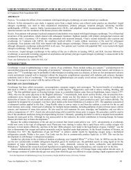
Presumed Sinus-Related StrabismusDISCUSSIONSIsolated strabismus cases due to acute sinusitis have beenreported in the literature. Two cases of acute-onsetBrown’s syndrome occurred due to pansinusitis, 4 andsuperior oblique palsy attributed to sinusitis 5 has also beendescribed.Ophthalmic complications of adjacent sinusitis havelong been known. <strong>The</strong>se include cellulitis, 6-9 orbitalabscess, 7-9 orbital myositis, 10 cavernous sinus thrombosis, 7-9and blindness. 7-9 Strabismus due to sinusitis was usuallyattributed to cranial nerve palsies owing to cavernoussinus involvement. 10-12 Most reported cases of ophthalmiccomplications of sinusitis were related to symptomatic,acute, fulminant sinus disease. Vertical diplopia due tochronic asymptomatic sinusitis has been described due toorbital floor collapse, which leads to enophthalmos andhypoglobus. 13Sinus lesions such as mucoceles, 14,15 osteomas, 16 andmalignancies 14 are known to cause diplopia due to directorbital extension. Damage to extraocular muscles withsecondary strabismus is a reported complication of sinussurgery. 17-19<strong>The</strong> strabismus cases in this study were different frompreviously reported cases in that most of the patients wereunaware of sinus disease. Few had clinical evidence ofsinus disease, although in retrospect, several reportedimproved health and decreased facial pain after clearingof sinusitis. One mother reported resolution of her son’schronic rhinorrhea, but before treatment, neither she norhis pediatrician had been concerned about his mild symptoms.Office otolaryngologic examination was notadequate to diagnose sinusitis in this group. Sinus CTscans were required to diagnose and monitor sinusitis.In a population-based study of childhood esotropia,idiopathic acquired nonaccommodative esotropia wasreported in 10.4%. 20 During data analysis of a large seriesof accommodative esotropia, a number of cases did not fitinto the accepted definitions of congenital or accommodativeesotropia. 21 <strong>The</strong>se may also represent acquired nonaccommodativeesotropia. <strong>The</strong> children with sinus-relatedesotropia in this series may be similar to the same subsetsof childhood esotropia as the other studies.Superior oblique palsy appearing in late childhoodwithout neurologic abnormality or trauma was felt to bedue to decompensation of a previously asymptomatic butcongenital defect. 22 Nineteen of the cases in this studycould have been classified as fourth cranial nerve palsybased upon alignment testing. <strong>The</strong> findings of contralateralinferior rectus fibrosis described above differentiatedthese cases from true fourth cranial nerve palsy.Gradual-onset adult strabismus is also often attributedto decompensation of congenital strabismus, 22 unlessneurologic or myopathic etiology is diagnosed. Some ofthe patients in this series had been wearing graduallyincreasing spectacle-mounted prisms for many years, andone had noticed the temporal coincidence between hisyearly sinus infection and the need for a yearly increase inprism strength.Some of the patients in this study had acute or subacuteonset of diplopia. Cranial nerve palsy was the clinicaldiagnosis until imaging disclosed sinusitis (Figure 4).Force generation testing of the suspected palsied musclewas a useful diagnostic test in the older children andadults. Motility limitation was less marked in these casesthan would be expected in cranial nerve palsy, andsaccades were not slowed.Since the advent of broad-spectrum antibiotics, theserious complications of acute sinusitis of the past havebecome rare. 7,9 <strong>The</strong> serious ophthalmic complications ofsinusitis that appeared in the early literature includedblindness and severe palsy of multiple cranial nerves.<strong>The</strong>se are not found in the modern literature but werefamiliar to clinicians prior to 1950. 12 Sinus disease itselfremains common, however. One study showed positiveCT scan findings of sinus disease in 15% of asymptomaticindividuals. 23 Another recent study found mucosal thickeningin 17% of a control group of CT scans for unspecifiedorbital disease, but only 2% had radiologically significantsinus disease. 3 A chronic, smoldering sinusitis couldbe predicted to cause a milder inflammation with secondaryfibrosis of adjacent orbital tissues and extraocularmuscles rather than palsy of the cranial nerves. A 1950study ascribed strabismus to sinusitis in 10 cases. 12Diplopia was attributed to cranial nerve palsy in all 10,and most had severe acute sinusitis with multiple symptoms.Sinus treatment improved motility without strabis-FIGURE 4Magnetic resonance imaging scan of a 73-year-old man with acute onsetof esotropia, showing bilateral ethmoid sinusitis. Esotropia fully resolvedafter 1 month of antibiotic treatment.163
Ludwig et almus surgery in each patient. Several, however, had subacutepresentations with milder strabismus and sinussymptoms, consistent with the cases in this series.It is possible that some of the patients in this seriesmay have experienced a component of cranial nerve palsyat some point, although none had muscle weakness at thetime of treatment. Otitis media and sinusitis may occursimultaneously, and a sixth cranial nerve palsy due tomastoiditis 14 must also be considered in a case of acuteacquired esotropia. <strong>The</strong> incomitance of acute cranialnerve palsy was not prominent in this series, however.<strong>The</strong> medial wall of the orbit contains three verticalfissures: the lacrimomallary, the lacrimoethmoidal, andthe sphenoethmoidal foramina. Vessels and nerves travelthrough these foramina. <strong>The</strong>re may also be congenitaldehiscences in the medial orbital wall. <strong>The</strong>se structuresmay allow transmission of inflammation or infection fromthe ethmoidal sinuses to the orbit. 7 <strong>The</strong> orbital floor isformed by the maxillary sinus roof, which is a thin bone,through which travels the infraorbital nerve. <strong>The</strong> inferiororbital wall may be dehiscent, with no bony covering.<strong>The</strong>se weaknesses may allow transmission of inflammationfrom the maxillary sinuses into the orbit. Chronicsinusitis itself may cause erosion of orbital bone. Orbitalcellulitis and orbital abscesses are well-known complicationsof sinusitis. It is logical to assume that subclinicalorbital infection or inflammation could exist withoutprogressing to manifest cellulitis or orbital abscess.Proving the link between the sinus disease and strabismusmay be impossible in this heterogeneous group ofpatients. Both disorders are chronic and common. <strong>The</strong>reare many individuals with recurrent sinusitis who neverdevelop strabismus and many strabismus patients whohave no sinusitis or unrelated sinusitis. <strong>The</strong> patientsreported here had atypical strabismus, which respondedpoorly to standard treatment until sinusitis was controlled.Some cases resolved with sinus treatment alone, and manyhad unusually severe sinus inflammation by radiologicexamination and at surgery. Although incidence figuresare not available in this study, it is the authors’ impressionsthat the incidence of positive CT scans exceeded 50%.<strong>The</strong> strabismus patients seen by the J.F.S. center had a90% incidence of sinus abnormalities by CT scan. Thisseries had many with complete pansinusitis or completeobliteration of one or more sinuses. That high degree ofseverity was previously reported in only 2% of asymptomaticcontrols with orbital CT scans. 3A normal sinus CT scan may not prove that earliersinusitis did not contribute to strabismus. Recurrent boutsof subacute sinusitis could cause progressive extraocularmuscle fibrosis, but then not show up on scans betweenepisodes.Patients with amblyopia or decreased fusion due toprior strabismus could be predisposed to worsening orrecurrence of strabismus if the extraocular musclesbecome inflamed by sinusitis. <strong>The</strong>y often have lessfusional reserve than normal and may be less able to adaptto changes in muscle tension.When the strabismus patient presents with an atypicalhistory and examination, which could representextraocular muscle fibrosis, it is worthwhile to investigatefor possible sinus disease. Sinus treatment may improvealignment if the strabismus is of recent onset. Sinusdisease management may also reduce the risk of strabismusrecurrence after successful surgery. Regular stretchingof the extraocular muscles to prevent shorteningseemed to assist some patients when they began to noticerecurrent diplopia. If unexpected fibrosis of the extraocularmuscles is detected at surgery, sinus evaluation maystill be of value to prevent strabismus recurrence.REFERENCES1. <strong>American</strong> Academy of Otolaryngology—Head and NeckSurgery. Clinical Indicators Compendium. Washington,DC: AAO—HNS; 2000:32-35.2. Guyton DL. Exaggerated traction test for the obliquemuscles. Ophthalmology 1981;88:1035-1040.3. Jones NS, Strobl A, Holland I. A study of the CT findingsin 100 patients with rhinosinusitis and 100 controls. ClinOtolaryngol 1997:22:47-51.4. Saunders RA, Stratas BA, Gordon RA, et al. Acute-onsetBrown’s syndrome associated with pansinusitis. ArchOphthalmol 1990;108:58-60.5. Lyons CJ, Lee JP. Bilateral consecutive superior obliquepalsy following fronto-ethmoidal sinusitis. J PediatrOphthalmol Strabismus 1990;27:233-236.6. Greenberg MF, Pollard ZF. Medical treatment of pediatricsubperiosteal orbital abscess secondary to sinusitis. JAAPOS 1998;2:351-355.7. Chandler JR, Langenbrunner DJ, Stevens ER. <strong>The</strong> pathogenesisof orbital complications in acute sinusitis.Laryngoscope 1970;80:1414-1428.8. Mortimore S, Wormald PJ. <strong>The</strong> Groote-Schuur hospitalclassification of the orbital complications of sinusitis. JLaryngol Otol 1997;111:719-723.9. Welsh LW, Welsh JJ. Orbital complications of sinus disease.Laryngoscope 1974;84:848-856.10. Duke-Elder S, ed. System of Ophthalmology. Vol 6. StLouis: Mosby; 1973:761-763.11. Deans JAJ, Welch AR. Acute isolated sphenoid sinusitis: adisease with complications. J Laryngol Otol 1991;105:1072-1074.12. Dimsdale H, Phillips DG. Ocular palsies with nasal sinusitis.J Neurol Neurosurg Psychiatr 1950;13:225-236.13. Borruat FX, Jaques B, Durig J. Transient vertical diplopiaand silent sinus disorder. J Neuroophthalmol 1999;19:173-175.14. Muneer A, Jones NS, Bradley PJ, et al. ENT pathology anddiplopia. Eye 1998;12:672-678.164
Presumed Sinus-Related Strabismus15. Hayasaka, S, Shibasaki H, Sekimoto M, et al. Ophthalmiccomplications in patients with paranasal sinus mucopyoceles.Ophthalmologica 1991;203:57-63.16. Biedner B, Monos T, Frilling F, et al. Acquired Brown’ssyndrome caused by frontal sinus osteoma. J PediatrOphthalmol Strabismus 1988;25:226-229.17. Penne RB, Flanagan JC, Stefanyszyn MA, et al. Ocularmotility disorders secondary to sinus surgery. OphthalmicPlast Reconstr Surg 1993;9:53-61.18. Pelletier CR, Jordan DR, Grahovac SZ. Inferior rectusentrapment following Caldwell-Luc surgery associated withan unrecognized hypoplastic maxillary antrum. Can JOphthalmol 1997;32:189-192.19. Rosenbaum AL, Astle WF. Superior oblique and inferiorrectus muscle injury following frontal and intranasal sinussurgery. J Pediatr Ophthalmol Strabismus 1985;22:194-202.20. Mohney BG. Common forms of childhood esotropia.Ophthalmology 2001;108:805-809.21. Ludwig IH, Imberman SP, Thompson HW, et al. Longtermstudy of accommodative esotropia. Trans AmOphthalmol Soc 2003;101:155-161.22. Miller MT, Urist MJ, Folk ER, et al. Superior oblique palsypresenting in late childhood. Am J Ophthalmol 1970;70:212-214.23. Calhoun KH, Waggenspack GA, Simpson CB, et al. CTevaluation of the paranasal sinuses in symptomatic andasymptomatic populations. Otolaryngol Head Neck Surg1991;104:480-483.DISCUSSIONDR DAVID R. STAGER, SR. Atypical or unexplainedacquired strabismus poses a challenge to the strabismusspecialist and it is therefore commendable that theauthors have undertaken this attempt to relate such caseswith possible sinus-related abnormalities. However, as asurvey supported by the <strong>American</strong> Academy ofOtolaryngology has found, 42 percent of people surveyedreported having at least one sinus infection in the last 12months. It has been estimated that 37 million people inthis country have sinus disease. This certainly creates adistinct possibility of a causal relationship between strabismusand sinus disease. From an epidemiologic standpoint,it would be important to identify the incidence ofstrabismus problems in patients who have sinus disease.How many patients in this study with acquired strabismushave a normal CT scan (and could serve as a controlgroup)? <strong>The</strong> answers to these questions would help usdetermine whether there is a cause-and-effect relationshipbetween sinus disease and atypical acquired strabismusor whether this relationship is coincidental. I foundsome of the clinical descriptions confusing. Do the measurementschange as the fibrotic muscle is placed onstretch? Would that help distinguish the 25 patients withcomitant esotropia or the 19 patients with suspectedfourth nerve palsy versus fibrosis of the inferior rectusmuscle? How does muscle fibrosis cause a high AC/Aratio?Secondly, is there a way of documenting an inflammatorybasis of eye muscle involvement such as high resolutionMRI or histopathology of adjacent tissue or experimentswith an animal model? Could one demonstrate animprovement of length-tension curves of the inflamedand fibrotic muscles before and after treatment of sinusdisease?Dr Ludwig has alerted us to a potential cause ofacquired incomitant strabismus. Historically, she hasproven herself to be a keen observer. If further investigationconfirms the association of sinus disease and strabismus,we will owe her our gratitude. This will provide uswith a non-surgical treatment that may be more effectivethan what is currently available. However, a great deal ofinvestigative work needs to be done. What level of sinusdisease can cause strabismus? What types of strabismusmay be due to this syndrome, as opposed to a coincidentalrelationship?Although this concept is in its infancy, and quite tenuous,Dr Ludwig’s paper is innovative and thought provoking.She does think “outside the box,” a talent that oftenleads to great progress.DR MALCOLM L. MAZOW. You might try to determine thefrequency or incidence of strabismus that occurs aftersignificant orbital cellulitis. In my practice, it does notseem to be very common.DR ALLAN J. FLACH. You seem to have converted a surgicaldisease into a medical disease and should be congratulated.In my adult practice, my most troublesomepatients are thyroid patients. Could you comment aboutwhat you think might occur in thyroid patients when theyhave an orbital decompression? Does the decompressiondo something to the sinus and might it have some impacton your study? I do not see anything wrong with a therapeutictrial of antibiotics in your patients with this conditionsince it might help someone avoid surgery.DR ROBERT RITCH. I came back from Germany inJanuary with bad sinusitis, losing all sense of olfaction andtaste. A couple of days after that, I developed diplopia,which turned out to be a comitant partial sixth nerve palsy.I was scheduled to get an MRI when, as Chair of this AOSprogram, your abstract came in the mail. A couple of dayslater, I was in a conference call with Dr Marilyn Millerand she mentioned she had similar cases. I performed aliterature search but really could not find very muchoutside of severe complications, like orbital cellulitis andorbital pseudotumor. I urge you to continue to delve165
Ludwig et alfurther into this problem and consider reporting youradditional findings.DR ALAN H. FRIEDMAN. Did any of the pre-op CT or MRIscans show any abnormalities in the extraocular muscles?In pseudotumor and in thyroid ophthalmopathy, we dosee abnormalities in the muscles. Is this a time measuresaccades or even EMG’s of suspected abnormal ocularmuscles? Was ocular pathology performed on any of yourcases?DR JOHN T. FLYNN. What muscle-stretching exercisesshould be performed in this context?DR GEORGE B. BARTLEY. <strong>The</strong> frequency in this cohort ofsinus surgeries seems pretty high. Did the culture resultsfrom these cases differ from what is typically seen in agerelatedcontrols that have sinusitis but yet do not requiresurgery?DR EDWARD L. RAAB. Many of us in pediatric ophthalmologysee children, especially in the hospital, who mighthave orbital cellulitis and actually turn out to have periorbitalcellulitis. In most children old enough to havesinuses, they have a lot of evidence of sinusitis, but hardlyever any restriction to motility. Any reason to think there’sleakage into the orbit? Where there is leakage, moreoften then not it would result in a subperiosteal abscess,and many of those do not result in any limitation of motility.So, it is hard to put this together as a cause-and-effectrelationship with that kind of evidence.DR. IRENE H. LUDWIG. With regard to the questions of DrStager, I don’t have good incidence figures on normal CTscans in this series. This was a gradual evolution of thinking,and I kept records on patients who had what Isuspected to be positive sinus-related strabismus. I havenow started to collect more data. My impression is thatwhen I do suspect possible sinus-related strabismus, I findsevere sinus disease about 50 percent of the time. Anumber of ENT colleagues have related that I have sentthem some of the most challenging sinus cases they haveseen. <strong>The</strong>re are other patients who have evidence ofextraocular muscular fibrosis without evidence of thyroiddisease and with negative CT scans. <strong>The</strong>y may have othercauses of acquired muscle fibrosis, such as rheumatologicdisorders. I have some patients with strabismus andrheumatologic disorders, which may be related. <strong>The</strong>irfindings are often similar to the findings in sinus-relatedstrabismus.It is difficult to measure young children for incomitance.<strong>The</strong> adults in this series often demonstrated mildincomitance, such as an esotropia of 25 diopters in theprimary position and 35 diopters on side gaze, suggestingtightness of the medial recti or mild weakness of thelateral recti. When I perform force generation testing onthese patients, they have strong muscles with no evidenceof muscle palsy.Alignment testing with prism and alternate cover forfourth cranial nerve palsy is inadequate without evaluationof torsion. <strong>The</strong> three-step test in a patient with acquiredvertical strabismus due to inferior rectus fibrosis is identicalto the measurements in a contralateral fourth cranialnerve palsy. <strong>The</strong> fundus exam, however, demonstratesextorsion in the lower eye when inferior rectus fibrosis ispresent. <strong>The</strong> forced duction test is very helpful, althoughthe force generation test for the superior oblique is verysubtle and very weak.How might you possibly cause a high AC/A ratio withthis problem? It might occur with a slight shortening ofthe extraocular muscles, not enough to affect the primaryposition or the distance alignment, but just enough thatthe patient can't control alignment at near. We know thatmedial rectus recession will decrease a high AC/A relationship,and we know that a low AC/A relationship can becollapsed with a slight resection of the medial rectus.<strong>The</strong>refore, it is certainly possible to develop a high AC/Arelationship from a slight shortening of the medial rectus.I do not have a good animal model or imaging methodyet to look at inflammation in the eye muscles. I haveconsulted with several investigators at LSU but still havenot been able to develop an animal model of sinusitis andstrabismus. We have been developing a convenientmuscle hook with a built-in strain gauge to measurelength-tension curves on all surgical strabismus patients.Sinus-related strabismus patients have stiffness in theirmuscles, but this is a subjective finding. It would be betterto have objective documentation of muscle stiffness.Sinus treatment probably does not improve musclecompliance in patients unless the process is caught early.Most of these people have had prolonged sinus diseaseand the best I hope for is arrest of progression. <strong>The</strong>re area number of people in this series who had strabismusrecurrences. I straightened their eyes, but six monthslater, the strabismus recurred due to unrecognized preoperativesinusitis. It was not until the sinus disease wascontrolled that the strabismus stabilized.<strong>The</strong> extraocular muscle may not be the only tissueaffected by the adjacent sinusitis. <strong>The</strong> motility restrictionsmay be created more from the orbital tissues, the orbitalseptae, and the connections to the eye muscles. <strong>The</strong>patients that develop sinusitis and then diplopia probablyhave different orbital defects. I have a few anecdotal casesnot in this series where patients have known orbitaltrauma with good alignment after surgery but thendevelop a sinus infection and strabismus.166
Presumed Sinus-Related StrabismusTo answer Dr Mazow’s question about orbital cellulitisand strabismus incidence, I have seen only a fewcellulitis cases, and they did not have strabismus. <strong>The</strong>patients who develop strabismus have chronic, long-terminflammation that may recur periodically over yearsbefore manifestation of strabismus. I have the impressionthat the patients who develop the most problems are theones with posterior sinus disease. <strong>The</strong>ir sinuses don'tdrain, and they are unaware of their sinus disease. Ascellulitis is treated promptly, chronic extra-ocular musclefibrosis probably does not have time to develop.In answer to Dr Flach’s question, I have one patientwho had thyroid ophthalmopathy with an orbital decompressionand extraocular muscle surgery. He indicatesthat every time he has a sinus infection, his strabismusreturns. With prompt use of oral antibiotics and extensiveexercise of his extraocular muscles, the strabismusresolves. This is just one anecdotal case, but it is an interestingone.In response to Dr Ritch’s discussion, there is onepatient in my series who developed what was diagnosed afourth cranial nerve palsy. Being a physician, he knew thatthe maxillary sinus was adjacent to the eye muscles, so hetreated himself with antibiotics. Within a day, the strabismusresolved. I would welcome any references you canadd to my search.To answer Dr Friedman’s question, I did not findobvious extraocular muscle changes on imaging study inmy patients. I do not have access to EMGs in my practice.I do not obtain tissue for histopathologic evaluation onthese patients since I have been taught to avoid disruptingthe extraocular muscle tissue. Obtaining a biopsy of anextra-ocular muscle could cause increased fibrosis andadhesion.Dr Flynn, the eye muscle stretching exercises in ayoung child are performed by having the parents use atarget, like a toy, and encouraging the child to rotate theeyes into side gaze, upgaze, and up and obliquely to thecorners since the superior obliques are often tight in thesecases. For adults, it’s easier to teach them to fixate on atarget, such as a television, and then have them rotate thehead and hold the eyes in extreme side and upgaze forprolonged periods of time.Dr Bartley, we did not perform cultures on thesepatients. <strong>The</strong>ir otolaryngologists, following prolongedantibiotic use, performed the sinus surgeries.Dr Raab commented on the number of sinusitispatients who don’t have motility restrictions. <strong>The</strong> onesthat do not develop problems with strabismus are the oneswho are draining or who are being treated promptly. It isthe patient who is unaware of his sinus disease who seemsto present with strabismus.167