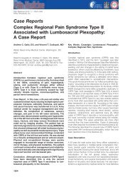
REVIEWSa b cFigure 4 | Neuron-to-glia communication. When pain processing is enhanced by inflammationor damage to peripheral tissues or peripheral nerves, signals must be relayed from sensorynerves to spinal cord glial cells to cause glial activation. There are at least two possible routes ofneuron-to-glia communication that could lead to glial activation and consequent enhancement ofnociception. First, neurotransmitters that relay information of the presence of peripheral noxiousstimuli could bind to and activate glia. Although probable, this has not yet been proven for spinalcord. Second, neurons could release a selective neuron-to-glia signal that binds to and activatesglia. This avenue of neuron-to-glia signalling has only very recently begun to be productivelyexplored. One candidate signal is fractalkine, a protein expressed on the extracellular surface ofneurons that, on strong neuronal activation, can be released into the extracellular fluid. In spinalcord, only microglia express receptors for fractalkine, making it a putative neuron-to-glia signal.Fractalkine, either injected exogenously or released endogenously in response to peripheral nervedamage, enhances nociception in animal models. The photomicrographs are of astrocyte andmicroglia mixed cultures. These photomicrographs demonstrate that microglia, but notastrocytes express fractalkine-binding sites. Green fluorescence (a and c) reveals glial fibrillaryacidic protein (GFAP), an astrocyte-specific marker. Red fluorescence (b and c) reveals bindingof fluorescent fractalkine. The lack of yellow (co-localization of green and red) indicates thatastrocytes do not express binding sites for fractalkine. By contrast, all microglia in the field bindthis putative neuron-to-glia signal. Panel c shows the mixed glial culture with superimposedfluorescence images. Modified with permission from REF. 133 © Springer-Verlag (2003).PERI-SPINAL INJECTIONAdministering a drug into thecerebrospinal fluid surroundingthe spinal cord; also called‘intrathecal’.their effects in brain 40 . Likewise, PERI-SPINAL INJECTION ofantagonists of pro-inflammatory cytokine functionprevents and/or reverses allodynia and hyperalgesia invirtually every animal model tested 17,19,34,36,46 .Suchmodels include inflammation and/or injury to peripheraltissues, peripheral nerves, spinal nerves and spinalcord.The fact that established allodynia and hyperalgesiacan be reversed by pro-inflammatory cytokineantagonists supports the conclusion that these glialproteins are involved in the maintenance, as well asthe initial induction, of these enhanced nociceptivestates. That is an important point when one is attemptingto identify drug targets for controlling pre-existingclinical pain syndromes.Recognition of the importance of pro-inflammatorycytokines in the induction and maintenance of allodyniaand hyperalgesia has led to the testing of variouscytokine-suppressive drugs in animal models. Oneapproach to control pro-inflammatory cytokines is toblock intracellular pathways that lead to their production.Another approach is to block intracellular pathwaysthat are activated by the binding of pro-inflammatorycytokines to their receptors. Although multiple intracellularsignalling cascades have been implicated in proinflammatorycytokine signalling and production, p38MAP kinase is crucially involved in both 33,47 .Indeed,p38 MAP kinase inhibitors were originally referred to ascytokine-suppressive anti-inflammatory drugs, reflectingthe importance of this signalling pathway. These compoundsinhibit allodynia and hyperalgesia induced byperipheral tissue inflammation, peripheral nerve injury,spinal nerve injury, spinal cord inflammation, andperi-spinal substance P and NMDA administration inanimal models 18,21,48–52 .At least some of these compoundscross the blood–brain barrier 51,52 , and so are effectivesystemically as well as after peri-spinal administration.An alternative approach to suppressing cytokines isto use xanthine derivatives, such as propentofylline.Propentofylline controls enhanced nociception inducedby spinal nerve transection 53 .Notably, it is equally effectivein reversing and preventing these changes, after bothsystemic and peri-spinal delivery 53 . Furthermore,propentofylline decreases both microglial and astrocyticactivation in spinal cord 53 .Other compounds that have been tested in animalmodels include the disease-modifying anti-rheumaticdrug leflunomide, the immunosuppressive drugmethotrexate and the immunomodulatory drug thalidomide.Systemic leflunomide was more effective inattenuating enhanced nociception induced by peripheralnerve injury than nociception caused by damage tospinal roots 54 .Methotrexate, delivered systemically aswell as at the site of spinal root injury, both preventedand reversed neuropathic pain behaviours in rats 55 ,anexciting outcome from a clinical perspective. Thalidomide,to date, has only been tested systemically. It hasproven effective in delaying enhanced nociceptiveresponses in rats induced by peripheral nerve damage 56 .However, its ability to reverse neuropathic pain behavioursin rats has been questioned 56 .Peripherally, thalidomideattenuated nerve-damage-induced TNF, but hadno effect on either IL-1 or IL-6 expression in the damagednerve 57 .Given that the focus of these investigators wasthalidomide-regulation of peripheral nerve changes,rather than spinal cord glia, they did not assess the effectof systemic thalidomide on central glial activation orpro-inflammatory cytokines. Because thalidomidecrosses the blood–brain barrier 58,59 ,changes in spinalcord glial function would be expected.The newest approach to controlling glially enhancednociception is upregulation of the expression of acytokine not yet discussed in this review, IL-10. Ratherthan being a pro-inflammatory cytokine, IL-10 is ananti-inflammatory cytokine. Like pro-inflammatorycytokines, anti-inflammatory cytokines are a family ofproteins that can be released by immune cells andimmunocompetent cells, such as glia 60,61 . Endogenously,anti-inflammatory cytokines serve as negative-feedbackregulators that keep potentially pathological activation ofimmune and immune-like cells under control 60,61 .Withinthis family, IL-10 is by far the most powerful member.IL-10 is attractive as a suppressor of glial pathologicalexcitation for a number of reasons: it can inhibit proinflammatorycytokine production at multiple levels,including transcription, translation and release; it candownregulate the expression of receptors for proinflammatorycytokines, so that even if pro-inflammatory978 | DECEMBER 2003 | VOLUME 2 www.nature.com/reviews/drugdisc
REVIEWSabTACETNF-αOthercellsCXCR4TNFRICa 2+ ERK1/2Glial cellFigure 5 | Pro-inflammatory cytokines are constitutively expressed in an inactive precursor form, allowing rapid release.Constitutive expression and rapid release of tumour-necrosis factor (TNF) is illustrated. a | Constitutive extracellular surfaceexpression of TNF on astrocytes. Exposure of living rat astrocytes (glial fibrillary acidic protein-positive, red) to an anti-TNF antibodyselectively reveals TNF sequences exposed on the extracellular surface (green) of about 50% of astrocytes. b | Molecular eventscoupling receptor activation of astrocytes (CX3CR4 shown in example) to TNF release. TNF is released in response to stimulationof various astrocyte receptors. Illustrated is TNF action on a G-protein-coupled receptor (CXCR4) by its endogenous ligand(SDF-1-α). The resultant intracellular signalling activates extracellular signal-regulated kinase (ERK1/2), which activatesTNF-α-converting enzyme (TACE). TACE is a specific enzyme required to cleave the extracellular domain of membrane-boundpro-TNF (a 26-kDa protein) to generate the released mature TNF (18-kDa protein) through a process known as ectodomainshedding. Once cleaved, the mature (active) TNF both exerts auto-stimulation of the same cell and diffuses away to exert paracrineactions on surrounding glia and neurons. Adapted with permission from REF. 134 © Oxford University Press (2002).SYNOVIAL TISSUESTissues encapsulating joints.cytokines are released they are less effective due todecreased availability of receptors; it can upregulateendogenous antagonists to pro-inflammatory cytokines,thereby limiting their effectiveness; and evidence to dateindicates that neurons in the spinal cord do not expressreceptors for IL-10, so normal neuronal functions wouldbe unaffected by the presence of IL-10 (REFS 60–62).Takentogether, this is a powerful profile of effects. Behaviourally,studies of IL-10 in rats demonstrate that it prevents orreverses every enhanced nociceptive state examined todate. These models include pain induced by spinalinflammation, inflammatory neuropathy, traumatic neuropathy,spinal trauma and peri-spinal dynorphin 19,36,63–65 .Drug discovery outlookThe potential efficacy of a drug is dependent on manyfactors. What follows is a summary of clinically relevantaspects of the various drugs that target glia and whichhave successfully controlled enhanced nociceptive statesin animal models. No drug presently available for use inhumans was developed to target glia. Rather, they weredeveloped to suppress the function of the peripheralimmune system. Indeed, their efficacy in suppressingpain in humans and enhanced nociceptive behavioursin rats following systemic administration supports theargument that suppressing pro-inflammatory cytokineproduction by SYNOVIAL TISSUES,Schwann cells and otherimmunocompetent cell types in peripheral tissues,peripheral nerves and/or dorsal root ganglia 3 candecrease transmission of nociceptive information to thespinal cord. In the present context, the discussion ofthese compounds will be focused on their potential foralso suppressing the pain-enhancing effects of spinalcord glial activation. There is a great need for new drugsto reach clinical trials for controlling the pathologicalside of spinal cord glial activation.Disrupting glial activation. The two drugs that havebeen examined in animal models for their ability todisrupt glial activation, to date, are fluorocitrate andminocycline. Fluorocitrate is a reversible glial poisonnot appropriate for human use. Although fluorocitrateis a selective glial inhibitor at low doses and short postdrugtime intervals 13,14 , higher doses and longerpost-drug times can indirectly affect neuronal functions.This indirect effect on neurons can result fromelevated extracellular concentrations of excitatoryamino acids due to the inhibition of glial transport 13,14 .Seizures have also been reported in response to glia-toxicdoses of this compound 66 .On the other hand, minocycline exhibits selectivityfor microglia. It is a tetracycline derivative that hasanti-inflammatory effects which are independent of itsantimicrobial actions. In rats, it can inhibit microglialactivation, p38-MAP-kinase activation, IL-1-convertingenzyme(caspase-1) activation, IL-1 release and theproduction of nitric oxide 28,67 .Although the animalliterature largely supports the conclusion that minocyclineinhibits activation of microglia independent ofdirect effects on astrocytes and neurons, neuroprotectiveeffects of minocycline on neuronal culturesexposed to toxic levels of nitric oxide have beenreported 68 .Although all of these indices are positivewith regards to minocycline’s potential for controllingglially driven allodynia and hyperalgesia, concern israised by the fact that minocycline fails to reverse, oris far less effective at reversing, established enhancednociceptive states in animal models, relative to agentsthat inhibit astrocyte as well as microglial activity 20,21 .These initial studies indicate that microglia mighthave a more important role in the initial creation ofenhanced nociceptive states in animals, whereas astrocytesmight become the key glial cell type as allodynia/NATURE REVIEWS | <strong>DRUG</strong> <strong>DISCOVERY</strong> VOLUME 2 | DECEMBER 2003 | 979