GLIA: A NOVEL DRUG DISCOVERY TARGET FOR CLINICAL PAIN
GLIA: A NOVEL DRUG DISCOVERY TARGET FOR CLINICAL PAIN
GLIA: A NOVEL DRUG DISCOVERY TARGET FOR CLINICAL PAIN

Create successful ePaper yourself
Turn your PDF publications into a flip-book with our unique Google optimized e-Paper software.
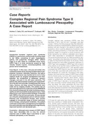
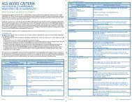
REVIEWSa b cFigure 4 | Neuron-to-glia communication. When pain processing is enhanced by inflammationor damage to peripheral tissues or peripheral nerves, signals must be relayed from sensorynerves to spinal cord glial cells to cause glial activation. There are at least two possible routes ofneuron-to-glia communication that could lead to glial activation and consequent enhancement ofnociception. First, neurotransmitters that relay information of the presence of peripheral noxiousstimuli could bind to and activate glia. Although probable, this has not yet been proven for spinalcord. Second, neurons could release a selective neuron-to-glia signal that binds to and activatesglia. This avenue of neuron-to-glia signalling has only very recently begun to be productivelyexplored. One candidate signal is fractalkine, a protein expressed on the extracellular surface ofneurons that, on strong neuronal activation, can be released into the extracellular fluid. In spinalcord, only microglia express receptors for fractalkine, making it a putative neuron-to-glia signal.Fractalkine, either injected exogenously or released endogenously in response to peripheral nervedamage, enhances nociception in animal models. The photomicrographs are of astrocyte andmicroglia mixed cultures. These photomicrographs demonstrate that microglia, but notastrocytes express fractalkine-binding sites. Green fluorescence (a and c) reveals glial fibrillaryacidic protein (GFAP), an astrocyte-specific marker. Red fluorescence (b and c) reveals bindingof fluorescent fractalkine. The lack of yellow (co-localization of green and red) indicates thatastrocytes do not express binding sites for fractalkine. By contrast, all microglia in the field bindthis putative neuron-to-glia signal. Panel c shows the mixed glial culture with superimposedfluorescence images. Modified with permission from REF. 133 © Springer-Verlag (2003).PERI-SPINAL INJECTIONAdministering a drug into thecerebrospinal fluid surroundingthe spinal cord; also called‘intrathecal’.their effects in brain 40 . Likewise, PERI-SPINAL INJECTION ofantagonists of pro-inflammatory cytokine functionprevents and/or reverses allodynia and hyperalgesia invirtually every animal model tested 17,19,34,36,46 .Suchmodels include inflammation and/or injury to peripheraltissues, peripheral nerves, spinal nerves and spinalcord.The fact that established allodynia and hyperalgesiacan be reversed by pro-inflammatory cytokineantagonists supports the conclusion that these glialproteins are involved in the maintenance, as well asthe initial induction, of these enhanced nociceptivestates. That is an important point when one is attemptingto identify drug targets for controlling pre-existingclinical pain syndromes.Recognition of the importance of pro-inflammatorycytokines in the induction and maintenance of allodyniaand hyperalgesia has led to the testing of variouscytokine-suppressive drugs in animal models. Oneapproach to control pro-inflammatory cytokines is toblock intracellular pathways that lead to their production.Another approach is to block intracellular pathwaysthat are activated by the binding of pro-inflammatorycytokines to their receptors. Although multiple intracellularsignalling cascades have been implicated in proinflammatorycytokine signalling and production, p38MAP kinase is crucially involved in both 33,47 .Indeed,p38 MAP kinase inhibitors were originally referred to ascytokine-suppressive anti-inflammatory drugs, reflectingthe importance of this signalling pathway. These compoundsinhibit allodynia and hyperalgesia induced byperipheral tissue inflammation, peripheral nerve injury,spinal nerve injury, spinal cord inflammation, andperi-spinal substance P and NMDA administration inanimal models 18,21,48–52 .At least some of these compoundscross the blood–brain barrier 51,52 , and so are effectivesystemically as well as after peri-spinal administration.An alternative approach to suppressing cytokines isto use xanthine derivatives, such as propentofylline.Propentofylline controls enhanced nociception inducedby spinal nerve transection 53 .Notably, it is equally effectivein reversing and preventing these changes, after bothsystemic and peri-spinal delivery 53 . Furthermore,propentofylline decreases both microglial and astrocyticactivation in spinal cord 53 .Other compounds that have been tested in animalmodels include the disease-modifying anti-rheumaticdrug leflunomide, the immunosuppressive drugmethotrexate and the immunomodulatory drug thalidomide.Systemic leflunomide was more effective inattenuating enhanced nociception induced by peripheralnerve injury than nociception caused by damage tospinal roots 54 .Methotrexate, delivered systemically aswell as at the site of spinal root injury, both preventedand reversed neuropathic pain behaviours in rats 55 ,anexciting outcome from a clinical perspective. Thalidomide,to date, has only been tested systemically. It hasproven effective in delaying enhanced nociceptiveresponses in rats induced by peripheral nerve damage 56 .However, its ability to reverse neuropathic pain behavioursin rats has been questioned 56 .Peripherally, thalidomideattenuated nerve-damage-induced TNF, but hadno effect on either IL-1 or IL-6 expression in the damagednerve 57 .Given that the focus of these investigators wasthalidomide-regulation of peripheral nerve changes,rather than spinal cord glia, they did not assess the effectof systemic thalidomide on central glial activation orpro-inflammatory cytokines. Because thalidomidecrosses the blood–brain barrier 58,59 ,changes in spinalcord glial function would be expected.The newest approach to controlling glially enhancednociception is upregulation of the expression of acytokine not yet discussed in this review, IL-10. Ratherthan being a pro-inflammatory cytokine, IL-10 is ananti-inflammatory cytokine. Like pro-inflammatorycytokines, anti-inflammatory cytokines are a family ofproteins that can be released by immune cells andimmunocompetent cells, such as glia 60,61 . Endogenously,anti-inflammatory cytokines serve as negative-feedbackregulators that keep potentially pathological activation ofimmune and immune-like cells under control 60,61 .Withinthis family, IL-10 is by far the most powerful member.IL-10 is attractive as a suppressor of glial pathologicalexcitation for a number of reasons: it can inhibit proinflammatorycytokine production at multiple levels,including transcription, translation and release; it candownregulate the expression of receptors for proinflammatorycytokines, so that even if pro-inflammatory978 | DECEMBER 2003 | VOLUME 2 www.nature.com/reviews/drugdisc