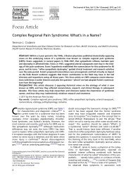
REVIEWSabIncomingA-δ/C fibre'pain' signalsNormalsubstance Prelease,EAA releaseQuiescent gliaPain messageto brain via PTNsNK-1receptorAMPAreceptorNMDAreceptorPain stimulusPTNcIncomingA-δ/C fibre'pain' signalsdViruses and bacteriaNonexistentgliaNK-1receptorEnhancedsubstance Prelease,EAA releaseAMPAreceptorCa 2+NMDAreceptorPTN: NO, PGs,fractal kineActivatedgliaPrimary afferent:Substance P, EAAs, ATP,fractal kinecNOSNOIL-1, TNF, IL-6, ROS, NO,PGs, EAAs, ATPL-argininePTNEnhance PTNexcitabilityEnhance primaryafferent substance Pand EAA releaseFigure 1 | Schematic of pain and pain modulation. a | Classical pain transmission pathway. When a noxious stimulus isencountered (such as stepping on a tack, as shown), peripheral nocisponsive nerves (A-δ and C fibres) are excited. These axonstransmit action potentials to their presynaptic terminals in the spinal cord dorsal horn. Neurotransmitters released here bind to andactivate postsynaptic receptors on pain transmission neurons (PTNs). In turn, the axons of PTNs ascend, predominantlycontralaterally, to the brain and carry information about the noxious stimulus to higher centres. The region of the sensory presynapticterminal and post-synaptic region of the PTN are shown in detail in (b–d). b | Normal pain. In normal, everyday situations in which painis experienced, glia are present but quiescent. Information about noxious stimuli arrives from the periphery along A-δ and C fibres,causing the release of substance P and excitatory amino acids (EAAs) in amounts appropriate to the intensity and duration of theinitiating noxious stimulus. Activation of neurokinin-1 (NK-1) receptors by substance P and activation of α-amino-3-hydroxy-5-methyl-4-isoxazole propionic acid (AMPA) receptors by EAAs cause transient depolarization of the PTNs, thereby generating action potentialsthat are relayed to higher brain areas. N-methyl-D-aspartate (NMDA)-linked channels are inoperative as they are chronically ‘plugged’by magnesium ions. c | Pathological pain: classic view. In reponse to intense and/or prolonged barrages of incoming nociceptiveinformation, the PTNs become sensitized and over-respond to subsequent incoming nociceptive signals. The intense and/orprolonged barrage depolarizes the PTNs such that the magnesium ions exit the NMDA-linked channel. The resultant influx of calciumions activates constitutively expressed nitric oxide synthase (cNOS), causing conversion of L-arginine to nitric oxide (NO). Because it isa gas, NO rapidly diffuses out of the PTNs. This NO acts presynaptically to cause exaggerated release of substance P and EAAs.Postsynaptically, NO causes the PTNs to become hyperexcitable. Glia have not been considered to have a role in creating painfacilitation by this neuronally driven model. d | Pathological pain: new view. Here, glial activation is conceptualized as a driving forcefor creating and maintaining pathological pain states. The role of glia is superimposed on the NMDA—NO-driven neuronal changesdetailed in c, so only the aspects added by including glia in the model are described here. Glia are activated (shown as hypertrophiedrelative to b, as this reflects the remarkable anatomical changes that these cells undergo on activation) by three sources: bacteria andviruses which bind specific activation receptors expressed by microglia and astrocytes; substance P, EAAs, fractalkine and ATPreleased by either A-δ or C fibre presynaptic terminals (shown) or by brain-to-spinal cord pain enhancement pathways (not shown);and NO, prostaglandins (PGs) and fractalkine released from PTNs. Following activation, microglia and astrocytes cause PTNhyperexcitability and the exaggerated release of substance P and EAAs from presynaptic terminals. These changes are created bythe glial release of NO, EAAs, reactive oxygen species (ROS), PGs, pro-inflammatory cytokines (for example, interleukin-1 (IL-1), IL-6,tumour-necrosis factor (TNF)), and nerve growth factor. Modified, with permission, from REF. 12 © Elsevier Science Ltd (2001).974 | DECEMBER 2003 | VOLUME 2 www.nature.com/reviews/drugdisc
REVIEWS<strong>GLIA</strong>L FIBRILLARY ACIDICPROTEIN(GFAP). An astrocyte-specificprotein. Increases in GFAP arefrequently used as a marker ofastrocyte activation.p38 MAP KINASEAn intracellular signallingcascade, activated in responseto pro-inflammatory cytokinereceptor binding. Activation ofthis cascade leads to productionof pro-inflammatorycytokines.astrocytes within the spinal cord. This review will firstexamine the evidence that activated glia drive the creationand maintenance of allodynia and hyperalgesia. Thisliterature review will demonstrate that astrocytes andmicroglia in the spinal cord must now be recognized asactive participants in the creation and maintenance ofpain facilitation induced by inflammation and damageto peripheral tissues, peripheral nerves, spinal nervesand the spinal cord. On activation, these glia release avariety of neuroexcitatory substances that potentiatepain transmission by neurons. Of these glial products,pro-inflammatory cytokines will be shown to be commonspinal mediators of allodynia and hyperalgesia.Given the failure of presently available drugs to provideadequate clinical pain management, this newly recognizedrole of glia in general, and pro-inflammatorycytokines in particular, is exciting because it predictsnovel approaches for effective pain control by targetingglial activation.A number of drugs will be introduced in the courseof this discussion that are effective in controlling gliallydriven exaggerated nociceptive states in laboratoryanimals. Each of these drugs will be discussed in thefinal section of this review, with regard to their potentialclinical application.What does it mean that glia are ‘activated’?In the following discussion, frequent reference will bemade to ‘activated’ glia. Activation is a fundamentallydifferent phenomenon in neurons compared with thatin glia. The term ‘activation’ refers to an enhanced abilityof a cell to perform a function beyond that present in abasal state. For neurons, activation is unidimensional, asit mainly relates to the production of action potentials.By contrast, activation of glia is multi-dimensionalbecause glia perform numerous functions (see below).So there are many different activational states, with variouscomponents expressed with different time-coursesand intensities that are dependent on the stimulus thattriggers activation.Astrocytes are basally active in carrying out a numberof functions, but unless there is an external stimulusthey do not become activated. Astrocytes serve severalfunctions in the normal central nervous system (CNS),including the regulation of: extracellular ion and neurotransmitterconcentrations; the availability of neurotransmitter/neuromodulatorprecursors to nearbyneurons; and extracellular pH. No evidence is apparentin the literature that these functions are regulated bystimuli that activate astrocytes or by substances releasedby activated glia. As such, they do not seem likely to beaffected by drugs that target these aspects of activatedastrocyte function.Astrocyte activation occurs in response to CNStrauma, ischaemia, tumours, neurodegeneration, andthe presence of immunogenic components of virusesand bacteria 5 .Activation is morphologically characterizedby hypertrophy and increased production of intermediatefilaments (<strong>GLIA</strong>L FIBRILLARY ACIDIC PROTEIN (GFAP),vimentin and/or nestin) 5 , and functionally by increasedproduction of a variety of pro-inflammatory substances 6 .Notably, functional changes and morphologicalchanges are not time-locked, so functional changescan be detected in the absence of increased intermediatefilaments, and vice versa.In contrast to astrocytes, microglia in the normalCNS are quiescent cells with no recognized function 7 .In this state, they exhibit a ramified morphology, noactivation of p38 MITOGEN-ACTIVATED PROTEIN (MAP) KINASE,and little or no expression of the receptors, cellsurfacemarkers or functional activities characteristicof activated microglia 7 .Microglial activation occurs in response to the samerange of stimuli that activate astrocytes. It is a gradedphenomenon, characterized by a specific morphology(retracted processes and hypertrophy; amoeboid morphologyunder strongly pathological circumstances),proliferation, increased expression of one or more cellsurfacemarkers or receptors (such as the complement 3receptor associated with adhesion, migration andphagocytosis and scavenger receptors associated withphagocytosis), and/or changes in functional activities(migration to areas of damage, phagocytosis, production/releaseof pro-inflammatory substances) 7 .Notably,the changes in receptors, cell-surface markers and/or theproduction of pro-inflammatory substances can occurin the absence of morphological changes, proliferationor phagocytosis 7,8 .So, as is the the case for astrocyteactivation, microglial activation is a multi-dimensionalprocess. The manner in which activation is expressed isdependent on the type and intensity of the inductivestimulus, and different patterns and time-courses ofresponses can occur.Spinal cord glia as powerful modulators of painUntil recently, glia were thought of simply as housekeepersfor neurons, regulating the extracellular ionicenvironment and removing debris. However, in recentyears it has become recognized that glia dynamicallymodulate the function of neurons under both physiologicaland pathological conditions 9 .In the discussion to follow, the term ‘glia’ will be usedto refer to both microglia and astrocytes. As will bereviewed below, both cell types are activated in thespinal cord in response to experimental manipulationsthat induce pain facilitation. Both cell types, on activation,can produce and release a variety of nociceptionenhancingsubstances. In addition, each stimulates thefurther activation of the other, perhaps forming afunctional unit. The use of the term ‘glia’ in the presentcontext reflects the fact that most presently availabledata support the notion that both microglia andastrocytes are involved in allodynia and hyperalgesia,but that these data cannot yet allow us to determine therelative contributions of each.Glia first came to the attention of pain researchersin the early 1990s when Garrison et al. reported thatperipheral nerve damage that created exaggerated nociceptiveresponses (neuropathic ‘pain’ behaviours) alsoactivated spinal cord glia (FIG. 2).Furthermore, theN-methyl-D-aspartate (NMDA) antagonist MK801,which blocks neuropathy induced allodynia andNATURE REVIEWS | <strong>DRUG</strong> <strong>DISCOVERY</strong> VOLUME 2 | DECEMBER 2003 | 975