Final Report - Strategic Environmental Research and Development ...
Final Report - Strategic Environmental Research and Development ...
Final Report - Strategic Environmental Research and Development ...

You also want an ePaper? Increase the reach of your titles
YUMPU automatically turns print PDFs into web optimized ePapers that Google loves.
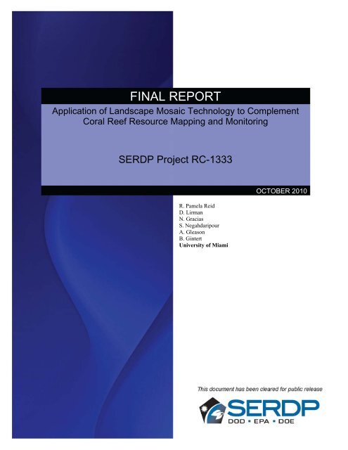
FINAL REPORTApplication of L<strong>and</strong>scape Mosaic Technology to ComplementCoral Reef Resource Mapping <strong>and</strong> MonitoringSERDP Project RC-1333R. Pamela ReidD. LirmanN. GraciasS. NegahdaripourA. GleasonB. GintertUniversity of MiamiOCTOBER 2010
This report was prepared under contract to the Department of Defense <strong>Strategic</strong><strong>Environmental</strong> <strong>Research</strong> <strong>and</strong> <strong>Development</strong> Program (SERDP). The publication of thisreport does not indicate endorsement by the Department of Defense, nor should thecontents be construed as reflecting the official policy or position of the Department ofDefense. Reference herein to any specific commercial product, process, or service bytrade name, trademark, manufacturer, or otherwise, does not necessarily constitute orimply its endorsement, recommendation, or favoring by the Department of Defense.
RC-1333 <strong>Final</strong> <strong>Report</strong> to SERDP 2010R.P.ReidA2.3 Analysis of mosaics .............................................................................................................41A2.3.1 Geometric analysis of l<strong>and</strong>scape mosaics ....................................................................42A2.3.2 Ecological analysis of l<strong>and</strong>scape mosaics ...................................................................44A2.3.2.1 Mosaic Selection ..................................................................................................44A2.3.2.2 Spatial Accuracy ..................................................................................................48A2.3.2.3 Percent Cover ......................................................................................................48A2.3.2.4 Colony Size ..........................................................................................................50A2.3.2.5 Change Detection ................................................................................................51A2.3.3 Benefits of second-generation mosaic technology to ecological analysis....................53A2.3.4 Recommendations for Field Implementation................................................................57A2.4 Applications of coral reef monitoring <strong>and</strong> mapping using l<strong>and</strong>scape mosaictechnology .....................................................................................................................................69A2.4.1 Ship Grounding ............................................................................................................69A2.4.1.1 Status <strong>and</strong> Trends of the Benthic Community......................................................72A2.4.1.2 Comments <strong>and</strong> Recommendations .......................................................................75A2.4.2 Spatio-temporal analysis of coral communities ...........................................................75A2.4.3 Monitoring <strong>and</strong> Assessment of Mesophotic coral communities ...................................80A2.4.4 Using L<strong>and</strong>scape Mosaics to Assess the Impacts of Hurricane Damage onAcropora palmata populations ..........................................................................................84A2.4.5 Use of 2D Video Mosaics for Assessing the Impacts of Mass-Bleaching Eventson Coral Communities .......................................................................................................87A2.5 Users/Partners .....................................................................................................................89A2.5.1 SERDP Coral Reef Monitoring <strong>and</strong> Assessment Workshop .........................................89A2.5.2 The National Park Service: Surveys of the threatened coral Acropora palmata .........91A2.5.3 University of Puerto Rico .............................................................................................91A2.5.4 The US Navy’s Coral Reef Monitoring Program at AUTEC .......................................92A2.5.5 The Nature Conservancy ..............................................................................................93A2.5.6 Rutgers University ........................................................................................................98A2.5.7 NOAA Restoration Group...........................................................................................101A2.5.8 NOAA Marine Heritage ..............................................................................................103A2.5.9 Florida Fish <strong>and</strong> Wildlife Conservation Commission ................................................105A2.5.10 Potential Department of Defense Collaborations/Users ..........................................106A2.5.11 L<strong>and</strong>scape Mosaics website .....................................................................................109A2.6 Software deliverables ........................................................................................................110A2.6.1 L<strong>and</strong>scape (2D) Mosaic Software .............................................................................110A2.6.1.1 Frame Extraction...............................................................................................111A2.6.1.2 Sunflickering Removal .......................................................................................112A2.6.1.3 Global Matching ................................................................................................112A2.6.1.4 Video <strong>and</strong> Still Registration...............................................................................113A2.6.1.5 Global Match Inspection ...................................................................................114A2.6.1.6 Mosaic Rendering with Improved Blending ......................................................115A2.6.1.7 Heading Integration Module .............................................................................115A2.6.1.8 Additional Features ...........................................................................................116A2.6.2 Ecological Analysis Module .......................................................................................118iii
RC-1333 <strong>Final</strong> <strong>Report</strong> to SERDP 2010R.P.ReidA2.6.3 External viewer ...........................................................................................................121A3. CONCLUSIONS AND IMPLICATIONS FOR FUTURERESEARCH/IMPLEMENTATION: .............................................................................. 123A4. ACTION ITEM ..................................................................................................... 128SECTION B: SINGLE OBJECT 3D RECONSTRUCTION ......................................... 133B1. MATERIALS AND METHODS ............................................................................. 133B2. RESULTS AND DISCUSSION ............................................................................ 135B3. CONCLUSIONS AND IMPLICATIONS FOR FUTURERESEARCH/IMPLEMENTATION ............................................................................... 142SECTION C: MULTISPECTRAL IMAGING ................................................................ 144C1. MATERIALS AND METHODS ............................................................................. 144C1.1 System <strong>Development</strong>, Data Acquisition, <strong>and</strong> Preliminary Processing .........................144C1.2 Experiments with Filter Set 1 ........................................................................................146C1.3 Experiments with Filter Set 2 ........................................................................................147C1.4 Experiments with Combined Spectral / Texture Classification .....................................147C2. RESULTS AND DISCUSSION ............................................................................. 149C2.1 System <strong>Development</strong> ......................................................................................................149C2.2 Experiments with Filter Set 1 ........................................................................................150C2.3 Experiments with Filter Set 2 ........................................................................................151C2.4 Experiments with Combined Spectral / Texture Classification .....................................152C3. CONCLUSIONS AND IMPLICATIONS FOR FUTURERESEARCH/IMPLEMENTATION ............................................................................... 153LITERATURE CITED .................................................................................................. 155APPENDIX 1. PUBLICATIONS AND PRESENTATIONS ACKNOWLEDGINGSERDP SUPPORT ...................................................................................................... 162APPENDIX 2. SUPPORTING DATA: LANDSCAPE MOSAICS ................................ 167APPENDIX 3. SUPPORTING DATA: LANDSCAPE MOSAIC CREATIONMANUAL ..................................................................................................................... 167iv
RC-1333 <strong>Final</strong> <strong>Report</strong> to SERDP 2010R.P.ReidAPPENDIX 4. SUPPORTING DATA: LANDSCAPE MOSAIC CREATIONSOFTWARE ................................................................................................................ 167APPENDIX 5. SUPPORTING DATA: 3D MOSAICING .............................................. 167APPENDIX 6. SUPPORTING DATA: MULTISPECTRAL DATA ............................... 167APPENDIX 7. SUPPORTING DATA: LANDSCAPE MOSAIC SURVEYINGTECHNOLOGY REQUIREMENTS ............................................................................. 168Software Requirements .............................................................................................................168Software Installation ..................................................................................................................169Acquisition Hardware ...............................................................................................................169Computing hardware <strong>and</strong> typical disk <strong>and</strong> CPU usage .........................................................170Guidelines for h<strong>and</strong>held image acquisition .............................................................................171APPENDIX 8. SUPPORTING DATA: ACTION ITEMS ............................................... 176APPENDIX9. OTHER MATERIALS (AWARDS) ........................................................ 176v
RC-1333 <strong>Final</strong> <strong>Report</strong> to SERDP 2010R.P.ReidTable of FiguresFigure A1. Early mosaicing results. ....................................................................................9Figure A2. Mosaics of the 4 tracks of Andros data ..........................................................10Figure A3. Mosaic creation example using Andros data. .................................................12Figure A4. Left: Mosaic created using the global mosaicing approach on a lowaltitude sequence alone. .....................................................................................................14Figure A5. Mosaic rendering. ...........................................................................................15Figure A6. Example of the watershed/graph-cut method. ................................................16Figure A7. Example of frame selection, used before the watershed/graph-cutblending method.................................................................................................................17Figure A8. Comparison of blending methods. ..................................................................18Figure A9. Example of application of the motion compensated filtering algorithm ........20Figure A10. Original frames of a shallow water sequence ...............................................21Figure A11. Sections of the same shallow water survey under strong refracted sunlight 21Figure A12. Performance of sunflicker removal methods under poor registration ..........23Figure A13. Completed enhanced imaging system. .........................................................27Figure A14. Plot of the Measured Voltage Transfer Function .........................................28Figure A15. Plot of the Angular transfer Function. ..........................................................29Figure A16. Heading Calibration Setup............................................................................29Figure A17. Table representing matches between video images <strong>and</strong> photo stills. ...........31Figure A18. Examples of audio samples ..........................................................................32Figure A19. Examples of audio samples <strong>and</strong> corresponding power spectral density .......33Figure A20. Illustration of the large settling time for a 360 to 0 degree transition. .........34Figure A21. Rotation angles obtained ..............................................................................35Figure A22. Calibration of the heading sensor. ................................................................36Figure A23. Example of a globally aligned mosaic with geometric distortions. ..............37Figure A24. Example of the use of heading on a globally aligned mosaic.. ....................38Figure A25. Example of use of heading in the estimation of the initial trajectory. ..........39FigureA26. Comparison of blending over a 3D surface, obtained using a simplegeometric criterion. ............................................................................................................41Figure A27. Mosaic image from the Key Biscayne site with selected marker positions. 43Figure A28. Histogram of the error components, with superimposed normal distributionfit. .......................................................................................................................................43vi
RC-1333 <strong>Final</strong> <strong>Report</strong> to SERDP 2010R.P.ReidFigure A29. Video mosaic created from h<strong>and</strong> held video images taken at the Brooke’sReef site in June 2004. .......................................................................................................45Figure A30. ROV Video Mosaic created from a high altitude. ........................................46Figure A31. ROV based video mosaic created from a low altitude pass. ........................47Figure A32. Abundance, spatial distribution, <strong>and</strong> sizes of stony corals obtained from alow-altitude. .......................................................................................................................50Figure A33. Example of co-registered mosaic sub-sections or tiles. ................................52Figure A34. A second-generation video mosaic from Brooke’s Reef, FL. ......................54Figure A35. of change detection potential using second-generation imagery. .................56Figure A36. <strong>Environmental</strong> data from Media Luna Integrated Coral Observing Network(ICON) station. ..................................................................................................................60Figure A37. Image of benthos from ~2m at Media Luna Reef. .......................................61Figure A38. Mosaic of Media Luna Reef video data from December 12 th , 2007. ...........62Figure A39. Maximum allowed speed of translation for the camera as a function of theshutter speed for different altitudes. ...................................................................................63Figure A40. Effect of light variations on image products. ...............................................64Figure A41. Example of a area coverage pattern comprising two lawnmower's patternsrotated 90 degrees. .............................................................................................................65Figure A42. Benefits of image pre-processing. ................................................................66Figure A43. A) Superficial damage to a coral colony caused by a small ship grounding.70Figure A44. Flow-charts depicting the differences between the Image-only method (A)<strong>and</strong> the Image-Plus GCP method (B).................................................................................72Figure A45. L<strong>and</strong>scape video mosaic surveys of the ship-grounding damage caused bythe Evening Star in 2005 <strong>and</strong> 2006. ...................................................................................73Figure A46. Encroachment of seagrass into the affected area. .........................................74Figure A47. Example of digitized h<strong>and</strong>-drawn species distribution map of a 10x10m reefpatch. ..................................................................................................................................76Figure A48. Partially digitized high resolution video mosaic of the 10x10m plot locatedat site S1-10........................................................................................................................77Figure A49. A) The 1972 (purple) <strong>and</strong> 2008 (orange) distributions of live coral cover atthe study site. .....................................................................................................................78Figure A50. A). Shows the 1972 <strong>and</strong> 2008 distributions of live coral cover within thestudy area. ..........................................................................................................................78Figure A51. The 1972 (blue) <strong>and</strong> 2008(purple) distributions of the coral species Poritesporites overlaid on top of a depth map of the study area. ..................................................79Figure A52. Site one from Sherwood Forest Reef, Dry Tortugas, Florida. ......................82vii
RC-1333 <strong>Final</strong> <strong>Report</strong> to SERDP 2010R.P.ReidFigure A53. Site two, a coral hillock from St. Thomas USVI. .........................................83Figure A54. Video mosaics from a study plot at Molasses Reef in the Florida Reef Tract............................................................................................................................................84Figure A55. a) Photograph of the reef section dislodged during Hurricane Rita. ............86Figure A56. L<strong>and</strong>scape video mosaics of site S1-10 located offshore of Andros Isl<strong>and</strong>,Bahamas. ............................................................................................................................88Figure A57. 2D video mosaics of Acropora palmata reefs at Buck Isl<strong>and</strong> NationalMonument, St. Croix, USVI. .............................................................................................91Figure A58. Example of coral reef monitoring at Andros Isl<strong>and</strong>, Bahamas. ....................92Figure A59. Location of the permanent sites established as part of the bleaching-mosaicassessment of the Biscayne Subregion of the Florida Reef Tract in the summer of 2008.93Figure A60. L<strong>and</strong>scape video mosaic of a permanent plot established within an OffshorePatch Reef habitat as part of the Florida Reef Resilience Program in the summer of 2008.94Figure A61. L<strong>and</strong>scape video mosaic of a permanent plot established within a Mid-Channel Patch Reef habitat as part of the Florida Reef Resilience Program in the summerof 2008. ..............................................................................................................................95Figure A62. L<strong>and</strong>scape video mosaic of a permanent plot established within an InshorePatch Reef habitat as part of the Florida Reef Resilience Program in the summer of 2008.96Figure A63. L<strong>and</strong>scape video mosaic of a permanent plot established within a Fore-Reefhabitat as part of the Florida Reef Resilience Program in the summer of 2008. ...............97Figure A64. L<strong>and</strong>scape mosaic of a Fore-Reef habitat showing the location of bleachedcolonies of Siderastrea siderea. .........................................................................................98Figure A65. Combined mosaic/FIRe product. Locations of individual organismsexamined with the underwater FIRe instrument are demarcated by the triangles (above).100Figure A66. Future of integrated mosaic/FIRe survey technology. ................................101Figure A67. South Carysfort Reef, Florida Keys. ..........................................................102Figure A68. Horseshoe Reef, Florida Keys. ...................................................................103Figure A69. L<strong>and</strong>scape mosaics of the Kyle Spangler. ..................................................105Figure A70. Video mosaic of 2mx20mtransect at Sombrero Reef, Florida Keys. .........106Figure A71. Screen-capture of l<strong>and</strong>scape mosaic website homepage. ...........................110Figure A72. Main Mosiacing Program GUI. ..................................................................111Figure A73. A screen capture of the video frame extraction GUI. .................................111Figure A74. Graphic user interface for the sunflicker removal module. ........................112Figure A75. The global matching interface. ...................................................................113Figure A76. The video frame <strong>and</strong> still image registration interface. ..............................114Figure A77. The match inspector GUI. ..........................................................................114viii
RC-1333 <strong>Final</strong> <strong>Report</strong> to SERDP 2010R.P.ReidFigure A78. Mosaic Rendering GUI. ..............................................................................115Figure A79. Heading integration module. ......................................................................116Figure A80. Basic point click GUI. ................................................................................116Figure A81. Basic Point Click GUI showing a selected video frame. ............................117Figure A82. Mosaic Info GUI. ........................................................................................117Figure A83. A mosaic with distance markers created by the mosaic_info GUI. ............118Figure A84. Screen-capture of a mosaic image being used with CPCe point countprogram. ...........................................................................................................................120Figure A85. Extracted still image of point I. ..................................................................121Figure A86. Screenshot of GUI that creates the file used for the external point <strong>and</strong> clickviewer. ..............................................................................................................................122Figure A87. External point click viewer interface. .........................................................122Figure A88. Result of clicking on mosaic on interface. ..................................................123Figure B1. A sample stereo pair, shown in (a) <strong>and</strong> (b), are processed by our method togenerate the 3D map. .......................................................................................................136Figure B2. Sample stereo pair with matching features. ..................................................137Figure B3. Sample left view of a stereo pair, showing the corals that have been processedfor 3D reconstruction. ......................................................................................................138Figure B4. Scan lines from the 3D reconstruction of corals in figure B3. .....................139Figure B5. Height measurements for various corals or reef rubble imaged by the stereocamera system. .................................................................................................................140Figure B6. Control points for 3D point <strong>and</strong> click calibration. ........................................141Figure B7. Independent measurements of the same coral colony or reef rubble by the 3Dpoint <strong>and</strong> click system (3 different stereo pairs) <strong>and</strong> 2 diver measurements. ..................142Figure C1. Proposed spectral b<strong>and</strong>s for coral reef mapping. ..........................................145Figure C2. (Left) Interior of multi-spectral camera. .......................................................149Figure C3. System rolloff factor as a function of angle from the center pixel. ..............150Figure C4. False color 600, 589, 568 nm RGB image of the .........................................150Figure C5. False color 600, 589, 568 nm RGB image of the ..........................................151Figure C6. Discriminant functions one (DF1) <strong>and</strong> two (DF2) computed from our fullspectrumdata base (left), from our partial-spectrum data base (center), <strong>and</strong> fromHochberg <strong>and</strong> Atkinson's (2003) database for their hypothetical CRESPO sensor.........151Figure C7. Four MSCAM scenes acquired with filter set two. ......................................152Figure C8. Summary of results for combined spectral / texture classification. ..............153ix
RC-1333 <strong>Final</strong> <strong>Report</strong> to SERDP 2010R.P.ReidTable of TablesTable A1. Table showing areas where speed <strong>and</strong> memory efficiency could be improvedin existing mosaic creation modules. ............................................................................................ 24Table A2. Table showing areas where of speed <strong>and</strong> memory efficiency could beimproved in enhanced mosaic capabilities. .................................................................................. 24Table A3. Measured Transfer Function. ...................................................................................... 27Table A4. Description of the three mosaics used in the monitoring assessment. ........................ 44Table A5. Mean cover (± S.E.M.) of the different benthic categories surveyed by divers<strong>and</strong> measured from video mosaics from a reef site in the northern Florida Reef Tract(depth = 7-10 m). .......................................................................................................................... 49Table A6. Comparison of coral size measurements between: (1) two divers measuringthe same colonies; <strong>and</strong> (2) between diver measurements <strong>and</strong> measurements of the samecolonies obtained directly from the video mosaics. ...................................................................... 50Table A7. Biological <strong>and</strong> <strong>Environmental</strong> characteristics of Brooke’s Reef <strong>and</strong> GrecianPatch permanent monitoring sites. ................................................................................................ 58Table A8. <strong>Environmental</strong> observations on dates in which permanent monitoring siteswere sampled. ............................................................................................................................... 58Table A9. Image quality characteristics for mosaic datasets. ...................................................... 66Table A10. Image quality information for mosaic data acquired at permanent monitoringsites Brooke’s Reef <strong>and</strong> Grecian Rocks. ....................................................................................... 67Table A11. Average st<strong>and</strong>ard deviations for mosaics that could not be created ( ....................... 68Table A12. Technology overlay <strong>and</strong> potential collaborations resulting from SERDP coralreef monitoring <strong>and</strong> assessment workshop. .................................................................................. 90Table A13. Military Facilities with Adjacent Coral Reef Resources. ....................................... 108Table A14. Summary of advancements to the state-of-the-art in coral reef monitoringtechniques. .................................................................................................................................. 124Table A15. Methods comparison. ............................................................................................... 129Table A16 Cost estimates of coral reef monitoring techniques. ................................................. 132Table C1. Images for Experiments with Filter Set 1. ................................................................ 146Table C2. Images for Experiments with Filter Set 2. ................................................................ 147Table C3. Thresholds Used For Each Algorithm <strong>and</strong> Dataset. .................................................. 148Table C4. Images for Experiments with Spectral / Texture Classification. ............................... 149Table C5. Image-Wide Accuracy. ............................................................................................. 152Table C6. Point-Based Overall Accuracy. ................................................................................. 152x
RC-1333 <strong>Final</strong> <strong>Report</strong> to SERDP 2010R.P.ReidKeywordsVideo mosaics; benthic surveys; image motion; reef condition; ROV; video surveys;benthic mapping; survey methods; second-generation l<strong>and</strong>scape mosaics; improvedmonitoring methods; archive; benthic assessment; coral monitoring; rapid fieldassessment; sunlight flickering; image filtering; mesophotic coral reef; image blending;Acropora palmata; hurricane damage; ship grounding; reef framework damage;watershed segmentation; 3D texture blending; image mosaicing; graph cuts; reefgroundings; damage assessment; Florida; global alignment; optical flow;3D mapping;color imagery; underwater image model; mixed adjustment model; stereovision;automated classification; multispectral imagery;List of acronymsAE – Absolute ErrorAFB – Air Force BaseAGRRA – Atlantic <strong>and</strong> Gulf Rapid Reef AssessmentANOVA – Analysis of Variance statistical testsAUTEC – Atlantic Undersea Test <strong>and</strong> Evaluation CenterAUV - Autonomous Underwater VehicleCCA – Crustose Coralline AlgaeC-MAN - Coastal-Marine Automated NetworkCPCe - Coral Point Count with ExtensionsCPU- Central Processing UnitCREMP – Coral Reef Evaluation <strong>and</strong> Monitoring ProgramDF – Discriminant FunctionDoD – Department of DefenseEIMS - <strong>Environmental</strong> Information Management SystemEGTTR - Eglin Gulf Test <strong>and</strong> Training RangeEPA – <strong>Environmental</strong> Protection AgencyESA – Endangered Species ActESTCP - <strong>Environmental</strong> Security Technology Certification ProgramFFT - fast Fourier transformFIRe – Fluorescence Induction <strong>and</strong> Relaxation SystemGB - GigabyteGCP – Ground Control PointGIS- Geographic Information SystemsGPS - Global Positioning SystemGUI – Graphical User InterfaceHDV – High-definition VideoICON – Integrated Coral Observing NetworkIEEE - Institute of Electrical <strong>and</strong> Electronics EngineersIFFT- Inverse fast Fourier transformLCD - Liquid Crystal DisplayGHz - Gigahertzxi
RC-1333 <strong>Final</strong> <strong>Report</strong> to SERDP 2010R.P.ReidKHz – KilohertzMCD – Marine Conservation DistrictMP – Mega-pixelMSCAM – Multi-Spectral CameraNAVFAC – Naval Facilities Engineering Comm<strong>and</strong>NEPA - National <strong>Environmental</strong> Policy ActNMS – National Marine SanctuariesNOAA - National Oceanic <strong>and</strong> Atmospheric AdministrationNRDA - Natural Resource Damage AssessmentPC – Personal ComputerPNG – Portable Network GraphicRAE – Relative Absolute ErrorPVC - Polyvinyl chlorideRAM- R<strong>and</strong>om-access memoryRGB – Red, Green, & BlueROV – Remotely Operated VehicleRSMAS – Rosenstiel School of Marine <strong>and</strong> Atmospheric ScienceSCUBA - Self Contained Underwater Breathing ApparatusSD – St<strong>and</strong>ard DeviationSDS – Scientific Diving ServiceSERDP – <strong>Strategic</strong> <strong>Environmental</strong> <strong>Research</strong> <strong>and</strong> <strong>Development</strong> ProgramSI – Sustainable InfrastructureSIFT - Scale-invariant feature transformSLR - single-lens reflexSPAWARSYSCEN-PAC – Space <strong>and</strong> Naval Warfare Systems Center PacificSWaPS – Shallow Water Positioning SystemSON – Statement of NeedUPRM – University of Puerto Rico MayaguezUS – United StatesUSVI – U.S. Virgin Isl<strong>and</strong>sUVI – University of the Virgin Isl<strong>and</strong>sVCO – Voltage Controlled OscillatorAcknowledgementsFunding for this project was provided by the <strong>Strategic</strong> <strong>Environmental</strong> <strong>Research</strong> <strong>and</strong><strong>Development</strong> Program (SERDP), Award CS1333 to Reid et al. Logistical support forfield work at the Atlantic Undersea Test <strong>and</strong> Evaluation Center (AUTEC) was providedby Mr. Tom Szlyk <strong>and</strong> Mr. Marc Ciminello. Dr. Humberto Guarin assisted withhardware development. Laboratory <strong>and</strong> field assistance were provided by M. Gonzalez,E. Martinez, R. Freedman, K. Cantwell, <strong>and</strong> P. Matich. We would also like to thank ournumerous collaborators for their support <strong>and</strong> continued interest in developing theunderwater mosaicing technology.xii
RC-1333 <strong>Final</strong> <strong>Report</strong> to SERDP 2010R.P.ReidAbstractThe primary objective of SI 1333 was to develop innovative technology to increase the speed<strong>and</strong> repeatability with which reef plots can be mapped <strong>and</strong> inventoried. Specifically, we usedunderwater images to create l<strong>and</strong>scape (2D) mosaics of reef plots in a highly automated way.The innovative aspect of the mosaicing technology is that the images provide both l<strong>and</strong>scapelevel(meter-scale) maps <strong>and</strong> high-resolution (sub-millimeter) images of individual coralcolonies. Users can collect imagery for areas of several hundred square meters in under an hourof in-water dive time to create mosaics that provide information on coral colony health <strong>and</strong>small-scale competitive interactions. The mosaic products are useful for extracting ecologicalindicators of reef health <strong>and</strong> for damage assessment; they also have excellent archive potential<strong>and</strong> are superior tools for tracking changes over time.As secondary goals, SI 1333 explored two techniques to assist or automate classification ofunderwater imagery: (i) 3D reconstruction of specific reef features <strong>and</strong> (ii) high resolutionmultispectral imaging. A 3D tool was developed that allows users to visualize <strong>and</strong> measuretopographic structure <strong>and</strong> heights of single objects, such as coral colonies, from l<strong>and</strong>scapemosaics created using stereo imagery. In addition, an automated seabed classification algorithmbased on texture analysis of narrow spectral b<strong>and</strong> images was identified that can reliably segmentcorals, algae <strong>and</strong> the non-photosynthetic background. These results suggest that high spectralresolution combined with texture-based image segmentation may be an optimal methodology forautomated classification of underwater coral reef imagery.ObjectivesThe development of underwater l<strong>and</strong>scape mosaicing capability addresses an emerging need toaugment diver surveys in an effort to efficiently inventory <strong>and</strong> monitor large areas of DoD-heldcoral reef resources. Mapping is a crucial component of establishing baseline environmental data<strong>and</strong> a primary goal of SON CSSON-03-02. For coral reefs, successful <strong>and</strong> legally defensiblemonitoring of reef condition requires estimates of basic ecological parameters, such as live coralcover, species diversity, <strong>and</strong> mortality/recruitment rates. Presently, such parameters are typicallymeasured during field surveys using trained divers. Airborne or satellite-based remotely senseddata are not currently able to reliably quantify coral condition at the required level of detail(Mumby et al. 1998). The most appropriate strategy for reef monitoring is detailed analysis ofmeter-scale plots at high spatial resolution.The aim of Project SI 1333 was to develop technology that will increase the speed <strong>and</strong>repeatability with which reef plots can be mapped <strong>and</strong> inventoried. Specifically, we are usingunderwater video to create l<strong>and</strong>scape mosaics of reef plots in a highly automated way. Ourobjective was is to construct spatially accurate l<strong>and</strong>scape (2D) video mosaics of reef plots <strong>and</strong> toextract meaningful ecological indices of reef condition from these mosaics. This l<strong>and</strong>scapemosaicing technology offers numerous advantages over traditional, diver-based video transects(1D) for coral reef mapping <strong>and</strong> monitoring. L<strong>and</strong>scape mosaics produce single, spatiallyaccurate, plot-scale, high-resolution images that can be georeferenced.1
RC-1333 <strong>Final</strong> <strong>Report</strong> to SERDP 2010R.P.ReidAs an extension to our original funded proposal we exp<strong>and</strong>ed our primary objective to includeexp<strong>and</strong>ed mosaicing capabilities <strong>and</strong> streamlined processing. The specific goals of this projectextension were to 1) reduce the impact of sunflickering interference on mosaic processing, 2)improve image blending to create seamless mosaics, 3) integrate a heading sensor for use in highrelief settings <strong>and</strong> 4) integrate a still camera for increased benthic resolution of l<strong>and</strong>scapemosaics. The resulting l<strong>and</strong>scape mosaics allow users to extract increased reef health informationin complex reef habitats <strong>and</strong> provide a better overall product with increased value to coral reefmonitoring users for change detection analyses. These are essential capabilities for the legallym<strong>and</strong>ated environmental documentation necessary for conducting military operations <strong>and</strong> couldprovide decision-makers with crucial information necessary to maintain compliance withrelevant statutes, regulations, <strong>and</strong> executive orders.In addition to the primary goal of developing an underwater l<strong>and</strong>scape mosaicing capability foruse in coral reef monitoring <strong>and</strong> mapping applications, we have explored two techniques assecondary project goals to assist or automate classification of underwater imagery: (i) 3Dreconstruction of specific reef features <strong>and</strong> (ii) underwater multispectral imaging.The objective of the 3D effort was to develop a 3D reconstruction tool that allows a user tovisualize <strong>and</strong> measure topographic structure <strong>and</strong> heights of single objects, such as coral colonies,from 2D mosaics created from stereo imagery. Our work on 3D reconstruction of reef featuresinvestigated two types of approaches: 1) an optical flow-based method applied to monocularvideo sequence for the dense estimation of a 3D map, <strong>and</strong> 2) a feature-based technique allowing3D reconstruction of single objects from stereo imagery. We determined that the secondapproach was more suitable for developing a tool for non-expert use in coral reef monitoringapplications. The materials <strong>and</strong> methods as well as results of this secondary goal are presentedin Section B of this report <strong>and</strong> can be read as a st<strong>and</strong>alone report.The objectives of the multispectral imaging effort were to build <strong>and</strong> deploy an underwatermultispectral camera to test whether the spectral b<strong>and</strong>s suggested in the literature (Holden <strong>and</strong>LeDrew 1998; Holden <strong>and</strong> LeDrew 1999; Clark et al. 2000; Hochberg <strong>and</strong> Atkinson 2000;Holden <strong>and</strong> LeDrew 2001; Holden <strong>and</strong> LeDrew 2002; Hochberg <strong>and</strong> Atkinson 2003; Hochberget al. 2003) could be used to automate the classification of underwater imagery from coral reefenvironments. A further objective, which developed during the course of the research, was to testwhether simple texture metrics in combination with narrow-b<strong>and</strong> spectral imagery couldautomatically classify basic bottom cover types associated with coral reefs, such as coral, algae,<strong>and</strong> s<strong>and</strong>. The materials <strong>and</strong> methods as well as results of this secondary goal are presented inSection C of this report <strong>and</strong> can be read as a st<strong>and</strong>alone report.BackgroundRecent declines in coral reefs across the globe underscore the need for new scientific tools tobetter underst<strong>and</strong> ecological patterns <strong>and</strong> rates of change. Of immediate interest to the Dept. ofDefense are mesoscale measurements that detect changes within reef systems by monitoringmeter-scale plots.2
RC-1333 <strong>Final</strong> <strong>Report</strong> to SERDP 2010R.P.ReidThe state-of-the-art in mesoscale coral reef assessment consists of a combination of diver-based<strong>and</strong> image-based measurements. The "gold st<strong>and</strong>ard" of reef monitoring, in terms of obtainingmaximum level of detail, is assessment by expert scientific divers. Diver-based assessments are,however, often time consuming in the water, require a high level of individual training, <strong>and</strong>provide a limited permanent record of the state-of-the-reef at the time of the survey.Technological aids, in the form of image-based techniques such as photo quadrats <strong>and</strong> 1D stripmosaics, have been adopted to complement diver-based assessments. Digital photographs orvideo images of small plots (< 1m 2 ) are commonly used to measure benthic cover, speciesdiversity, <strong>and</strong> coral condition within reef habitats. Similarly, photographs of permanent plots <strong>and</strong>coral colonies marked with metal stakes or nails are used to track changes over time. Videosurveys of the reef benthos can be analyzed by extracting individual frames, which are thentreated as photo-quadrats or by stitching the frames together into a 1-dimensional ‘strip’ mosaic,which provides a limited l<strong>and</strong>scape view of the bottom.At least one coral reef monitoring program, the Coral Reef Monitoring Program of the FloridaKeys National Marine Sanctuary, is using strip-mosaic software (RavenView by Observera) toprovide exp<strong>and</strong>ed views of the bottom <strong>and</strong> extract ecological information (Jaap et al., 2002). Inthis program, divers collect video <strong>and</strong> the imagery is stitched together to provide a strip mosaicof 22 m x 40 cm. The video mosaics are then analyzed to extract information on the benthiccover of reef organisms. Because these images are collected at a short distance from the bottom(generally 40 cm) <strong>and</strong> only partial views of larger organisms are obtained, size-estimation islimited to the smallest coral colonies that are completely imaged within a frame or a transect.Moreover, the lack of image registration limits spatial accuracy <strong>and</strong> precludes the estimation ofspatial patterns within plots.Although photo-quadrats <strong>and</strong> video surveys improve the efficiency of diver-based surveys byshifting expert analysis <strong>and</strong> species identifications from the field to the lab, these image basedmonitoring tools have many limitations. In particular, photo-quadrats <strong>and</strong> 1D strip mosaics havelimited footprints, thereby restricting the assessment area <strong>and</strong> often requiring placement ofnumerous markers <strong>and</strong> tags for repeated surveys of the plots or colonies of interest.Work in terrestrial systems has successfully implemented image-based mosaicing covering largeareas. State-of-the-art aerial photogrammetric mapping methods using digital technology arehighly automated <strong>and</strong> efficient. These techniques are, however, not applicable to underwaterimaging because they rely on input from Global Positioning Systems (GPS), which do notoperate underwater. Aerial imaging algorithms can be modified for underwater mosaicingthrough use of acoustic networks to provide navigation information. This approach has been usedfor well-funded deep-sea projects (e.g. archaeology with the WHOI Jason submersible; Foley<strong>and</strong> Mindell, 2002); it is, however, not suitable for routine reef monitoring because of theexpense of the equipment <strong>and</strong> the time required to install a sonar network, combined with thenumber of sites that are normally surveyed in a routine monitoring program.Image panorama software that blends components of several images taken from a stationarypoint (such as PTgui by NHIS, Panorama Composer by FirmTool or Cool360 by ULead) isuniversally unsuited to coral reef mapping applications because the algorithms cannot h<strong>and</strong>lemovement of the camera over an area of interest. Algorithms designed for this type of3
RC-1333 <strong>Final</strong> <strong>Report</strong> to SERDP 2010R.P.Reidapplication assume all images are acquired from the same point, with camera rotation being thevariable between images. Due to the attenuation of light by water, underwater mapping imagesmust be acquired by moving the camera between images. The resulting images vary not only inrotation but also significantly in translation. Algorithms that assume no translation fail with thistype of data input.Many manual photogrammetric systems that allow users to define common points betweenimages are available, <strong>and</strong> some have been used for underwater 2-D <strong>and</strong> even 3-D applications(Gifford, 1997; Courtney et al., 2006). These systems produce good results, but require intensiveuser input <strong>and</strong> are not practical for producing mosaics comprised of several hundred to over athous<strong>and</strong> images on a routine basis.2D l<strong>and</strong>scape mosaic technology, as developed during this project, presents significant advancesin state-of-the-art capabilities for reef mapping <strong>and</strong> monitoring. The most significant advancesresult from the fact that large areas can now be imaged at high spatial resolution (on the order of400 m 2 at 1-2 mm/pixel), resulting in spatially accurate, l<strong>and</strong>scape views of the bottom that werepreviously unobtainable. These l<strong>and</strong>scape mosaics will be useful for DoD reef monitoringrequirements <strong>and</strong> open doors for new applications in reef mapping <strong>and</strong> change detection.Project SI 1333 is based on the premise that the use of large-scale, 2D mosaic images of reefplots can circumvent the limitations of diver transects, photo-quadrats, <strong>and</strong> 1D strip mosaics,while simultaneously maintaining the strengths of the diver approach for the purposes of coralreef monitoring by the Navy.Secondary goal: 3D reconstructionsOne of the defining <strong>and</strong> most important attributes of coral reef communities is the 3D structureprovided by stony corals. Within coral reefs, the topographical complexity created by coralsprovides essential habitat for the multitude of organisms that form part of one of the most diverse<strong>and</strong> productive ecosystems in the planet. In addition to supporting these diverse communities, thestructures created by corals provide shoreline protection that prevents storm damage <strong>and</strong> coastalerosion.Because of the key role of habitat complexity in reef health, monitoring tools able to document3D topography can provide valuable information on reef structure <strong>and</strong> function. Theseparameters are not commonly obtained by st<strong>and</strong>ard monitoring methodologies, which measurecoral cover as the proportion of the bottom occupied by corals in a planar view.Previous research has demonstrated that a 3D approach provides a more realistic quantificationof coral structure available as essential habitat <strong>and</strong>, when combined with estimates of living coraltissue, quantifies the amount of live coral tissue available (Fisher et al. 2007). In colonialorganisms like corals, most physiological processes, such as calcification, growth, mortality, <strong>and</strong>fecundity, are directly linked to the surface area of coral colonies. Accordingly, the estimation ofcolony size based on single planar dimensions (e.g., maximum diameter, projected surface area)commonly underestimates the amount of coral tissue.4
RC-1333 <strong>Final</strong> <strong>Report</strong> to SERDP 2010R.P.ReidAlthough 2D video mosaics are effective tools to measure the size of coral colonies based onmaximum diameter, the added capability of extracting colony height measurements from stereoviews can provide all of the information needed to calculate both the volume <strong>and</strong> the surface areaof coral colonies. Most coral colonies approximate hemispherical shapes <strong>and</strong>, therefore, bymeasuring colony diameter <strong>and</strong> colony height <strong>and</strong> incorporating these measurements intost<strong>and</strong>ard geometric formulae, both the volume of the skeletal structures <strong>and</strong> the surface area oflive tissue available can be easily calculated (Fisher et al. 2007).Secondary goal: Multispectral ImagingUnderwater video <strong>and</strong> still imagery are useful tools for coral reef monitoring programs becausethey speed up the data acquisition process, allowing more sites to be visited for a given allotmentof field time <strong>and</strong> because imagery can be acquired by divers with minimal biological training.The disadvantage of an image-based approach is the time required to extract useful ecologicalinformation from the underwater imagery. Current state-of-the-art techniques require identifyingorganisms <strong>and</strong> substrate through the use of either r<strong>and</strong>om points placed on each image (pointcounting) or tracing individual objects. Such manual processing of each video/still frame is notonly labor intensive, but also requires an analyst able to identify coral reef organisms.Extracting data from underwater imagery is thus a major portion of the budget for coral reefmonitoring programs. Any procedure that automates or streamlines part of the image analysisprocess could, therefore, reduce costs associated with monitoring reef resources. Increasing theefficiency of information extraction from underwater imagery is the DoD regulatory problemthat this technology addresses. Several software packages designed to streamline the pointcounting process have been released in the past decade 1 (Kohler <strong>and</strong> Gill 2006). These packagesdo not actually automate the extraction of data; however, they simply facilitate manual data entry<strong>and</strong> storage.Color matching, laser line scan imagery, <strong>and</strong> texture segmentation are three approaches that havebeen explored previously in attempts to automate the classification of underwater imagery.Classification using color segmentation of st<strong>and</strong>ard broad-b<strong>and</strong> imagery has not been successful(Bernhardt <strong>and</strong> Griffing 2001). Interactive classification using color matching was moresuccessful (Bernhardt <strong>and</strong> Griffing 2001), but very time consuming. Underwater laser line scanimagery showed potential for automatically classifying the seabed in coral reef environments(Mazel et al. 2003), but laser line scan instruments are currently far too expensive for routinecoral reef monitoring projects. Recently, several groups have initiated efforts to automateclassification of underwater imagery using image texture in combination with neural network(Konotchick et al. 2006; Pizarro et al. 2006) or support vector machine (Mehta et al. 2007)classifiers. Such an approach may prove fruitful, but requires many training images (or portionsof images) <strong>and</strong> significant computing resources.Several studies have shown that, at least in principle, the hyperspectral reflectance of coral reeforganisms can be used to discriminate functional groups (e.g. corals, algae, <strong>and</strong> sediment)(Hochberg <strong>and</strong> Atkinson 2000; Hochberg <strong>and</strong> Atkinson 2003; Hochberg et al. 2003), identify the"health" of coral tissue (live, dead, bleached) (Holden <strong>and</strong> LeDrew 1998; Holden <strong>and</strong> LeDrew1 For example PointCount99 (http://www.cofc.edu/~coral/pc99/pc99.htm) <strong>and</strong> Vidana(http://www.projects.ex.ac.uk/msel/vidana/)5
RC-1333 <strong>Final</strong> <strong>Report</strong> to SERDP 2010R.P.Reid1999; Clark et al. 2000; Holden <strong>and</strong> LeDrew 2001; Holden <strong>and</strong> LeDrew 2002), <strong>and</strong> map coralreef communities (Louchard et al. 2003; Mobley et al. 2004). The major contribution of theseefforts has been to suggest spectral b<strong>and</strong>s that might be optimum for mapping <strong>and</strong> monitoringcoral reefs from satellite or airborne imagery.A first step in our effort in multispectral imaging was to test whether the spectral b<strong>and</strong>ssuggested in (Holden <strong>and</strong> LeDrew 1998; Holden <strong>and</strong> LeDrew 1999; Clark et al. 2000; Hochberg<strong>and</strong> Atkinson 2000; Holden <strong>and</strong> LeDrew 2001; Holden <strong>and</strong> LeDrew 2002; Hochberg <strong>and</strong>Atkinson 2003; Hochberg et al. 2003) could be used to automate the classification of underwaterimagery (Figure C1). A computer-controlled underwater camera with a filter wheel that holds sixnarrow-b<strong>and</strong> (10 nm) interference filters was used to acquire multispectral images both in saltwatertanks at the University of Miami <strong>and</strong> on coral reefs in the Bahamas <strong>and</strong> Florida Keys.Attempts to classify the multispectral images using the algorithms suggested by (Holden <strong>and</strong>LeDrew 1998; Holden <strong>and</strong> LeDrew 1999; Clark et al. 2000; Hochberg <strong>and</strong> Atkinson 2000;Holden <strong>and</strong> LeDrew 2001; Holden <strong>and</strong> LeDrew 2002; Hochberg <strong>and</strong> Atkinson 2003; Hochberget al. 2003) were not successful. As a byproduct of those experiments, however, it was noted thatspectral b<strong>and</strong>s near 545 <strong>and</strong> 570 nm were repeatedly identified as significant in discriminatinglive coral from other spectra. A ratio of the two b<strong>and</strong>s chosen in that spectral range (546 <strong>and</strong> 568nm) was able to segment coral <strong>and</strong> algae, together, from other objects but was not able toseparate coral from algae.Since segmenting coral <strong>and</strong> algae, together as one class, is not especially useful for ecologicalpurposes, image texture analysis methods were investigated to determine if a simple spatialtechnique could assist the spectral processing by separating coral <strong>and</strong> algae into two distinctclasses.A second step, we then investigated whether simple texture metrics in combination with narrowb<strong>and</strong>spectral imagery could automatically classify basic bottom cover types associated withcoral reefs, specifically: coral, algae, <strong>and</strong> "other". The general approach was first to segmentcoral <strong>and</strong> algae from the background using the normalized difference ratio of images at 546 <strong>and</strong>568 nm, then to segment coral from algae using texture metrics computed from grey level cooccurrencematrices.Methods, Results, <strong>and</strong> ConclusionsMethods, results <strong>and</strong> conclusions pertaining to the primary <strong>and</strong> secondary goals of project SI1333 are presented in three independent sections. Section A presents information relevant to theprimary goal: creation <strong>and</strong> analysis of l<strong>and</strong>scape (2D) mosaics for coral reef mapping <strong>and</strong>monitoring. Sections B <strong>and</strong> C summarize research associated with the secondary goals, namelysingle object 3D reconstruction (Section B) <strong>and</strong> underwater multispectral imaging (Section C) astools to assist mosaic classification <strong>and</strong> analysis.6
RC-1333 <strong>Final</strong> <strong>Report</strong> to SERDP 2010R.P.ReidSection A: L<strong>and</strong>scape MosaicsOur research on l<strong>and</strong>scape (2D) mosaics is based on the premise that use of large-scale, 2Dimages of reef plots can circumvent the limitations of current state-of-the-art methods in coralreef monitoring (i.e. diver transects, photo-quadrats, strip mosaics), while simultaneouslymaintaining the strengths of a diver-based approach. We have developed techniques to constructspatially accurate mosaics ~ 20 m x 20 m in extent with millimeter-scale resolution.Our mosaic algorithm involves three major steps (1) estimating image motion by matchingsequential video frames, (2) global alignment to match non-sequential frames, <strong>and</strong> (3) blendingto render the mosaic image. The first generation mosaics provided centimeter-scale resolution,but lacked sufficient detail needed for species-level identification of benthic organisms. In 2007the mosaicing system was upgraded by integrating a high-resolution still camera with the videoacquisition system. This ‘second generation’ mosaicing system produces mosaics with mmscaleresolution.The resulting mosaics excel in several areas limited by poor performance using traditional, stateof-the-art, reef monitoring techniques:• Mosaics provide a l<strong>and</strong>scape view of coral reefs that has previously been unobtainable.• Mosaics are superior tools to track patterns of change over time.• Mosaics have a spatial accuracy on the order of centimeters to millimeters.As a consequence of the capabilities above, mosaics are superior tools for monitoring disease,bleaching, <strong>and</strong> partial mortality--all important indicators of reef health.A1. MethodsThe basic algorithm used in this project for creating underwater mosaics stems from work byGracias <strong>and</strong> Santos-Victor (2000; 2001). The basic algorithm involves three main steps. The firststep computes the sequential estimation of image motion, using a subset of the images capturedby the video camera. The set of resulting consecutive homographies (i.e., coordinate mappingbetween two image projections of the same 3D plane) is cascaded to infer the approximatetrajectory of the camera. The trajectory information is then used to predict the areas where therewill be image overlap from non-consecutive images (i.e., neighboring video strips). To reducethe algorithmic complexity <strong>and</strong> memory requirements, a set of key frames are selected based onan image superposition criterion (typically 65%-80%). Only such key frames are used in thefollowing optimization steps.In the second step, a global alignment is performed where the overall camera trajectory is refinedby performing the following: (1) point correspondences are established between non-adjacentpairs of images that present enough overlap; <strong>and</strong> (2) the trajectory is updated by searching for theset of trajectory parameters that minimizes the overall sum of distances in the point matches. Aleast squares criterion is minimized using a non-linear optimization algorithm.7
RC-1333 <strong>Final</strong> <strong>Report</strong> to SERDP 2010R.P.ReidThe final step of the mosaicing process consists of blending the images (i.e., choosingrepresentative pixels from the spatially registered images to render the mosaic image). Themosaic is created by choosing the contributing pixels that are closest to the center of theirframes. This method preserves the texture of the seafloor <strong>and</strong> reduces artifacts due to registrationmisalignments of 3D structure.The method for mosaic creation was initially tailored for processing video acquired by a singlecamera, either deployed by a diver or by an underwater vehicle. The method was furtherextended to deal with a two-camera configuration, comprising a video <strong>and</strong> a high-resolution stillcamera. The extended method performs the registration of stills against the video frames in orderto find the adequate placement of the stills in the mosaic. Upgrades <strong>and</strong> extensions to the originalalgorithm are described in Section A2A2. Results <strong>and</strong> DiscussionMajor accomplishments for project SI 1333, as summarized by year, include the following: Year1 (June-Dec 2003) focused on system development (camera resolution improvement <strong>and</strong> betterpositioning capability) <strong>and</strong> collection of initial data sets at Florida <strong>and</strong> Andros sites. Year 2(2004), was devoted to creating 2D mosaics of reef plots, assessing the geometric accuracy of themosaics <strong>and</strong> performing ecological analysis of the plots. Accomplishments in Year 3 (2005)focused on exploratory enhancements of the 2D mosaicing algorithm, groundtruthing ecologicalanalyses from mosaic products, <strong>and</strong> applying mosaics to non-traditional reef monitoringapplications. In Year 4 (2006), 2D video mosaic technology was applied to different reefmonitoring needs such as hurricane damage assessment, grounding <strong>and</strong> coral bleaching recovery.Year 5 (2007) focused on upgrading the mosaicing imaging platform <strong>and</strong> integrating newfeatures into the 2D mosaicing software; the new platform with enhanced capability is referred toas a ‘second generation’ system, which produces ‘high resolution’ mosaics. In addition, userfriendlysoftware for 2D mosaic creation <strong>and</strong> extraction of ecological indices was developed inYear 5. Work during Year 6 (2008) focused on continued development <strong>and</strong> utilization of thesecond-generation mosaic technology for coral reef monitoring applications, development <strong>and</strong>integration of enhanced capabilities in the mosaicing software deliverable, <strong>and</strong> continuingcollaborations with other government <strong>and</strong> scientific users. Year 7 (2009) has focused oncontinued research with governmental <strong>and</strong> educational collaborators as well as a finalization ofthe mosaic module deliverable <strong>and</strong> manual. Below is a summary of major accomplishmentsthroughout the project. These accomplishments are subdivided with respect to 1) mosaicalgorithm development, 2) mosaic products, 3) analysis of mosaic products, 4) applications ofl<strong>and</strong>scape mosaic technology to reef monitoring <strong>and</strong> mapping, 5) collaborations withgovernmental <strong>and</strong> educational users, <strong>and</strong> 6) project deliverables.A2.1 Mosaic algorithm developmentA2.1.1 Initial mosaicing capabilityThe first mosaics constructed in project SI 1333 were based on techniques outlined byNegahdaripour et al (1998). A low-resolution, real-time algorithm was used to estimate theimage motion <strong>and</strong> to create a live preview of small areas of the benthos (in the order of tens of8
RC-1333 <strong>Final</strong> <strong>Report</strong> to SERDP 2010R.P.Reidsquare meters). This algorithm was built for fast operation, <strong>and</strong> was targeted at real-time imagemotion estimation for ROV position control <strong>and</strong> station-keeping.The initial mosaicing capability was restricted to low-resolution black <strong>and</strong> white images of shortduration such as those shown in Figure A1. Although this was a necessary first step in creatinguseful mosaics for reef resource mapping <strong>and</strong> monitoring, black <strong>and</strong> white video images did notprovide the benthic detail necessary for ecological analysis. Therefore, our cameras wereupgraded to color video cameras with single mega-pixel frames (~1000x800), rather than thest<strong>and</strong>ard 640x480 interlaced video. The difference in benthic identification ability is shown infigure A1. Objects that were unidentifiable, such as the brick in the top portion of the image, arebecame clear using megapixel color imagery.Figure A1. Early mosaicing results. Low-resolution mosaic (a) constructed with the real-time vision system ofour ROV in comparison with a high-resolution color mosaic generated offline (b). The red line in (a) showsthe trajectory.The Negahdaripour et al (1998) algorithm had difficulty creating a 2D “l<strong>and</strong>scape” view overcomplex seabeds, so the first mosaics for this project had to be assembled as a series of stripscovering the area of interest (Figure A2).9
RC-1333 <strong>Final</strong> <strong>Report</strong> to SERDP 2010R.P.ReidFigure A2 . Mosaics of the 4 tracks of Andros data, collected over a 3mx3m grid. Misalignments in certainparts of each view are directly tied to the 3D nature of the terrain, as current mosaicing algorithms assume aflat terrain model.10
RC-1333 <strong>Final</strong> <strong>Report</strong> to SERDP 2010R.P.ReidImage motion estimation in the early mosaics was based in optical-flow techniques, which arenot able to deal with departures from the underlying assumption of a flat sea floor. Using thistechnique, image motion estimation <strong>and</strong> mosaic previews were being made concurrently. Themotion estimation used the mosaic as a reference map. As a result, any misalignments created byimproper image motion estimation were immediately incorporated into the resulting mosaic,which would further hinder the motion estimation. This process, although fast, did not producethe geometrically accurate results needed for repeated mosaic surveys of the same area. This incombination with the of low resolution of the real-time image processing made the approachunsuited for mosaicing areas larger than tens of square meters.A2.1.2 Refined algorithmIn 2004, we implemented a new technique for off-line creation of 2D mosaics, which enabledcreation of mosaics with extended area coverage. This capability was developed by building onthe Ph.D. work of project member Nuno Gracias (Gracias et al 2003). The technique involvesthree processing phases, as follows:i. Sequential Estimation. In this step, image motion between time consecutive video frames isestimated by finding corresponding image points across pairs of images. This results in aset of ordered image-to-image mappings. The correspondences are found using a mixtureof Scale Invariant Feature Transform (SIFT) (Lowe, 2004) <strong>and</strong> the Harris corner detector(Harris <strong>and</strong> Stephens, 1988) with normalized cross-correlation. Robust estimationtechniques are used to filter out wrongly matched pairs of points, which may appear dueto strong 3D or to moving algae or fish. These techniques make the newer mosaicingcapability much more resilient to a non-flat benthos. The mappings are cascaded in orderto infer the approximate camera trajectory. The accuracy of this step is limited becausesmall unavoidable errors in motion estimation lead to growth of errors in cameratrajectory. This feature-based approach to estimating image motion replaced the flowbasedmotion estimation technique described in our original proposal.ii. Alignment. In this step, an estimate of the camera trajectory is used to predict areas of imageoverlap resulting from loops. When such regions are found, additional constraints on thecamera trajectory can be imposed, thereby improving the spatial correctness of the finalmosaic. The algorithm performs a batch optimization on the global set of motionconstraints. (Gracias et al 2003, Lirman et al 2007).iii. Rendering. The final mosaic image is created by warping <strong>and</strong> blending the original videoframes, using the optimization result of the previous step (Gracias et al 2006, Gracias etal 2009).Figure A3 shows results using the Gracias et al 2003 mosaicing algorithm. To illustrate the needfor the global estimation phase, the upper image was rendered using only sequential estimation(first phase only), while the lower one was created with the full algorithm. The erroraccumulation is visible on the repetitive pattern (marked by the dotted lines), which correspondsto the same area of the seabed: a single cross over point of two cables.11
RC-1333 <strong>Final</strong> <strong>Report</strong> to SERDP 2010R.P.Reid1 mAcousticcablesFigure A3. Mosaic creation example using Andros data. Top: Mosaic created using only sequential motionestimation. Bottom: Mosaic incorporating global alignment. The circled areas designate a single cross-overpoint of two cables. Note that this single point is repeated several times in the top, unaligned mosaic due toaccumulated error.12
RC-1333 <strong>Final</strong> <strong>Report</strong> to SERDP 2010R.P.ReidThe implementation of the revised mosaicing approach allowed for mosaic creation of colorimagery in a single l<strong>and</strong>scape image that was previously unattainable.A2.1.3Enhancements to the 2D Mosaicing AlgorithmConsiderable effort from 2004-2009 was devoted to enhancing the Gracias et al 2003 2Dmosaicing algorithm. An important limitation to this algorithm is an underlying assumption thatthe area being imaged is dominantly flat. We were able to mosaic the sea floor for moderatedepartures of this assumption such as areas where the height of the benthic structures is roughlyless than ¼ of the distance to the sea floor. However 3D content can lead to registrationinaccuracies that degrade the appearance of the resulting mosaics <strong>and</strong> hinder analysis. To betterdeal with topography, improve the quality of the mosaic products <strong>and</strong> increase processing speed,a variety of enhancements to the 2D mosaicing technique were developed, as described below.A2.1.3.1 Topography: Combining Different Altitude SequencesIn order to make more accurate mosaics of areas with topographic relief, we devised <strong>and</strong> testedtwo different methods. One method creates a higher altitude mosaic against which lower altitudeimages are directly registered. When using 2D registration methods, higher altitude images aremore reliably registered. This is due to the smaller topography variations, scaled by the distanceto the scene. Conversely, lower altitude images provide higher resolution views, but are moredifficult to register. Combining both can significantly increase the robustness <strong>and</strong> efficiency ofthe mosaicing process, since higher altitude sequences can be used to guide the registration of thelower ones.The second method for dealing with topography uses the displacement of the images on acommon spatial reference frame. The images of the lower altitude sequences are matcheddirectly against the higher altitude ones. For both methods the resulting set of geometricconstraints is used in a bundle adjustment step, which promotes the final geometric accuracy.The methods were evaluated in terms of accuracy <strong>and</strong> computational efficiency. We found thefirst method to be significantly faster while marginally less precise.When compared to the case of single altitude sequences, we show that by combining geometricinformation from different sequences, we are able to successfully estimate the topology of muchlower altitude sequences. This allows for creating benthic views with higher resolution <strong>and</strong>geometric accuracy.An illustrative result is presented with a low altitude sequence, captured at approx. 1.4 m abovesea floor. It contains benthic structures with distinct 3D content, such as colonies that protrude50-70 cm above the floor. When using this sequence alone, the 3D content <strong>and</strong> the low overlapamong strips leads to inaccurate mapping (Figure A4 Left). By combining with a secondsequence captured at approx. 2.5 m, we are able to create a mosaic of much higher geometricaccuracy (Figure A4 Right).13
RC-1333 <strong>Final</strong> <strong>Report</strong> to SERDP 2010R.P.ReidFigure A4. – Left: Mosaic created using the global mosaicing approach on a low altitude sequence alone. Thelow overlap between strips leads to large registration errors which are clearly visible. The first two strips onthe left h<strong>and</strong> side should be overlapping. Note the broken image of the quadrat, made of white PVC tubing.Right: The improved low altitude mosaic resulting created using geometric information from both the low<strong>and</strong> high altitude sequences. Improvements are easily visualized when comparing the shapes of the 4 PVCquadrats in the two examples.This work resulted was presented orally at the IEEE conference on Ocean Engineering inWashington DC in 2005 <strong>and</strong> was published in the following journal:Gracias, N. <strong>and</strong> Negahdaripour, S. (2005) Underwater Mosaic Creation Using Video Sequencesfrom Different Altitudes. Proc. IEEE/MTS Oceans’05, Washington, DC.A2.1.3.2 Efficient Image BlendingImage blending is an integral final step in mosaic processing in which information from thehundreds or thous<strong>and</strong>s of component frames used in the image matching steps are combined tocreate a single large image. The most straightforward method for assembling a single mosaicfrom multiple registered images is to simply fill the mosaic with the portion of each single imageclosest to its center. This approach was implemented in the basic mosaicing package, but it oftenleft visible seams in the mosaic at the sudden transitions between component images. Thesevisible seams from blending are distracting to the user <strong>and</strong> can interfere with interpretation of themosaic (Figure A5).14
RC-1333 <strong>Final</strong> <strong>Report</strong> to SERDP 2010R.P.Reid.Figure A5. Mosaic rendering. Left: Area detail illustrating a mosaic rendered using a purely geometricmethod – closest distance to each image center. Right: Result using our improved blending approach; thismethod uses geometric <strong>and</strong> photometric information, to compute image boundaries that avoid cutting benthicstructures.During 2006 <strong>and</strong> 2007, a new approach was devised to blend images using watershedsegmentation <strong>and</strong> graphcut optimization (Figure A6.). Watershed segmentation allows forgrouping sets of neighboring pixels into clusters which are then treated as indivisible units in theblending process. This leads to substantial time saving since the number of clusters is muchsmaller than the number of pixels. Graphcut optimization is a recent energy minimizationmethod that is particularly well adapted to image segmentation applications. Our improvedblending method presents important advantages for underwater mosaics over existing state-ofthe-artapproaches. It combines geometric <strong>and</strong> photometric criteria to effectively erase seamsbetween images. The algorithm stores data in an efficient way, thereby reducing memoryrequirements, <strong>and</strong> is suited to parallel implementation. It allows the efficient blending of largemosaics, with no user intervention. The method, referred to as watershed/graph-cut blending, hasbeen presented at an international conference (Gracias et al. 2006) <strong>and</strong> later published in thefollowing journal publication:Gracias, N., M. Mahoor, S. Negahdaripour, <strong>and</strong> A. Gleason. (2009) Fast Image Blending usingWatersheds <strong>and</strong> Graph Cuts, Image <strong>and</strong> Vision Computing, 27(5): 597-607.15
RC-1333 <strong>Final</strong> <strong>Report</strong> to SERDP 2010R.P.ReidR 2020 R 10R inter10(a)(b)(c)(d)Figure A6. Example of the watershed/graph-cut method. The region of intersection of the two images(marked as R inter in (a)) is divided into small segments using watershed segmentation (b). The white regions in(b) represent the areas where the two images are most similar in terms of color <strong>and</strong> intensity. These are theareas where the seams should be placed. The graph-cut optimization is used to assign each segment to eachimage in order to reduce the overall visibility of the seam (c). The resulting blended image is shown in (d).Two additional improvements to the blending procedures have been incorporated into the mosaicdeliverable:i. A new selection scheme to reduce the number of images to be blended. Themosaicing of large areas may involve the registration <strong>and</strong> joint optimization of severalthous<strong>and</strong> images. This number can be particularly high under difficult mosaicingconditions, namely in the presence of high topography, moving algae <strong>and</strong> reducedvisibility, where the amount of overlap between images needs to be high to insure properregistration. Blending many images with high overlap often leads to each image havinga small contribution on the final mosaic <strong>and</strong> to a large number of seams amongneighboring images. To address this issue, an image selection scheme was devised tofind a much smaller sub-set of images to be blended. The selection insures that the area16
RC-1333 <strong>Final</strong> <strong>Report</strong> to SERDP 2010R.P.Reidcovered by the mosaic remains the same, <strong>and</strong> leads to substantial time savings on thesubsequent blending. An example is given in figure A7.Figure A7. Example of frame selection, used before the watershed/graph-cut blending method. The originalset of 691 images (a) is reduced to 145 images (b). The frame selection method rejects images with smallfootprints while ensuring enough overlap among neighboring images required by the watershed/graph-cutblending method. The total area of the mosaic is not affected.ii. A new complementary blending method using image gradients. A new method wasdeveloped to improve the final rendering of the mosaics. It uses the derivatives of thepixel values to impose smooth transitions between neighboring images. This methodcomplements the existing watershed/graph-cut blending module. The purpose of thewatershed/graph-cut blending method is to place the seams (i.e. the common borderamong pairs of neighboring images) where these seams are the least visible. This meansthat the seams are placed in the areas where the neighboring images have similar pixelcolor <strong>and</strong> intensity. However, in the cases where the neighboring images have verydifferent intensities (for example due to uncorrected refracted sunlight); thewatershed/graph-cut method is not able to totally eliminate the visibility of the seams.Conversely, the purpose of this new gradient blending module is to smooth the transitionof pixel color <strong>and</strong> intensity over the pre-established seam. The smoothing is done by (1)computing the horizontal <strong>and</strong> vertical derivatives of the mosaic, (2) imposing zeroderivatives along the seams, <strong>and</strong> (3) integrating the derivative field to obtain a smoothedmosaic. The last step involves performing an approximation, since the derivatives17
RC-1333 <strong>Final</strong> <strong>Report</strong> to SERDP 2010R.P.Reidmodified in (2) cannot be directly integrated. This approximation is done by solving aPoisson equation using Fourier analysis. An example of the result of the new gradientblending module is given in Figure A8.Figure A8. Comparison of blending methods. Fig (a) was obtained with the st<strong>and</strong>ard blending using allimages. For each point in the mosaic, the st<strong>and</strong>ard method selects the contribution from the closest image,which results in a large number of visible seams. Fig (b) presents the result of the watershed/graph-cutmethod after selecting a subset of representative images (see text). The watershed/graph-cut method placesthe seams over the areas where they are the least visible. However seams are still visible between imagescaptured under different illumination. Fig (c) show the result of the new gradient blending module applied tothe result of (b). The image transitions are smoothed out, thus making the seams practically invisible.A2.1.3.3 Removal of Sunflickering interference18
RC-1333 <strong>Final</strong> <strong>Report</strong> to SERDP 2010R.P.ReidA common problem in video surveys in very shallow waters is the presence of strong lightfluctuations, due to sunlight refraction. Refracted sunlight casts fast moving patterns, which cansignificantly degrade the quality of the acquired data.A new module for the removal of refracted sunlight from shallow water video was created. Thismodule is based on a method developed earlier at a proof-of-concept level.The method of sunflickering removal exploits the fact that video sequences allow severalobservations of the same area of the sea floor, over time. The image difference between a givenreference frame <strong>and</strong> the temporal median of a registered set of neighboring images is computed<strong>and</strong> used to correct the original image frame. A key observation is that this difference will havetwo components with separable spectral content. One is related to the illumination field (lowerspatial frequencies) <strong>and</strong> the other to the registration error (higher frequencies). The illuminationfield, recovered by lowpass filtering, is used to correct the reference image. In addition toremoving the sunflickering patterns, an important advantage of our approach is the ability topreserve the sharpness in the flicker-free image, even in the presence of registration inaccuracies.The intermediate results of the method are illustrated in figure A9. This example was constructedfrom stack of nine registered frames over a relatively flat area. A low resolution median wasconstructed, <strong>and</strong> then subtracted from the original image. The two components of the differenceimage are easily seen. They can be effectively separated into the low frequency illumination field<strong>and</strong> the high frequency residues (resulting from imprecise registration <strong>and</strong> the low resolutionmedian).19
RC-1333 <strong>Final</strong> <strong>Report</strong> to SERDP 2010R.P.Reid(a)(b)(c)(d)(e)(f)Figure A9. Example of application of the motion compensated filtering algorithm -Original (reference) frame(a), sub--sampled temporal median of the registered stack after projection into the reference frame (b), imagedifference between the median image <strong>and</strong> the original for one of the color channels(c), low--pass filteredversion of the difference (d), result of the illumination correction to the original image (e), <strong>and</strong> differencebetween the corrected image <strong>and</strong> the median (f). The values of images (c), (d) <strong>and</strong> (f) have been stretched toimprove visualization.The method was tested on several underwater video sequences. Figure A10 provides an exampleof the input <strong>and</strong> output of the method on a shallow water sequence (2.5 meter depth), where thecamera was close to the surface <strong>and</strong> moving at approximately 0.5 m/s. The method is able toremove the sunlight patterns without degrading image detail. Figure A11 illustrates the impact ofrefracted sunlight <strong>and</strong> the benefit of its removal, on a mosaic section of a shallow water survey.20
RC-1333 <strong>Final</strong> <strong>Report</strong> to SERDP 2010R.P.ReidFigure A10. Original frames of a shallow water sequence (2–3 meter) with refracted sunlight (upper row)<strong>and</strong> resulting frames after applying our filtering technique (lower row).Figure A11. Sections of the same shallow water survey under strong refracted sunlight, using the originalimages (a) <strong>and</strong> the output images of our algorithm for sun-flickering removal. For both cases the sameblending method was used.21
RC-1333 <strong>Final</strong> <strong>Report</strong> to SERDP 2010R.P.ReidThe motivation behind the sunflickering work was mainly practical, due to the large number ofshallow water surveys carried out on sunny days. Unexpectedly only one previous work, bySchechner <strong>and</strong> Karpel (2004), exists on the topic of multi-image sunflicker removal. Theirmethod is based on the median of gradients <strong>and</strong> is inspired by effective methods for removingshadows in open-air scenes (Weiss 2001). This approach is based on the observation that thespatial intensity gradients of the caustics tend to be sparse (i.e. affect small areas of the image).Therefore, by performing the temporal median over the gradients of a small number of images, itis possible to obtain a gradient field where the effects of the varying illumination are eliminatedor at least greatly reduced. The flicker-free image is obtained by integrating the median gradientfield. As the gradient field is likely to be inconsistent, a least squares approximation is performedusing reversed derivative kernels. This approach does not attempt to estimate any particular timeinstance of the refracted sunlight illumination field. Rather, the flicker-free image isreconstructed using contributions from all images, as determined by the temporal median.Therefore this method can lead to blurry results under camera motion, since small registrationerrors are practically unavoidable.In order to compare performance we implemented the median of gradients method, closelyfollowing the algorithm provided in (Schechner <strong>and</strong> Karpel 2004). Figure A12 provides anexample using 5 registered images where the 3D content of the scene is clearly not wellrepresented by planar image motion. The method based on the median of gradients, lead toblurry results due to the local misalignments from the 3D content, whereas our method is able toproduce a sharp flicker-free image.22
RC-1333 <strong>Final</strong> <strong>Report</strong> to SERDP 2010R.P.ReidFigure A12. Performance of sunflicker removal methods under poor registration — Original frame from asequence of 5 images registered under the assumption of flat seafloor (a), result of the median of gradientsmethod (Schechner <strong>and</strong> Karpel 2004) showing blurring <strong>and</strong> color bleeding (b), <strong>and</strong> result of our methodwhich is capable of removing the illumination artifacts without degrading the image quality (c).Our work on sunflickering resulted in a research paper (Gracias et al., 2008) that was submitted,accepted <strong>and</strong> presented orally at the IEEE conference on Ocean Engineering in Quebec City,Canada in September 2008. Details on the method <strong>and</strong> a formal description of the approach aregiven in the paper. An extended version is in preparation for journal submission.The initial research code (used for producing the paper results) was streamlined into an easy-tousesoftware module that is part of the project deliverable. The streamlining was done at twolevels: (1) several input parameters now have data-driven values that are automatically adjustedfrom analysis of the images, thus releasing the user from that requirement, <strong>and</strong> (2) a graphicaluser interface was created for the complete process.A2.1.3.4 Profiling <strong>and</strong> identification of bottlenecksAs part of improving the overall efficiency of mosaic creation, a profiling of processingbottlenecks was conducted. The purpose was to evaluate the entire mosaic production pipeline<strong>and</strong> identify the areas that need improvement. This effort lead to improvements conducted in23
RC-1333 <strong>Final</strong> <strong>Report</strong> to SERDP 2010R.P.Reid2008 <strong>and</strong> 2009, which involved recoding some parts of the software with gains at two levels –execution speed <strong>and</strong> memory limits.The execution speed <strong>and</strong> memory limitations were addressed in two ways, <strong>and</strong> are detailed insections A5.1.3.5 <strong>and</strong> A5.1.3.6. The following tables (Table A1 <strong>and</strong> Table ) summarize theprocessing steps <strong>and</strong> indicate the level of improvement expected after re-coding. The first tablerefers to the existing mosaic creation modules <strong>and</strong> the second to the newer added capabilities.Table A1. Table showing areas where speed <strong>and</strong> memory efficiency could be improved in existing mosaiccreation modules.Task Subtask Time presentlyrequired, <strong>and</strong> %of total (smallsize mosaic)ImageextractionGlobalMatchingGlobalMatchingGlobalMatchingSuitability forParallelImplementationImpact ofParallelImplem.Convertibleto C / MexImpact ofconversionto C / MexImage extraction 660 sec (1.8%) Low Low No Very lowComputingSIFTs608 sec (1.6%) High Medium No LowImage Matching 34600 sec (95%) High Very High Low LowOptimization 70 sec (0.2%) None None No NoneBlending Image mapping 80 sec (0.2%) Low Low Yes MediumBlending Image Warping 300 sec (0.8%) Low Low No NoneTable A2. Table showing areas where of speed <strong>and</strong> memory efficiency could be improved in enhanced mosaiccapabilities.AddedCapabilityReduction ofSun flickeringUse of HeadingSensorGraphcutBlendingIntegration ofStills <strong>and</strong>VideoSuitability for Impact of Convertible Impact ofParallelImplementationParallelImplem.to C / Mex conversionto C / MexLow Low Partially LowNone None No LowHigh Low Partially MediumHigh High No NoneThe following routines were identified as key c<strong>and</strong>idates that would benefit from recoding:Image mapping in Blending, <strong>and</strong> Graphcut Blending – These steps contain very repetitive lowleveloperations which can be suitably implemented in C/C++.Computing SIFTs, Image matching, <strong>and</strong> Integration of video <strong>and</strong> stills – A substantial part ofthese routines operate on pairs of images. The processing of each pair is totally independent ofany other pair. Thus there is a potential for large speed increase after parallelization.24
RC-1333 <strong>Final</strong> <strong>Report</strong> to SERDP 2010R.P.ReidThe memory limitations affect mostly the global optimization <strong>and</strong> the blending part of themosaic creation. The limitations were imposed by the Matlab version 6.5, now outdated, forwhich the code was developed.A2.1.3.5 Memory EnhancementsMemory limitations of the original mosaic deliverable product stemmed from problems inherentin running the mosaicing algorithm in an outdated version of Matlab. Although version 6.5 ofMatlab was the programming environment in which the mosaicing processing algorithms werecreated, this version is bound to 32bits architectures with a maximum size for a single datastructure of 1GByte. As an example, this environment precludes the rendering of mosaics largerthan 50 Mpixel thereby limiting the size <strong>and</strong> resolution of all mosaic products produced in thisformat. Conversion <strong>and</strong> adaptation of the code to the newer version of Matlab <strong>and</strong> to a 64 bitversion of the operation system raised this limit considerably.All components of the mosaic creation code were successfully converted from Matlab version6.5 (released in 2002) to version 2007b/64 bit (released in late 2007). Among other advantages,the newer 64 bit version allows for significantly larger data structures, thus releasing the existingmemory limitation. With this conversion, the processing is only limited by the amount ofphysical memory in the computer. In a PC with 8GB RAM, the mosaic software in now able toproduce mosaics in excess of 300 Mpixel, which are six times larger than previously possible.This allows us to not only process larger datasets but to incorporate higher resolution imageryinto our mosaic processing systems. Both changes have resulted in improved products for coralreef monitoring.Apart from the conversion to the 64 bits platform, several improvements were made on the datastructures to promote a lower use of memory. Among these was a new tiling scheme for thegradient blending method. The new gradient blending module requires the use of the 2D FastFourier Transform algorithm (FFT) <strong>and</strong> its inverse (IFFT). The FFT <strong>and</strong> IFFT are applied tomatrices that are as large as the final mosaic size. For very large mosaics (of more than hundredMpixels) this requires enormous amounts of memory which are not available in st<strong>and</strong>ard or highendPCs. In order to address this issue, we have created a scheme to divide the mosaic space intosmaller tiles. Each tile is processed individually, taking into account the tile border conditions (toavoid creating seams along these borders). This scheme allow for running the gradient blendingmethod for very large mosaics in PCs with st<strong>and</strong>ard memory capacity (3GByte). Furthermore theprocessing of all the tiles is significantly faster than a single run on the whole mosaic.A2.1.3.6 Speed EnhancementsParallelizationA central limitation of our previous mosaic processing capability was the single threadprocessing of many key routines. As such we have recoded many of these routines into Clanguage, using multithreaded programming. Multithread programming allows for low-levelparallelization in processors with several cores. For example the lower-level blending routines25
RC-1333 <strong>Final</strong> <strong>Report</strong> to SERDP 2010R.P.Reidoperate at pixel-level, <strong>and</strong> perform a very large number of independent operations which can beexecuted in parallel. The recoding into multi-threaded C language resulted in an eight-foldincrease of speed in our dual quad core PC.Improvement in the linear optimization module - Implementation of a rigid sub-mosaic approachA central component in the mosaic creation process is the so-called global optimization wherethe relative displacements of all images are computed. This process is carried out in two stages:(1) linear optimization to provide a fast, approximate solution <strong>and</strong> (2) non-linear optimization toimprove on that solution. The linear optimization module requires the inversion of very largematrices, <strong>and</strong> sets the limit on the number of images that can be simultaneously mosaiced. Due tothe memory limitations of version 6.5 of Matlab, the matrix inversion could not be performed formosaics of more than 2000 images. This became a limiting factor for mosaicing larger areas. Theconversion to Matlab version 2007b/64bit allowed for inverting matrices of much larger sizes.However the execution time for matrix inversion grows cubically with the size of the matrix.When doubling the number of images the processing time increases 8-fold.To address this issue, a new linear optimization module was created in which the number ofvariables is reduced by considering several small rigid sub-mosaics. Using sub-mosaics of 5 to10 images each, the linear solution is 125 to 1000 times faster, without adverse effects to thenon-linear optimization stage. With the new module, more than 7000 images can be processedefficiently.Improvement in the non-linear optimization module - Computation of analytic derivativesinstead of numerical approximationsThe non-linear optimization stage searches for the best displacement of images in the mosaic, byminimizing a given cost function. It requires the repeated computation of the derivatives of thecost function with respect to the parameters being minimized. On our initial implementation, thiscomputation took most of the processing time, since it was done by numerical approximations.The non-linear optimization module was re-coded to use sparse matrix structures <strong>and</strong> to computethe derivative from symbolic formulas, instead numerical approximations. As a result, theexecution time was cut by a factor of 3 for the typical number of images in our mosaics (about800 to 1200). For larger sets, the speed increase is even larger.A2.1.4 Second-generation l<strong>and</strong>scape mosaicsThe second-generation system was designed <strong>and</strong> built to incorporate high resolution still imagery<strong>and</strong> real time heading information into a st<strong>and</strong>ardized video acquisition system that would beeasily deployed <strong>and</strong> utilized by a single research diver. The purpose of this upgraded system wasto increase benthic resolution of the st<strong>and</strong>ard 2D video mosaic products (as developed during thisproject) <strong>and</strong> incorporate real world heading information into the video acquisition process toassist mosaic processing for high relief reef areas (See Section A2.1.4.2).The upgraded mosaicing system was created using commercially available imaging componentsincluding: a Nikon D200 10MP Digital SLR camera <strong>and</strong> Ikelite underwater housing, Sony HDVvideo camera <strong>and</strong> Amphibico housing, fluxgate compass, <strong>and</strong> 3.5” LCD external color videomonitor (Figure A13). All components of the enhanced mosaicing system were mounted in an26
RC-1333 <strong>Final</strong> <strong>Report</strong> to SERDP 2010R.P.Reidaluminum/stainless steel frame.3.5 “ LCD color monitorFluxgate compass, Ikelitehousing, <strong>and</strong> monitorNikon D200 10MP still camera <strong>and</strong>housingSony HDV camera <strong>and</strong> AmphibicohousingFigure A13. Completed enhanced imaging system.Synchronization of the digital still frames with the video was achieved by installing an audiomicrophone inside the still digital camera housing to detect the click noise produced when thecamera shutter is activated when a still picture is taken. This audio signal is routed to one of theaudio channels of the video camera using a bulkhead waterproof connector.To integrate heading information, a compass interface electronic circuit was designed, tested <strong>and</strong>calibrated. The heading information is provided by a KVH fluxgate electronic compass thatmeasures the Earth’s magnetic field <strong>and</strong> produces an analog voltage output proportional to thecompass heading. This voltage is converted to a frequency in the audio range <strong>and</strong> stored in theaudio channel #1 of the high definition video camera. The transfer function of the electroniccircuit was measured on the bench obtaining the values shown in Table A3 <strong>and</strong> plotted asFigures A14<strong>and</strong> Figure A15.Table A3. Measured Transfer Function27
RC-1333 <strong>Final</strong> <strong>Report</strong> to SERDP 2010R.P.ReidDegrees Vin (Volts) Fout (Hertz)0 0.1 23621.4 0.207 48543.2 0.316 73760.6 0.403 94081.4 0.507 1185101 0.605 1410123 0.715 1660140 0.8 1860163.6 0.918 2140193.4 1.067 2480224 1.22 2840241.6 1.308 3040260.6 1.403 3260281.2 1.506 3500300.4 1.602 3730323 1.715 4000341.2 1.806 4200359 1.895 4400Figure A14. Plot of the Measured Voltage Transfer Function28
RC-1333 <strong>Final</strong> <strong>Report</strong> to SERDP 2010R.P.ReidFigure A15. Plot of the Angular transfer Function.To calibrate the compass, the system was mounted on a rotating table with the real time LCDcompass display mounted in front of the cameras enabling the video camera to record thedisplayed compass heading while recording in one of its audio channels the tone correspondingto this particular heading <strong>and</strong> also recording in the second audio channel the instant that a stillpicture was taken with the Nikon camera. The system was rotated following a pre-establishedprotocol while the video camera was recording the heading <strong>and</strong> a still was taken every twoseconds. Figure A16 illustrates the calibration setup.Figure A16. Heading Calibration Setup29
RC-1333 <strong>Final</strong> <strong>Report</strong> to SERDP 2010R.P.ReidThe upgraded mosaic imaging system was designed for deployment by a single research diver,<strong>and</strong> is neutrally buoyant in saltwater. The imaging system is designed to record up to an hour ofvideo per tape <strong>and</strong> up to 1300 10MP still images before downloading. The interval timer modeof the Nikon D200 allows users to change the rate of image acquisition depending on surveyrequirements. Field testing of the enhanced mosaic imaging system has shown that surveysperformed with still images taken once every 2 seconds provided sufficient overlap to cover a10x10m area.A2.1.4.1 Integrating high resolution stills with video image acquisitionThe objective of this effort was to increase the resolution of the mosaics by combining imagesfrom two coupled optic sensors (i.e. cameras) with different resolutions <strong>and</strong> frame-capture rates.The mosaic algorithm requires a high overlap between sequential images to accurately estimatecamera motion. In the field, high overlap between images is achieved by using video cameraswith a high rate of frame capture. In addition, the ability to detect <strong>and</strong> identify small organisms(< 10 cm) from the mosaic requires a high image resolution. Current camera technology does notprovide a system that can simultaneously capture frames at a high rate with high imageresolution.The proposed solution was to combine information from two cameras: (1) a high-definitionvideo camera capable of high frame-capture rates (30 fps) with lower image resolution ( 10Mpixel) at low capture rates (one image every sec).The enhanced acquisition hardware allows for the simultaneously collecting video <strong>and</strong>photographic stills using two independent cameras mounted on the same frame. The video sensorcan provide images of 0.7 Mpixel at 30 frames per second, while the still camera provides 10Mpixel images at 1 frame per second. The sensors are not synchronized, so the correspondencebetween the video <strong>and</strong> the still frames is not known during acquisition. However, the sound ofthe shutter release of the still camera is recorded in one of the audio channels of the videocamera. The approach for combining video <strong>and</strong> stills comprises the following three steps:1 – Find correspondences between individual video frames <strong>and</strong> stills.2 – Create a mosaic from the video images.3 – Relate the stills with the mosaic.Step 1 deals with determining which video frame most closely relates to each photo still. Twoapproaches were tested:1 - Detecting the audio signatures of the shutter release.2 – Using image matching between video <strong>and</strong> stills.The first method requires the user to establish the first corresponding pair of video <strong>and</strong> still. Itassumes that all subsequent shutter releases are successfully detected. The second method usesthe image matching tools already developed for the mosaic creation. It does not require userintervention, is significantly more robust in finding the correct correspondences, <strong>and</strong> provides the30
RC-1333 <strong>Final</strong> <strong>Report</strong> to SERDP 2010R.P.Reidgeometric displacement between images. Albeit slower (taking a few hours instead of fewminutes), it was the method of choice.Step 2 is the st<strong>and</strong>ard mosaic creation procedure. In a final step the information of the first twosteps is combined to establish the position of each still in the mosaic frame.Results of the joint processing of video images <strong>and</strong> photo stills were obtained using datacollected in Andros, Bahamas in October 2007. Figure A17 illustrates the results of matchingpairs of images from the two different sensors. For each video frame, this matching allowedestablishing the closest corresponding still, by choosing the largest overlap between frames.HDV/Stills Matching table50Photo still index10015020025050 100 150 200 250 300 350 400 450 500HDV video frame indexFigure A17. Table representing matches between video images <strong>and</strong> photo stills. Successful matches arerepresented by dark dots.A simple point <strong>and</strong> click tool was created to illustrate the usefulness of combining video <strong>and</strong>stills. The tool allows for retrieving the video image <strong>and</strong> the still photo that are closest to a pointof interest in the mosaic, defined by the user. Figure A17 illustrates the concept.This tool was further developed into a st<strong>and</strong>-alone Windows application. It provides the samepoint <strong>and</strong> click functionality to retrieve <strong>and</strong> inspect individual video <strong>and</strong> still images. Since itdoes not require the presence of the Matlab environment in the computer. It can easily bedistributed <strong>and</strong> used in any windows machine. The application is described in Section A2.6.3A2.1.4.2 Integration of heading information in 2D mosaic processingIn a further attempt to improve mosaicing in areas of high topographic relief, we investigatedintegration of heading information to help track the trajectory of the video cameras. Thedetection of camera trajectory is a crucial step needed to obtain high quality mosaics with lowgeometric distortions. The proposed solution was to integrate a heading sensor with the video31
RC-1333 <strong>Final</strong> <strong>Report</strong> to SERDP 2010R.P.Reidcamera, in order to provide absolute orientation of each image frame with respect to North. Theheading sensor allows all frames to be oriented, thus increasing the chances of tracking thecorrect camera trajectory.Integration of a heading sensor for improved mosaicing in high relief areas involved two steps:i – Develop algorithms to decode the compass output information. This information is recordedin one of the audio channels of the High Definition Video (HDV) camera.ii– Develop a calibration procedure to obtain a relationship between the output of the compass<strong>and</strong> the heading values <strong>and</strong> to assess the sensor accuracy.i. Decoding the headingThe flux gate compass outputs a voltage proportional to the heading angle. This voltage isconverted into a squared wave of varying frequency using a Voltage Controlled Oscillator(VCO). The frequency ranges from 250Hz to 4400Hz, approximately corresponding to headingangles of 0 to 360 degrees. The camera is set to record at 48 KHz thus being able to reproducesignals of frequencies up to its Nyquist cut-off frequency of 24 KHz. Thus the frequencies of theheading signal are well within the range adequately recorded in the audio channel of the camerawithout aliasing.Each cycle of a squared wave comprises a high value output <strong>and</strong> a low value output. The dutycycle is defined as the ratio of the duration of the high value output with the duration of thecomplete cycle. The squared wave produced by the VCO has a duty cycle that varies with thefrequency. Figure A18 illustrates audio samples for a low frequency <strong>and</strong> a high frequency case.0.5Audio Sample (window index 5510)Audio value0-0.5-10 50 100 150 200 250 300Time(samples at 48Khz)Audio Sample (window index 2340)10.5Audio value0-0.5-10 50 100 150 200 250 300Time(samples at 48Khz)Figure A18. Examples of audio samples for a low frequency case (top, 250Hz corresponding to heading ofapproximately 0 deg) <strong>and</strong> high frequency case (bottom, 4400Hz corresponding to approximately 360 deg).Our goal was to determine the base frequency of the square wave (first harmonic). The fact thatthe duty cycle is not constant results in distinct spectral content for wave of different basefrequencies. To determine the base frequency, the audio signal is divided into non-overlappingsegments of 1600 samples. Each segment has the duration of one frame of the original HDvideo, at 30 frames per second (fps), <strong>and</strong> is processed independently.32
RC-1333 <strong>Final</strong> <strong>Report</strong> to SERDP 2010R.P.ReidTo analyze the frequency content of each segment, three approaches were considered. These arebased on computing the values of: 1 – the power spectral density, 2 – the temporalautocorrelation <strong>and</strong> 3 – the magnitude of the Fast Fourier Transform. A representation of thesecomputations is given in Figure A19 for low frequency <strong>and</strong> high frequency examples.Power Spectral Density (dB/Hz)Corr. Coeff.Audio valueValueAudio Sample (window index 5510)10-10 50 100 150 200 250 300Time(samples at 48Khz)Welch PSD Estimate0-100-2000 0.5 1 1.5 2Frequency (Hz)x 10 4Temporal autocorrelation100-100 50 100 150 200 250 300Delay (samples at 48Khz)Abs Value of FFT1005000 50 100 150 200 250 300FFT Coeff Idx (samples at 48Khz)Corr. Coeff. Power Spectral Density (dB/Hz)Audio valueValueAudio Sample (window index 2340)10-10 50 100 150 200 250 300Time(samples at 48Khz)Welch PSD Estimate0-50-1000 0.5 1 1.5 2Frequency (Hz)x 10 4Temporal autocorrelation1000-1000 50 100 150 200 250 300Delay (samples at 48Khz)Abs Value of FFT40020000 50 100 150 200 250 300FFT Coeff Idx (samples at 48Khz)Figure A19. Examples of audio samples (first row), <strong>and</strong> corresponding power spectral density (second row),temporal autocorrelation (third row) <strong>and</strong> magnitude of the FFT (forth row), for the low frequency case (leftcolumn) <strong>and</strong> the high frequency case (right column).All three approaches can be used to determine the base frequency, by localizing the first peak.However the best results were obtained by the magnitude of the FFT. This approach is the mostrobust to artifacts caused by changes of base frequency within the segment. Changes infrequency occur when the sensor outputs a new heading value.An unexpected issue occurs when the heading sensor crosses the 0 to 360 degree boundary. Theheading sensor has relatively large settling time <strong>and</strong> thus outputs intermediate values over aninterval of approximately one second. These intermediate values need to be filtered out, sincethey will lead to largely inconsistent heading readings. In addition, the sensor has gaps in theoutput when it changes its output values. These gaps last for approximately 50 msec, duringwhich the frequency is close to zero, <strong>and</strong> can corrupt the segments to the point of resulting inwrong base frequency estimation.The issues of the large settling times <strong>and</strong> the output gaps were solved by (1) performing afiltering of the estimated base frequency using the median over a temporal sliding window, <strong>and</strong>(2) explicitly detecting the north crossing transitions <strong>and</strong> removing intermediate outputs duringthe settling time. An illustration of this process is given in Figure A20.33
RC-1333 <strong>Final</strong> <strong>Report</strong> to SERDP 2010R.P.Reid1Audio Values for Transition at North (360 to 0 deg)0.80.60.4Audio value0.20-0.2-0.4-0.6-0.8-10 0.5 1 1.5 2 2.5Time (samples at 48Khz, 0.5 sec interval)x 10 4250Frequency of first FFT peak for transition at North (360 to 0 deg)Frequency of first FFT peak for transition at North (360 to 0 deg) after filtering250200200first FFT peak150100first FFT peak150100505000 10 20 30 40 50 60 70Time (segments of 1600 sample, 2 sec interval)00 10 20 30 40 50 60 70Time (segments of 1600 sample, 2 sec interval)Figure A20. Illustration of the large settling time for a 360 to 0 degree transition. The top figure contains anaudio sample for an interval of approximately one second. The lower left figure shows the raw value of thedecoded heading signal, which is proportional to the audio frequency. The gaps <strong>and</strong> the multiple intermediatefrequencies are visible in the several peaks. The lower right figure shows the raw decoded heading afterfiltering, with these artifacts removed.ii. Heading sensor calibrationThe purpose of the sensor calibration is twofold: (1) to obtain a way to convert the frequencyoutput values into heading angles, <strong>and</strong> (2) check for systematic errors caused by the magneticinterference of the hardware surrounding the compass, <strong>and</strong> estimate sensor accuracy.A calibration procedure was devised. It comprises the following steps:1. Acquire video under pure vertical rotation – The acquisition is done with the completesetup, in the water, with the camera roughly pointing towards the same area in thebottom.2. Use image registration to obtain rotation angles between images – This involvesperforming the global matching step of the 2D mosaic creation software. Since all imageslook at the same area of the bottom, it is possible to perform the image registrationbetween any pair of images. Therefore, there we can estimate the rotation angle of eachimage with respect to the first image (considered the reference frame) withoutaccumulating errors. This measurement is taken as ground-truth <strong>and</strong> compared against thecompass measurements.3. Fit a curve to the pairs of compass reading <strong>and</strong> image rotation angles.4. Characterize distribution of residues, to check for systematic errors <strong>and</strong> obtain a sensoraccuracy estimate.The image rotation angles measured from the calibration sequence <strong>and</strong> the sensor readings areplotted in Figure A21.34
RC-1333 <strong>Final</strong> <strong>Report</strong> to SERDP 2010R.P.Reid300Image rotation angle200Deg1000-1000 50 100 150 200 250 300 350 400 450250Heading sensor outputRaw heading2001501005000 50 100 150 200 250 300 350 400 450Time (video frames)Figure A21. Rotation angles obtained from the calibration sequence using image registration (upper row)<strong>and</strong> raw sensor readings decoded from the compass audio recording (lower row).Figure A22 (left) shows that the pairs of compass readings <strong>and</strong> image angle measurements havea close linear relationship. Additionally, no large deviation from this line is observed, whichindicates the absence of large systematic errors. As such, there was no need to correct for theseerrors.The uncertainty of the heading sensor was estimated by checking the distribution of residues ofthe linear fit. The histogram is presented in Figure A22 (right), <strong>and</strong> approximately follows aNormal distribution with st<strong>and</strong>ard deviation of 2.2 degrees. This value is consistent with therated precision reported by the manufacturer, <strong>and</strong> is well suited for our needs.35
RC-1333 <strong>Final</strong> <strong>Report</strong> to SERDP 2010R.P.Reid240220Image angles vs Sensor outputData from compass <strong>and</strong> image registrationLinear fit5045Distribution of Residues from Linear FitNormal fit (std = 2.2426deg)2004018035Raw heading value160140120100806040-50 0 50 100 150 200 250 300Angle from image registration (deg)Frequency302520151050-10 -5 0 5 10DegreesFigure A22. Calibration of the heading sensor – The left figure shows pairs of compass readings <strong>and</strong> imageangle measurements (blue dots) with a linear fit (red line). The right figure presents the histogram ofresidues from the linear fit. The red line is a Normal distribution with the same zero mean <strong>and</strong> the samest<strong>and</strong>ard deviation of 2.24 degrees as the residues.To investigate the usefulness of the heading information, we performed two experiments. Theexperiments differ regarding the stage of the mosaic creation process where the headinginformation was used.The current implementation of the 2D mosaicing algorithm assumes that a set of images havebeen extracted from a video sequence. In the first step, called initial trajectory estimation, theimages are matched consecutively in pairs along time. The purpose of this step is to removeimages that have high overlap with the previous ones, thus reducing the number of total imagesto be mosaiced. In the second step, called global matching, the selected images are matchedagainst the others in order to establish as many links as possible. These links are geometricconstraints among images that result from a successful image matching. In the third step, calledglobal alignment, all the image links are taken into account to establish the best geometricarrangement for all images.The first result is an a posteriori application of the heading information to a globally alignedmosaic that was created using information from the image matching alone. We selected anexample where the presence of high topography prevented the mosaicing algorithm fromproducing geometrically accurate results (Figure A23). The survey pattern consists of horizontalstrips. The two central strips have very low overlap. A close inspection of the right h<strong>and</strong> side ofthe mosaic reveals that the mosaic section above <strong>and</strong> below the wedge-shaped dent should beslightly overlapping. Due to the low superposition of these images <strong>and</strong> high topography area inthe middle, the mosaic algorithm did not join these sections.36
RC-1333 <strong>Final</strong> <strong>Report</strong> to SERDP 2010R.P.ReidFigure A23. Example of a globally aligned mosaic with geometric distortions. The gap inside the areamarked in yellow should not exist since the upper <strong>and</strong> lower portions of mosaic overlap. The arrows markthe same benthic structures. The distortions are due to high relief (in the central area of the mosaic) <strong>and</strong> lowoverlap among the images inside the marked region.The heading information is used by replacing the orientation of the images usually obtained bythe global alignment, with the compass readings. The relative translation among neighboringimages along the trajectory was kept intact. This process changes both the position <strong>and</strong> theorientation of each image, <strong>and</strong> has the effect of restoring the parallelism among the horizontalstrips that form the survey pattern.A schematic of the image displacement before <strong>and</strong> after the use of heading is given in FigureA24. The use of heading leads to a small improvement, since the two sides of the gap are closer.However it is not sufficient to remove the gap <strong>and</strong> join the overlapping areas.37
RC-1333 <strong>Final</strong> <strong>Report</strong> to SERDP 2010R.P.ReidFigure A24. Example of the use of heading on a globally aligned mosaic. The mosaic on the left was obtainedfrom image matching alone <strong>and</strong> is the same in Figure A23. The mosaic on the right was created bycombining the heading information from the compass with the image translation provided by the imagematching. The heading improved the results but not enough to avoid the gap on the right. These mosaicswere rendered with visible image frames to emphasize differences in orientation.The second example illustrates a potential benefit of using heading during the step of initialtrajectory estimation. Figure A25 shows an example from a short video sequence where thecamera moved sideways while keeping roughly the same heading. The survey pattern consistedof 4 parallel strips. Without heading information the initial trajectory rapidly accumulates errors(namely in the orientation of the images), which leads to large displacements in the trajectory.Using the heading information the error growth is much smaller, resulting in a trajectory closerto what is obtained after the complete mosaicing process.As mentioned previously, in the current implementation of the mosaicing algorithm, the initialtrajectory estimation step is only used to check the superposition among time-consecutiveimages, <strong>and</strong> to discard redundant images. However, if the trajectory is accurate enough, it can beused to predict areas of possible image overlap. The global matching step (that follows) canconcentrate on attempting to match images in these areas, where successful matches are morelikely to be found. This will potentially have a large impact in reducing the amount ofprocessing time required for the global matching step.38
RC-1333 <strong>Final</strong> <strong>Report</strong> to SERDP 2010R.P.Reid(B)(A)(C)Figure A25. Example of use of heading in the estimation of the initial trajectory. In this short video, thecamera moved sideways, keeping roughly the same heading. The survey pattern consists of 4 strips, whichare approximately parallel. (A) is the initial trajectory estimate without heading information. The motionestimation errors accumulate rapidly resulting in non-parallel strips. (B) is the initial trajectory where theheading was obtained from the compass <strong>and</strong> the translation from the image matching. The erroraccumulation is smaller since the strips remain parallel. (C) is the result of the complete mosaicing processusing global matching <strong>and</strong> global estimation, <strong>and</strong> serves here as ground-truth. The heading informationleads to a trajectory (B) closer to (C), than (A) is.A2.1.5 Collaborative work on further algorithm developmentMotivated by the scope of our SERDP project, exploratory work on image processing algorithmswas carried in collaboration with the University of Girona, in Spain. Several publicationsacknowledging SERDP resulted from this effort. Given the exploratory nature of this research,the outcomes were not intended to be incorporated into the software deliverables. Nonetheless itresulted in several algorithmic improvements that benefitted the final software deliverables.The exploratory work focused on three topics: 1) Selection of best c<strong>and</strong>idates for imagematching, 2) Automatic change detection <strong>and</strong> 3) Extension of the graph-cut blending to 3Dsurfaces.A2.1.5.1 Selection of best c<strong>and</strong>idates for image matchingRecent advances in image matching methods, such as the SIFT, allow for registering pairs ofimages in the absence of prior information on orientation, scale or overlap between images.Evaluating the benefit of potential matches between images before the actual matching operationis an important issue that has not yet been fully addressed in the literature <strong>and</strong> which can have alarge impact in the computational efficiency of batch mosaic algorithms. A new approach forsolving this issue was investigated using the Observation Mutual Information as a criterion toevaluate the benefit. This allows for ranking <strong>and</strong> ordering a list of potential matches in order tomake the loop-closing process more efficient. The criterion was compared against other39
RC-1333 <strong>Final</strong> <strong>Report</strong> to SERDP 2010R.P.Reidproposed strategies <strong>and</strong> results are presented using underwater imagery. This work was presentedat a conference <strong>and</strong> published as the following paper:Elibol, A., N. Gracias <strong>and</strong> R. Garcia (2009) Match Selection in Batch Mosaicing using MutualInformation. Proc. of the 4th Iberian Conference on Pattern Recognition <strong>and</strong> Image Analysis(IbPRIA2009), Povoa do Varzim, Portugal, June 2009An outcome of this work was the development of a fast method to find the most similar imagepairs, using SIFT descriptors (Lowe 2004). The most similar image pairs are matched first. Sincethe image matching step is the most time consuming part of all the mosaicing process, the use ofthis similarity criterion results in a significant saving in computation time. The method wasimplemented in multithreaded C <strong>and</strong> incorporated in the project deliverables.A2.1.5.2 Automated change detectionThe automated detection of changes between images of the same area of the benthos is of greatinterest for improving the speed of environmental monitoring. However, the existing algorithmsfor change detection were developed mainly for aerial surveying <strong>and</strong> require highly accurateregistration in 3D. This requirement has precluded their use in underwater imagery.To deal with this problem, a new approach was developed, based on the creation of 3D modelsof small areas. Using image-to-3D-model registration <strong>and</strong> local photometric adjustment, itattained the required levels for accuracy in geometric <strong>and</strong> photometric registration. This work isthe first successful attempt to detect changes in underwater image sets, of small coral reefpatches. It was published <strong>and</strong> presented at the leading conference in ocean engineering <strong>and</strong>published in the following paper:Delaunoy, O, N. Gracias, R. Garcia, (2008) Towards detecting changes in underwater imagesequences, Proceedings of the IEEE / MTS Oceans 2008 conference. Kobe, Japan, April 2008.A2.1.5.3 Extension of the graph-cut blending to 3D surfacesThe method for efficient image blending (Gracias et al, 2009), described above as part of theenhancement of the 2d mosaicing algorithm, was extended for blending multiple images over a3D surface for creating ortho-mosaics. As before, the objective was to produce seamless imageblending that preserves the clarity of object textures, even in the presence of illuminationchanges <strong>and</strong> image misalignments.The blending is performed by optimizing two distinct geometric <strong>and</strong> photometric criteria. Ameaningful geometric criterion was considered to select the images where the camera position<strong>and</strong> orientation are the best to provide texture to each triangle in the 3D model. This criterionprovides a balance between choosing nearby cameras <strong>and</strong> reducing perspective distortionsinduced by the slanting. The photometric criterion minimizes the visibility of the seams thatoccur at the boundaries of neighboring image contributions. The balance between theseindependent <strong>and</strong> conflicting criteria is obtained by graphcut optimization.40
RC-1333 <strong>Final</strong> <strong>Report</strong> to SERDP 2010R.P.ReidFigure A26 provides an example of the blending over a 3D surface for creating an ortho-mosaic.This extended blending method was published in the Journal of Field Robotics (Nicosevici et al2009).Figure A26. Comparison of blending over a 3D surface, obtained using a simple geometric criterion (a) <strong>and</strong>the improved graph-cut blending method extended to 3D, which combines geometric <strong>and</strong> photometric criteria(b). The seams visible in the middle <strong>and</strong> bottom of (a) are eliminated in (b). An orthomosaic using theimproved method is presented in (c).A2.2 Mosaic ProductsBetween 2003 <strong>and</strong> 2009, numerous reef sites were imaged with our 2D mosaicing technology.These surveys resulted in the production of 93 mosaic images for use in traditional <strong>and</strong> nontraditionalmonitoring applications. These mosaics are presented in Appendix 2, withinformation on ground resolution (pixel size) <strong>and</strong> total benthic area.A2.3 Analysis of mosaics41
RC-1333 <strong>Final</strong> <strong>Report</strong> to SERDP 2010R.P.ReidA2.3.1 Geometric analysis of l<strong>and</strong>scape mosaicsAn important aspect in evaluating the utility of the 2D mosaics is to determine their geometricaccuracy. To some extent, our current mosaicing algorithm can successfully cope with violationsof the assumption that the bottom is static <strong>and</strong> flat. However, significant topography cancontribute to geometric distortions. The amount of distortion needs to be quantified forecological analysis involving spatial measurements. In order to assess geometric accuracy, wehave established a method <strong>and</strong> performed the analysis over our two test sites.The method involves two steps.1. In the first step, divers establish the metric positions (XY coordinates) of a set of markersplaced on the sea floor. Once established, the location of these markers is consideredground-truth. Since it is difficult to directly measure the XY coordinates of pointsunderwater, the creation of the ground-truth data is done indirectly using a network ofdistance measurements between points. Field data for measuring these distances werecollected by divers.2. In the second step, the locations of the markers as measured from the mosaic arecompared with diver ground truth. To do this, the mosaic image has to be georeferenced.The georeferencing is done by computing the coordinate mapping between the mosaicimage <strong>and</strong> the world metric frame, using a subset (or all) of the ground-truth. After this,the image locations of the markers are projected onto the world coordinate frame. Anerror vector is defined as the XY differences of the projections with the ground-truth. Asmeasures of accuracy, we consider (1) the st<strong>and</strong>ard deviation of the error components,<strong>and</strong> (2) the maximum error magnitude.An initial accuracy assessment was performed on a mosaic from the Key Biscayne site usingvideo data acquired in June 2004. A ground-truth set of 24 positions was estimated. Theresiduals approximate a Normal distribution with 2 cm st<strong>and</strong>ard deviation, which is consistentwith the measurement noise. A subset of 20 marker points, visible on the mosaic (Figure A27),was used both for georeferencing <strong>and</strong> error measurements. The error vector presents a st<strong>and</strong>arddeviation of 5.1 cm. The maximum error magnitude is 14.2 cm. The histogram of the error vectoris shown in Figure A28.42
RC-1333 <strong>Final</strong> <strong>Report</strong> to SERDP 2010R.P.Reidm14m7m19m3m18m17m13m6m21m2m9m12 m16m20m15 m5 m8m22m1m782Figure A27. Mosaic image from the Key Biscayne site with selected marker positions.Figure A28. Histogram of the error components, with superimposed normal distribution fit.43
RC-1333 <strong>Final</strong> <strong>Report</strong> to SERDP 2010R.P.ReidGeometric accuracy analysis of the Key Largo site used 22 visible ground truth points <strong>and</strong>produced comparable results. The error st<strong>and</strong>ard deviation is 7.7 cm <strong>and</strong> the maximum errormagnitude is 18 cm.Two factors influence the accuracy <strong>and</strong> applicability of this analysis. One is the fact that theground-truth positions are obtained in 2D, thus assuming constant elevation. Although care wastaken in choosing locations for the markers that were approximately on the main plane of theterrain, there are always elevation differences that are not accounted for. The other is the numberof markers <strong>and</strong> the area covered. Current results correspond to around 20 markers over 5x5 meterareas.A2.3.2 Ecological analysis of l<strong>and</strong>scape mosaicsA subset of our existing mosaics was selected to evaluate the utility of the mosaicing techniquein coral reef monitoring applications. The performance of the mosaics was assessed in terms ofthe ability to 1) produce spatially accurate coral reef scenes, 2) extract percent covermeasurements of major benthic categories, 3) accurately measure colony size from videomosaics, <strong>and</strong> 4) detect changes in benthic composition over time. These measures of reefcondition were measured by divers <strong>and</strong> then compared directly to the same indicators obtainedfrom mosaics created with video sequences collected at the same time. The indicators measuredby a single diver (D. Lirman) were used as the st<strong>and</strong>ard against which all other measurementswere compared.A2.3.2.1 Mosaic SelectionThree mosaics were used in the monitoring assessment. The three mosaics are of the same reefplot; they were collected using different survey platforms <strong>and</strong> represent a range of altitudes <strong>and</strong>ground resolution (Table A4).Table A4. Description of the three mosaics used in the monitoring assessment. Each of the mosaics wascreated from video taken at the Brooke’s Reef site in the northern Florida Reef Tract (depth = 7-10 m).Survey Date Survey Platform(Camera Resolution)1 June 04 Diver(720 x 530 pixels)2 April 05 ROV(1024 x 768 pixels)3 April 05 ROV(1024 x 768 pixels)Altitude AreaCoveredGroundResolution2 m 53 m 2 3.0 mm/pixel2.5 m 400 m 2 2.5-3.0 mm/pixel1.5 m 45 m 2 1.4 mm/pixelFor the first mosaic (Figure A29), video footage was acquired in June 2004 using a Sony TVR900 DV camcorder in an underwater camera housing. This first survey is included to illustratethat the mosaicing algorithm can produce geometrically accurate mosaics from a st<strong>and</strong>ard, low-44
RC-1333 <strong>Final</strong> <strong>Report</strong> to SERDP 2010R.P.Reidcost, h<strong>and</strong>held camera. The mosaic was created from 365 key-frames selected using a criterion of75% overlap between consecutive images. Frame resolution is 720 x 530 pixels..Figure A29. Video mosaic created from h<strong>and</strong> held video images taken at the Brooke’s Reef site in June 2004.For all mosaics, the camera followed a “lawnmower pattern” of side-by-side strips,complemented by the same pattern rotated 90° to ensure full coverage of the area <strong>and</strong> highsuperposition among the strips. The colors on all mosaics were adjusted by manually selectingboth a white <strong>and</strong> a black reference <strong>and</strong> linearly interpolating the red, green, <strong>and</strong> blue intensities.45
RC-1333 <strong>Final</strong> <strong>Report</strong> to SERDP 2010R.P.ReidThe algorithms were coded in Matlab 6.2, <strong>and</strong> the overall processing took between 6-12 hoursper mosaic using a 3.0GHz PC.For the second <strong>and</strong> third mosaics (Figures A30 <strong>and</strong> A31), video was collected using a Pt GreyFlea camera mounted on a Phantom remotely operated vehicle (ROV) (Xu, 2000) in April 2005.Both high <strong>and</strong> low altitude datasets were used for assessing ecological indices. The cameraswere internally calibrated to reduce image distortion from the lens <strong>and</strong> housing (Bouguet, 2002).Frame resolution is 1024 x 768 pixels.Figure A30. ROV Video Mosaic created from a high altitude (2.5m) pass of the Brooke’s Reef site in April2005.For the second mosaic (Figure A30), 496 key frames were selected out of the complete set of5061 images, using 72% overlap. The registration parameters for the non key-frames wereobtained by linear adjustment of the sequential matching, constrained by the registration46
RC-1333 <strong>Final</strong> <strong>Report</strong> to SERDP 2010R.P.Reidparameters of the two closest key-frames. For the third mosaic (Figure A31), 872 key frameswere selected from a set of 3439 images with a 75% overlap criterion.Figure A31. ROV based video mosaic created from a low altitude pass (1.5m) of the Brooke’s Reef site inApril 2005.47
RC-1333 <strong>Final</strong> <strong>Report</strong> to SERDP 2010R.P.ReidA2.3.2.2 Spatial AccuracyThe geometric accuracy of three of the 2D mosaics was analyzed by comparing the locations ofpoints measured from the mosaics with diver measurements of the locations of the same points.Distortion indicators showed an improvement in spatial accuracy (i.e., decreases in the st<strong>and</strong>arddeviations of the residuals <strong>and</strong> maximum distance errors) going from video collected by a diverholding a digital camcorder (first mosaic) to video collected by a high-resolution cameramounted on the ROV (second mosaic). Distortion indicators did not improve with increasedimage resolution (i.e. a smaller pixel size), however, because to obtain smaller pixel size requiresimage acquisition closer to the bottom, thereby increasing the distortions due to 3D structure ofthe scene. St<strong>and</strong>ard deviations of the residuals were 5.1, 3.9, <strong>and</strong> 5.5 cm, while maximumdistance errors were 12.9, 10.7, <strong>and</strong> 13.5 for the first, second, <strong>and</strong> third mosaics respectively.The ability to extract accurate distance measurements from the mosaics was evidenced by thelow values calculated for the distortion indicators. Moreover, the spatial accuracy of the videomosaics presented here was similar or lower than the measurement uncertainty of divermeasurements, which typically exhibits a st<strong>and</strong>ard deviation of 5 cm (Holt, 2000). While animprovement in camera resolution resulted in a reduction in spatial distortion, the higherdistortion of the low-altitude mosaic highlighted a present limitation of the mosaic algorithm.The sources that contribute to spatial distortions in mosaics include: (1) departures from themodel assumption of a flat environment; (2) amount of superposition among strips during theacquisition; (3) limited visibility underwater; (4) limited resolution of the imaging sensors; <strong>and</strong>(5) limited accuracy of the image matching algorithm. The higher distortion recorded for thethird mosaic, collected closest to the bottom, can be likely attributed to the fact that the planarityassumptions were clearly violated at the low altitude at which the video sequence was collected.A2.3.2.3 Percent CoverThe percent cover of the dominant benthic organisms measured by trained divers in situcompared favorably with those measured directly from the video mosaics. Five out of the eightcategories chosen (hard corals, octocorals, Palythoa, sponges, <strong>and</strong> s<strong>and</strong>) showed no significantdifferences between diver based surveys <strong>and</strong> high <strong>and</strong> low altitude video mosaics in terms ofpercent cover (Table A5, p > 0.05). The remaining three categories, all corresponding tofunctional forms of reef macroalgae (erect macroalgae, turf, <strong>and</strong> crustose coralline algae) didshow significant differences among survey methodologies (p < 0.05). However, when thesecategories are grouped together into a single macroalgae group, no significant differences werefound among survey methodologies (p > 0.05).48
RC-1333 <strong>Final</strong> <strong>Report</strong> to SERDP 2010R.P.ReidTable A5. Mean cover (± S.E.M.) of the different benthic categories surveyed by divers <strong>and</strong> measured fromvideo mosaics from a reef site in the northern Florida Reef Tract (depth = 7-10 m). Divers surveyed twentyfive0.25 m 2 quadrats. For comparison, a subset of twenty-five quadrats (0.25 m 2 ) was sampled at r<strong>and</strong>omfrom the video mosaics collected at 2 different resolutions. High-resolution mosaics were collected at adistance of 1.5 m to the bottom (2.5-3.0 mm/pixel). Low-resolution mosaics were collected at a distance of 2.5m to the bottom (1.4 mm/pixel). CCA = Crustose Coralline Algae. p-values from a Kruskal-Wallis test.Benthic Diver Mosaic - High Mosaic - Low pCategoriesResolution ResolutionStonyCorals 1.4 (0.5) 2.0 (0.7) 1.8 (1.0) 0.6Octocorals 7.5 (2.6) 6.2 (1.6) 4.7 (1.6) 0.6Macroalgae 38.1 (3.4) 31.7 (3.0) 21.2 (3.1) < 0.01CCA 1.1 (0.4) 0.3 (0.2) 0 0.02Sponges 3.4 (1.2) 12.9 (1.9) 13.6 (1.9) < 0.01Palythoa 4.2 (2.6) 1.2 (0.5) 2.7 (1.7) 0.3S<strong>and</strong> 5.8 (2.0) 9.2 (2.0) 7.5 (1.7) 0.6Turf 38.9 (2.9) 36.5 (3.0) 41.6 (3.9) 0.3As an additional measurement of coral cover, the boundaries of all stony corals found within thearea imaged by the low-altitude mosaic were digitized <strong>and</strong> analyzed using the “particle analysis”feature in the ImageJ software that calculates the total area of polygon features within an area ofinterest. The coral cover value obtained by digitizing the boundaries of all of the coral colonieswithin the area imaged by the high-resolution mosaic (2.8 %) was within the 95% confidenceintervals of the values obtained by divers (1.4%) <strong>and</strong> from video mosaics (2.0 <strong>and</strong> 1.8%) usingthe point-count method (Table A5).<strong>Final</strong>ly, the abundance of juvenile corals (< 4 cm in diameter) measured by divers within benthicquadrats was compared to the abundance of juvenile corals measured from the mosaic tiles.While the mean abundance of juvenile corals (< 4 cm in diameter) documented by divers duringvisual surveys were 1.1 <strong>and</strong> 1.4 juveniles m -2 , no juvenile corals were detected from the mosaics.We conclude that a major limitation of video-mosaic surveys is the ability to detect <strong>and</strong> identifyjuvenile corals (< 4 cm in diameter). This limitation of video mosaic surveys is usually due tothe fact that these small corals are often found on cryptic habitats <strong>and</strong> can only be seen in visualsurveys where the observer can shift the angle of view. Future improvements in cameraresolution should enhance the ability to detect juvenile corals from 2D mosaics <strong>and</strong> facilitate theclassification of additional benthic categories. These issues have been mostly addressed with theimplementation of the second-generation mosaicing system <strong>and</strong> its benefits are discussed insection A2.3.3.49
RC-1333 <strong>Final</strong> <strong>Report</strong> to SERDP 2010R.P.ReidFigure A32. Abundance, spatial distribution, <strong>and</strong> sizes of stony corals obtained from a low-altitude (1.5 mfrom the bottom) high-resolution (1.4 mm/pixel) video mosaic (a). The boundaries of each coral colony (b)were digitized <strong>and</strong> the benthic coverage of stony corals was measured using the ImageJ software. The coralcover obtained by this method (2.8 %) was within the 95% confidence intervals of the values obtained bydivers (1.4%) <strong>and</strong> from video mosaics (2.0 <strong>and</strong> 1.8%) using the point-count method.A2.3.2.4 Colony SizeTo determine the accuracy of diver surveys <strong>and</strong> video mosaics to estimate coral colony size, thedifferences between the values obtained by Lirman <strong>and</strong> those obtained by a second diver (B.Gintert), or directly from the video mosaics were measured. Accuracy of the size measurementswas ascertained by calculating two measurements of error as described by Harvey et al. (2000):Absolute Error = AE = ( ⎜Diver 1 - Diver 2⎜) <strong>and</strong> ( ⎜Diver 1 - Mosaic⎜)Relative Absolute Error = RAE = [( ⎜Diver 1 - Diver 2⎜) / Diver 1]<strong>and</strong> [( ⎜Diver 1 - Mosaic⎜) / Diver 1]To compare the size data collected by divers <strong>and</strong> mosaics, an ANOVA with two factors, surveymethod <strong>and</strong> coral size category, was performed using the AE values. When the accuracy of thetwo methods was compared using the AE, significant differences were found among the sizecategories, with AE increasing with colony size <strong>and</strong> height (ANOVA, p < 0.01) (Table A5).However, no significant differences were documented based on survey method (ANOVA, p >0.05). The mosaic-diver deviations were generally higher for the diver-mosaic comparisons forsmall colonies (< 20 cm) but higher for diver-diver comparisons for larger colonies (Table A5).Table A6. Comparison of coral size measurements between: (1) two divers measuring the same colonies; <strong>and</strong>(2) between diver measurements <strong>and</strong> measurements of the same colonies obtained directly from the videomosaics. AE 1 = Absolute Error = (⎜Diver 1 - Diver 2⎜), RAE 1 = Relative Absolute Error = [( ⎜Diver 1 - Diver50
RC-1333 <strong>Final</strong> <strong>Report</strong> to SERDP 2010R.P.Reid2⎜) / Diver 1 ]. AE 2 = Absolute Error = (⎜Diver 1 - Mosaic⎜), RAE 2 = Relative Absolute Error = [( ⎜Diver 1 -Mosaic⎜) / Diver 1 ]. Measurements taken by Diver 1 (Lirman) were considered here as the st<strong>and</strong>ard againstwhich all other measurements were compared. Values reported are means (± S.D.).Coral Sizes(cm)Diver-Diver Comparison 1 Diver-Mosaic Comparison 2AE 1 RAE 1 N AE 2 RAE 2 N20-30 4.8 (1.2) 17.7 7 3.4 (0.8) 14.2 19>30-80 5.4 (2.7) 11.1 7 5.6 (1.4) 13.1 20The capability of identifying individual coral colonies <strong>and</strong> measuring their size directly fromeach mosaic is one of the most important benefits of 2D video mosaics. While the accuracy ofthe mosaic measurements relative to the diver-based measurements was influenced by colonysize, these patterns result from the difficulty that divers commonly encounter while trying tomeasure coral colonies in the field. Colony boundaries are easily distinguished in small (< 20cm) colonies that commonly exhibit circular shapes, but larger colonies with irregular shapespose a challenge for divers trying to delimit live tissue boundaries. Future improvements in the3D representation of benthic mosaics, such as the work presented in Section B (Negahdaripour<strong>and</strong> Madjidi, 2003; Negahdaripour et al., 2005), are expected to substantially improve theaccuracy of colony size measured from mosaics, particularly for larger colonies with morecomplex topographies. The analysis of mosaics constructed over two spatial dimensions hashighlighted several advantages over strip mosaics constructed along a single spatial dimension.For example, the sizes of coral colonies were accurately measured from two-dimensionalmosaics, even though they are typically hard to acquire from one-dimensional mosaics whereonly the smallest coral colonies are completely imaged along a single transect.A2.3.2.5 Change DetectionAn additional benefit of using spatially accurate mosaics is our ability to easily return to a fixedpoint within the resulting image, making it easy to follow single colonies over time. This abilityto track changes in single colonies may help to identify the effects of disease or bleaching morequickly <strong>and</strong> precisely than if using percent cover alone. The removal of coral colonies or otherbenthic organisms <strong>and</strong> changes in the composition of the substrate can be easily discerned bylooking at the same section of the reef in a time series of video mosaics (Figure A33).51
RC-1333 <strong>Final</strong> <strong>Report</strong> to SERDP 2010R.P.ReidFigure A33. Example of co-registered mosaic sub-sections or tiles (1 m x 1 m) used to assess patterns ofchange in the abundance <strong>and</strong> distribution of benthic organisms between 2004 (a) <strong>and</strong> 2005 (b). The boxhighlights the removal or mortality of a small (< 10 cm in diameter) coral colony between surveys.Using this method, the mortality or removal of four coral colonies (out of 50 colonies) wasdocumented between 2004-2005 (Mosaics 1 <strong>and</strong> 3) from an area of approximately 16 m 2 (FigureA33). Moreover, two-dimensional imagery from repeated surveys was accurately referenced toassist with change-detection, unlike linear transects that are exceedingly difficult to duplicateprecisely over time.Previous research on the design of field programs aimed at documenting patterns of change inbenthic resources over time has highlighted the increased statistical power gained by surveyingprecise specific locations repeatedly compared to the survey of r<strong>and</strong>om locations (Van de Meer,1997; Ryan <strong>and</strong> Heyward, 2003). The demarcation of permanent plots on hard benthic substrateis commonly achieved by attaching pipes or nails on the bottom, <strong>and</strong> the number of markersneeded to mark multiple colonies, quadrats, or transects at a given site can be quite large. Videomosaics provide an alternative to these labor-intensive methods. By placing a limited number ofpermanent markers to provide a reference frame within each video mosaic (only four permanentmarkers were used in this study to accurately survey an area of 400 m 2 ), the technique describedin this study can reduce significantly the bottom-time needed to collect ecological information inthe field. Moreover, by providing the ability to survey specific sub-plots repeatedly within alarger area of the benthos, video mosaics provide increased statistical power to detect smallchanges in abundance, cover, <strong>and</strong> size of benthic organisms. However, a trade-off exists betweenwithin-site precision <strong>and</strong> the ability to survey large areas, making the video mosaic technique anideal method to survey areas < 500 m 2 but impractical for documenting changes in the extent <strong>and</strong>condition of benthic resources at larger spatial scales within a single image. Of course multiplemosaics can be used to statistically sample larger areas. It is expected that further improvementsin the mosaicing algorithms combined with the use of improved positioning systems (e.g.,acoustic transponder networks) will make construction of even larger 2-D mosaics practical at inthe near future.52
RC-1333 <strong>Final</strong> <strong>Report</strong> to SERDP 2010R.P.ReidA2.3.3 Benefits of second-generation mosaic technology to ecological analysisFirst-generation video mosaics were an innovative reef survey technology that provided largescale(up to 400 m 2 ), spatially accurate, high-resolution images of the reef benthos withoutextensive survey times or a need for scientific divers. Despite these advances, first-generationmosaic products were insufficient for species-level identification of many benthic taxa, therebylimiting the monitoring potential of the technique. The second-generation mosaic surveytechnology that was developed in 2007 integrates high-resolution still-image acquisition withhigh-definition video surveys of the reef benthos (see section A2.1.4 for methods).The first-generation l<strong>and</strong>scape mosaics were created from video taken with either a Sony TRV900 DV camera h<strong>and</strong>-held by a diver, or a Point Grey <strong>Research</strong> Flea camera mounted on aRemotely Operated Vehicle. These two camera systems provided spatial resolution ranging fromone to four mm/pixel, which was sufficient to identify taxonomic groups, such as stony corals,sponges, gorgonians, macroalgae <strong>and</strong> s<strong>and</strong>, but was not fine enough to allow species-levelidentification in most cases (Lirman et al. 2007). Organisms smaller than 5 cm in diameter weretypically not detectable using first-generation mosaic products (Lirman et al. 2007).To overcome the resolution limits of the first-generation mosaic survey technology whilemaintaining the high frame rate <strong>and</strong> altitude requirements of the mosaic processing algorithms, atwo-camera system was adopted. This second-generation mosaic imaging system combinesinformation from (1) a high-definition video (HDV) camera capable of full video frame rate (30fps) <strong>and</strong> (2) a digital still camera capable of acquiring very high-resolution images (10 MP) atlow capture rates (1 fps).In the field, the second-generation imaging system is deployed with both cameras in a downlookingposition. Cameras are swum by a single diver 2 m above the reef area-of-interest in adouble lawn-mower pattern as described previously (Lirman et al. 2007).Data processing of the second-generation mosaic data is described previously (section A2.1.4.1).Display software was developed that allows users to select any point within the l<strong>and</strong>scape mosaicproduced by the second-generation imaging technology <strong>and</strong> retrieve the corresponding 10 MPstill image <strong>and</strong> corresponding video frame of the area surrounding that point (Reid et al, 2007).Testing of the second-generation mosaicing system continued in 2008, with the mapping of aportion of Brooke’s Reef, FL (25 o 40.508’N, 80 o 5.908’W) in June. The l<strong>and</strong>scape mosaicgenerated from the high-definition video of the second-generation system covered 156 m 2(Figure A34A). The first-generation l<strong>and</strong>scape mosaic of Brooke's Reef taken in June 2004 has aspatial resolution of 3 mm/pixel (Figure A34B), whereas the second generation version has aspatial resolution of 2 mm/pixel (Figure A34C). The second-generation still camera images wereacquired at a frame rate of 1 fps, resulting in approximately 1,800 10 MP images of the reefbenthos. The 1,800 still images were then matched to key video frames. The average spatialresolution of the still images from the second-generation system was 0.4 mm/pixel, almost anorder of magnitude improvement over the video of the first generation system (Figure A34D, E).53
RC-1333 <strong>Final</strong> <strong>Report</strong> to SERDP 2010R.P.ReidFigure A34. (A) A second-generation video mosaic from Brooke’s Reef, FL taken in June 2008. Images of aMe<strong>and</strong>rina me<strong>and</strong>rites colony (shown in A) selected from mosaic surveys created using: (B) a Sony DVcamcorder in June 2004, (C) high definition Sony camcorder from June 2008, <strong>and</strong> (D-E) 10 MP Nikon D200still camera. All images were acquired at a distance of 2 m from the benthos. The combination of highresolution still images with video acquisition provides increased reef health information such as (D) tissuepartial mortality <strong>and</strong> (E) cyanobacterial, macroalgal, <strong>and</strong> sponge interactions.54
RC-1333 <strong>Final</strong> <strong>Report</strong> to SERDP 2010R.P.ReidThe 2 mm/pixel spatial resolution of the second- generation l<strong>and</strong>scape mosaic, produced fromthe high-definition video, enabled improved analysis of the mosaic. Boundaries betweensponges, corals <strong>and</strong> gorgonians were clearer <strong>and</strong> large colony identification (> 25 cm) waspossible for most Caribbean species (Figure A34C).Analysis of the 10 MP still images from the second-generation system provided high taxonomicresolution. The sub-millimeter resolution of the still images allowed for species-levelidentification of colonies as small as 3 cm in diameter. In addition, macroalgal groups wereidentifiable to genus with species level identification possible for macroalgae with obviousdefining characteristics such as Halimeda tuna <strong>and</strong> H. opuntia. Coral colony health informationsuch as partial mortality boundaries <strong>and</strong> evidence of bleaching <strong>and</strong> disease were all recognizableusing the still images. Small-scale indicators of reef health such as cyanobacteria, macroalgal,<strong>and</strong> sponge competition were also visible using high-resolution still data (Figure A34D, E).The first-generation video mosaics demonstrated that mosaic-based monitoring could beaccomplished without trained, expert scientific divers, long dive times, or extensive tagging ofcoral colonies. This technique also allowed users to assess basic reef health indices <strong>and</strong> monitorcoral colonies using a large-scale image-based approach that limited the need for extensive divetime<strong>and</strong> provided long lasting visual information on the state-of the reef at the time of thesurvey (Lirman et al. 2007).Second-generation mosaics retain these monitoring strengths while providing increased benthicresolution over the entire survey area; enabling species level information for most coral colonies.This allows greater taxonomic information with respect to percent cover <strong>and</strong> diversity indicesthan was available from first-generation mosaics. In addition, change detection analysis usingsecond-generation mosaic products provides information on conspicuous indices of reef health(such as bleaching <strong>and</strong> tissue loss) while also allowing users to monitor fine scale stressors suchas macroalgal <strong>and</strong> cyanobacterial interactions <strong>and</strong> predation events (Figure A35). This representsa significant monitoring improvement over first-generation products that could only discerndramatic indicators of colony health such as bleaching, <strong>and</strong> significant tissue loss (Gleason et al.2007).55
RC-1333 <strong>Final</strong> <strong>Report</strong> to SERDP 2010R.P.ReidFigure A35. Example of change detection potential using second-generation imagery. High resolution stillimages were acquired from approximately 2 m water depth during video mosaic surveys. Images wereautomatically matched to their geographic locations within the survey area for change detection analyses.Between February 2008 (A) <strong>and</strong> September 2008 (B) the Porites porites (center) <strong>and</strong> Stephanocoeniaintersepta (right) colonies shown above have undergone considerable paling. Evidence of partial mortality<strong>and</strong> an increase of cyanobacteria within the P. porites colony are also identifiable by comparing the surveyimages. Small indicators of coral health such as fish bite predation are also clearly present on both the P.porites <strong>and</strong> the S. intersepta in February 2008 <strong>and</strong> can be followed through time to monitor long-termimpacts (A).Although many monitoring programs take pictures of the reef for analysis <strong>and</strong> documentationpurposes, the extensive cataloging <strong>and</strong> database management required in the laboratory oftenlimits the amount of photographic data acquired during a survey. The automated matching ofhigh resolution still images to their geographic location within a second-generation mosaicremoves the need for user-intensive laboratory cataloging <strong>and</strong> allows the archiving of severalthous<strong>and</strong> high resolution images of a reef area with minimal user input.56
RC-1333 <strong>Final</strong> <strong>Report</strong> to SERDP 2010R.P.ReidImage-based techniques for reef monitoring have an excellent archive potential that is vastlysuperior to written notes by divers. The second-generation mosaic survey technology describedhere provides both l<strong>and</strong>scape level <strong>and</strong> colony level (as small as 3 cm) permanent records of thestate-of-the-reef at the time of the survey without increasing the dive-time of first-generationmosaic products. The simultaneous capture of both of these scales of information allows users tomonitor large-scale changes in reef communities such as hurricane or ship grounding damage, aswell as small scale interactions such as sponge <strong>and</strong> macroalgal competition using the samesurvey design <strong>and</strong> the same raw data set.The incorporation of high-resolution still imagery into the second-generation imaging equipment<strong>and</strong> mosaic processing resolved the species identification limitations of the first-generationsystem. The ability to capture high resolution images over 100’s of square meters <strong>and</strong>georeference these images within video mosaics without user input is a major advantage of thel<strong>and</strong>scape mosaic technology. The combination of large-scale image maps <strong>and</strong> high-resolutionbenthic information provides a unique tool that maintains the strengths of most traditionalmonitoring methods, such as the ability to extract indices of reef health (percent cover, coralcolony sizes, <strong>and</strong> diversity indices), while also providing a rapid method to document <strong>and</strong> assesschanges at both large <strong>and</strong> small scales in coral communities without extensive tagging. Thiscombined with the extensive archive potential inherent to this technology provides a significantadvancement in coral reef community monitoring technologyThe above information is presented in the following paper:Gintert, B., Gracias, N., Gleason, A.C.R., Lirman, D., Dick, M., Kramer, P., Reid, R.P. (2008)Second-Generation L<strong>and</strong>scape Mosaics of Coral Reefs. Proceedings of the 11 th InternationalCoral Reef Symposium. Fort Lauderdale, FL, July 7-11, 2008. (In-press)A2.3.4 Recommendations for Field ImplementationThe collection, processing, <strong>and</strong> analysis of l<strong>and</strong>scape video mosaics may be affected by waterquality parameters at the time of collection. In addition, because of the mosaic algorithm thatcalculates camera trajectory based on pixel location within sequential images, camera motion<strong>and</strong> movement of objects on the bottom are issues of concern.Over the past six years, video footage has been collected for mosaic development under a widevariety of environmental conditions. The range of conditions <strong>and</strong> the quality of the resultingmosaics have allowed us to develop a set of guidelines or best practices for the collection of fielddata that can be use to determine under which conditions the likelihoodof creating a good mosaic can be maximized <strong>and</strong>, more importantly, which conditions to avoid ifpossible.These guidelines are as follows:1. <strong>Environmental</strong> conditions: At the inception of project SI1333 two fixed monitoring sites wereestablished in Florida at Brooke’s Reef <strong>and</strong> Grecian Patch. These two sites were57
RC-1333 <strong>Final</strong> <strong>Report</strong> to SERDP 2010R.P.Reidcharacterized by both their environmental <strong>and</strong> biological characteristics in an attempt toparameterize successful data collection of the prototype system (Table A7).Table A7. Biological <strong>and</strong> <strong>Environmental</strong> characteristics of Brooke’s Reef <strong>and</strong> Grecian Patch permanentmonitoring sites.Visibility <strong>and</strong> water clarity were different at the two sites. The large tidal influence <strong>and</strong>proximity to Biscayne Bay at Brooke’s Reef caused visibility to be generally lower <strong>and</strong> meansedimentation to be more than 20 times higher than at the Grecian Patch site (Table A7).Visibility at the Grecian Patch site is usually excellent <strong>and</strong> only after severe storms doessediment resuspension result in reduced water clarity. Despite the disparity in environmentalcharacteristics of these two sites, the in-water conditions were never severe enough toprevent mosaic creation at these fixed sites. Table A8 gives a summary of environmentalconditions on days in which permanent monitoring sites were sampled.Table A8. <strong>Environmental</strong> observations on dates in which permanent monitoring sites were sampled.58
RC-1333 <strong>Final</strong> <strong>Report</strong> to SERDP 2010R.P.ReidAir Temp Avg wind Barometric PracticalDate ( O C) speed (knots) pressure (mb) Salinity3/30/2004 21.3 11.4 1018.3 24.24/1/2004 22.2 17.3 1014.4 24.54/6/2004 21.5 8.6 1017.4 24.110/20/2004 27.7 7.8 1015.1 28.13/5/2005 19.7 10.2 1019.5 23.04/1/2005 24.3 14.2 1016.1 24.25/9/2005 23.4 10.7 1018.3 25.212/9/2005 25.3 14.3 1020.8 24.45/9/2006 26.1 12.3 1014.1 26.43/2/2007 25.0 12.4 1012.2 24.36/16/2008 28.5 8.6 1014.7 28.28/7/2008 30.0 5.0 1016.1 30.28/11/2008 28.5 9.7 1014.9 29.39/15/2009 28.5 9.3 1013.7 29.7In general, successful mosaic sampling occurred when average wind speeds were below15knots. The one exception to this trend occurred on 4/1/2004 in which the Brooke’s Reefsite was successfully sampled on a day with an average wind speed of 17.3 knots. The successof this mosaicing is attributed to the greater water depth <strong>and</strong> decreased dependence on surfacewave conditions for water clarity at this site.In addition to the permanent monitoring sites established in Florida, nearly 40 other sites weresampled during the term of the SI 1333 project. Although environmental data were notcollected for each of these additional sites due to heavy processing costs <strong>and</strong> limitations ofcollaborative projects, some important environmental observations were extracted from nonpermanentsites to establish optimal working conditions for the prototype system. Of the datacollected, conditions were suitable for creating mosaics in all but one set of field collections.During a collaborative effort with the University of Puerto Rico, in-water properties wereencountered that prevented mosaic creation. In association with NOAA, environmentalparameters for the study area near Media Luna Reef, Puerto Rico were obtained <strong>and</strong> analyzedto provide optimum environmental parameters for mosaic acquisition.On December 11, 2007 an out of season tropical storm (Tropical Storm Olga) passed over theisl<strong>and</strong> of Puerto Rico. Due to inflexible travel constraints mosaic data was collected onDecember 12 th 13 th at 4 reef sites near the Media Luna Reef Integrated Coral ObservationNetwork (ICON) station in Puerto Rico.Due to the passage of the storm the previous day, water clarity (as measured by the diffuselight attenuation coefficient K d ) was very low. K d varied between 0.3 <strong>and</strong> 0.34 both duringthe tropical storm <strong>and</strong> the two sampling days that followed (Figure A36). In clear tropicalwaters K d is typically 0.05, indicating that the waters in Puerto Rico were substantially more59
RC-1333 <strong>Final</strong> <strong>Report</strong> to SERDP 2010R.P.Reidturbid than normal during the entire study period <strong>and</strong> for several days afterwards.Additionally, wind speed was near the upper end of operational limits experienced at theFlorida fixed sites during mosaic sampling (average wind speed was between 14-15knots)(Figure A36). While these wind speeds are not out of range of those successfullysurveyed in Florida (Table A8), wind gusts during the study period were above 25 knots <strong>and</strong>during the previous day were near 50 knots. At the Media Luna site the cumulative effect ofmultiple high wind days caused decreased visibility to persist even after average wind speedsfell back within operational ranges (Figure A36 see K d data).Figure A36. <strong>Environmental</strong> data from Media Luna Integrated Coral Observing Network (ICON) station forthe month of December 2007). Average <strong>and</strong> Maximum wind speeds are shown in knots while visibility isgiven by the light attenuation coefficient K d. December 11th, the date in which Tropical Storm Olga passedover Puerto Rico is highlighted in Red; survey dates are highlighted in blue.The result was decreased underwater visibility <strong>and</strong> consequently decreased contrast in theacquired images. An example image from Media Luna Reef collected on December 12 th ,2007 is shown below (Figure A37).60
RC-1333 <strong>Final</strong> <strong>Report</strong> to SERDP 2010R.P.ReidFigure A37. Image of benthos from ~2m at Media Luna Reef on December 12 th , 2007.The high degree of sediment suspended in the water column created a very low-contrastdataset for the mosaic creation algorithms. As such, only small portions of high contrastimages from the study sites were successfully matched <strong>and</strong> assembled into a compositemosaic image. It should also be noted that these limited results were only available afterconsiderable color <strong>and</strong> contrast adjustments to the original video frames (Figure A38).61
RC-1333 <strong>Final</strong> <strong>Report</strong> to SERDP 2010R.P.ReidFigure A38. Mosaic of Media Luna Reef video data from December 12 th , 2007. Only a h<strong>and</strong>ful of framescontaining high contrast objects (such as the large Montastrea colony above) were successfully matched usingthis dataset.The low-contrast of the majority of the dataset prevented an entire l<strong>and</strong>scape video mosaic tobe created with this dataset. As a result it is recommended that video mosaic data only beacquired when average wind speeds are less than 15 knots <strong>and</strong> that sufficient time be givenafter multiple high wind days to allow for sediment settlement before mosaic acquisition isattempted.<strong>Final</strong>ly it is recommended that while it is possible to successfully mosaic video imageryacquired under a range of environmental conditions, the best products with the highestbenthic resolution are acquired under the best visibility conditions.2. Camera motion: Guidelines for collecting good video for mosaic creation are no differentthan those required for collecting good photographs underwater. Blurry images with a lot ofmotion will impede the mosaic algorithm. Camera motion can be controlled conductingsurveys at low speed (this applies to divers, ROVs, <strong>and</strong> AUVs) <strong>and</strong> avoiding conditions inwhich high surge or currents cause rapid shifts in position along the survey track.62
RC-1333 <strong>Final</strong> <strong>Report</strong> to SERDP 2010R.P.ReidThe blur caused by camera motion can be quantified by the length (in pixels) that a givenimage point moves during the image exposure interval. For our image processing algorithmsit is not advisable to have motion blur more than 4 pixels, since it reduces significantly theability to properly register the images. To have a guideline on the maximum speed allowedwe consider our typical case where the camera is straight looking down at the bottom at aheight of h meters <strong>and</strong> translating at a speed of v meters per second. The camera lensesprovide a horizontal field of view of a degrees <strong>and</strong> the image sensor has r pixels along thehorizontal. The shutter speed is s second -1 . With these parameters the motion blur b isvapproximated by b =. By imposing that the blur should be smaller than b 0 =4sin( a / r)⋅ h ⋅ spixels, we can establish a condition for the maximum allowed translation speed for a givenshutter speed: v < sin( a / r)⋅ h ⋅b0⋅ s . In practical conditions the shutter speed should be setto the highest possible value that does not produce a noticeable darkening of the image.Therefore it should be adjusted in the field to match the particular illumination conditions ofthat day. Figure A39 presents the plots for a high definition video camera with 60 deg field ofview, for different heights above the sea floor.Max. velocity (m/sec)1.510.5Shutter speed vs Maximum camera velocityh = 1mh = 1.5mh = 2mh = 2.5mh = 3m00 100 200 300 400 500 600Shutter speed (sec -1 )Figure A39 –Maximum allowed speed of translation for the camera as a function of the shutter speed fordifferent altitudes. The plots were computed for a high definition video camera with 60 deg horizontal field ofview, <strong>and</strong> motion blur under 4 pixels.In addition, camera motion should be minimized by not making any sudden turns ormovements with the video camera during acquisition. This is accomplished by the divermoving around the camera to push it in a new direction while swimming the lawn-mowerpattern over the area of interest while keeping the camera in the same orientation throughoutthe survey.3. Motion of organisms on the bottom: The same issues that apply to camera motion apply tothe motion of organisms like fish, macroalgal fronds, <strong>and</strong> soft corals that can translate intoblurry images that can cause problems for the mosaicing algorithm. To minimize these63
RC-1333 <strong>Final</strong> <strong>Report</strong> to SERDP 2010R.P.Reidimpacts, surveys should be conducted at times where the motion of organisms on the bottomcaused by surge <strong>and</strong> currents is minimized. Due to the natural surge of areas of interest, themosaicing algorithms are designed to find matches between two images that follow the samemotion pattern. This eliminates some of the problems with small movements of individualsoft corals or fish within the field of view as the matching algorithms are designed to followthe features of the more static benthos. However, this feature of the matching algorithm willnot work if the moving object fills the entire field of view. Therefore, it is beneficial to avoidlarge areas of swaying soft corals or swimming divers while acquiring mosaic data. Softcorals in particular will also often block the view of other benthic organisms <strong>and</strong> hinder theanalysis of benthic features. Thus areas of high density of swaying soft corals should beavoided. In addition, the deployment of flexible tapes that demark areas of interest can swaywith the currents <strong>and</strong> cause problems during processing. Markers used to identify areas ororganisms of interest should be weighted <strong>and</strong> stationary to provide minimal artifacts duringprocessing. Numbered ceramic tiles are heavy enough that they do not move in normal surge<strong>and</strong> present a large surface area for numbers to be read from video, making them idealmarkers for mosaic acquisition.4. Lighting: Imaging of benthic features is best completed under moderate sunlight conditions.While light is needed for imaging any set of benthic data, high sunlight can often causeartifacts during mosaic processing. Even lighting has been found to create the best mosaicproducts, as shown in Figure A40. In this figure we have compared two sites of similardepth (~2.5m) under different light conditions. The site on the left was taken under overcastconditions with no direct sunlight reaching the benthos, while the example on the right wastaken on a sparsely cloudy day with ample direct sunlight. While sunflickering effects (asseen in the right image) can be removed using the module developed in this project (Seesection A2.1.3.3), it is still recommended that video footage (if possible) be collected at timesor conditions when lighting of the benthos is even. This is usually best accomplished onovercast days. Deeper sites often limit light penetration <strong>and</strong> thus lighting <strong>and</strong> image qualityare less variable during the day. At deeper sites color loss is often more important, <strong>and</strong> ifpossible, images should be taken with a red filter at depths deeper than 10m.Figure A40. Effect of light variations on image products. Still camera image from Carysfort reef (~2.5mdeep) on an overcast day (Left). Video image from shallow (~2.5m) Acropora palmata site in St. Croix on asunny day (Right). Light refraction against the bottom is clearly seen at the St. Croix site.64
RC-1333 <strong>Final</strong> <strong>Report</strong> to SERDP 2010R.P.Reid5. Image overlap: The mosaicing algorithm depends on a minimum of 60% overlap betweenimages to process effectively. A lack of sufficient image overlap may lead to failure of imageregistration which results in parts of the survey being rejected during processing, or in visibleartifacts in the mosaic (such as the same benthic objects appearing repeated in differentlocations). Most of these failures can be compensated by manually performing the imagematching. However this is a tedious process requiring user intervention.To adequately cover the area of interest it is suggested that surveyors use a doublelawnmower’s pattern. This pattern comprises two simple lawnmower’s patterns rotated 90degrees between each other. Figure A41 illustrates this pattern. The double lawnmowerpattern provides extensive overlap among perpendicular strips. To increase the ability of thediver to follow the intended coverage pattern, it is recommended that markers are deployedon the bottom to delimit the boundaries of the area <strong>and</strong> that these l<strong>and</strong>marks are used toensure that each survey track provides adequate overlap.Figure A41 - Example of a area coverage pattern comprising two lawnmower's patterns rotated 90 degrees.6. Image processing: As discussed above, low visibility is the primary limiting factor for thecollection of good video footage to be used for mosaics. As a rule of thumb, mosaic surveysshould be avoided when sedimentation or water color impedes a clear view of the bottom(See part 1 of this section). In cases where sediment re-suspension is high, the video camera<strong>and</strong> the mosaic algorithm may focus on the moving sediment particles <strong>and</strong> impedeprocessing. Due to the fact that field data is often expensive to collect, many datasets havebeen collected <strong>and</strong> successfully mosaiced under less-than optimal conditions. As such anumber of steps have been put in place to deal with less than ideal conditions of water clarity.In particular pre-processing of video data using st<strong>and</strong>ard color <strong>and</strong> contrast correction oftenprovides much clearer images than were possible to extract in the field on the day ofcollection. Several sub-routines have been developed in Matlab to do batch processing of thelarge sets of video data, but image manipulation software such as Photoshop are also capableof the same results. Below is an example of mosaicing results before <strong>and</strong> after color <strong>and</strong>brightness correction (Figure A42). In this example, raw video, taken in the field, wasdegraded by the presence of sediment suspended in the water column, decreasing visibility.When attempting to mosaic this dataset without pre-processing only 10 frames were able tobe matched (Figure A42.A). After pre-processing the extracted frames using color <strong>and</strong>65
RC-1333 <strong>Final</strong> <strong>Report</strong> to SERDP 2010R.P.Reidcontrast correction the entire set of images was able to be assembled into a single mosaicimage (Figure A42.B). In this case, the sediment <strong>and</strong> decreased water clarity preventedenough benthic texture for the successful use of the l<strong>and</strong>scape mosaic feature matchingalgorithms in the original video frames. However, simple procedures for increasing thebenthic clarity were able to overcome these limitations <strong>and</strong> allow the creation of a mosaicimage of higher clarity than the original video frames (Figure A42). This ability to preprocessextracted video frames has allowed the l<strong>and</strong>scape mosaicing software to be used in amore robust set of working environments including areas where the visibility was less than20 feet. However it must be noted that optimum sampling will occur with high water clarity<strong>and</strong> visibility.Figure A42. Benefits of image pre-processing. Video data from a shallow patch reef in the Florida Keys washampered by low visibility <strong>and</strong> suspended sediments in the water column during data collection.Unprocessed video frames only produced matches between 10 video frames from the dataset (A) while color<strong>and</strong> contrast corrections allowed the entire dataset to be used <strong>and</strong> mosaiced despite poor initial conditions(B).7. Image quality. As discussed above, many factors can influence the successful mosaicingof an image dataset. In an attempt to define parameters for the successful use of thel<strong>and</strong>scape mosaicing algorithms we compared the image quality of several datasets collectedduring the course of this project. The results of this comparison are shown in Table A9 <strong>and</strong>Table A10.Table A9. Image quality characteristics for mosaic datasets.66
RC-1333 <strong>Final</strong> <strong>Report</strong> to SERDP 2010R.P.ReidIn this analysis, the average <strong>and</strong> st<strong>and</strong>ard deviation of the digital numbers of the red, green,<strong>and</strong> blue channels were calculated for a subset of images extracted from each dataset. Inaddition, the average histograms <strong>and</strong> example images are shown. This comparison allows usto determine the range of image quality conditions with which the l<strong>and</strong>scape mosaicalgorithms are capable of h<strong>and</strong>ling. In general, the two datasets that did not allow for mosaiccreation (in their original form) were characterized by relatively narrow sharp peaks (TableA9 grey shaded rows). The low st<strong>and</strong>ard deviation of digital numbers observed in each colorchannel of these datasets indicates very low-contrast images. The position of the sharp peaksto the left of the image also indicates a preponderance of shadows in the dataset. Thepresence of a single narrow peak can indicate the preponderance of a single color in thedataset (Table A9 blue shaded row). While this can create a skewed result, the presence of asingle narrow peak may not preclude successful mosaic creation <strong>and</strong> maybe corrected usingcolor <strong>and</strong> contrast adjustments. The presence of sharp narrow b<strong>and</strong>s were absent in thedatasets that were considered excellent for mosaic creation (Table A9 orange shaded rows).These datasets are characterized by very wide peaks across all color b<strong>and</strong>s, indicating highcontrast color images. These results produced the best mosaic images for ecologicalanalysis. It should also be noted that the use of color <strong>and</strong> contrast correction in generalcreated images that had wider histogram b<strong>and</strong>s after processing (Table A9; the green shadedrows show the pre-processed <strong>and</strong> processed data). <strong>Final</strong>ly, an examination of the datasetscollected from the permanent sites Brooke’s Reef <strong>and</strong> Grecian Rocks showed a wide varietyof image quality examples that were successfully matched using the l<strong>and</strong>scape mosaicalgorithms (Table A10).Table A10. Image quality information for mosaic data acquired at permanent monitoring sites Brooke’s Reef<strong>and</strong> Grecian Rocks.67
RC-1333 <strong>Final</strong> <strong>Report</strong> to SERDP 2010R.P.ReidWhile these datasets were mosaiced under a wide range of image quality conditions, ingeneral b<strong>and</strong>s were wide <strong>and</strong> shallow. This range of successful processing parametersindicates that it is the presence of sharp narrow peaks among multiple channels that prohibitssuccessful mosaicing (Table A10). The average st<strong>and</strong>ard deviation in each b<strong>and</strong> of themosaics that were not successfully processed was between 13.7 <strong>and</strong> 22.5, whereas theaverage st<strong>and</strong>ard deviation of the mosaics that were either assessed as excellent or acceptableranged from 35.4 to 46.7. The color-skewed mosaic from the deep site in the Dry Tortugashad one channel (red) with low contrast (S.D. = 24.1) but two channels with high contrast(green <strong>and</strong> blue with S.D. = 39.7 <strong>and</strong> 40.7, respectively). From these data, we can suggest arule of thumb that images with st<strong>and</strong>ard deviations in all b<strong>and</strong>s less than about 20 digitalnumbers will not produce acceptable mosaics (Table A11).Table A11: Average st<strong>and</strong>ard deviations for mosaics that could not be created (“poor mosaics”) comparedwith mosaics that were successfully created (the other rows). Data taken from Tables A9 <strong>and</strong> A10.Dataset St<strong>and</strong>ard deviation St<strong>and</strong>ard deviation St<strong>and</strong>ard deviationin red b<strong>and</strong> in green b<strong>and</strong> in blue b<strong>and</strong>Poor mosaics 22.5 21.6 13.7Color skewed 24.1 39.7 40.7Excellent mosaics 37.7 39.6 35.4Mosiacs from FL 41.5 46.7 39.768
RC-1333 <strong>Final</strong> <strong>Report</strong> to SERDP 2010R.P.ReidA2.4 Applications of coral reef monitoring <strong>and</strong> mapping using l<strong>and</strong>scapemosaic technologyA2.4.1 Ship GroundingVessel groundings are a major source of disturbance to coral reefs worldwide. Documenting theextent of damage caused by groundings is a crucial first step in the reef restoration process.Video mosaics, created by merging thous<strong>and</strong>s of video frames, combine quantitative <strong>and</strong>qualitative aspects of damage assessment <strong>and</strong> provide a geo-referenced, l<strong>and</strong>scape, highresolution,spatially accurate permanent record of an injury.The active rehabilitation <strong>and</strong> restoration of damaged reef habitats in the US relies largely on theability of authorities with jurisdiction over the resources to prosecute the parties responsible forthe damage <strong>and</strong> retain monetary recoveries that can be used directly for restoration (Precht <strong>and</strong>Robbart 2006; Shutler et al. 2006). To determine the proper amount of restoration required, atwo-stage Natural Resource Damage Assessment (NRDA) is conducted to determine: (1) the“primary” actions needed to return the habitat to its original baseline structure <strong>and</strong> function; <strong>and</strong>(2) the “compensatory” actions needed to compensate the public for the loss of resources <strong>and</strong>services until primary restoration is completed (Symons et al. 2006). Central to the NRDAprocess is the accurate <strong>and</strong> comprehensive quantification of the damage caused by a vessel on abenthic community. In this study, we describe the application of a novel methodology, l<strong>and</strong>scapevideo mosaics that are ideally suited for the quantification of damage caused by vesselgroundings on coral reefs as well as subsequent recovery patterns. This methodology can, withlimited time in the field, satisfy the crucial initial damage assessment needs that are required forthe subsequent recovery of funds from responsible parties as well as establish a visual baselineof the damage against which future recovery can be ascertained.On 5 December 2002, the 49-foot vessel “Evening Star” ran aground on a hardbottomcommunity dominated by stony <strong>and</strong> soft corals within the waters of Biscayne National Park,Florida (25º 23.332’ N, 80º 09.874’ W, 3 m of depth). On May 23, 2005 <strong>and</strong> again on July 19,2006, video data of the damaged <strong>and</strong> surrounding areas was collected using a Sony TRV900 DVcamcorder placed in an underwater housing following the methods described by Lirman et al.(2007). The camera operator swam a lawnmower's pattern of side-by-side strips followed by asimilar pattern rotated 90º. A bubble level taped to the back of the camera housing helped thediver keep the camera pointed in a nominally nadir angle. The camera operator used a digitaldepth gauge to keep a consistent depth during the surveys. The time required for a single diver tocollect the video used for mosaic creation was less than one hour in both years.During the 2006 survey, positional (GPS) information was obtained for the outline of the injuryas well as 25 ground-control points (GCPs) along the periphery of the scar using the diverplatform of the Shallow Water Positioning System (SWaPS) (Figure A43.D). The GPS tracksrecorded by the diver were used to demark the perimeter of the scar, <strong>and</strong> the area of the scar was69
RC-1333 <strong>Final</strong> <strong>Report</strong> to SERDP 2010R.P.Reidthen computed from the polygon delimited by the scar perimeter using linear distances betweenGCPs. Positions of the 25 GCPs, identified using numbered ceramic tiles <strong>and</strong> painted disks easilyvisible in the video (Figure A43.C), were captured by the SWaPS platform <strong>and</strong> used for mosaiccreation. The deployment of the tiles used to establish the position of the GCPs as well as theSWaPS survey took a single operator less than one hour.Figure A43. A) Superficial damage to a coral colony caused by a small ship grounding. B) Severe reefframework damage caused by a large vessel grounding. C) Numbered tiles <strong>and</strong> painted disks used as GroundControl Points (GCPs) for mosaic creation. D) Diver version of the Shallow Water Positioning System(SWaPS) used to determine the location of GCPs along the scar boundary. The unit integrates a GPS unit<strong>and</strong> a video camera to provide get-tagged frames of the bottom.The scar, imaged in 2005 <strong>and</strong> 2006 in a Florida reef impacted by a 49-foot vessel, covered anarea of 150 m 2 (total imaged area was > 600 m 2 ). The impacted coral community showed limitedsigns of coral recovery more than three years after the initial impact; the cover of corals was stillsignificantly higher in the undamaged areas compared to the scar. However, seagrasscolonization of the scar was observed.Accurately documenting patterns of physical damage (<strong>and</strong> subsequent recovery patterns) tobenthic habitats can be especially challenging when the spatial extent of injuries exceeds tens ofsquare meters. These large injuries are often too difficult to measure in situ by divers <strong>and</strong> toosmall or costly to be quantified effectively using aerial <strong>and</strong> satellite remote sensing tools.Moreover, in cases where immediate action is required to initiate recovery efforts <strong>and</strong> avoidsecondary damage to the resources, damage assessment activities need to be conducted quickly.L<strong>and</strong>scape mosaics capture data at a scale between diver observations <strong>and</strong> aerial imagery,thereby providing an ideal approach to assess grounding injuries because: (1) the images arerecorded close to the seabed (< 2 m from the bottom) thus capturing detailed visual information,70
RC-1333 <strong>Final</strong> <strong>Report</strong> to SERDP 2010R.P.Reid(2) the resulting mosaics cover large areas of the bottom at scales commensurate with thedamage caused by large vessel groundings; <strong>and</strong> (3) the imagery needed to document patterns canbe collected quickly with an underwater video camera <strong>and</strong>, optionally, a surface GPS. Using thismosaic-based (or image-based) methodology, the dimensions of the injury caused by the 49-footcabin cruiser “Evening Star” in December 2002 in the waters of Biscayne National Park, Florida,as well as the condition of the affected benthic community were documented in 2005.In addition to providing a method for measuring the extent of injuries, l<strong>and</strong>scape mosaics createa spatially accurate map of the distribution <strong>and</strong> condition of benthic organisms so that patterns ofrecovery (or further damage) can be more easily assessed than by diver-based methods alone.Repeat mosaics taken over time at the same location can be used to measure changes to a studysite without requiring extensive tagging of individual organisms. In the present study, a secondmosaic of the same grounding scar was constructed in 2006 to assess patterns of communitysuccession <strong>and</strong> further damage caused by the passage of four hurricanes (Dennis, Katrina, Rita,<strong>and</strong> Wilma) during the summer of 2005 (Manzello et al. 2007).The image-plus-GCP mosaicing method differs from the image-only method in the globaloptimization step (Figure A44). Under the image-only method, the cost function that isminimized uses only the image-to-image registration points (Gracias et al. 2003). In contrast,under the image-plus-GCP method, the cost function to be minimized uses terms for both theimage-to-image registration points <strong>and</strong> the image-to-GPS registration points (Farrer et al. 2007).In both the image-only <strong>and</strong> image-plus-GCP algorithms, the image registration process estimatesthe 3D position <strong>and</strong> orientation of the camera for each image thus accommodating for changes inaltitude <strong>and</strong> pitch <strong>and</strong> roll. In addition, the image-plus-GCP algorithm georeferences the mosaicto a world coordinate system (Universal Transverse Mercator Zone 17N in this case). Therefore,following the blending step, the mosaics created with GPS input are directly exportable to GISsoftware or Google Earth (Geotiff® <strong>and</strong> KMZ formats).71
RC-1333 <strong>Final</strong> <strong>Report</strong> to SERDP 2010R.P.ReidFigure A44. Flow-charts depicting the differences between the Image-only method (A) <strong>and</strong> the Image-PlusGCP method (B).The use of mosaics to survey the damage caused by groundings shifts the bulk of time needed tocomplete a diver-based classic damage assessment from the field to the lab. The time required tocollect both the video (< 1 hour) <strong>and</strong> the GCPs (< 1 hour) in the field was minimal <strong>and</strong> waseasily achieved with one pair of divers. The processing time for the completion of the l<strong>and</strong>scapemosaics ranged from 5-10 days. However, it is important to note that most of the processingsteps are automated <strong>and</strong> therefore require only minimal operator input, so the actual operatortime required was only a few hours for each mosaic. More importantly, significant improvementsto the mosaic algorithms have been made over the past three years, <strong>and</strong> total processing times formosaics similar to those presented here are now 1-2 days. The processing time is roughly dividedinto the following portions: sunflickering removal (33 % of the time), global matching (64 %),optimization (1%), blending (2%). For the 2006 mosaic, documenting the position of the GCPsfrom the geo-tagged video took approximately 3 hours.A2.4.1.1 Status <strong>and</strong> Trends of the Benthic CommunityThe mosaics of the grounding site created in 2005 <strong>and</strong> 2006 were analyzed to assess the percentcover of benthic organisms in both the grounding scar <strong>and</strong> adjacent, undamaged areas as well asthe patterns of permanence <strong>and</strong> removal of stony corals between 2005 <strong>and</strong> 2006.72
RC-1333 <strong>Final</strong> <strong>Report</strong> to SERDP 2010R.P.ReidFigure A45. L<strong>and</strong>scape video mosaic surveys of the ship-grounding damage caused by the Evening Star in2005 <strong>and</strong> 2006.All information on the percent cover of the dominant benthic taxa (i.e., stony corals, soft corals,sponges, seagrass) was measured using the point-intercept method within replicate 1 m x 1 msections of the mosaics (Lirman et al. 2007). For this assessment, a set of r<strong>and</strong>om points wassuperimposed onto each mosaic using the image analysis software CPCe (Kohler <strong>and</strong> Gill 2006).The r<strong>and</strong>om points were used as the central locations of simulated 1 m x 1 m quadrats used assampling units. Once the quadrats had been positioned within each mosaic, a set of 25 quadratswithin the grounding scar <strong>and</strong> 25 quadrats from the adjacent, undamaged areas were selected (thefirst 25 r<strong>and</strong>om points were selected for each habitat type). Each individual quadrat was thenanalyzed by superimposing 25 r<strong>and</strong>om points within its boundary using CPCe (Kohler <strong>and</strong> Gill2006). The identity of the organism or bottom type immediately under each point wasdetermined <strong>and</strong> the percent cover of each category was calculated as the proportion of the pointsoccupied by a given taxon over the total number of points (i.e., 25 points per quadrat) asdescribed by Lirman et al. (2007). The percent cover data were analyzed in a 2-way ANOVAwith time (2005, 2006) <strong>and</strong> habitat type (scar, undamaged area) as factors. <strong>Final</strong>ly, a subset oforganisms (corals, sponges) visible in the 2005 mosaic were identified <strong>and</strong> relocated in the 2006mosaic to determine permanence or removal/mortality between surveys. Because mosaics of thesame area collected over time are easily referenced to each other, it is possible to determine thelocation of organisms or features in different surveys without the need to deploy markers. Thus,removal of organisms can be easily determined by locating their initial position in a prior survey.In the area of overlap between the 2005 <strong>and</strong> 2006 mosaics, a total of 69 coral colonies wereidentified in 2005. Of the 69 colonies identified in 2005, 62 (90%) were relocated in the 2006mosaic showing that limited physical damage was experienced by this site due to the 200573
RC-1333 <strong>Final</strong> <strong>Report</strong> to SERDP 2010R.P.Reidhurricanes (or any other potential source of physical damage like swells or additionalgroundings). A total of seven colonies were removed or died completely between surveys (10%). Lastly, two surviving colonies, both within the scar, appeared to have become dislodged <strong>and</strong>moved from their original location between surveys.Three years after the initial vessel impact, the benthic communities in damaged <strong>and</strong> undamagedareas were still significantly different in the percent cover of the dominant taxa. The cover ofstony corals <strong>and</strong> soft corals was significantly higher in the undamaged areas, while no significantdifferences in the cover of sponges between habitats were detected. No significant differences inthe cover of corals <strong>and</strong> sponges were documented between 2005 <strong>and</strong> 2006. The most strikingfeature of the inter-annual comparison was the evident encroachment of seagrass (Thalassiatestudinum) into the scar from the surrounding, unaffected habitat (Figure A46). The cover ofseagrass within the scar increased significantly between 2005 (3.7 %, S.D. = 4.4) <strong>and</strong> 2006 (8.2%, S.D. = 8.5).Figure A46. Encroachment of seagrass into the affected area between the 2005 survey (A) <strong>and</strong> the 2006survey (B) as assessed through repeat video-mosaics.74
RC-1333 <strong>Final</strong> <strong>Report</strong> to SERDP 2010R.P.ReidA2.4.1.2 Comments <strong>and</strong> RecommendationsProviding a rapid <strong>and</strong> accurate assessment of the damage caused by vessel groundings on coralreef habitats is a crucial first step in the reef restoration process. This new application ofunderwater mosaics covered a larger area (> 600 m 2 ) than previous surveys with this technology<strong>and</strong> demonstrated the potential to incorporate external navigation into the mosaic processing,thereby enhancing the spatial accuracy of the resulting l<strong>and</strong>scape map. The video mosaicsprovided a means to accurately <strong>and</strong> efficiently collect information on the size of the damage areaas well as the status <strong>and</strong> trends of the impacted biological communities, thereby exp<strong>and</strong>ing thequality <strong>and</strong> diversity of information that can be collected during field surveys. The damagedportion of the reef surveyed in Florida covered an area of 150 m 2 <strong>and</strong> the impacted coral reefcommunity showed limited convergence to the undisturbed community in the same habitat morethan three years after the initial impact.For a complete assessment of the use of l<strong>and</strong>scape mosaic technology for the documentation <strong>and</strong>monitoring of ship-grounding disturbance on coral reefs please see:Lirman, D., N. Gracias, B. Gintert, A. C. R. Gleason, G. Deangelo, M. Gonzalez, E. Martinez, R.P. Reid (in press): Damage <strong>and</strong> Recovery Assessment of Vessel Grounding Injuries on CoralReef Habitats Using Georeferenced L<strong>and</strong>scape Video Mosaics. Limnology <strong>and</strong> Oceanography:Methods.A2.4.2 Spatio-temporal analysis of coral communitiesAs part of an ongoing collaboration with the coral reef monitoring program at the AtlanticUndersea Test <strong>and</strong> Evaluation Center (AUTEC) changes in the abundance <strong>and</strong> spatialarrangement of benthic organisms of an inshore patch reef at Andros Isl<strong>and</strong>, Bahamas wereexamined over a 35-year time period.Recent decades have been marked by extreme changes in coral reef communities both in termsof abundance <strong>and</strong> community composition. Despite extensive research into the causes <strong>and</strong>effects of recent changes in coral communities, little is known about how the spatial distributionsof benthic organisms within reefs have changed over recent decades. In association with thecoral reef monitoring program at the U.S. Navy’s Atlantic Undersea Test <strong>and</strong> Evaluation Center(AUTEC) at Andros Isl<strong>and</strong>, Bahamas, the spatial arrangement of benthic organisms within a10x10-m reef plot has been mapped over a 35 year period, providing a unique historical datasetto evaluate long-term changes in abundance <strong>and</strong> spatial distribution of reef organisms.The h<strong>and</strong> drawn coral species distribution map used in this collaboration consists of a 10m x10marea of an inshore patch reef just offshore of the Atlantic <strong>and</strong> Undersea Test <strong>and</strong> EvaluationCenter (AUTEC) (Figure A47). This 10x10m reef plot was installed in the late 1960’s for use ina Navy coral reef monitoring program. Within the 10x10m reef plot all sessile benthicinvertebrates were surveyed <strong>and</strong> mapped to species level for corals <strong>and</strong> genus for sponges,gorgonians <strong>and</strong> zoanthids.75
RC-1333 <strong>Final</strong> <strong>Report</strong> to SERDP 2010R.P.ReidFigure A47. Example of digitized h<strong>and</strong>-drawn species distribution map of a 10x10m reef patch. The area hasbeen digitized by coral species with the following color codes: orange (Porites porites), purple (Montastreaannularis complex), gray (Montastrea cavernosa), red (Agaricia agaricites), green (Porites astreoides).A second survey of the inshore patch reef site was made in February 2008 using secondgenerationmosaic survey technology (Figure A48). The resulting video mosaic was used todocument the location <strong>and</strong> areal extent of coral species within the study area.Both the h<strong>and</strong> drawn <strong>and</strong> mosaic derived species distribution maps were georeferenced usingArcGISv9.0. The boundaries of individual coral colonies were digitized by h<strong>and</strong> <strong>and</strong> categorizedby species. Analysis of spatial distribution patterns of individual species was accomplished byexamining the nearest neighbor statistics, spatial means <strong>and</strong> st<strong>and</strong>ard deviational ellipses, <strong>and</strong> atest for local spatial autocorrelation (the Getis-Ord G i *).76
RC-1333 <strong>Final</strong> <strong>Report</strong> to SERDP 2010R.P.ReidFigure A48. Partially digitized high resolution video mosaic of the 10x10m plot located at site S1-10. Coralspecies are coded by color: Orange (Porites porites), Purple (Montastrea annularis complex), Green (Poritesastreoides), <strong>and</strong> Light Blue (Montastrea cavernosa).Live coral cover within the 10mx10m study plot showed a slight increase over the 35 year timeperiod (14.0%-15.7%). Despite this increase the number of colonies found in the study plotdeclined significantly (from 350 to 260) while the average colony size was found to increaseover the time period (from 3.8cm 2 to 6.0cm 2 ). These changes indicate not only a loss of smallcolonies but also significant growth of large colonies resulting in the slightly higher coral cover.Examination of the spatial distribution maps of the two time periods (Figure A49.A) indicatessignificant changes in the arrangement of coral colonies within the site despite the similar levelsof abundance at the two time periods. For comparison purposes the digitized area coverages ofthe two distributions were gridded <strong>and</strong> the 1972 distribution was subtracted from the 2008 arealdistribution map. Changes in coral cover are shown in figure A49.B. Areas of high mortality areshown in red <strong>and</strong> areas of growth are depicted in dark blue.77
RC-1333 <strong>Final</strong> <strong>Report</strong> to SERDP 2010R.P.ReidABFigure A49. A) The 1972 (purple) <strong>and</strong> 2008 (orange) distributions of live coral cover at the study site. Thegrid shows the ½ m grid cells used to create the change in area map (B). B) An interpolated area map basedon the change in live coral cover values from 1972-2008. Areas in red represent areas of high coral cover losswithin grid cells (-2500 cm2- -1000 cm2) orange represents moderate loss (-1000 cm2 -230 cm2) green is littleto no loss (-230 cm2 -350 cm2) light blue is moderate growth (350 cm2 -1000 cm2) <strong>and</strong> dark blue is highgrowth (1000 cm2 -2500 cm2).In order to determine if local areas within the 10x10m plot were prone to coral mortality orgrowth (as opposed to a r<strong>and</strong>om pattern) a clustering analysis was performed on the change inarea information. The results were mapped over the original area (Figure A50.B)ABFigure A50. A). Shows the 1972 <strong>and</strong> 2008 distributions of live coral cover within the study area. B) Depictsthe results of the local indicators of spatial autocorrelation Getis-ORD G test over the same study area as seenin (A). Significant negative changes in area (high mortality) are shown in red. Significant areas of highpositive changes in area (high growth) are shown in dark blue.78
RC-1333 <strong>Final</strong> <strong>Report</strong> to SERDP 2010R.P.ReidThe local indicators of spatial autocorrelation test (Getis-Ord G i * ) indicate that neither mortalitynor growth during the 35 year time period occurred in a r<strong>and</strong>om pattern. The top left of thequadrat seems to have undergone significant mortality while growth was localized in the lowerright portion of the study area.One of the most promising features of the spatial analysis of coral communities is that bymapping clusters of high mortality <strong>and</strong> high growth characteristics over time we may be able toidentify local environmental or habitat characteristics that maybe influential in the persistence ofcoral communities in these locations. From the present study we found that the finger coralPorites porites had the highest change in spatial distribution over time at the study site. Theseanalyses also showed distinct regions of high growth <strong>and</strong> mortality characteristics for this speciesover the 35-year time period. As an initial attempt to underst<strong>and</strong> these patterns we overlaid thetwo distributions over a depth map of the area (Figure A51).Figure A51. The 1972 (blue) <strong>and</strong> 2008(purple) distributions of the coral species Porites porites overlaid on topof a depth map of the study area. Graph shows the % live coral cover of P. porites by depth for 1972 (green)<strong>and</strong> 2008(purple).From this it was apparent that the majority of the 2008 distribution was found in deeper waterthan the 1972 distribution. In addition the only areas of overlap between the two distributions(found in the lower right portion of the study area) are again located in slightly deeper watersthan the majority of the 1972 distribution. The smaller colonies located in 2008 (<strong>and</strong> those mostlikely newest) are located in deeper portions of the reef than the rest of the 2008 distribution.Although not conclusive these findings suggest that depth, or other environmental variablesrelated to depth (light intensity, temperature etc) may have played a role in the survivorship <strong>and</strong>persistence of Porites porites over the last 35 years at this site.The results of the spatial analysis represent some of the first analyses of changes in spatialdistributions over time for coral species. Although limited in its spatial extent these results show79
RC-1333 <strong>Final</strong> <strong>Report</strong> to SERDP 2010R.P.Reidthat despite being long-lived species, corals do show changes in their distributions over time.Additionally the use of local cluster analyses such as the Getis-ORD G i * have provided coralecologists with a new tool in which to view <strong>and</strong> map local areas of mortality <strong>and</strong> growth that canprovide more information on coral community dynamics than changes in abundance alone. Thisis especially apparent at the study site discussed here in which measures of abundance stayedrelatively constant over the 35 year time span while the distributions of corals within this areachanged significantly. In addition the ability to create local maps of high mortality <strong>and</strong> highgrowth characteristics may allow researchers to identify local environmental or habitatcharacteristics that maybe beneficial to the persistence of coral reef communities into the future.A2.4.3 Monitoring <strong>and</strong> Assessment of Mesophotic coral communitiesL<strong>and</strong>scape mosaic products provide an excellent record of the state-of–the-reef at the time of asurvey <strong>and</strong> can be used over time to monitor changes in reef communities. Because of the shortdive times required for acquisition, these tools are uniquely suited for use in reef environmentsthat are not easily accessible. As such we have collaborated with other researchers at theUniversity of Miami <strong>and</strong> at the University of the Virgin Isl<strong>and</strong>s to survey <strong>and</strong> create baselinedata for mesophotic reef communities.Well-developed coral communities in mesophotic habitats have been historically underrepresentedin regional reef monitoring programs due to difficulties associated with access oftrained observers to deep areas. Mesophotic coral reef communities may play a significant roleas essential habitat for associated reef organisms <strong>and</strong> as potential refugia (Riegl <strong>and</strong> Piller 2003)from which shallow-water coral ecosystems might recover from disturbance events that havedecimated shallow-water counterparts (Gardner et al. 2003). In this study, we describe l<strong>and</strong>scapevideo mosaics, which have several advantages for monitoring the status <strong>and</strong> trends of mesophoticreef habitats. L<strong>and</strong>scape mosaics can help circumvent the limitations to diver-based monitoringimposed by limited bottom time at mesophotic depths because they consist of high-resolution,spatially accurate imagery that can be analyzed to extract key metrics of coral communitycondition such as cover, prevalence of disease <strong>and</strong> bleaching, <strong>and</strong> colony sizes.The effectiveness of using l<strong>and</strong>scape mosaics for mapping <strong>and</strong> monitoring shallow reefs,suggests that l<strong>and</strong>scape mosaics may also prove useful for mosaicing mesophotic coralecosystems. The goals for this study were to mosaic video data from two regions containingextensive coral ecosystems at mesophotic depths (the US Virgin Isl<strong>and</strong>s <strong>and</strong> the Dry Tortugas,USA), analyze the mosaics to measure the cover of dominant benthic taxa, <strong>and</strong> determine ifcover from mosaics was comparable to diver-based measurements for the same areas.As part of the collaborative effort between the University of Miami <strong>and</strong> UVI, two mesophoticreef sites were surveyed <strong>and</strong> analyzed. Site one (24° 42.295’ N, 83° 2.622’ W, 23 m depth) inthe Sherwood Forest region of the Dry Tortugas, FL was surveyed on 17 May 2005. This generalarea has been described by Miller et al. (2001). Video data were acquired with a Sony TRV900digital video camera in an Aquavideo housing using ambient lighting. The corners of anapproximately 7 x 7 m survey plot were marked with inflatable diver signaling devices to help80
RC-1333 <strong>Final</strong> <strong>Report</strong> to SERDP 2010R.P.Reidguide the diver while swimming, <strong>and</strong> three 25 x 25 cm PVC quadrats were placed within thesurvey plot for scale.Site two (18° 11.8083’ N, 65° 04.7451’ W, 38.5 m depth) in the Hind Bank Marine ConservationDistrict (MCD), US Virgin Isl<strong>and</strong>s (USVI) was surveyed on 4 December 2007. This area hasbeen described by Smith et al. (2009). Video data were acquired with a Sony TRV900 digitalvideo camera in a Light <strong>and</strong> Motion Stingray II housing. A Halcyon Apollo light system withdual 50 W HID lamps was used to provide supplementary artificial illumination. The corners ofan approximately 5 x 5 m survey plot were marked with Styrofoam floats <strong>and</strong> a single 25 x 25cm PVC quadrat was placed within the survey plot for scale.At both sites, a single diver swam transects over the survey plot while holding the video cameraas vertical as possible. One set of parallel transects was completed with high (at least 50%)overlap between the swaths <strong>and</strong> then a second set of transects with little overlap between theswaths was acquired perpendicular to the first set.L<strong>and</strong>scape mosaics of both sites were created using the procedure described by Lirman et al.(2007). The cover <strong>and</strong> composition of the coral communities on the two survey plots wereassessed directly from each mosaic using the point-intercept method (Lirman et al. 2007). Fourhundred r<strong>and</strong>om points were superimposed onto each image using the CPCe software (Kohler<strong>and</strong> Gill 2006) <strong>and</strong> the identity of the organisms or bottom type found directly under each pointwas determined.In the Dry Tortugas, in addition to collecting the video for the construction of the l<strong>and</strong>scapemosaics, divers estimated the percent cover of stony corals along four 10 m line-intercepttransects following the Atlantic <strong>and</strong> Gulf Rapid Reef Assessment (AGRRA) protocol (Kramer<strong>and</strong> Lang 2003). One of these four transects was at the site of the mosaic, the other three werewithin 500 m of the mosaic. Additional data at the USVI site included habitat type, benthiccomposition, <strong>and</strong> coral health (Smith et al. 2009). Benthic cover was extracted from four 20 - 30m video transects, producing 63 to 87 non-overlapping frames. Percent cover of benthiccategories was determined under 10 r<strong>and</strong>omly placed points per frame (Smith et al. 2008). Oneof these four transects was at the site of the mosaic, other three were within 1 km of the mosaic.The Dry Tortugas mosaic (Figure A52) covered 52 m 2 with an approximate spatial resolution of2.3 mm/pixel. The USVI mosaic (Figure A53) covered 31 m 2 with an approximate spatialresolution of 1.8 mm/pixel. Cover of stony coral was high at both sites but coral cover at theUSVI plot (42.5%) was higher than the coral cover at the Dry Tortugas plot (23.1%). Plating orencrusting forms of colonies of the genus Montastraea dominated both communities. The meanrelative cover of this genus was 85% at the USVI <strong>and</strong> 54% at the Dry Tortugas. Other coralgenera found at lower abundances in both plots included Agaricia, Colpophyllia, Helioseris,Mycetophyllia, <strong>and</strong> Porites. The dominant macroalgae was Lobophora variegata, seen growingon dead coral skeletons at both locations. The cover of both soft corals <strong>and</strong> sponges was low atboth sites (< 2 %). Measurements of coral cover derived from the mosaics are within onest<strong>and</strong>ard deviation of diver line-intercept transects at <strong>and</strong> within 500 m of the Dry Tortugas site(25.4 %, ± 8.1 S.D.) <strong>and</strong> diver video transects at <strong>and</strong> within 1 km of the USVI site (41.0 %, ± 1.9S.D.).81
RC-1333 <strong>Final</strong> <strong>Report</strong> to SERDP 2010R.P.ReidFigure A52. Site one from Sherwood Forest Reef, Dry Tortugas, Florida (23m water depth).82
RC-1333 <strong>Final</strong> <strong>Report</strong> to SERDP 2010R.P.ReidFigure A53. Site two, a coral hillock from St. Thomas USVI (38.5m water depth).In this study, the software previously used to create l<strong>and</strong>scape mosaics for shallow coral reefswas applied to mesophotic coral reef ecosystems. L<strong>and</strong>scape mosaics are well suited to thisenvironment, in part due to the plating <strong>and</strong> encrusting growth forms of coral colonies atmesophotic depths that lend themselves to down-looking image-based monitoring.One benefit of l<strong>and</strong>scape mosaics is that they capture a permanent record from which percentcover, size, diversity indices, <strong>and</strong> the status of hundreds of individual colonies may be extractedas needed. A second benefit of l<strong>and</strong>scape mosaics, like other image-based survey methods, isthat shifting analysis to the lab saves dive time, which is critical at the depths of mesophotic reefecosystems. Dive time <strong>and</strong> diver training were not obstacles to video acquisition for l<strong>and</strong>scapemosaics, which in each case took 25 minutes. Different divers acquired the footage for eachmosaic. Although both were experienced science divers, the diver for the Dry Tortugas surveyhad never acquired footage for video mosaics before, <strong>and</strong> the diver for the USVI survey had83
RC-1333 <strong>Final</strong> <strong>Report</strong> to SERDP 2010R.P.Reidmade just one practice survey. The ease of data acquisition, in combination with the ability to useoff-the-shelf hardware that most coral reef ecologists probably have already (i.e. a video camera<strong>and</strong> possibly lights), suggests that l<strong>and</strong>scape mosaics could be applied widely for inexpensivesurveys of mesophotic coral ecosystems.A portion of the above work was presented in the following paper:Gleason, A. C. R., N. Gracias, D. Lirman, B. E. Gintert, T. B. Smith, M. C. Dick, R. P. Reid(2009). L<strong>and</strong>scape video mosaic from a mesophotic coral reef, Coral Reefs, doi:10.1007/s00338-009-0544-2.A2.4.4 Using L<strong>and</strong>scape Mosaics to Assess the Impacts of Hurricane Damage on Acroporapalmata populationsTo document the impacts of storm damage on populations of Acropora palmata we used videomosaictechnology to document a population of the branching coral Acropora palmata atMolasses Reef (25º 0.609, N 80º 22.397 W, depth = 3.5-4.5 m) both before <strong>and</strong> after the 2005hurricane season.Mosaics of the study plot (approximately 10 m x 10 m) were constructed from underwater videocollected before <strong>and</strong> after the 2005 hurricane season (Figure A54).A) B)Figure A54. Video mosaics from a study plot at Molasses Reef in the Florida Reef Tract (depth= 3.5 – 4.5 m).A) Mosaic from May 2005, prior to the start of the 2005 hurricane season. B) Mosaic from February 2006following the passage of Hurricanes Dennis (July 9-10), Katrina (August 25-26), Rita (September 19-20), <strong>and</strong>Wilma (October 24-25). The yellow line A-B shows where the reef framework was dislodged duringhurricane Rita causing sections of the reef marked C <strong>and</strong> D to collapse. The section labeled C also appears inthe figure bellow.Video data were collected at Molasses Reef in May 2005 <strong>and</strong> in February 2006 after the passageof Hurricanes Dennis (dates of influence over the Florida Keys = July 9-10, peak wind gusts atMolasses Reef (C-MAN station) = 90 kph), Katrina (August 25-26, 116 kph), Rita (September19-20, 100 kph) <strong>and</strong> Wilma (October 24-25, 147kph).84
RC-1333 <strong>Final</strong> <strong>Report</strong> to SERDP 2010R.P.ReidThe video-mosaics produced in this study have high spatial accuracy <strong>and</strong> thereby provide thecapability to measure distances <strong>and</strong> sizes directly from the images once a scale has beenestablished (Lirman et al. 2006). The scale in these mosaics is provided by PVC segments <strong>and</strong>ceramic tiles scattered throughout the images. The size of the A. palmata colonies found withineach mosaic was measured as: (1) the maximum colony diameter; <strong>and</strong> (2) the projected surfacearea of live tissue using the image-analysis software Image-J.The direct physical damage caused by hurricanes <strong>and</strong> tropical storms can vary significantlyacross multiple spatial scales, ranging from minimal to severe (Harmeline-Vivien 1997).Whereas changes in coral cover, abundance, <strong>and</strong> condition can be easily discerned fromtraditional before-<strong>and</strong> after surveys, changes to the structure of reefs are harder to quantify. Thevideo mosaics created in this study provide a unique view of the reef benthos that facilitates thedocumentation of colony-level impacts as well as large-scale structural changes to the reefframework.If only colony-based information such as coral cover, abundance, <strong>and</strong> size-structure had beencollected prior to the onset of the 2005 hurricane season, the damage report for the A. palmatapopulation at Molasses Reef would have revealed limited damage. A total of 19 A. palmatacolonies were identified from the video mosaics from May 2005, prior to the onset of the 2005hurricane season, <strong>and</strong> 17 of these colonies remained (in the same location) in the study plot inFebruary 2006 (Figure A54). The two colonies that were removed from the plot were located onthe sections of the reef framework that was dislodged during Hurricane Rita (Figure A55.a).These two colonies remained attached to the dislodged reef section but ended up in contact withbottom sediments <strong>and</strong> died shortly after the storm (Figure A55.b). The tissue on these largecolonies (110 <strong>and</strong> 115cm in maximum diameter) represented 14% of the total live Acroporatissue on the plot prior to the storms. For those colonies that remained, the net tissue lossesbetween surveys were only 10%. Fifty-two percent of colonies lost live tissue; the maximumtissue loss for an individual colony was 46%, <strong>and</strong> the maximum tissue gain was 47% of theoriginal tissue area. The mean diameter of colonies decreased slightly from 88.4cm (SD ±70.1)to 79.6 (±63.3) cm. Tissue losses were mainly attributed to the removal of branches (FigureA55.c, d).85
RC-1333 <strong>Final</strong> <strong>Report</strong> to SERDP 2010R.P.Reida) b)c) d)Figure A55. a) Photograph of the reef section dislodged during Hurricane Rita. b) Photograph of two A.palmata colonies attached to the dislodged reef section. These colonies ended up facing the sediments <strong>and</strong>died shortly after the storm. Sections of video mosaics from May 2005 (c) <strong>and</strong> Feb 2006 (d) showing an A.palmata colony (labeled E in the figure above) that experienced fragmentation <strong>and</strong> tissue mortality due to the2005 hurricanes.Previous studies have documented an increase in the abundance of A. palmata colonies throughfragment formation <strong>and</strong> reattachment after storms (Fong <strong>and</strong> Lirman 1995). This was notobserved within the 2005-2006 study plot at Molasses Reef. Fragment reattachment requirestime (Lirman 2000) <strong>and</strong> the succession of storms during the summer of 2005 may have impededthis process.Although there was limited impact from the 2005 storms on individual coral colonies, largechanges to the reef framework were documented at this site. Within the study plot, a largesection of the reef (surface area = 12.7 m 2 , diameter = 6m) was dislodged <strong>and</strong> deposited on thes<strong>and</strong> at the bottom of the reef spur (Figure A54, A55a). The shift in orientation of these sectionsresulted in the smothering <strong>and</strong> burial of coral colonies <strong>and</strong> the exposure of reef framework,which may be further weakened by the future activities of bioeroders (Glynn 1988). The precisedocumentation of such large-scale modifications to the structure of the reef would not have beenpossible without the availability of the l<strong>and</strong>scape view provided by the video-mosaics.The methods used to assess damage <strong>and</strong> recovery patterns of reef communities commonly entailthe construction of underwater maps of the benthos based on diver-collected distancemeasurements <strong>and</strong> drawings, <strong>and</strong> the deployment of survey markers <strong>and</strong> permanent tags for coralcolonies within plots. Assessing the impacts of severe physical disturbance on coral reefs can beespecially challenging when large-scale modifications to the reef structure takes place <strong>and</strong> bothcoral colonies <strong>and</strong> survey markers are removed (Hudson <strong>and</strong> Diaz 1988; Jaap 2000). The data86
RC-1333 <strong>Final</strong> <strong>Report</strong> to SERDP 2010R.P.Reidpresented in this study show that l<strong>and</strong>scape video-mosaics provide the tools needed to accuratelyassess reef damage <strong>and</strong> recovery patterns at multiple scales <strong>and</strong> represent a significantimprovement over existing survey techniques, which require extensive bottom time <strong>and</strong> the useof trained research divers.The above research was included in the following publication:Gleason, A.C., Lirman, D., Williams, D., Gracias, N.R., Gintert, B.E., Madjidi, H., Reid, R.P.,Boynton, G.C., Negahdaripour, S., Miller, M. <strong>and</strong> Kramer, P. (2007) Documenting hurricaneimpacts on coral reefs using two-dimensional video-mosaic technology. Marine Ecology 28:1-5.A2.4.5 Use of 2D Video Mosaics for Assessing the Impacts of Mass-Bleaching Events onCoral CommunitiesOur efforts in 2006 showed the benefits of mosaics in assessing the impact of mass bleachingevents on coral communities. In 2005, Caribbean reefs experienced a major bleaching event thatresulted in significant coral mortality throughout the region. The magnitude of the 2005bleaching event has prompted a federal response by the US Coral Reef Task Force to initiatelarge scale assessments of short <strong>and</strong> long term impacts of this event on Caribbean coralcommunities.During December 2005, we surveyed a shallow patch reef offshore of the AUTEC naval base onAndros Isl<strong>and</strong>, Bahamas. Although our survey was not conducted at the peak of the bleachingevent (August-October), bleached colonies were still observed at this site. 2D video mosaicswere made from video footage acquired in December of 2005 <strong>and</strong> again in October 2006.By rectifying the resulting mosaics, pale or bleached colonies identified in the 2005 mosaic wererelocated in the October 2006 mosaic to document patterns of coral mortality <strong>and</strong>/or recovery(Figure A56.a). The analysis of these mosaics indicates that bleaching status in 2005 was not agood predictor of mortality as examples of both partial mortality <strong>and</strong> colony recovery occurredwithin the site (Figure A56.b & c). Several of the coral colonies that were completely bleachedin December 2005, had recovered their pigmentation by October 2006 (Figure A56.c), whileother colonies that showed only slight paling in December 2005 showed significant partialmortality 8 months later (Figure A56.c).The inability to use bleaching severity as a predictor of coral recovery potential highlights theappeal of using l<strong>and</strong>scape video mosaics that capture the health of all colonies >5 cm within agiven area to follow changes over time. This feature allows users to follow individual coloniesthrough time with limited field effort. Although st<strong>and</strong>ard tagging based approaches thatr<strong>and</strong>omly tag large numbers of colonies will yield similar information on the health of bleached<strong>and</strong> non-bleached colonies over time, these approaches are time consuming underwater <strong>and</strong> areoften plagued by tag loss that can cause coral health information to be lost.87
RC-1333 <strong>Final</strong> <strong>Report</strong> to SERDP 2010R.P.Reida)December 2005 October 2006b)b)c)c)b)c)Figure A56. (a) L<strong>and</strong>scape video mosaics of site S1-10 located offshore of Andros Isl<strong>and</strong>, Bahamas inDecember 2005 (left column) <strong>and</strong> October 2006 (right column). Affected colonies were identified using the 2Dvideo mosaics shown in (a). Red arrows indicate colonies that underwent partial mortality between December2005 <strong>and</strong> October 2006. Blue arrows indicate colony recovery during the same time period.The application of our 2D mosaic products to the assessment of mass bleaching eventsindicates that this technology provides a large scale permanent record of colony location, <strong>and</strong>health that can be followed over time to assess coral colony mortality <strong>and</strong> recovery withoutextensive field efforts.88
RC-1333 <strong>Final</strong> <strong>Report</strong> to SERDP 2010R.P.ReidA2.5 Users/PartnersThroughout the term of this project our group has developed several collaborations with bothgovernment <strong>and</strong> scientific users of l<strong>and</strong>scape video mosaics. The results of these collaborationsare outlined below:A2.5.1 SERDP Coral Reef Monitoring <strong>and</strong> Assessment WorkshopA SERDP-sponsored workshop was held at the Rosenstiel School of Marine <strong>and</strong> AtmosphericScience, University of Miami Nov 18-19, 2008. The primary focus for the workshop was to: (1)underst<strong>and</strong> the DoD client perspective on assessment <strong>and</strong> monitoring needs; (2) underst<strong>and</strong> otherpotential user perspectives (i.e., in addition to DoD) regarding their coral reef monitoring <strong>and</strong>assessment needs <strong>and</strong> how the two SERDP-developed technologies (l<strong>and</strong>scape mosaics <strong>and</strong>FIRe) may help address those needs; <strong>and</strong> (3) identify how the two approaches/technologies arecomplementary to each other <strong>and</strong> how they might be integrated to meet end-user needs.Presentations by DoD personnel, representatives from key governmental <strong>and</strong> non-governmentalorganizations/offices actively involved in coral reef management <strong>and</strong> research, <strong>and</strong> the researchteams from the University of Miami <strong>and</strong> Rutgers were interspersed with active discussion.Results from the workshop are summarized in a comprehensive report (SPAWARSYSCENPAC. 2008). Key findings include the following:1) Federal policy m<strong>and</strong>ates that DoD characterize, assess, <strong>and</strong> monitor underwaterbenthic communities at Air Force, Army, <strong>and</strong> Navy bases in order to documentcompliance with national policy <strong>and</strong> to ensure that DoD operations do not lead to naturalresource degradation, particularly with respect to coral reefs. DoD is looking fortechnologies <strong>and</strong> methodologies that will enable the collection of the data with less divetime, reproduce data collection transects reliably year after year, <strong>and</strong> retain flexibility tobe modified based on expert evaluation of site conditions at the time of the survey. DoDis also interested in exploring how emerging technologies may foster new opportunitiesto develop productive partnerships between the NAVY <strong>and</strong> other organizations.2) Workshop participants were in agreement that current monitoring <strong>and</strong> assessmentstrategies conducted by the agencies are, in general, adequate to meet present m<strong>and</strong>ates,but there was broad interest from all agencies in developing methodologies that reducedive time <strong>and</strong> cost efficiency. Specific challenges <strong>and</strong> needs to exp<strong>and</strong> presentmethodologies include capabilities for detailed mapping with improved accuracycompared to currently used strip (1D) mosaics, monitoring deep reefs, studies of infectionpatterns of coral disease, <strong>and</strong> non destructive methods for determining coral physiology.3) There was consensus regarding the usefulness of l<strong>and</strong>scapes mosaics <strong>and</strong> FIRetechnologies for advancing coral reef monitoring <strong>and</strong> assessment practices. Individually89
RC-1333 <strong>Final</strong> <strong>Report</strong> to SERDP 2010R.P.Reidor combined, these techniques offer potential for more efficient methods of monitoringcoral cover, colony size, mortality, bleaching <strong>and</strong> disease prevalence, coral populationstructure, extent of injury <strong>and</strong> recovery patterns. There was also agreement that thetransition of both technologies to the end-user community would be valuable.The potential for the two SERDP technologies to augment <strong>and</strong> enhance the specific reefmonitoring <strong>and</strong> assessment activities of the participating agencies is summarized in Table A12.Column 1 of Table A12 lists the governmental <strong>and</strong> non-governmental agencies represented byworkshop presenters <strong>and</strong> other participants. Columns 2, 3 <strong>and</strong> 4 are color coded to indicatepotential contributions of mosaics (green), FIRe (yellow) or both technologies (purple) toaugment or enhance monitoring of present metrics (Column 2), enable new desired capabilities(Column 3), or provide new opportunities for partnerships (Column 4). Text in Column 2identifies indices of reef health presently monitored by each agency that could benefit from theuse of mosaics <strong>and</strong>/or FIRe. Text in Column 3 identifies desired enhanced monitoringcapabilities that could be accomplished using mosaics <strong>and</strong>/or FIRe. Column 4 summarizespotential collaborations using mosaics <strong>and</strong>/or FIRe.Table A12. Technology overlay <strong>and</strong> potential collaborations resulting from SERDP coral reef monitoring<strong>and</strong> assessment workshop.Following the SERDP coral reef monitoring <strong>and</strong> assessment workshop several collaborationswith reef monitoring user groups were created <strong>and</strong>/or extended. Overviews of our collaborativeefforts are described in the following sections.90
RC-1333 <strong>Final</strong> <strong>Report</strong> to SERDP 2010R.P.ReidA2.5.2 The National Park Service: Surveys of the threatened coral Acropora palmataIn conjunction with the National Park Service in St. Croix, USVI, mosaic surveyswere completed at four sites within the Buck Isl<strong>and</strong> Reef National Monument.Sites were chosen to document the current state of the threatened coral speciesAcropora palmata at Buck Isl<strong>and</strong> reefs <strong>and</strong> provide baseline data from whichimpacts of future hurricanes, bleaching, <strong>and</strong> disease events could be assessed.These sites at Buck Isl<strong>and</strong> are known to be some of the best remaining Acroporapalmata reefs in the Caribbean. The four reefs surveyed varied in terms ofexposure <strong>and</strong> depth providing a comprehensive survey of the types of Acropora palmata reefspresent at Buck Isl<strong>and</strong>.A. B.1m1mFigure A57. 2D video mosaics of Acropora palmata reefs at Buck Isl<strong>and</strong> National Monument, St. Croix,USVI. The reef in (A) is a deep patch reef consisting of very large individual colonies of Acropora palmata.B) Is a shallow inshore patch reef site.A2.5.3 University of Puerto RicoIn December 2007 we were invited to participate in a large multi-organizationalhyperspectral mission headed by the University of Puerto Rico at Mayagüez(UPRM). As part of this mission, different resolution airborne hyperspectralimagery was acquired over designated reef science areas. During the mission2D video mosaics were acquired in four different reef environments to providespatially explicit groundtruth information for the hyperspectral images.Additional spectral data <strong>and</strong> precision gps information was taken during mosaic acquisition. Theimage based groundtruthing will allow for an in-laboratory assessment of the accuracy ofhyperspectral image classification at various resolutions. This is the first attempt at using 2Dvideo mosaicing technology to groundtruth hyperspectral imagery.91
RC-1333 <strong>Final</strong> <strong>Report</strong> to SERDP 2010R.P.ReidA2.5.4 The US Navy’s Coral Reef Monitoring Program at AUTECIn association with the US Navy’s coral reef monitoringprogram at the Atlantic Undersea Test <strong>and</strong> Evaluation Center(AUTEC) we have begun using 2D video mosaics to monitor anumber of their 10x10m permanent reef monitoring sites.Installed in the late 1960’s these sites provide a long history ofreef health information. 2D video mosaics have been acquiredrepeatedly at a number of these sites between 2005 <strong>and</strong> 2009.As a result of this monitoring effort we have been able to monitor Andros Isl<strong>and</strong> coralpopulations following the 2005 Caribbean-wide mass bleaching event, the 2009 region-widebleaching <strong>and</strong> disease event, <strong>and</strong> assess the spatio-temporal dynamics of Andros Isl<strong>and</strong> coralcommunities over the last 40 years (See sections A5.4.2 <strong>and</strong> A5.4.5).Figure A58. Example of coral reef monitoring at Andros Isl<strong>and</strong>, Bahamas. In the images above all coralcolonies within a 10x10m permanent reef plot were surveyed <strong>and</strong> monitored during the 2009 bleaching <strong>and</strong>disease event using second-generation video mosaicing technology. This high resolution imagery allowed forthe identification of diseased coral colonies (such as the one shown above) without the use of tagging.Colonies at this site have been continuously monitored for changes since the inception of the collaboration in2005.92
RC-1333 <strong>Final</strong> <strong>Report</strong> to SERDP 2010R.P.ReidA2.5.5 The Nature ConservancyIn collaboration with the Florida Reef Resilience Program weproposed to construct video mosaics of reef plots (5 x 5 m)within four reef habitats (i.e., inshore, mid-channel, fore-reef,<strong>and</strong> offshore patch reef habitats) to: (1) test the potentialapplication of this methodology in documenting reefresilience <strong>and</strong> resistance to bleaching impacts; <strong>and</strong> (2)establish eight permanent plots (replicated by habitat type) to monitor future temporal, colonybasedpatterns of bleaching, mortality, <strong>and</strong> recovery.As part of the Florida Reef Resilience Program’s yearly bleaching surveys, we identified eightsites (n = 2 per habitat type) where permanent plots (5 m x 5 m) were established by hammering<strong>and</strong> cementing sections of rebar into the reef substrate. The center of each plot was marked witha buoy <strong>and</strong> a GPS location was established from the surface (Figure A59). At each site, digitalvideo <strong>and</strong> still images were collected at 1.5 – 2 m from the bottom.Figure A59. Location of the permanent sites established as part of the bleaching-mosaic assessment of theBiscayne Subregion of the Florida Reef Tract in the summer of 2008.Reef habitats appear in yellow. Red =Inshore sites, green = mid-channel sites, blue = fore-reef sites, black = offshore patch reef sites. Trianglesmark the locations of the permanent sites for which video mosaics were constructed (Figs. A60-A63).For four of the eight permanent plots, video mosaics were created using the algorithm describedin detail by Lirman et al. (2007). The four mosaics constructed for this study have a ground93
RC-1333 <strong>Final</strong> <strong>Report</strong> to SERDP 2010R.P.Reidresolution of 1-2 mm.pixel -1 (Figs. A60-A63). These video mosaics cover areas of roughly 100-125 m 2 <strong>and</strong> allow for the tracking of hundreds of coral colonies without the need to tagindividual colonies. The spatial accuracy of the mosaics also provide a way to measure coralsizes <strong>and</strong> patterns of tissue mortality that would be important indicators of individual <strong>and</strong>population responses to stress. Future surveys require only that divers relocate the permanentmarkers <strong>and</strong> video surveys can be easily completed in < 30 min of bottom time.Figure A60. L<strong>and</strong>scape video mosaic of a permanent plot established within an Offshore Patch Reef habitatas part of the Florida Reef Resilience Program in the summer of 2008. Site number = 1188, Site location = 25°24.088’ N, 80° 9.208’ W, depth = 12 m.94
RC-1333 <strong>Final</strong> <strong>Report</strong> to SERDP 2010R.P.ReidFigure A61. L<strong>and</strong>scape video mosaic of a permanent plot established within a Mid-Channel Patch Reefhabitat as part of the Florida Reef Resilience Program in the summer of 2008. Site number = 2197, Sitelocation = 25° 30.525’ N, 80° 7.232’ W, depth = 4 m.95
RC-1333 <strong>Final</strong> <strong>Report</strong> to SERDP 2010R.P.ReidFigure A62. L<strong>and</strong>scape video mosaic of a permanent plot established within an Inshore Patch Reef habitat aspart of the Florida Reef Resilience Program in the summer of 2008. Site number = 3161, Site location = 25°27. 819’ N, 80° 10.085’ W, depth = 3 m.96
RC-1333 <strong>Final</strong> <strong>Report</strong> to SERDP 2010R.P.ReidFigure A63. L<strong>and</strong>scape video mosaic of a permanent plot established within a Fore-Reef habitat as part of theFlorida Reef Resilience Program in the summer of 2008. Site number = 3202, Site location = 25° 40.508’ N,80° 5.908’ W, depth = 11 m.While the principal objective of this project was to exploit the benefits of mosaic images to trackspatial patterns of coral bleaching response <strong>and</strong> recovery, no significant coral bleaching wasobserved in 2008 (only a few colonies showed signs of bleaching within the plots established;Figs. A60-A63). Nevertheless, the eight permanent sites established, the video <strong>and</strong> stillscollected, <strong>and</strong> the four mosaics created provide the tools <strong>and</strong> resources needed to, in the event ofa future bleaching event, document bleaching impacts rapidly <strong>and</strong> accurately <strong>and</strong> establish abaseline against which patterns of mortality <strong>and</strong> recovery (i.e., resilience) can be accuratelyassessed over time.The four mosaic sites surveyed in 2008 were re-visited in the summer of 2009. Bleachingprevalence in 2009 (as was the case in 2008) was limited. Nevertheless, a small number of97
RC-1333 <strong>Final</strong> <strong>Report</strong> to SERDP 2010R.P.Reidcolonies within the survey plots exhibited signs of bleaching. The coral colonies showing signsof paling or bleaching were identified <strong>and</strong> marked with numbered tiles that appear in themosaics, tagged with numbered aluminum tags, <strong>and</strong> measured. These tiles were deployed withinthe area appearing in the mosaic <strong>and</strong> thus provide a way of finding <strong>and</strong> identifying these coloniesfor future assessments (Figure A64). Subsequent mosaic surveys will enable us to assess thepatterns of growth, survivorship, <strong>and</strong> mortality of these bleached colonies, thus providing a goodtool to evaluate bleaching impacts <strong>and</strong> reef resilience for the different reef habitats of the FloridaReef Tract.Figure A64. L<strong>and</strong>scape mosaic of a Fore-Reef habitat showing the location of bleached colonies of Siderastreasiderea. Bleached colonies within mosaics were identified with a numbered or colored tile.A2.5.6 Rutgers UniversityAs part of the project extension, we were asked to collaboratewith Rutgers University to evaluate the potential for integratingl<strong>and</strong>scape mosaic technology with another methodologydeveloped under SERDP: Fluorescence Induction <strong>and</strong>Relaxation technique, or FIRe, developed by Drs. MaximGorbunov <strong>and</strong> Paul Falkowski at Rutgers.On June 16-18, 2008, researchers from the University of Miami <strong>and</strong> Rutgers Universityconducted a joint sampling effort in the northern section of the Florida Reef Tract to evaluate thepotential for integrating these two distinct coral monitoring methodologies into an integratedsampling strategy. As an initial evaluation of this potential integrated approach, two coral reefssites were selected <strong>and</strong> surveyed jointly. At each reef, high-resolution video <strong>and</strong> still imageswere collected from a reef area roughly 10 x 10 m. The data collected will be used to construct a98
RC-1333 <strong>Final</strong> <strong>Report</strong> to SERDP 2010R.P.Reidspatially accurate, high-resolution video mosaic using the methods described by Lirman et al.,2007. In addition, 20-30 benthic organisms dominant in the area (including hard corals, softcorals, sponges, macroalgae) were identified within the imaged plot <strong>and</strong> FIRe fluorescencemeasurements were taken for each organism using the diver-operated FIRe instrument asdescribed in (Gorbunov et al, 2001; Gorbunov <strong>and</strong> Falkowski, 2005).The first reef surveyed, Brooke’s Reef, is located at 25 40.508 o N, 80 5.908 o W. This reef is foundat 10 m of depth <strong>and</strong> it has been surveyed annually since 2004 by University of Miamiresearchers using the video mosaic technology. The plot surveyed within Brooke’s Reef isdemarcated by four pipe sections permanently attached to the bottom. These markers are easilyidentified within the video mosaic, which allows for multiple mosaics to be referenced againsteach other for change-detection analyses. Prior to the collection of the video imagery forconstructing the video mosaics, 30 benthic organisms were identified within the plot bydeploying numbered tiles. These tiles are commonly used to identify organisms or featureswithin the completed video mosaics. In this case, the tiles will be used to link the organismswithin the video mosaic with the physiological measurements obtained for each one. After thecompletion of the image acquisition (~30 min), researchers from Rutgers University collectedfluorescence measurements for each of the organisms identified by the numbered tiles. The timerequired to collect the fluorescence information from 30 objects (with 3-4 replicates on eachtarget) was approximately 40-60 min.The same procedure was repeated at a second, shallower reef (5 m) (25 35.255 o N 80 6.778 o W).In this case, a permanent plot, roughly 5 m x 5 m, was established by cementing rebar stakesonto the reef bottom. Within this plot, 20 benthic organisms were identified with numbered tilesas described, <strong>and</strong> video images were collected. As described, fluorescence measurements of allof the tagged benthic organisms were collected after the collection of the video. The two researchgroups worked well as a team <strong>and</strong> permanent sites were established <strong>and</strong> surveyed within twohours of bottom time.The results of the field experiment resulted in a combined mosaic-FIRe product as shown inFigure A65.99
RC-1333 <strong>Final</strong> <strong>Report</strong> to SERDP 2010R.P.ReidFigure A65. Combined mosaic/FIRe product. Locations of individual organisms examined with theunderwater FIRe instrument are demarcated by the triangles (above). The color of the triangles indicates therelative photosynthetic stress on the organism as determined by Dr. Gorbunov (green= healthy; yellow=potential stress; red= photosynthetic stress).Individual FIRe measurements of corals, sponges, <strong>and</strong> gorgonians were spatially mapped bymatching numbers from tiles placed near the test organism at the time of mosaic acquisition tothose indicated by diver notes. Measurements were evaluated by Dr. Gorbunov <strong>and</strong> given arelative health index based on the 5 measurements obtained from individual FIRe measurements(F o , F m , F v /F m , sigma, <strong>and</strong> Tau1). Health indices were separated into three groups; green, yellow<strong>and</strong> red based on their FIRe values. Green indicated limited stress on the target organism, yellowindicated possible stress, <strong>and</strong> red indicated significant stress on the target organism.Analysis of the combined products was difficult due to the limited number of sample points ateach site. Despite this limitation a vision of a future integrated product was developed as part ofthe collaboration (shown below in Figure A66).100
RC-1333 <strong>Final</strong> <strong>Report</strong> to SERDP 2010R.P.ReidFigure A66. Future of integrated mosaic/FIRe survey technology. 1) Vision of integrated platform withsecond-generation mosaic system combined with new distance FIRe instrument. Both will be acquiredsimultaneously at a distance of 2m from the bottom. 2). Results of integrated instrumentation. Highresolution mosaic products will be combined with 100’s to thous<strong>and</strong>s of FIRe measurements taken during asingle survey. 3). Using information from the combined product we can produce dense benthic health mapsto locate hot spots of stress in coral environments.The possible combination of the two survey techniques provides a significant enhancement tocurrent reef monitoring technology by providing three levels of reef health information:l<strong>and</strong>scape level (mosaic products), colony level (integrated stills) <strong>and</strong> physiological (FIRe) froma single survey platform. Our vision of the future technology allows for a diver to swim a singleplatform with FIRe <strong>and</strong> video imaging hardware enabling the capture of both video mosaic <strong>and</strong>FIRe data simultaneously. This acquisition of 100’s to 1000’s of FIRe measurements during asingle dive survey will provide much greater health information about the reef as a whole than iscapable using the single colony approach (Figure A66.2). The dense FIRe measurements <strong>and</strong> thespatial reference data available from the high resolution mosaics will also allow users to createdense probability maps of reef health which could lead to the identification of local hotspots ofstress along affected reefs (Figure A66.3). Over time, these maps could be used to track bothvisible <strong>and</strong> physiological changes in reefs at a l<strong>and</strong>scape level <strong>and</strong> help determine the impacts ofreef stressors in a way that is currently inaccessible to modern reef scientists.A2.5.7 NOAA Restoration GroupIn May of 2006 two species of coral, Acropora palmata <strong>and</strong> Acroporacervicornis, were listed as “threatened” on the endangered species list due totheir widespread declines throughout the Caribbean. Under the EndangeredSpecies Act (ESA), 16 USC §§1531-1544 Federal agencies must “insure thatany action authorized, funded, or carried out by such agency is not likely tojeopardize the continued existence of any endangered species or threatenedspecies or result in the destruction or adverse modification of habitat of such species.” Inaccordance with this Federal protection, governmental stakeholders are responsible for thedocumentation <strong>and</strong> monitoring of these threatened species with respect to any DoD or NOAAactions. To ensure this protection, the NOAA restoration group in association with the101
RC-1333 <strong>Final</strong> <strong>Report</strong> to SERDP 2010R.P.ReidUniversity of Miami/RSMAS has begun to monitor two local populations of Acropora palmatain the Florida Keys using l<strong>and</strong>scape video technology.The two monitoring sites in the Upper Florida Keys were chosen to represent different stages ofA. palmata population health. Both sites were marked <strong>and</strong> surveyed in June 2009. Site one atSouth Carysfort Reef is a shallow site (~2m deep) that experienced significant declines overrecent decades but is now showing moderate recovery (Figure A67). Colonies are sparselydistributed <strong>and</strong> smaller in overall size than Site two. Numerous small colonies are distributedthroughout the site representing recent cementation <strong>and</strong> growth events.Figure A67. South Carysfort Reef, Florida Keys. Large dead skeletons <strong>and</strong> mostly sparse small coloniesdistributed throughout the site characterize this population of threatened coral species Acropora palmata.Site two at Horseshoe Reef is perhaps the largest grouping of live Acropora palmata left in theFlorida Keys (Figure A68). This st<strong>and</strong> is deeper (~3m) than the Carysfort site <strong>and</strong> consists ofvery densely packed Acropora palmata colonies. Colonies at this site appear healthy with someevidence of fish predation. This st<strong>and</strong> is believed to have survived much of the catastrophicdamage to their population as seen elsewhere in the Florida Keys during the last few decades.Colonies are densely packed with several layers of healthy A. palmata understory. The site wassurveyed as two overlapping sections (both ~400m2) <strong>and</strong> will be used along with mosaics at siteone to inventory, map, <strong>and</strong> follow these st<strong>and</strong>s of threatened Acropora palmata through time.102
RC-1333 <strong>Final</strong> <strong>Report</strong> to SERDP 2010R.P.ReidFigure A68. Horseshoe Reef, Florida Keys. This population of Acropora palmata is one of the largest <strong>and</strong>healthiest st<strong>and</strong>s remaining in the Florida Keys National Marine Sanctuary. The image above represents650m 2 <strong>and</strong> is only half of the entire mosaic study site.The branching growth form <strong>and</strong> life-history characteristics of A. palmata pose a considerablechallenge to traditional reef monitoring methods. Acroporids often grow in shallow, high-energyreef areas as thickets of interlaced colonies. This often limits our ability to distinguish individualcolonies <strong>and</strong> makes the deployment of lines <strong>and</strong> quadrats cumbersome <strong>and</strong> damaging to theresources that can be fragmented easily. Furthermore, the main mode of reproduction ofAcroporid species is through branch detachment <strong>and</strong> fragmentation (Lirman, 2000), which canproduce very quick <strong>and</strong> dynamic changes in colony size <strong>and</strong> abundance that are not easilytracked through conventional diver-based methods.Due to these difficulties, mapping methodologies that cover areas larger than single colonies willbe beneficial for assessing the status of populations of these threatened species <strong>and</strong> their recoverythrough time. The use of video mosaic monitoring technology in areas of threatened A. palmata<strong>and</strong> A. cervicornis species provides a non-invasive <strong>and</strong> large area method of recording thecurrent status of these species in a given area. Individual colonies <strong>and</strong> thickets can be monitoredthrough time without the need for tagging of individual colonies (Gleason et al., 2007). Al<strong>and</strong>scape mosaic will permanently acquire an overall detailed view/image of a given reef habitat<strong>and</strong> its benthic components including ESA-listed corals Acropora cervicornis <strong>and</strong> A. palmata<strong>and</strong> their designated critical habitat. These mosaics will yield information on thepresence/absence of ESA-listed corals A. cervicornis <strong>and</strong> A. palmata, as well as accurateestimates of A. cervicornis <strong>and</strong> A. palmata cover or density per sampled area, <strong>and</strong> colony size. Asuccession of mosaics over time will enable the accurate monitoring of the dynamics of A.cervicornis <strong>and</strong> A. palmata at a given site.A2.5.8 NOAA Marine HeritageThe National Oceanic <strong>and</strong> Atmospheric Administration Office of National Marine Sanctuaries(NMS) coordinates efforts to document national maritime heritage resources. L<strong>and</strong>scape mosaicsmay help documentation <strong>and</strong> communication efforts for many of the underwater archaeologicalsites they survey.103
RC-1333 <strong>Final</strong> <strong>Report</strong> to SERDP 2010R.P.ReidTrained underwater archaeologists have intensively surveyed only a relative h<strong>and</strong>ful of highprioritysites, such as the USS Monitor. For such high-priority sites, l<strong>and</strong>scape mosaics cancomplement traditional methods of underwater archaeology by helping document large-scalechanges to the site over time <strong>and</strong> facilitate effective communication, for example in educationaldisplays or web sites describing the site. NOAA NMS does not have the resources to conductintensive excavations or create base maps for the vast majority of underwater heritage sites,however. For these numerous lower-priority sites, at which imagery is likely to be the onlysource of data, l<strong>and</strong>scape mosaics can provide crucial documentation of the artifacts <strong>and</strong> sitecondition.We conducted a pilot effort to evaluate whether the l<strong>and</strong>scape mosaic software could contributeto the NMS mosaic efforts. The schooner Kyle Spangler was chosen as a test site because NMSarchaeologists had previously created both a detailed archaeological base map <strong>and</strong> a h<strong>and</strong>-madel<strong>and</strong>scape mosaic (http://thunderbay.noaa.gov/shipwrecks/spangler.html). The resulting mosaicproduced from our software (Figure A69.A) resembles the h<strong>and</strong>-made mosaic made by NMSarchaeologists (Figure A69.B). The two primary differences between the mosaics were, first, thatthe main (aft) mast has been cropped out of the automated mosaic, <strong>and</strong>, second, that the scale isnot constant in the automated mosaic. Differences in data acquisition between the methodnormally employed by the RSMAS team for coral reef surveys <strong>and</strong> the method used for theSpangler survey explain the differences in the resulting mosaics.The cropped mast is due to an extreme violation of one of the assumptions used in the automatedmosaic algorithm, namely that the maximum relief in the scene is small relative to the altitude ofthe camera above the seabed. Although there is no hard-<strong>and</strong>-fast rule about what constitutes“small” relief, the normal rule of thumb used by the RSMAS team when acquiring imagery forcoral reef surveys is to place the camera about 1.5 to 2 times as high as the highest relief objectin the scene. For example, if there were a 1 m high coral head in the area surveyed we wouldnormally acquire data 1.5 to 2 m above the bottom. The Spangler images containing the mastsobviously violate this “small relief” assumption as the camera was actually below the tops of themasts. Fortunately, most of the images in the dataset did not have such extreme relief. Theblending scheme used by the automated mosaic method has cropped out the mast because it wasdesigned to reduce the deleterious effects of parallax on the presentation of the final mosaic. Thereason that it removed only the main mast <strong>and</strong> not the forward mast is probably due to thenumber of images available. Near the ends of the ship there were probably fewer potentialimages that could have been placed in the mosaic, so cutting out the forward mast might haveleft a hole in the mosaic.The scale variation in the automated mosaic, visible as the front portion of the ship appearingwider than the rear, was caused by a different approach to data acquisition. The automatedalgorithm relies on substantial overlap in both along-track <strong>and</strong> across-track directions <strong>and</strong>performs best when “tie-lines” (i.e. multiple passes over the same portion of the scene) areavailable. NMS acquired the data, however, as just a single pass down one side of the vessel <strong>and</strong>a second pass up the other side. This approach minimizes time in the water <strong>and</strong> gave sufficientimages for a manual match, but was sub-optimal for the automated algorithm. The automated104
RC-1333 <strong>Final</strong> <strong>Report</strong> to SERDP 2010R.P.Reidmosaic of the Spangler was created from only 24 frames, whereas the coral reef mosaicstypically use between 800 - 5000 frames to ensure sufficient overlap.The experience with the Spangler mosaic suggests that automated l<strong>and</strong>scape mosaics have thepotential to assist NMS marine heritage resource inventory personnel. Results indicate twoavenues to pursue to improve future results. First, we should coordinate with the divers acquiringdata to make some small adjustments to the survey methodology to increase overlap <strong>and</strong> add tielines.Second, modifications to the algorithm might be possible to detect <strong>and</strong> retain areas of highparallax, as opposed to the current method of detecting <strong>and</strong> discarding areas of high parallax.Modifying the algorithm in this way would require additional research effort <strong>and</strong> would probablydegrade results over reefs, which could be overcome by reverting to the current version of thealgorithm for reef work <strong>and</strong> using the modified version only when necessary.Figure A69. L<strong>and</strong>scape mosaics of the Kyle Spangler. Mosaic created using the automated proceduresdeveloped in this project (Top). Mosaic created by NMS by h<strong>and</strong>-matching each image (Bottom).A2.5.9 Florida Fish <strong>and</strong> Wildlife Conservation CommissionThe Coral Reef Evaluation <strong>and</strong> Monitoring Project (CREMP, funded byEPA, NOAA, <strong>and</strong> the State of Florida) is an annual survey of over 40permanent monitoring sites throughout the Florida Reef Tract. As part oftheir coral survey methodology, their team uses underwater videotransects that are subsequently subsampled by extracting individualvideo frames. These video frames are analyzed using point-countmethods to determine percent cover of corals. This monitoring program105
RC-1333 <strong>Final</strong> <strong>Report</strong> to SERDP 2010R.P.Reidrepresents one of the longest running programs in the Florida Keys <strong>and</strong> was one of the originalprograms to implement video-based reef monitoring into their survey methodologies.In November 2009, a pilot collaboration effort was conducted between the University ofMiami <strong>and</strong> CREMP to co-survey two sites (consisting of two stations each) using CREMPst<strong>and</strong>ard methods as well as UM’s Video Mosaic method. Both data sets, collected at the sametime, will be analyzed <strong>and</strong> the metrics of coral condition obtained will be compared betweenprograms to determine whether video mosaics can: 1) provide comparable estimates of coralcover, <strong>and</strong> 2) exp<strong>and</strong> the number of indicators of coral condition that can be extracted from eachsite using video-based survey methodologies. In addition the collaboration hopes to utilizehistorical video data to create a 10+ year comparison of video mosaic sites in the Florida Keys.Figure A70. Video mosaic of 2mx20mtransect at Sombrero Reef, Florida Keys.A2.5.10 Potential Department of Defense Collaborations/UsersThe following report was compiled by Lorri Schwartz <strong>and</strong> colleagues, NAVFACHQ.DEPARTMENT OF DEFENSE NEEDS FOR MAPPING, INVENTORY, AND ASSESSMENTOF BENTHIC MARINE COMMUNITIESThe Department of Defense (DoD) needs to inventory, identify, document <strong>and</strong> assess benthicreef communities <strong>and</strong> other benthic habitats in order to have baseline information to comply withregulations <strong>and</strong> resource management requirements in proximity to installations <strong>and</strong> operationalareas. DoD utilizes tools such as Habitat Equivalency Analysis for performing analysis ofpotential impacts for construction activities. Additionally, DoD needs to conduct monitoring of106
RC-1333 <strong>Final</strong> <strong>Report</strong> to SERDP 2010R.P.Reidbenthic habitats in order to fulfill NEPA or permit mitigation, Trustee obligations or otherconservation commitments. The benthic reef community includes corals, algae, <strong>and</strong> other sessile<strong>and</strong> mobile invertebrates <strong>and</strong> associated substrates.Technologies fulfilling these needs will provide operators <strong>and</strong> natural resources personnel withcomprehensive knowledge of benthic habitats <strong>and</strong> coral reef communities under DoD purview.This information is necessary for operational <strong>and</strong> environmental planning <strong>and</strong> provides decisionmakerswith crucial information needed to maintain compliance with statutes, regulations, <strong>and</strong>executive orders directly related to operations conducted in benthic areas, including:• Clean Water Act (33 USC §1251 et seq.)• Coastal Zone Management Act of 1972(16 USC §§1451-1465)• Comprehensive <strong>Environmental</strong> Response, Compensation, <strong>and</strong> Liability Act (42 USCChapter 103)• Coral Reef Conservation Act of 2000 (16 USC §6401 et seq.)• Magnuson-Stevens Fisheries Conservation <strong>and</strong> Management Act (16 USC §§1801-1882)• Marine Protection, <strong>Research</strong> <strong>and</strong> Sanctuaries Act (16 USC §§1431-1445a)• National <strong>Environmental</strong> Policy Act as amended (42 USC §§4321-4347)• Oil Pollution Act of 1990, 33 USC §2701• Rivers <strong>and</strong> Harbors Act (33 USC §403)• Sikes Act Improvement Act of 1997 (16 USC §670a-o)• Executive Order 13089, Coral Reef Protection• Executive Order 12114, <strong>Environmental</strong> Effect Abroad• Executive Order 12777, Oil Pollution Act Implementation• Executive Order 13158, Marine Protected AreasObtaining baseline data is an important element not only for Federal coastal management ofprotected resources but to provide a foundation for environmental documentation necessary toconduct operations. Such documentation requires the assessment of environmental conditionsprior to any incidents possibly resulting in damage to or loss of habitat. Successful <strong>and</strong> legallydefensibly documentation requires the assessment of environmental conditions prior toconducting operations <strong>and</strong> implementation of mitigation measures. Assessment information isalso necessary in resolving Federal trustee matters related to damage assessments. Legallydefensible data is necessary to communicate <strong>and</strong> negotiate all regulatory actions in the marineenvironment.• Efficient assessment of benthic habitats to support routine activity planning• Reduced time <strong>and</strong> expense for data collection• Reasonable operator experience <strong>and</strong> dive time requirements• Experts spend more time in lab analyzing data than in field collecting data• Applicable in a wide range of locations (see military facility table below)• Support day or night data collection as required• Data quality to support compliance requirements• Quantitatively <strong>and</strong> qualitatively characterize the diversity, abundance, temporalvariation <strong>and</strong> spatial distribution of corals, algae <strong>and</strong> other invertebrates• Support Habitat Equivalency Analysis tool <strong>and</strong> NEPA analyses, as well as permit<strong>and</strong> mitigation compliance107
RC-1333 <strong>Final</strong> <strong>Report</strong> to SERDP 2010R.P.Reid• Provide a common monitoring protocol for the benthic community wasformulated with regards to location <strong>and</strong> frequency of surveys• Robust, reliable <strong>and</strong> legally defensible• Locate survey start <strong>and</strong> end points located using Global Position System (GPS)sensors• Provide data/image archival capability <strong>and</strong> data compatibility with existingsoftware including military GIS applications (EIMS <strong>and</strong> PMAP)• Facilitate interoperability between DoD components <strong>and</strong> cooperation with other Federal<strong>and</strong> State agencies for compliance <strong>and</strong> stewardship efforts.• Mutual benefit to use same tools• Cost savings to share the same data for regulatory needs.• Low cost, high benefit, ease of deployment will allow exp<strong>and</strong>ed benthic habitatassessment <strong>and</strong> monitoring• Potential to leverage research needsTable A13. Military Facilities with Adjacent Coral Reef ResourcesBranch Facility Name LocationAir Force Anderson Air Force Base GuamAir Force Cape Canaveral Air Force Station FloridaAir Force Eglin Gulf Test <strong>and</strong> Training Range (EGTTR) FloridaAir Force Hickam Air Force Base HawaiiAir Force Tyndall AFB FloridaAir Force Bellows Air Force Station HawaiiAir Force Patrick Air Force Base FloridaAir Force Wake Atoll (Wake Isl<strong>and</strong>) US TerritoryAir Force/NavyEglin AFBFloridaArmy Fort Buchanan Puerto RicoArmy Fort Shafter HawaiiArmyJohnston Atoll Chemical Agent Disposal SystemUS TerritoryFacility (JACAD)Army Kwajelein Atoll, Reagan Test Site, Marshall Isl<strong>and</strong>s US TerritoryArmy Pohakuloa Training Area HawaiiArmy Schofield Barracks HawaiiArmy Tripler Army Medical Center HawaiiMarineCorpsMarine Corps Base HawaiiHawaiiMarineCorpsMarine Corps Base Hawaii RangesHawaiiNavy Andros Isl<strong>and</strong>, AUTEC BahamasNavy Awase Transmitter Site, Okinawa JapanNavy Barbers Point Family Housing <strong>and</strong> Support HawaiiNavy Diego Garcia Navy Support Facility BIOTNavy Diego Garcia Range Complex BIOT108
RC-1333 <strong>Final</strong> <strong>Report</strong> to SERDP 2010R.P.ReidBranch Facility Name LocationNavy Farallon De Madinilla (FDR) CNMINavy Ford Isl<strong>and</strong> Naval Station Annex HawaiiNavy Guam Naval Activities GuamNavy Guantanamo Bay Naval Station CubaNavy Guantanamo Complex CubaNavy Gulf of Mexico Training Area FloridaNavy Hawaiian Range Complex HawaiiNavy Japan Range Complex JapanNavy Key West Range Complex HawaiiNavy Key West Naval Air Station FloridaNavy Marianas Range Complex CNMINavy NAMFI Complex MediterraneanNavy NASD, EMA & AFWTF Puerto RicoNavy Naval Supply Center Red Hill HawaiiNavy Okinawa Naval Activities JapanNavy Okinawa Complex JapanNavy Pachino Complex MediterraneanNavyPacific Missile Range Facility (PMRF) BarkingHawaiiS<strong>and</strong>s, KauaiNavy Panama City Coastal Systems Center FloridaNavy Pearl Harbor Naval Station HawaiiNavy Pensacola, Naval Air Station FloridaNavy Tinian Isl<strong>and</strong>, Military Leased Areas CNMINavy White Beach Naval Facility, Okinawa JapanA2.5.11 L<strong>and</strong>scape Mosaics websiteIn 2008 a website detailing information on underwater l<strong>and</strong>scape mosaics was constructed <strong>and</strong>put on-line for users to have free access to basic information on mosaic creation, current coralmonitoring projects using this technology, <strong>and</strong> contact information for any interested parties.The website is continually being updated to include more information <strong>and</strong> better imagery as theybecome available. Lists of our publications <strong>and</strong> PDF's of many of our posters are also availablehere. The web address is:www.rsmas.miami.edu/groups/reidlab109
RC-1333 <strong>Final</strong> <strong>Report</strong> to SERDP 2010R.P.ReidFigure A71. Screen-capture of l<strong>and</strong>scape mosaic website homepage www.rsmas.miami.edu/groups/reidlab.A2.6 Software deliverablesA2.6.1 L<strong>and</strong>scape (2D) Mosaic SoftwareA significant portion of our efforts in the final two years of the project focused on thedevelopment of user-friendly software for use by external users for the creation of 2D videomosaics. As such, a set of 2D mosaicing programs has been written to run under thecommercially available Matlab computation platform. Additionally a detailed user manual wasproduced to cover the use of all 2D mosaic creation programs <strong>and</strong> is provided in Appendix 3. Abrief introduction to these programs is given below.The Mosaic Creation program allows a user to create a 2D video mosaic from an “AVI” mediafile. The program <strong>and</strong> interface consists of six main modules: 1) Frame Extraction, 2) SunFlicker Correction, 3) Global Matching, 4) Video <strong>and</strong> Still Registration, 5) Global Match110
RC-1333 <strong>Final</strong> <strong>Report</strong> to SERDP 2010R.P.ReidInspection <strong>and</strong> 6) Mosaic Rendering. When the Mosaic Creation toolbox is executed, a toolboxmenu will appear showing the main steps of mosaic creation as the user’s options (Figure A72).Figure A72. Main Mosiacing Program GUIA2.6.1.1 Frame ExtractionAs a first step, users extract individual video frames from an input “AVI” media file,compensate for camera lens distortion <strong>and</strong> remove interlaced scan lines. This is accomplished byclicking on the “Correct AVI Frames” option in the toolbox menu (shown above). This thenopens a new “Frame Extractor” interface (Figure A73).Figure A73. A screen capture of the video frame extraction GUI.The user can correct the color balancing <strong>and</strong> enhance the contrast, by providing black <strong>and</strong> whitereferences. The output is a set of undistorted individual video frames that are the basis for thenext steps in the mosaic creation process.111
RC-1333 <strong>Final</strong> <strong>Report</strong> to SERDP 2010R.P.ReidA2.6.1.2 Sunflickering RemovalThe second module is targeted at removing sunflickering <strong>and</strong> is optional. The processing is donein two steps: (1) Sequential matching <strong>and</strong> (2) sunflicker filtering. The first step of sequentialmatching finds the registration parameters among time-consecutive images. In this step,normalized cross correlation is used as a similarity measure between images, since it outperformsthe Scale Invariant Feature Transform (SIFT) method for images heavily affected by refractedsunlight. The SIFT method is used in our mosaicing software for matching images under normalillumination conditions. The second step of sunflicker filtering step uses the registrationparameters to estimate <strong>and</strong> remove the instantaneous illumination field caused by the sunlight.The user interface is shown in Figure A74. The separation of the processing in two steps allows,if needed, for re-running filtering step with different parameters without having to re-run thematching, which is the most time consuming part of the method.Figure A74. Graphic user interface for the sunflicker removal module.A2.6.1.3 Global MatchingThe third module, called Global Matcher performs three main tasks:• Image selection: computes image features, performs sequential matching <strong>and</strong> removesframes that are found to have very large overlap• Global matching: computes the fast similarity measure among all images <strong>and</strong> performsmatching for all pairs of high similarity112
RC-1333 <strong>Final</strong> <strong>Report</strong> to SERDP 2010R.P.Reid• Global optimization: finds the best displacement for all images, using the result of theglobal matching.The Global Matcher interface (Figure A75) allows for setting all the relevant mosaicingparameters, such as the range of image to be matched, <strong>and</strong> the thresholds for image selection <strong>and</strong>matching. These thresholds can be adjusted to improve the matching performance forparticularly difficult image sets (for example containing strong topography). Upon finishing, datafiles are created with all the information needed to blend the mosaic.Figure A75. The global matching interface.A2.6.1.4 Video <strong>and</strong> Still RegistrationThe forth module allows for registering high definition stills against the video frames that wereused in a run of the Global Matching module, using the user interface shown in Figure A76.113
RC-1333 <strong>Final</strong> <strong>Report</strong> to SERDP 2010R.P.ReidFigure A76. The video frame <strong>and</strong> still image registration interface.A2.6.1.5 Global Match InspectionThe fifth module is the Global Match Inspector (Figure A77). This optional module allows theuser to inspect the results of the global matching. More specifically, the user will be able to viewindividual pairs of matched images (with the corresponding feature points overlaid), manuallyadd or clear global matches to correct optimization, as well as create new optimization files forthe rendering process.Figure A77. The match inspector GUI.114
RC-1333 <strong>Final</strong> <strong>Report</strong> to SERDP 2010R.P.ReidA2.6.1.6 Mosaic Rendering with Improved BlendingThe sixth module is the Mosaic Rendering (Figure A78). It renders a complete mosaic using theresults of the global matching <strong>and</strong> optimization.Figure A78. Mosaic Rendering GUI.The user may select an image subset to render by clicking on a section of the global matchingtable. This table is useful to check the parts of the sequence that contain loops, which typicallycorrespond to the intended survey area. The user may also perform a variety of image blendingtechniques: Closest, Graph-Cut <strong>and</strong>/or Gradient Blending. The final outcome is a “PNG” formatpicture file created at a desired resolution.A2.6.1.7 Heading Integration ModuleThe heading integration module is intended to be used with the hardware developed for thesecond-generation mosaics. This module processes the audio file that contains the raw headinginformation (recorded in the video camera audio channel) <strong>and</strong> converts the information intoheading angles (GUI shown in Figure A79).115
RC-1333 <strong>Final</strong> <strong>Report</strong> to SERDP 2010R.P.ReidFigure A79: Heading integration module.A2.6.1.8 Additional FeaturesA few extra features have been added to complement the mosaic creation deliverable <strong>and</strong> allowsome ecological analysis of the final mosaic. The first is the “Point <strong>and</strong> Click” feature, allowingthe user to select a desired point/area on the mosaic <strong>and</strong> view the corresponding full resolutionvideo frames. Calling the “point_click” function in the Matlab comm<strong>and</strong> line will display thefollowing interface (Figure A80).Figure A80: Basic point click GUI.With the pathname of the mosaic <strong>and</strong> the corresponding registration file, the user would simplyclick the “Select a Point” button <strong>and</strong> select a point/area in the mosaic of interest. The closest116
RC-1333 <strong>Final</strong> <strong>Report</strong> to SERDP 2010R.P.Reidframe to the desired area will be displayed next to the mosaic as well as a feature box showingthe frame’s place in the mosaic, as seen below. This provides users with an un-manipulated viewof the area or point of interest. Often this provides a clearer image than the 2D mosaic <strong>and</strong> canprovide additional health information (Figure A81).Figure A81. Basic Point Click GUI showing a selected video frame.The second ecological analysis feature is the “Mosaic Info” function. Calling “mosaic_info” inthe Matlab comm<strong>and</strong> line will call the following interface to the user (Figure A82)Figure A82. Mosaic Info GUI.117
RC-1333 <strong>Final</strong> <strong>Report</strong> to SERDP 2010R.P.ReidA total description of the mosaic is displayed to the right, including information of the size of themosaic (in Pixels) <strong>and</strong> the area covered during the mosaic survey (in meters). The user c<strong>and</strong>etermine the distance (in meters) between two points within the mosaic by clicking the “FindDistance/Marker” button. This has been used to assess the size of coral colonies within a videomosaic as well as spatial patterns of coral distributions. After selecting desired points of interestin the area, the user is given the option to save a copy of the mosaic with the points marked on itfor reference, as well as an excel/csv output file that contains the distances of the markers to eachboundary corner (Figure A83).Figure A83. A mosaic with distance markers created by the mosaic_info GUI.For further information on the Mosaic Creation steps <strong>and</strong> processes, please see Appendix 3,“Mosaic Creation Manual”.A2.6.2 Ecological Analysis ModuleAn objective of the l<strong>and</strong>scape mosaicing system is to provide end users with improvedmonitoring products that provide important information on reef health. One index of reef healthcommonly used by monitoring <strong>and</strong> scientific purposes is that of percent cover. When usingbenthic imagery to extract this index, users typically upload images to point count software (e.g.CPCe, Kohler <strong>and</strong> Gill, 2006) that displays a number of r<strong>and</strong>om points within the image that arethen assessed for benthic composition. L<strong>and</strong>scape mosaics have been used with pre-existingsoftware (namely CPCe) to extract indices of percent cover directly from mosaics or fromportions of mosaic images. However, due to the limited resolution of first-generation mosaicproducts percent cover indices were often limited to major benthic categories such as s<strong>and</strong>,118
RC-1333 <strong>Final</strong> <strong>Report</strong> to SERDP 2010R.P.Reidmacroalgae, coral, soft coral <strong>and</strong> zoanthids. Thus information on species-level cover or differentmacroalgal groups was not available using first-generation products.The creation of the second-generation of l<strong>and</strong>scape mosaics provides a product with thous<strong>and</strong>s ofincorporated still images. These high resolution still images provide sub-millimeter resolution forend-users, species level identification capabilities for most corals, <strong>and</strong> genera level for mostmacroalgae. Because these improvements in resolution are substantial, it is important to be ableto utilize the information within the georeferenced images in point counting analyses. Up untilthis point the only way to use the georeferenced still images within common point count analysissoftware was to identify the area in which analysis is needed <strong>and</strong> using Matlab attempt to clickon the area where the point was for identification purposes. To make this process easier, <strong>and</strong> tobetter incorporate second-generation mosaic products with existing image analysis software, wehave created an automated extraction program that uses CPCe point count information to notonly identify benthic points within a mosaic image but also within the closest corresponding stillimage.CPCe Points Extraction moduleThe Coral Point Count program is a Windows-based program that provides a tool for thedetermination of coral cover using transect photographs (in this case a rendered mosaic). Note:CPCe program developed by Kevin E. Kohler (http://www.nova.edu/ocean/cpce/)To use the CPCe points extraction module the user will first open a mosaic in the CPCe program,select an area of interest <strong>and</strong> establish the number of r<strong>and</strong>om points that will be created (FigureA84). Each point is given a unique name (usually letters A, B, C etc) that is used to identify thesubstrate under each set of crosshairs.119
RC-1333 <strong>Final</strong> <strong>Report</strong> to SERDP 2010R.P.ReidFigure A84. Screen-capture of a mosaic image being used with CPCe point count program. The user hasselected an area of interest (yellow box) <strong>and</strong> 20 r<strong>and</strong>om points within the image (points shown in green). Theuser can then save a .cpc file with the locations of each point within the image that is then used to extract thecorresponding still images.Once the points have been established on the mosaic file, the user will save the set of points as a*.cpc file. This creates a location file for each of the points created by the program.After the CPC file has been created for the desired mosaic, users can click the ‘Extract Framesfrom CPC file’ button on the “Point <strong>and</strong> Click Video <strong>and</strong> Stills” GUI. Matlab will prompt theuser to find <strong>and</strong> select the appropriate *.cpc file <strong>and</strong> choose an output folder in which theextracted images will be saved to. This feature will proceed to read all the r<strong>and</strong>om points fromthe selected .cpc file <strong>and</strong> extract the closest video/still frame for each point. The exact location ofthe r<strong>and</strong>om point will be computed for the still frame <strong>and</strong> a cross-hair corresponding to ther<strong>and</strong>om point will be placed on the designated still image (Figure A85). All still images neededfor this analysis will be extracted to their own folder <strong>and</strong> renamed with the name of the r<strong>and</strong>ompoint designated by CPCe. These images can then be analyzed in CPCe or any other imageviewer. The result of this process is the ability to incorporate second-generation mosaic productsinto commonly used image analysis software without extensive user intervention.120
RC-1333 <strong>Final</strong> <strong>Report</strong> to SERDP 2010R.P.ReidFigure A85. Extracted still image of point I. The location of the r<strong>and</strong>om point is computed on the extractedframe <strong>and</strong> displayed with the blue crosshairs shown in the bottom right of the image. Images can be analyzedusing CPCe or any other image viewing program.A2.6.3 External viewerThe external viewer allows the user to view a specific area in a mosaic <strong>and</strong> retrieve the closestoriginal frame <strong>and</strong> high-resolution stills. This viewer runs outside of the Matlab environment,<strong>and</strong> can be easily distributed as a st<strong>and</strong>alone windows applications.The input required by the external viewer is generated using a simple interface shown below.121
RC-1333 <strong>Final</strong> <strong>Report</strong> to SERDP 2010R.P.ReidFigure A86. Screenshot of GUI that creates the file used for the external point <strong>and</strong> click viewer.When open, the external mosaic viewer allows the user to click on the mosaic to retrieveindividual video frame <strong>and</strong> high resolution still.Figure A87. External point click viewer interface.122
RC-1333 <strong>Final</strong> <strong>Report</strong> to SERDP 2010R.P.ReidFigure A88. Result of clicking on mosaic on interface. The two screens brought up are the still image (on theleft) <strong>and</strong> the video frame (on the right).A3. Conclusions <strong>and</strong> Implications for Future <strong>Research</strong>/Implementation:SERDP project SI 1333 has developed <strong>and</strong> assessed the use of a novel technology-underwater,l<strong>and</strong>scape (2D) image mosaics- for use in coral reef mapping <strong>and</strong> monitoring. A major goal inthis project was to determine whether l<strong>and</strong>scape mosaics address critical limitations oftraditional, diver-based assessment (considered to be the ‘gold st<strong>and</strong>ard’ in reef monitoring),while simultaneously retaining the strengths of the diver-based approach (Table A14). Ouroverall assessment is that mosaic technology accomplishes this role, thereby advancing state-ofthe-artcapabilities for reef mapping <strong>and</strong> monitoring in a variety of important ways.L<strong>and</strong>scape mosaics are automatically created from numerous, overlapping, nadir-view,underwater images acquired within a few meters of the seabed. The algorithms used to create themosaics evolved over the duration of the project (Section A2.1). Initial mosaicing capability usedalgorithms developed by Neghadaripour <strong>and</strong> Gracias prior to the start of the project to stitchtogether frames grabbed from a video camera. Refinements <strong>and</strong> enhancements to the Graciasalgorithm as part of SI 1333 during 2003-2007 created what we refer to as our ‘first-generation’mosaicing algorithm. The first-generation algorithm improved mosaicing capability to the pointwhere the hardware used for data acquisition became the limiting factor for mosaic quality.During 2007-2009, improvements were made to address these limitations <strong>and</strong> create ‘secondgeneration’ mosaics. These improvements involved integration of a high resolution still camera<strong>and</strong> a heading sensor with the video acquisition system. The integration provided increased123
RC-1333 <strong>Final</strong> <strong>Report</strong> to SERDP 2010R.P.Reidspatial resolution <strong>and</strong> improved performance in areas with high topographic relief. In addition,the mosaicing algorithm was upgraded to accommodate the additional data. Underwaterl<strong>and</strong>scape mosaics created using our ‘second generation’ system cover areas of up to 400 m 2with sub-millimeter pixel resolution.BCATable A14: Summary of advancements to the state-of-the-art in coral reef monitoring techniques.Capabilities <strong>and</strong> characteristics of various techniques are rated as ‘excellent’ (green), indicating a desiredlevel of performance; ‘adequate’ (yellow) for an acceptable level of performance; <strong>and</strong> ‘poor’ (purple),indicating unsatisfactory performance. See text for details.Performance of the mosaicing algorithms improved dramatically throughout the course of SI1333 as the algorithm was refined <strong>and</strong> upgraded (Section A2.1.2). L<strong>and</strong>scape mosaics created atthe start of the project (i.e. ‘initial capability’ or arrow A Table A14) addressed the limitations ofdiver-based surveys <strong>and</strong> resulted in improved capabilities relative to other reef-monitoringimage-based techniques, but performed poorly in terms of analysis of benthic indicators. ‘Firstgeneration’ mosaics, produced with our initial upgraded algorithm, resulted in improvements inanalysis capability, such that traditional benthic indicators could be extracted from the mosaicswith a degree of detail that was adequate for many applications (arrow B, Table A14). Despiteimprovement relative to the initial capability, resolution was insufficient for detailed benthicanalysis. Incorporation of a still camera in the data acquisition system, to generate ‘secondgenerationl<strong>and</strong>scape mosaics’, resulted in performance that was comparable to the ‘goldst<strong>and</strong>ard’ of diver-based transects (arrow C, Table A14). The increases in capabilities shown inTable A14 are summarized in the following paragraphs (for further details, see Section A2.3.3).124
RC-1333 <strong>Final</strong> <strong>Report</strong> to SERDP 2010R.P.ReidAn important component of project SI 1333 was to assess whether l<strong>and</strong>scape mosaics retainedthe strengths of diver-based coral reef assessment. Five classic indicators of reef condition wereconsidered in addressing this question (Table A14): percent benthic cover, diversity index,juvenile coral density, disease/bleaching/partial mortality, <strong>and</strong> coral colony size. Benthicindicators extracted from our initial mosaics compared poorly with diver-based measurements(see ‘initial capability’, Table A14). Refinements <strong>and</strong> enhancements performed under SI 1333 tocreate the ‘first-generation mosaic algorithm’ (Sections A5.1.1, 5.1.2) improved the ability toextract benthic indicators of reef condition (see ‘first generation mosaics’ Table A14). Thepercent cover of corals <strong>and</strong> major reef taxa, diversity indices, <strong>and</strong> bleaching/disease/mortalitymetrics analyzed from first-generation mosaics were not significantly different from thoseanalyzed by divers as long as analysis was restricted to major groups of biotic taxa (e.g. corals,macroalgae, s<strong>and</strong>, sponges; Lirman et al. 2007). As coarse levels of classification are acceptablefor many reef monitoring purposes, first-generation performance was deemed ‘adequate’ (TableA14). With the higher resolution available from the second-generation mosaics, analysis wasfurther improved such that species-level identification was possible (see ‘second generationmosaics’, Table A14). As a result, second-generation performance was considered ‘excellent’(i.e. on a par with divers; Table A14). Sizes of coral colonies measured from both first <strong>and</strong>second-generation mosaics did not significantly differ from diver-based measurements, <strong>and</strong> thusmosaic measurements were assessed as ‘excellent’ (Table A14; Lirman et al. 2007).One category where ecologic analysis of even ‘second generation mosaics’ was deemed‘adequate’, rather than ‘excellent’, was documenting juvenile coral density. Documenting theabundance <strong>and</strong> fate of juvenile corals is a key monitoring need, as the ability of reef communitiesto persist over time <strong>and</strong> recover from disturbances is dependent on the continuous influx of coralrecruits. Image-based approaches in general are, however, not as good at documenting recruits asdivers because recruits <strong>and</strong> juvenile corals are very small (order of mm to cm) <strong>and</strong> can be easilyobscured from view by larger corals, or occur on bottom surfaces not imaged with downwardlooking cameras. Nevertheless, identification <strong>and</strong> monitoring of larger juvenile corals (> 10cm)can be accomplished using 2D mosaics <strong>and</strong> other image-based approaches (Lirman et al. 2007).Identification of juveniles as small as 5 cm is better on second-generation than first-generationmosaics, but in neither case is as good as diver identification. We cannot envision developmentof a technology in the near future that could replace divers for monitoring very small or crypticrecruits. Mosaics are, however, useful for monitoring juveniles on the order of 5-10 cm, whichcould be adequate for many coral monitoring needs.SI 1333 also assessed whether l<strong>and</strong>scape mosaics were useful in overcoming limitations of adiver-based approach to coral reef assessment. Six metrics were evaluated (required experience;data collection time; archive potential; depth limitations; mapping large scale features; <strong>and</strong>spatial resolution; Table A14). As indicated on Table A14, even the initial mosaics excelled inmost categories. Advantages of mosaics compared to the diver based approach <strong>and</strong> other imagingtechniques (i.e. photo quadrats <strong>and</strong> 1D strip mosaics) are summarized below.Field personnel lacking the scientific expertise required for diver-based measurements canconduct Mosaic surveys. Imagery can be acquired using regular divers or with unmannedplatforms, such as a remotely operated vehicle (ROV). This may be an advantage at DoD125
RC-1333 <strong>Final</strong> <strong>Report</strong> to SERDP 2010R.P.Reidfacilities, where trained ROV operators may be more available than divers trained as coral reefbiologists!Data acquisition with image-based approaches to reef monitoring is rapid relative to dataacquisition by trained divers. Relative speeds of the photo-quadrat, strip mosaics, <strong>and</strong> full 2-Dmosaics are difficult to compare because times will be dependent on survey sampling design. Asan indication of speed, however, it takes about 30 minutes to acquire the video for a 10 m x 10 m2-D mosaic. Diver surveys of a similar site could take hours to days.Image-based techniques for reef monitoring have excellent archive potential, providing acapability that is vastly superior to that offered by written notes made by divers. Image-basedtechniques excel because variables can be extracted at any time by different people. In addition,mosaics are superior tools to track patterns of change over time. Mosaics collected in repeatsurveys can be easily referenced to one another with only four permanent markers <strong>and</strong> largenumbers of coral colonies can be easily monitored over time without the need for extensivetagging (Lirman et al. 2007). This way, individual colonies can be followed to documentchanges, such as burial, or removal from plots (Lirman et al. 2006). In contrast, tracking changesover time with photo-quadrats requires that several permanent markers be deployed per image,which is tedious to set up <strong>and</strong> easily disturbed. Strip mosaics can be re-run with permanentmarkers, but it is not possible to ensure that exactly the same area is covered every time. Thefield of view is very narrow with strip mosaiced imagery (~0.5 m), <strong>and</strong> consequently even smalldrifts can result in large portions of the strips not being covered in a repeat survey.Mosaics are also ideal for documenting reefs at or beyond the depth limit for scientific SCUBAdiving. These deep reefs are increasingly being recognized as essential reef habitats. Weprocessed mosaics from the "Sherwood Forest" in the Dry Tortugas National Park, Florida at adepth of 30 m <strong>and</strong> the Marine Conservation District of the U.S. Virgin Isl<strong>and</strong>s at depths up to 45m (Gleason et al. 2009). Since image data may be acquired from platforms such as ROVs orAUVs, there are no theoretical depth limits to data collection, thereby adding considerableflexibility to a reef monitoring program.As a further benefit, mosaics provide a l<strong>and</strong>scape view of coral reefs that has previously beenunobtainable. In this sense, mosaics bridge a gap between macro-scale coral reef mapping fromairborne imagery <strong>and</strong> detailed underwater monitoring by trained divers. Strip mosaics over coralreefs cover typically < 10m2 (Jaap et al., 2002). In contrast, 2-D mosaics can cover in excess of400m2 of reef habitat at mm scale pixel size (ground resolution). This l<strong>and</strong>scape view of the reefmakes new measures of reef health practical, such as documenting spatial relationships ofdisease patterns, or the effects of hurricane damage (Gleason et al. 2007) <strong>and</strong> ship groundings(Lirman et al. in press).<strong>Final</strong>ly, mosaics have a spatial accuracy on the order of a few centimeters (Lirman et al. 2007).This high level of performance, which greatly improves upon the accuracy of strip mosaics,combined with the large footprint, enables accurate measurements of large features, such asgrounding scars, as well as small features such as the size of individual coral colonies. In corals,key life-history processes such as growth, mortality, <strong>and</strong> reproduction are directly related withcolony size. Moreover, the age-structure of coral populations is increasingly being used as an126
RC-1333 <strong>Final</strong> <strong>Report</strong> to SERDP 2010R.P.Reidindicator of disturbance levels on reef communities (Bak <strong>and</strong> Meesters 1999). Lirman et al.(2007) showed that coral colony sizes measured from mosaics were as accurate as thosemeasured by divers. Lirman et al. (in press) showed that two mosaics of a grounding scar inBiscayne National Park, FL, could be accurately georeferenced <strong>and</strong> could be used to measure thesize of the damaged area with no significant difference from diver-based measurements.The results above indicate that the l<strong>and</strong>scape mosaics developed within project SI 1333 addressthe limitations of diver-based coral reef assessment, while simultaneously retaining most of thestrengths of a diver-based approach. These strengths were further demonstrated through practicalapplications to a variety of coral reef monitoring efforts (Section A2.4). The spatial accuracy <strong>and</strong>archive potential of l<strong>and</strong>scape mosaics, for example, allows individual coral colonies to bemonitored without extensive tagging; this characteristic was exploited to monitor damage due tohurricanes (Gleason et al. 2007; Section A2.4.4) <strong>and</strong> mass bleaching events (Section A2.4.5). Inaddition, the l<strong>and</strong>scape view of mosaics enables visualization <strong>and</strong> measurement of large features;this characteristic was exploited to map a scar from a ship grounding (Lirman et al. in press;Section A2.4.1) <strong>and</strong> to measure the size of a large slumping feature (Gleason et al. 2007; SectionA2.4.4). The short dive times <strong>and</strong> utility of mosaics for monitoring deep sites was also exploredin the imaging of mesophotic reefs (Gleason et al. 2009; Section A2.4.3).The practical applications of the l<strong>and</strong>scape mosaic technology, as outlined above, allowed us totest the system, <strong>and</strong> also served as an outreach tool to coral reef science community (SectionA2.5). Each of the applications was conducted in partnership with scientists from differentorganizations. Over the course of the project we conducted field operations with personnel fromeight agencies (or groups within agencies). The SERDP Coral Reef Monitoring <strong>and</strong> AssessmentWorkshop <strong>and</strong> the project web site were sources for further interactions with other agencies.Additional examples illustrate the near-term future implications of the mosaic technology.NAVFAC Engineering <strong>and</strong> Support Center Scientific Diving Services (SDS) would like totransition the l<strong>and</strong>scape mosaicing technology to an operational product to use in routinemonitoring of coral reefs near Naval bases. As a first step in making this transition, SDS, alongwith partners from SPAWAR, the University of Miami, <strong>and</strong> the University of Girona, Spain,have submitted a proposal to the <strong>Environmental</strong> Security Technology Certification Program(ESTCP) to evaluate the operational costs <strong>and</strong> performance of the mosaics. In addition, NOAAAssessment & Restoration Division is planning to evaluate the use of l<strong>and</strong>scape mosaics fordocumenting the December 2009 grounding of a 920’ tanker on a reef off the south coast ofPuerto Rico. <strong>Final</strong>ly, future use of l<strong>and</strong>scape mosaics may extend to applications beyond coralreef mapping <strong>and</strong> monitoring. For example, NOAA’s Ocean Service <strong>and</strong> Office of NationalMarine Sanctuaries are considering the use of mosaics to assist mapping <strong>and</strong> monitoring ofdiscarded military munitions off Oahu Hawaii.In summary, l<strong>and</strong>scape mosaics developed under SI 1333 provide scientists <strong>and</strong> resourcemanagers with novel, powerful survey tools. Project deliverables include software to generatemosaics, tools to facilitate viewing the mosaics <strong>and</strong> assist in the extraction of ecologicalinformation, <strong>and</strong> user manuals. L<strong>and</strong>scape mosaics can now be used to routinely map largeunderwater areas at high spatial resolution (on the order of 400 m 2 at sub-mm resolution),resulting in spatially accurate, l<strong>and</strong>scape views of the bottom that were previously unobtainable.127
RC-1333 <strong>Final</strong> <strong>Report</strong> to SERDP 2010R.P.ReidThese l<strong>and</strong>scape mosaics will be useful for DoD reef monitoring requirements <strong>and</strong> open doorsfor new applications in reef mapping <strong>and</strong> change detection.A4. Action ItemIn your final <strong>Report</strong> provide an operational cost analysis between the ROV/mosaic/3D technique<strong>and</strong> the current industry st<strong>and</strong>ard using divers <strong>and</strong> 2D data.Response:As an action item from a previous IPR, we were asked to provide an operational cost analysis ofthe l<strong>and</strong>scape mosaic technique developed in the present project (SI 1333) in comparison tocurrent industry st<strong>and</strong>ard reef-survey methodologies that utilize divers to conduct assessments ofreef condition.At present there is not a single industry st<strong>and</strong>ard for reef surveys – the method for any reefmonitoring protocol necessarily depends on the particular questions being asked. We havechosen to compare the cost of conducting reef surveys using l<strong>and</strong>scape video mosaics withtraditional diver-based methods, such as those that were used to document reef status at AUTECin the 70s <strong>and</strong> 80s, <strong>and</strong> a more recent rapid reef survey methodology (AGRRA), used currently atAUTEC.A direct comparison of the cost of each survey methodology is hindered by the fact that whilesome of the indicators collected are common to all three methods (see Table A15 for a list ofindicators), there are unique, method-specific indicators that need to be considered in the costcomparison. The cost of each survey needs to be weighed according to the diversity, accuracy,<strong>and</strong> precision of the indicators being collected as well as the potential application of the datacollected. In this report, we provide a list of the indicators collected by each method as well as adescription of the applications <strong>and</strong> advantages <strong>and</strong> disadvantages of each methodology that helpqualify the cost estimates shown in Table A16.Diver-Based Mapping MethodologyThe goal of the diver-based mapping methodology, used by NAVO at AUTEC during the 70s<strong>and</strong> 80s to monitor status <strong>and</strong> trends of coral reefs, is to map the abundance <strong>and</strong> distribution ofcorals <strong>and</strong> other organisms within marked, permanent plots. These plots are re-visitedperiodically to evaluate changes over time. The emphasis of this methodology is on within-siteprecision <strong>and</strong> accuracy as well as repeatability. The final product of this methodology is a h<strong>and</strong>drawnmap of the reef bottom. The location <strong>and</strong> size of individual coral colonies or other featuresis determined by divers in the field using flexible underwater tapes. The metrics collected duringthese surveys include coral abundance, diversity, <strong>and</strong> colony sizes. Other organisms such assponges <strong>and</strong> soft corals are also included in the maps. Once developed, these maps can be usedto measure the benthic coverage of the main reef organisms.The positive aspects of the diver-based mapping methodology are: (1) the maps collected usingthis methodology provide a l<strong>and</strong>scape sketch of the reef bottom that can be archived for128
RC-1333 <strong>Final</strong> <strong>Report</strong> to SERDP 2010R.P.Reidreference <strong>and</strong> change detection over time; <strong>and</strong> (2) the identification of individual coral colonieswithin plots provides a high accuracy <strong>and</strong> precision to detect changes over time.The negative aspects of this methodology are: (1) the time required to complete an assessment ofa typical permanent site (2 days for a plot roughly 20 m x 20 m) limits the number of sites thatcan be practically surveyed within a region during a monitoring mission, (2) the diversconducting the surveys need to be trained in species identification; (3) the spatial accuracy of themaps is limited because they are h<strong>and</strong>-drawn by divers in the field; (4) the field maps need to betransferred into digital format for analysis using digitizing software; <strong>and</strong> (5) the lack ofreplication due to the long time required to survey each site influences the representative-ness ofthe observed patterns, which can not be easily extrapolated to the regional level.Table A15. Methods comparison. Overview of the characteristics of each survey method, including the reefcondition indicators extracted, the effort required for data collection, the training required for personnel, <strong>and</strong>other survey characteristics.AGRRA (Atlantic <strong>and</strong> Gulf Rapid Reef Assessment)129
RC-1333 <strong>Final</strong> <strong>Report</strong> to SERDP 2010R.P.ReidThe main goal of the AGRRA methodology is to document large-scale patterns of reef conditionusing rapid in-water surveys to collect indicators of coral reef condition. The informationgathered during AGRRA surveys, which is focused on stony corals, includes coral diversity,abundance, condition (recent <strong>and</strong> old partial mortality), <strong>and</strong> colony sizes. Other metrics includepercent cover of corals, macroalgae, <strong>and</strong> other organisms, as well as macroalgal cover canopyheight <strong>and</strong> reef topography. The prevalence of coral diseases, bleaching, <strong>and</strong> the abundance ofsea urchins (Diadema) are also noted. <strong>Final</strong>ly, the potential causes of coral mortality (e.g.,sponge overgrowth, macroalgal overgrowth, sediments) are documented.The positive aspects of this methodology are: (1) the bottom time at a given site is commonlyshort (< 2 hrs) allowing for multiple sites to be surveyed in one day; (2) to date, > 700 sites havebeen surveyed with this methodology, providing a regional baseline against which new surveyscan be compared; <strong>and</strong> (3) during AGRRA surveys, all of the reef condition indicators arecollected in situ. Data transcription <strong>and</strong> analyses are conducted at a later time in the lab, but nolab time is required for extracting indicators (e.g. coral cover) from raw data (e.g. video).The negative aspects of this methodology are: (1) within-site accuracy <strong>and</strong> precision are deemphasizedin lieu of spatial coverage <strong>and</strong> number of sites sampled; (2) the ability of thismethodology to detect changes over time has not been tested, as the number of repeat surveys ofthe same areas has been limited to date; (3) corals < 10 or 20 cm in diameter are often excludedto expedite the surveys; <strong>and</strong> (4) the divers conducting AGRRA surveys need to be trained inspecies identification <strong>and</strong> go through a rigorous calibration training exercise.L<strong>and</strong>scape Video MosaicsL<strong>and</strong>scape video mosaics are used to obtain a high resolution (mm-scale pixels) compositeimage of a large area (hundreds of m^2) of the seabed that allows for the direct extraction ofcoral indicators such as coral cover <strong>and</strong> colony sizes. This methodology provides a bridgebetween rapid <strong>and</strong> comprehensive reef survey methodologies by requiring only a short time ofdata collection in the field <strong>and</strong> shifting the assessment load from the field to the lab. Moreover,video mosaics can be analyzed to extract the same general suite of indicators of reef conditionthat are collected with the AGRRA <strong>and</strong> diver-based mapping methods, but they also provide aunique set of metrics <strong>and</strong> products that can’t be collected using the former methods (Table A15).The final product of a video mosaic survey is a spatially accurate l<strong>and</strong>scape digital image of thereef bottom that can be analyzed to extract indicators of reef condition.The positive aspects of this methodology are: (1) digital video mosaics offer all of theadvantages of diver-drawn maps such as high precision <strong>and</strong> accuracy to detect within-sitechanges over time; (2) mosaics provide the added capability to extract accurate size <strong>and</strong> distancemeasurements directly from the images, a unique tool to document <strong>and</strong> assess the impacts ofdisturbances such a ship groundings or anchor damage that are hard to assess in the field; (3) reefplots can be surveyed very quickly (< 40 min by a single camera operator), thus allowing formultiple sites to be surveyed in a given mission; (4) camera operators do not need any priorecological training; (5) this methodology shifts the analysis time form the field to the lab whereoperations are generally cheaper <strong>and</strong> less time-constrained; <strong>and</strong> (6) the end product provides apowerful visual tool that can be easily archived for future reference.130
RC-1333 <strong>Final</strong> <strong>Report</strong> to SERDP 2010R.P.ReidThe negative aspects of this methodology are: (1) the construction of mosaics can take up to 48hrs in the lab <strong>and</strong> requires qualified technicians; (2) because only the top surface of the reef iscaptured in the video, cryptic habitats can’t be surveyed; <strong>and</strong> (3) video surveys can’t beconducted in conditions of limited visibility.Cost Comparison Among Survey MethodologiesThe relative costs of each survey method were estimated from four components of the surveyprocess: boat time, diver time, data processing time, <strong>and</strong> analysis time. Boat cost was computedas the price of boat rental for one day (assumed to be $1000) divided by the number of sites thatcould be surveyed in one day. The number of sites possible in one day was based on fieldexperience <strong>and</strong> assumed favorable conditions (e.g. short distances between sites, no problemsanchoring, good weather <strong>and</strong> so forth). Diver cost was computed as the number of diversrequired times the cost per day per diver (assumed to be $500) divided by the number of sites perday. Data processing time includes data transcription from field sheets to electronic format (forNAVO <strong>and</strong> AGRRA methods) <strong>and</strong> mosaic creation (Mosaic method only). For the mosaics,computer time required to create the mosaic is not included, only the time required for interactiveinput from the technician. Extracting indicators from the NAVO <strong>and</strong> AGRRA data is rapid, oncethe data are in digital form. Extracting indicators from the mosaics involves point counting sotakes longer than the other methods. Technician costs for data processing <strong>and</strong> indicatorextraction were estimated at $500/day. Costs for consumable supplies such as waterproof paper,video tapes, PVC quadrats <strong>and</strong> so on are small when calculated on a per-site basis ($10 or so), sowere not included in these calculations.The depth of survey sites is an important variable to consider for planning diving operations.Depth will dictate the required surface intervals between dives <strong>and</strong> therefore impact the numberof sites that can be visited in a single day. In order to capture this element of cost we have madeestimates of the number of sites possible per day in three depth ranges: 0-30 feet, 30-50 feet, <strong>and</strong>50-90 feet. The cutoff at 30' was chosen because shallower than 30' bottom time is not a limitingfactor. The cutoff at 50' was chosen because virtually no AGRRA surveys have been conductedbelow that depth. The cutoff at 90' was chosen as the maximum depth at which a diver on airwould have enough time to realistically complete acquisition of a 10 m x 10 m mosaic in onedive.As shown on Table A16, the most expensive method is diver-based maps, at an estimated cost of$7700 per site. Below 30 feet it is not practical to complete diver-based maps. In shallow water,the AGGRA method is the least expensive, at an estimated cost of $1100 per site. The mosaicingmethod is slightly more expensive than AGGRA, at a cost of approximately $1350 per site inshallow water. In the middle depth range, 30-50', the mosaics become slightly less expensivethan AGRRA. Below 50 feet, the mosaics are the most cost effective method.131
RC-1333 <strong>Final</strong> <strong>Report</strong> to SERDP 2010R.P.ReidTable A16: Estimates costs per site of three reef survey methodologies. The costs assumed were: Boat =$1000/day, Diver = $500/day, Data processing <strong>and</strong> indicator extraction = $500/day.ConclusionThe products of the mosaicing method are comparable to the diver-based maps, but are muchcheaper to produce. AGRRA surveys are less expensive than mosaics in shallow water butbecome more expensive than mosaics in deeper water. Additionally, mosaics overcome somelimitations of the AGGRA method, providing within-site accuracy, ability to detect changes overtime through repeat surveys, detection of small corals <strong>and</strong> no need for scientifically traineddivers.132
RC-1333 <strong>Final</strong> <strong>Report</strong> to SERDP 2010R.P.ReidSection B: Single Object 3D ReconstructionTo assist the classification of l<strong>and</strong>scape mosaics, a secondary goal of project SI 1333 was toinvestigate 3D reconstruction of single objects within the l<strong>and</strong>scape mosaics. 3D metrics such asheight <strong>and</strong> rugosity can provide users with information on the topographical complexity createdby corals, which can be used to identify key habitat features for a variety of organisms.Additionally, the 3D structure of coral colonies is important in many physiological processessuch as coral calcification, growth, mortality, <strong>and</strong> fecundity. Despite importance of topography,3D coral colony structure is not commonly incorporated in monitoring programs because it isextremely difficult to measure.<strong>Research</strong> conducted within SI 1333 developed a 3D reconstruction tool that allows a user tovisualize <strong>and</strong> measure topographic structure <strong>and</strong> heights of single objects, such as coral colonies,from l<strong>and</strong>scape mosaics created from stereo imagery. With a user-friendly interface, the softwarerelies on the camera trajectory <strong>and</strong> indexed images used for mosaic construction. Starting withthe identification of an object of interest in the mosaic, the user follows a number of steps thatlead to the construction of a 3D map of the coral colony or specific area of interest. The resultsobtained using the 3D reconstruction software compare favorably with diver measurements.Overall our research has demonstrated that the 3D system is a useful tool that can automaticallydetermine object sizes <strong>and</strong> small scale rugosity measurements from stereo views.B1. Materials <strong>and</strong> MethodsIn order to make 3D measurements, a 3D map of an object or region of interest must beconstructed. As part of SI 1333, we investigated two approaches: 1) an optical flow-basedmethod applied to monocular video sequence for the dense estimation of a 3D map, <strong>and</strong> 2) afeature-based technique allowing 3D reconstruction of single objects. We determined that thesecond approach was more suitable for developing a tool for non-expert use in coral reefapplications.Simple geometric triangulation provides the foundation for feature based 3D reconstruction ofstereo video imagery. Consider a pair of overlapping images taken from slightly different points.If the baseline between the focal points of the two images <strong>and</strong> the transformation from pixel row<strong>and</strong> column to angle from the camera optical axis are known, then the distances from eachcamera to a single point visible in both images can be computed geometrically.Given stereo images acquired in a down-looking configuration from above an object, thedifferences between the z-coordinates of a point near the top of the object <strong>and</strong> one near the baseof the object give the approximate height of the object. When only a few points are matchedbetween a pair of images, the result is a "sparse depth map". On the other h<strong>and</strong>, when nearlyevery pixel in one image can be matched with a pixel in the other image the result is a "densedepth map", which is essentially an elevation model or terrain model of the surface in theimages. Even a dense depth map, however, is not a full three dimensional reconstruction likethose computed by Bythell et al. (2001) <strong>and</strong> Cocito et al. (2003) because in a single overheadstereo pair, there will generally be occluded areas that are not imaged <strong>and</strong> can therefore not betriangulated (reconstructed).133
RC-1333 <strong>Final</strong> <strong>Report</strong> to SERDP 2010R.P.ReidOur methodology for feature-based 3D reconstruction of single objects is as follows: First,construct <strong>and</strong> calibrate an underwater stereo camera. Second, acquire overhead stereo images ofthe survey area. Third, process the stereo pairs to form dense depth maps <strong>and</strong> then select pointsnear the top <strong>and</strong> bottom of each object of interest to get the heights of the objects. Further detailsare given below.The stereo imaging system used for this work was custom-built using commercially availablecomponents as much as possible. The imaging device was a Videre Designs MDCS2 variablebaseline digital stereo head set to an approximate baseline of 20 cm. A VIA EPIA Mini ITXcomputer running RedHat Linux 9.0 performed all control functions. The stereo head was drivenover an IEEE-1394 connection by the Small Vision Systems (SVS) version 4.1 software librarythat was provided with the cameras to perform data acquisition. The st<strong>and</strong>ard interface to thislibrary had to be modified to accept control from a touch pad rather than the default mousedriveninterface. During acquisition, images were written in raw format to an 80 GB hard disk.Image acquisition averaged about 5 frames per second. When downloaded at the end of the daythe images were converted to color at 1280x960 resolution.The stereo video system was calibrated using the Camera Calibration Toolbox for Matlab(Bouguet 2002). The calibration routine solved for the intrinsic parameters for each camera(focal length, lens distortion, pixel aspect ratio, center pixel coordinates) as well as the extrinsicparameters (rotation <strong>and</strong> translation) relating the coordinate systems of the two cameras.Image processing consists of six steps for each stereo pair: image selection, image preprocessing,feature matching, match propagation, interpolation, <strong>and</strong> height extraction. The firstthree steps create a "sparse depth map" as defined above. The fourth <strong>and</strong> fifth steps fill in theareas between the matched features to create a "dense depth map".Image selection involves reviewing the captured images <strong>and</strong> extracting only the stereo pairs to beprocessed: namely, those containing the objects marked by the divers. We have built a graphicalinterface for image selection, within which the images are first stitched together into a mosaicusing the methods described in Lirman et al. (2007) <strong>and</strong> then a user clicks on objects of interest(numbered tiles in this case) to bring up all stereo pairs that overlapped that point.The second step is image pre-processing, in which the camera calibration parameters are appliedto each stereo pair to a) remove lens distortion effects, <strong>and</strong> b) rectify the images such thatmatching features would appear on the same row in each image (Bouguet 2002).The third step is feature matching, in which specific features visible in both images are identifiedas belonging to the same physical point on the seabed. The Shift-Invariant Feature Transform(SIFT) algorithm is used to automatically match features in each pair of images (Lowe 2004).The SIFT-generated matches are reviewed to discard any incorrect matches <strong>and</strong> flag any areas ofthe image where SIFT failed to find matches.Match propagation, the fourth step, uses each of the SIFT-generated matches as a seed point tolook for additional matches within a small window around that point. The idea is to search for134
RC-1333 <strong>Final</strong> <strong>Report</strong> to SERDP 2010R.P.Reidadditional matching points near points known to be good matches. The algorithm implementedfor match propagation is guided local disparity computation (Chen <strong>and</strong> Medioni 1999).Theoretically, if the distances to the cameras vary slowly, <strong>and</strong> if there is adequate texture acrossthe image, then match propagation could fill in matches across the entire image from a singleseed point. In practice, depth map discontinuities due to occlusions or illumination artifactsviolate these constraints <strong>and</strong> match propagation can fill in only a local region around each seedpoint. Therefore, having many SIFT-generated seed points to start with helps this processsucceed.The fifth step interpolates between matched points to fill in gaps following match propagation.The result after the fifth step is a dense depth map computed for the area of overlap between thetwo images. The final step is to identify the top <strong>and</strong> bottom of the object(s) of interest <strong>and</strong>compute the depth difference between those points. A graphical interface was designed tofacilitate this process. The analyst clicks with a mouse on the pixels at the top of the object ofinterest <strong>and</strong> on the benthos surrounding the object to calculate <strong>and</strong> record the vertical height ofthe object.B2. Results <strong>and</strong> DiscussionAs a culmination of our work in 3D mosaicing we developed a “point <strong>and</strong> click” system thatallows users to select features of interest from a 2D video mosaic (created from stereo imagery)<strong>and</strong> create a small-scale 3D interpolation of that object. The steps involved in using the “point<strong>and</strong> click” system are described in methods.Sample results are given in Figures B1-B7. In Figure B1, elevation has been encoded by color(red highlights low elevation, <strong>and</strong> purple is the highest elevation); the elevation display isgenerated using the GeoZui system (University of New Hampshire website). The representationsallow users to click on map features <strong>and</strong> examine their elevations, or click on pairs of points onthe map to determine the elevation difference. This feature allows us to determine the 3D heightof corals or other benthic objects directly from mosaic products.Figure B2 depicts an example of various steps of the point-click system. In (b), we have shownboth features detected <strong>and</strong> matched by the algorithm, as well as those added manually over thecoral (within rectangular region). The disparity map in (c) has been shown by gray-level coding,with the gray bar code given to the right of the map. The horizontal <strong>and</strong> vertical distancesbetween various points in (d) are determined by clicking on pairs of points on the image. Usingthis technology we can select two points within an image <strong>and</strong> view the regional rugosity betweenthe points of interest. Figure B3 shows the selection of various lines of interest (for examiningregional rugosity) in four different reef scenes. Figure B4 shows the results of the rugositycomputations of these scenes from the 3D map.135
RC-1333 <strong>Final</strong> <strong>Report</strong> to SERDP 2010R.P.ReidFigure B1. A sample stereo pair, shown in (a) <strong>and</strong> (b), are processed by our method to generate the 3D mapin (c). Here, colors encode height, with red the low points <strong>and</strong> purple the highest elevation.136
RC-1333 <strong>Final</strong> <strong>Report</strong> to SERDP 2010R.P.Reida)b)11cm62 cm53cmc) d)Figure B2. Sample stereo pair with matching features (a). Features over the coral reef, within yellowrectangle have been added manually (b). Computed disparity map in (c) allows the reconstruction of 3Dmap, <strong>and</strong> measurement of D distances (d).137
RC-1333 <strong>Final</strong> <strong>Report</strong> to SERDP 2010R.P.ReidFigure B3. Sample left view of a stereo pair, showing the corals that have been processed for 3Dreconstruction.138
RC-1333 <strong>Final</strong> <strong>Report</strong> to SERDP 2010R.P.ReidFigure B4. Scan lines from the 3D reconstruction of corals in figure B3.Accuracy of the 3D system has been assessed by comparing measurements with those made bytwo divers, as well as known sizes (14.5” <strong>and</strong> 19”) of a number of flower pots placed on the seafloor. The height has been determined from the top of the object to a nearby point on the seafloor. For the corals, these two locations were marked by a 2’’ round metallic washer <strong>and</strong> a4.3’’x 4.3’’ tile, respectively. Height estimates using the point <strong>and</strong> click system for coral <strong>and</strong>reef rubble are compared to two independent diver measurements in Figure B5. Comparison ofthe point <strong>and</strong> click system to objects of known size are shown in Figure B6.139
RC-1333 <strong>Final</strong> <strong>Report</strong> to SERDP 2010R.P.ReidFigure B5. Height measurements for various corals or reef rubble imaged by the stereo camera system.Heights are given for measurements calculated by the point <strong>and</strong> click (3D column) as well as 2 independentdiver measurements (D1 <strong>and</strong> D2).140
RC-1333 <strong>Final</strong> <strong>Report</strong> to SERDP 2010R.P.ReidFigure B6. Control points for 3D point <strong>and</strong> click calibration. Flower pots of known dimension were placedon the seafloor <strong>and</strong> measured in the same way as coral colonies or rubble. Height estimates from the point<strong>and</strong> click system (3D column) are compared to that of laboratory measurements of the pots (G-truth column).In all but three cases, the results show that the estimates from the 3D system are in closeagreement with the two diver measurements, as well as the pot sizes. In one case, tile 51 in the2 nd data set, one diver measurement agrees well with the estimate from the 3D system (97.1 [cm]–vs- 99 [cm]), but varies by roughly 10% from the 2 nd diver measurement (108 [cm]). In anothercase, tile 20 of 2 nd data set, the discrepancy is roughly 25% (25.8 [cm] –vs- average divermeasurement of 33 [cm]). Only in one case, tile 44 in data set 1, the 3D system <strong>and</strong> divermeasurements disagree significantly (58.5 [cm] –vs- average diver measurement of 39 [cm]).Given that 3 independent estimates by the 3D system have produced consistent results (see nextparagraph) <strong>and</strong> the two diver measurements are within 2 [cm], such a large discrepancy can onlybe attributed to some unknown factor that we have not been able to identify (such as recordingerror).The consistency <strong>and</strong> (or) repeatability of the 3D system was investigated by determining theheight for selected corals from different stereo pairs. More precisely, we have applied ourmethod to 3 different stereo pairs of the same object recorded from nearby positions of the stereoplatform. Figure B7 shows these results. The st<strong>and</strong>ard deviation of the three measurements foreach object is equal or less than 2.5% of the average estimate in all but one case, where it is5.8%. From these results, one can conclude that the 3D system is a useful tool to determine themissing height dimension automatically from a pair of stereo views.141
RC-1333 <strong>Final</strong> <strong>Report</strong> to SERDP 2010R.P.ReidFigure B7. Independent measurements of the same coral colony or reef rubble by the 3D point <strong>and</strong> clicksystem (3 different stereo pairs) <strong>and</strong> 2 diver measurements.B3. Conclusions <strong>and</strong> Implications for Future <strong>Research</strong>/ImplementationOur overall mosaicing research has targeted the development of image-based technologies, toincrease the speed <strong>and</strong> repeatability with which reef plots can be mapped <strong>and</strong> inventoried.Because of habitat complexity, monitoring tools that document the 3D topography can providevaluable information on reef structure <strong>and</strong> function which are not commonly obtained byst<strong>and</strong>ard monitoring methodologies for measuring coral cover as the proportion of the bottomoccupied by corals in a planar view. More precisely, utilizing our tool for the 3D sizemeasurements, the user can more readily acquire the minimum technical knowledge forassessing the performance of the underlying methods comprising the entire system.The 3D tool, which has a user-friendly GUI, is applied to a 2D mosaic of a reef area. It relies onthe camera trajectory <strong>and</strong> the indexed images that are utilized in the construction of the mosaic.Starting with the identification of an object of interest in the mosaic, the user can follow anumber of simple consecutive steps leading to the construction of the final 3D map. These stepsallow the user to examine each intermediate result, which as a product can be readily assessed bya non-expert. For example, choosing between two different methods for establishing the142
RC-1333 <strong>Final</strong> <strong>Report</strong> to SERDP 2010R.P.Reidcorrespondences in the two stereo images, he/she can verify if the stereo matches have beenidentified correctly. The user can readily remove outliers if they exist. Alternatively, in regionswhere our system fails to identify correspondences, the user can manually add matched features.The results obtained from our 3D system have been compared to the heights measured by twodivers using flexible tapes, as well as the flower pots of know sizes laid on the sea floor. The 3Destimates are in good agreement with independent diver measurements <strong>and</strong> the true pot sizes (seeAppendix 5). Furthermore, repeatability of the estimates from the 3D system has beendemonstrated by applying our method to three different stereo pairs for each of several objects(Appendix 5). Our investigation has demonstrated that the 3D system is a useful tool todetermine object sizes automatically from stereo views.143
RC-1333 <strong>Final</strong> <strong>Report</strong> to SERDP 2010R.P.ReidSection C: Multispectral ImagingA method for automated classification of reef benthos would improve coral reef monitoring byreducing the cost of data analysis. Spectral classification of st<strong>and</strong>ard (three-b<strong>and</strong>) colorunderwater imagery, however, does not work well for distinguishing major bottom types. Recentpublications of hyperspectral reflectance of corals, algae, <strong>and</strong> sediment, on the other h<strong>and</strong>,suggest that careful choice of narrow (~10 nm) spectral b<strong>and</strong>s might improve classificationaccuracy relative to the three wide b<strong>and</strong>s available on commercial cameras. We built anunderwater multispectral camera to test whether narrow spectral b<strong>and</strong>s were actually superior tost<strong>and</strong>ard RGB cameras for automated classification of underwater images. A filter wheel wasused to acquire imagery in six 10 nm spectral b<strong>and</strong>s, which were chosen from suggestions in theliterature.Results indicate that the algorithms for classifying underwater imagery suggested in the literaturerequire careful compensation for variable illumination <strong>and</strong> water column attenuation for evenmarginal success. On the other h<strong>and</strong>, research in project SI 1333 suggests that a new algorithm,based on the normalized difference ratio of images at 568 nm <strong>and</strong> 546 nm can reliably segmentphotosynthetic organisms (corals <strong>and</strong> algae) from non-photosynthetic background. Moreover,when this new algorithm is combined with very simple texture segmentation, the general coverclasses of coral <strong>and</strong> algae can be discriminated from the image background with accuracies onthe order of 80%.These results suggest that a combination of high spectral resolution <strong>and</strong> texture-based imagesegmentation may be an optimal methodology for automated classification of underwater coralreef imagery.C1. Materials <strong>and</strong> MethodsC1.1 System <strong>Development</strong>, Data Acquisition, <strong>and</strong> Preliminary ProcessingAn underwater, six b<strong>and</strong>, multispectral camera (MSCAM) was constructed for image acquisition.The main components of the system included an Apogee Instruments Inc. AP47p CCD camera,However motorized filter wheel with space for six 25.4 mm interference filters, a Tamron 24mm,f/2.5 lens, <strong>and</strong> a single board computer for controlling the camera <strong>and</strong> filter wheel as well asstoring images during acquisition. All of these components were housed in a watertight canisterthat was tethered to the surface via a cable supplying power <strong>and</strong> communication. The MSCAMdesign is almost identical to the NURADS camera (Voss <strong>and</strong> Chapin 2005) except the latter useda fish-eye lens <strong>and</strong> dome port to acquire hemispherical images. In contrast, the MSCAM fullfield of view was only about 45 degrees.Recent literature on the hyperspectral reflectance of biotic organisms <strong>and</strong> substrates associatedwith coral reefs (Holden <strong>and</strong> LeDrew 1998; Holden <strong>and</strong> LeDrew 1999; Clark et al. 2000;Hochberg <strong>and</strong> Atkinson 2000; Holden <strong>and</strong> LeDrew 2001; Holden <strong>and</strong> LeDrew 2002; Hochberg<strong>and</strong> Atkinson 2003; Hochberg et al. 2003) was consulted for guidance the spectral b<strong>and</strong>sconsidered most useful for optical seabed classification. In Figure C1 each row of points144
RC-1333 <strong>Final</strong> <strong>Report</strong> to SERDP 2010R.P.Reidcorresponds to the b<strong>and</strong>s used by a particular study. Solid points mark b<strong>and</strong> centers, whilesquares <strong>and</strong> diamonds mark the locations of derivatives (see legend for details). Only six filtersfit in the MSCAM at once, so two sets of six filters were used; these are marked by the verticalblue <strong>and</strong> red rectangles. B<strong>and</strong>s centered at 546, 568, <strong>and</strong> 589 nm were used in both filter sets.Figure C1. Proposed spectral b<strong>and</strong>s for coral reef mapping. See text for description.A full system calibration was performed (see Voss <strong>and</strong> Chapin 2005), but the only correctionused in these experiments was for the camera lens rolloff, defined as the relative response of theimaging system as a function of location within the field of view.During data acquisition, the camera rested on a tripod in a downward looking configuration <strong>and</strong>acquired multiple sets of images with each of the six filters. A panel of spectralon (LabSphere,Inc.) with 20 % Lambertian reflectance was placed in the camera field of view during dataacquisition. For each scene imaged, several complete sets of light (shutter open) <strong>and</strong> dark(shutter closed) images were acquired for each filter. The AP47p has 1024 x 1024 pixelresolution, but a bin factor of two was used in some cases, so the resulting images are either 1024x 1024 or 512 x 512 pixels.After data acquisition with the MSCAM, images of each scene were acquired with a Sony P-93in order to have traditional three-b<strong>and</strong> (RGB) underwater images for comparison. Images withthe Sony camera were also taken from the tripod in order to maintain the same distance to theseabed.Initial data processing steps were common to all experiments. The first step in data processingwas to correct the raw image digital numbers (DNs) for the CCD dark current <strong>and</strong> system rollofffactor (1).DN c = (DN L - DN D ) / R off . (1)DN L <strong>and</strong> DN D are the digital numbers in the light <strong>and</strong> dark current images, respectively. R off isthe correction for system rolloff (Figure C3). DN c are the corrected digital numbers. Thecorrected DN c were then converted to reflectance (R) by dividing all pixels in the image by DN cs ,(the average corrected digital numbers observed over the spectralon panel)(2). All calculations in(1) <strong>and</strong> (2) are performed on a pixel-by-pixel basis for each b<strong>and</strong>, so all terms except for DN csare functions of row, column, <strong>and</strong> wavelength.145
RC-1333 <strong>Final</strong> <strong>Report</strong> to SERDP 2010R.P.ReidR = DN c / DN cs . (2)<strong>Final</strong>ly, the average reflectance image (R A ) for each b<strong>and</strong> was then computed as the mean of all(N) images acquired in each dataset.C1.2 Experiments with Filter Set 1Experiments with filter set 1 were designed to test algorithms proposed by Clark et al. (2000)<strong>and</strong> Holden <strong>and</strong> LeDrew (2001; 2002). Four datasets were acquired, two in the field <strong>and</strong> two insaltwater tanks at the University of Miami (Table C1). Four rules-based classification algorithmswere tested, two from Clark et al. (2000), <strong>and</strong> one each from Holden <strong>and</strong> LeDrew (2001; 2002).Clark et al. (2000) algorithm A:• If the first derivative at 596 nm > -0.1 then live coral• Else if the first derivative at 596 nm > -0.19 <strong>and</strong> < -0.1 then recently dead coral• Else old dead coralClark et al. (2000) algorithm B:• If the second derivative over 515-572 nm > 0.00253 then live coral• Else if the first derivative at 536 nm > 0.07 then recently dead coral• Else old dead coralHolden <strong>and</strong> LeDrew (2001) algorithm:• If the first derivative between 605-625 nm > 0.0 then s<strong>and</strong>• Else if the second derivative at 545 nm > 0.0 then live coral• Else if the first derivative between 585-605 nm > 0.0 then dead coralHolden <strong>and</strong> LeDrew (2002) algorithm:• If the first derivative between 605-625 nm > 0.0 then s<strong>and</strong>• Else if the first derivative between 585-605 nm > 0.0 then bleached coral• Else if the second derivative at 545 nm > 0.0 then live coral• Else dead coral or algaeA fifth algorithm was tested with decision steps identical to Clark et al. (2000) algorithm B butwith thresholds for each decision tuned to the particular scene.Table C1. Images for Experiments with Filter Set 1Shorth<strong>and</strong> Location Lighting NTank 1 UM Tank Ambient 1Pickles 24°59.07' N / 80°24.97' W Ambient 6Tank 2 UM Tank Ambient 5Shorth<strong>and</strong> is the name used to refer to the dataset within the text. Location is the decimal latitude/ longitude of the dataset in degrees or "UM Tank" for a saltwater flow though tank on theUniversity of Miami / RSMAS campus. The sun provided "Ambient" lighting; N is the numberof image sets (all six filters) that were averaged to create the single image used for analysis.146
RC-1333 <strong>Final</strong> <strong>Report</strong> to SERDP 2010R.P.ReidC1.3 Experiments with Filter Set 2Hochberg <strong>and</strong> Atkinson (2000; 2003) used stepwise b<strong>and</strong> selection <strong>and</strong> linear discriminantfunction analysis to demonstrate that spectral separation of coral, algae, <strong>and</strong> s<strong>and</strong> reflectancespectra is possible with as few as four non-contiguous narrow waveb<strong>and</strong>s. Experiments withFilter Set 2 were designed to see if their suggested b<strong>and</strong>s could be used to classify underwaterimages.The actual discriminant functions used by Hochberg <strong>and</strong> Atkinson (2000; 2003) were notreported, so step one was to assemble hyperspectral reflectance measurements of live coral,algae, <strong>and</strong> calcareous sediment that we had made with underwater point spectrometers (both theDiveSpec Mazel 1997 <strong>and</strong> the GER1500 were used) <strong>and</strong> to extract the portions of thosemeasurements corresponding to the six spectral b<strong>and</strong>s in our camera. Step two was to calculatethe multivariate discriminant functions that best separated the spectra belonging to the threegroups (manova1 function in MATLAB 2006b, Mathworks, Natick, MA). <strong>Final</strong>ly, thediscriminant functions were applied to four MSCAM datasets from the field (Table C2) to createthe classified images.Table C2: Images for Experiments with Filter Set 2Shorth<strong>and</strong> Location Lighting NGrecian 1 25.10603 N / 80.31810 W Ambient 31Grecian 2 25.10603 N / 80.31810 W Ambient 11Andros 1 24.71900 N / 77.77012 W Ambient 16Andros 2 24.71260 N / 77.74770 W Ambient 14Shorth<strong>and</strong> is the name used to refer to the dataset within the text. Location is the decimal latitude/ longitude of the dataset in degrees or "UM Tank" for a saltwater flow though tank on theUniversity of Miami / RSMAS campus. The sun provided “Ambient” lighting; N is the numberof image sets (all six filters) that were averaged to create the single image used for analysis.C1.4 Experiments with Combined Spectral / Texture ClassificationThe method used for combined spectral / texture classification had three steps. First, a spectralb<strong>and</strong> ratio was used to separate coral <strong>and</strong> algae from other, "background" portions of the image.Second, texture metrics were used to separate coral from algae only for the part of the image thathas not been masked as background. <strong>Final</strong>ly, the output was smoothed to combine multipletexture measures <strong>and</strong> to reduce blocky artifacts introduced when small subsections of the imageare passed to the texture computation. These steps are described in detail in Gleason et al.(2007), <strong>and</strong> will be outlined below.The normalized difference ratio of b<strong>and</strong>s centered at 568 <strong>and</strong> 546 nm (ND 568-546 ) was computedfrom the averaged reflectance values, R A (3).ND 568-546 = ( R A,568 - R A,546 )/( R A,568 + R A,546 ). (3)A threshold for ND 568-546 was chosen interactively to segment pixels containing coral <strong>and</strong> algaefrom "background" pixels containing anything else (Table C3). Photosynthetic organisms such as147
RC-1333 <strong>Final</strong> <strong>Report</strong> to SERDP 2010R.P.Reidcorals <strong>and</strong> algae tend to have higher values of ND 568-546 than other organisms, sediments, <strong>and</strong>bare rock. All pixels below the threshold were set to zero.The grey level co-occurrence matrix (GLCM) algorithm provided with MATLAB (R2006b; TheMathWorks, Inc. Natick, MA) was used to compute three texture metrics from the thresholdedND 568-546 image. The MATLAB image processing toolbox documentation 2 contains details onthe calculations <strong>and</strong> provides two references (Haralick et al. 1973; Haralick <strong>and</strong> Shapiro 1992).In addition, descriptions of the algorithm are available in many image processing textbooks (e.g.Jain et al. 1995). Thresholds were visually established for the other three texture metrics at alevel that maximized the discrimination between coral <strong>and</strong> algae (Table C3).Table C3. Thresholds Used For Each Algorithm <strong>and</strong> DatasetThresholdsSpectralGrey Level Co-occurrence MetricsDataset ND 568-546 Correlation Energy HomogeneityTank 1 0.10 0.37 + 0.12 * 0.64 *Grecian 2 0.05 0.75 + 0.05 * 0.63 +Andros 1 0.08 0.35 + 0.20 * 0.50 +Tank 3 0.05 0.35 + 0.20 * 0.68 *Thresholds are all unitless. Pixels with values less than the spectral threshold were masked to azero value (background). Pixels with values greater than the spectral threshold were thenassigned to coral or algae classes based on their value relative to each of the GLCM thresholds.Values less than the GLCM thresholds marked with an asterisk (*) were assigned to the coralclass <strong>and</strong> values greater than the GLCM thresholds marked with an asterisk (*) were assigned tothe algae class. The reverse is true for GLCM thresholds marked with a cross (+).Output of the three GLCM computations was smoothed using a sliding window <strong>and</strong> majorityfilter in order to produce a single output image.The accuracy of the classified images was assessed by point counting the original input images.For each of the datasets, an analyst used CPCe software (Kohler <strong>and</strong> Gill 2006) to place 400r<strong>and</strong>om points over a false color image created from three of the six MSCAM b<strong>and</strong>s. The analystthen classified each of the points as coral, algae or background.Results from point counting were compared with the classified images in two ways. First, for animage-wide comparison, the percentage of points identified by the analyst as coral, algae <strong>and</strong>background was compared to the percentage of pixels classified by the spectral / texturealgorithm in those classes. For each of the three classes ( C ), accuracy (A C ) for that class was thencomputed following (4), where P I,C is the percentage of pixels in the image classified in class C,<strong>and</strong> P A,C is the percentage of points identified by the analyst as class C.A C = 1 - (P I,C - P A,C ) / P A,C . (4)Second, for a point-based comparison, the class for each of the 400 points identified by theanalyst was extracted from the corresponding pixel in the classified image. An error matrix wasthen constructed for each dataset (Congalton <strong>and</strong> Green 1999) <strong>and</strong> the overall accuracy was2 http://www.mathworks.com/access/helpdesk/help/pdf_doc/images/images_tb.pdf148
RC-1333 <strong>Final</strong> <strong>Report</strong> to SERDP 2010R.P.Reidcomputed following (5), where A O is the total overall accuracy, N C is the number of points thatwere correctly classified in class (C), <strong>and</strong> N T is the total number of points counted (400). Theanalysis was repeated both on the final classified image, which had coral, algae, <strong>and</strong> backgroundclasses, as well as on the thresholded ND 568-546 image, which is also a simple classification withjust two classes: coral <strong>and</strong> algae combined, <strong>and</strong> background.A O = Σ N C / N T . (5)Four datasets were used for testing the spectral / texture approach (Table IV).Table C4: Images for Experiments with Spectral / Texture ClassificationShorth<strong>and</strong> Location Lighting NTank 1 UM Tank Ambient 1Grecian 2 25.10603 N / 80.31810 W Ambient 11Andros 1 24.71900 N / 77.77012 W Ambient 16Tank 3 UM Tank Artificial 5Shorth<strong>and</strong> is the name used to refer to the dataset within the text. Location is the decimal latitude/ longitude of the dataset in degrees or "UM Tank" for a saltwater flow though tank on theUniversity of Miami / RSMAS campus. The sun provided "Ambient" lighting; Two Deep SeaPower <strong>and</strong> Light 500 W MultiSeaLite halogen lamps provided the “Artificial” lighting. Imageswith artificial lighting were acquired at night. N is the number of image sets (all six filters) thatwere averaged to create the single image used for analysis.C2.1 System <strong>Development</strong>C2. Results <strong>and</strong> DiscussionSystem integration was accomplished during 2004 (Figure C2). Most cameras have maximumresponse at the principal point <strong>and</strong> decreased response towards the edges of the field of view; theMSCAM rolloff was typical in this regard (Figure C3).Figure C2. (Left) Interior of multi-spectral camera. (Right) Camera in operation.149
RC-1333 <strong>Final</strong> <strong>Report</strong> to SERDP 2010R.P.ReidFigure C3. System rolloff factor as a function of angle from the center pixel. For clarity, only two of the sixb<strong>and</strong>s are shown: 487.1 nm (sold) <strong>and</strong> 589.0 nm (dashed). The response of the other b<strong>and</strong>s falls between theseextremes.C2.2 Experiments with Filter Set 1Efforts to classify imagery from the multi-spectral camera based on the schemes tested with filterset 1 were not successful. Negative results were probably due to two factors. First, the spectraused by Holden <strong>and</strong> LeDrew (2001; 2002), <strong>and</strong> Clark et al. (2000) were not corrected for watercolumn attenuation, which is significant even at very short distances. Second, the classesinvestigated by Holden <strong>and</strong> LeDrew (2001; 2002), <strong>and</strong> Clark et al. (2000) include only live,bleached, <strong>and</strong> dead coral, <strong>and</strong> s<strong>and</strong>. Real images include additional classes (algae, silt substrate,reflectance st<strong>and</strong>ard etc..) that are not accommodated in the Holden <strong>and</strong> LeDrew (2001; 2002),<strong>and</strong> Clark et al. (2000) schemes.Results from only the "Tank 1" dataset are presented here (Figures C4, C5), but they weresimilar for the other three images.Figure C4. False color 600, 589, 568 nm RGB image of the "Tank 1" dataset (Far left). Clark 2000 A, B, <strong>and</strong>tuned classifications for this dataset (Center left, center right, far right, respectively). In the classified imageswhite = live coral; red = recently dead coral; blue = old dead coral.150
RC-1333 <strong>Final</strong> <strong>Report</strong> to SERDP 2010R.P.ReidFigure C5. False color 600, 589, 568 nm RGB image of the "Tank 1" dataset (Far left). Holden LeDrew (2001)classification for this dataset (Center; white = s<strong>and</strong>; red = live coral; blue = old dead coral; black =unclassified). Holden LeDrew (2002) classification for this dataset (Right; white = s<strong>and</strong>; red = live coral; blue= bleached coral; black = algae/rubble).C2.3 Experiments with Filter Set 2The discriminant functions (DFs) computed from our spectra appeared similar to those calculatedby Hochberg et al. (2000) when applied to the training data (the point spectrometer data; FigureC6). The overall variance along each DF axis was similar, as was the ability of the DFs todiscriminate coral, algae, <strong>and</strong> s<strong>and</strong>.Figure C6. Discriminant functions one (DF1) <strong>and</strong> two (DF2) computed from our full-spectrum data base(left), from our partial-spectrum data base (center), <strong>and</strong> from Hochberg <strong>and</strong> Atkinson's (2003) database fortheir hypothetical CRESPO sensor. In all cases red points are for spectra of live corals, green points forspectra of algae, <strong>and</strong> blue points for spectra of sediment. Note that the sign of DF1 is flipped between our data<strong>and</strong> Hochberg <strong>and</strong> Atkinson (2003), but the ranges of both DF1 <strong>and</strong> DF2 are approximately the same.When the discriminant functions were applied to the multispectral images (Figure C7), in allcases the ranges (max-min values) of both DF1 <strong>and</strong> DF2 were much larger in the MSCAM datathan they had been in the spectrometer data (top row of Figure C7). This suggests that there isadditional variance in the images not accounted for by the model. Displaying the projected dataas images, with DF1 in red <strong>and</strong> DF2 in green (bottom row of Figure C7), reveals that there arestructured patterns related to the geometry of the scene, not the benthic cover. The conclusion isthat geometry is contributing at least as much to the variance of the scene as variations in thebottom reflectance.151
RC-1333 <strong>Final</strong> <strong>Report</strong> to SERDP 2010R.P.ReidFigure C7. Four MSCAM scenes acquired with filter set two. Top row: results of applying the discriminantfunctions to the image data for each scene. Note the range of the DF1 <strong>and</strong> DF2 axes are much larger than inFig. 6. Bottom row: DF1 displayed in red <strong>and</strong> DF2 displayed in green. Note variations in color related to thestructure of the scene <strong>and</strong> not just the benthic cover; this is particularly noticeable in the middle two images.C2.4 Experiments with Combined Spectral / Texture ClassificationFalse color RGB images of the MSCAM datasets as well as the resulting classified images areshown for all four datasets in Figure C8.The classified images agree well with the point count data when considering image-wideaccuracy, which assesses estimates of the total percentage of each class present in the scene. Theimage-wide accuracy, defined by (4), was greater than 70% for 11 of 12 classes <strong>and</strong> greater than75% for 9 of 12 classes (Table C5).The classified images also agree well with the point count data when considered on a point-bypointbasis. The overall accuracies, as computed from error matrices using (5), were greater than80% for three of the four datasets (Table C5).Table C5: Image-Wide AccuracyDataset Coral Algae BackgroundTank 1 74 % 71 % 98 %Grecian 77 % 96 % 77 %Andros 78 % 11 % 81 %Tank 2 94 % 93 % 98 %Image-wide accuracy was computed using (4) for each dataset.Table C6: Point-Based Overall AccuracyDataset Overall AccuracyTank 1 86 %Grecian 67 %152
RC-1333 <strong>Final</strong> <strong>Report</strong> to SERDP 2010R.P.ReidAndros 84 %Tank 2 87 %Overall accuracy was computed using (5) for each dataset.Figure C8. Summary of results for combined spectral / texture classification. Top row: False color imagescomposed of three multispectral camera b<strong>and</strong>s (589, 548, 487 nm) displayed in RGB. Bottom row: <strong>Final</strong>smoothed classified images with three classes: coral (red), algae (green), background (black). The datasets areplaced in columns in the same order as Table C5: Tank 1, Grecian, Andros, Tank 2, from left to right.C3. Conclusions <strong>and</strong> Implications for Future <strong>Research</strong>/ImplementationAs mentioned in Section C3, initial experiments testing whether the spectral b<strong>and</strong>s suggested in(Holden <strong>and</strong> LeDrew 1998; Holden <strong>and</strong> LeDrew 1999; Clark et al. 2000; Hochberg <strong>and</strong> Atkinson2000; Holden <strong>and</strong> LeDrew 2001; Holden <strong>and</strong> LeDrew 2002; Hochberg <strong>and</strong> Atkinson 2003;Hochberg et al. 2003) could be used for automated classification of underwater imagery were notsuccessful. The results presented above, indicate, however, that high spectral resolution maynevertheless be useful for underwater image classification because such data can greatly simplifysubsequent processing by a texture classification algorithm.High spectral resolution is an improvement over coarse spectral b<strong>and</strong>s for classifying underwaterimagery; the evidence for this is the ability of ND 568-546 to more accurately segment out coral +algae than color matching with st<strong>and</strong>ard underwater photography.Narrow spectral b<strong>and</strong>s alone are not a complete solution for automated underwater classificationof reef images, however. Segmenting coral <strong>and</strong> algae, together as one class, from the backgroundis not a useful end in itself. On the other h<strong>and</strong>, segmenting coral <strong>and</strong> algae with a narrow spectralb<strong>and</strong> ratio was useful in this study when combined with image texture measures. The evidencefor this was that thresholds of texture images computed with the GLCM algorithm were able toseparate coral <strong>and</strong> algae after the background had been identified with the narrow b<strong>and</strong> ratio, butthey could not do so without the prior application of the narrow b<strong>and</strong> ratio.153
RC-1333 <strong>Final</strong> <strong>Report</strong> to SERDP 2010R.P.ReidThe approach followed here was semi-automated in the sense that some user intervention wasrequired to select the four thresholds that need tuning for each dataset. Although a fullyautomated system would be ideal, the semi-automated approach was still faster than pointcounting, <strong>and</strong> there are at least three reasons to expect additional progress could be made towarda fully automated system, as follows:First, the range of thresholds to search was fairly small. The thresholds for ND 568-546 , GLCMCorrelation, <strong>and</strong> GLCM Homogeneity varied by about a factor of two. It would be easy to createan interface to facilitate threshold picking so that many images could be analyzed quickly.Second, no theoretical work has been performed on the texture properties of reef benthos that isparticularly relevant for guiding the choice of GLCM parameters. This differs significantly fromthe situation for spectral parameters, for which a number of previous studies have suggested thatwavelength regions around 546 <strong>and</strong> 568 nm were good for discriminating corals, algae, <strong>and</strong>substrate. Upon investigation, it may be possible to find better values for the GLCM block size,offset, <strong>and</strong> smoothing parameters than the nominal values used above.Third, the GLCM algorithm was chosen simply for convenience due to its existingimplementation in MATLAB. Better results may be expected with more sophisticated texturealgorithms, such as those being pursued by (Konotchick et al. 2006; Pizarro et al. 2006; Mehta etal. 2007), but the point is that regardless of the texture algorithm used, acquiring data in narrowspectral b<strong>and</strong>s is likely to improve the results.High spectral resolution was an improvement over coarse spectral b<strong>and</strong>s for classifyingunderwater imagery, but narrow spectral b<strong>and</strong>s alone were not a complete solution for automatedclassification of reef images. The important result was that acquiring underwater imagery innarrow spectral b<strong>and</strong>s <strong>and</strong> pre-processing the data with a b<strong>and</strong> ratio greatly simplified textureclassification. Texture classification became easy enough, in fact, that the GLCM approach,which on its own was not useful for segmenting these images, was found to produce reasonableresults when combined with ND 568-546 .The potential for full automation of underwater high spectral resolution imagery is promising<strong>and</strong> will benefit from three future areas of research: 1) careful correction for scene geometry, 2)combination of spectral <strong>and</strong> more sophisticated texture algorithms, 3) additional research on thetexture properties of corals, algae, other organisms <strong>and</strong> the substrate associated with coral reefenvironments.154
RC-1333 <strong>Final</strong> <strong>Report</strong> to SERDP 2010R.P.ReidLiterature CitedBernhardt, S. P. <strong>and</strong> L. R. Griffing (2001). An Evaluation of Image Analysis at Benthic SitesBased on Color Segmentation. Bulletin of Marine Science 69(2): 639-653.Bouguet, J.Y. (2002). Matlab camera calibration toolbox. (http://www.vision.caltech.edu/bouguetj/calib.doc).Brown, M., <strong>and</strong> D. Lowe. (2003) Recognizing panoramas. Proc Int Conf Comp Vision,Washington, DC, USA, 1218 pBythell, J., P. Pan, <strong>and</strong> J. Lee. (2001) Three-dimensional morphometric measurements of reefcorals using underwater photogrammetry techniques. Coral Reefs 20(3): 193-199.Chen, Q. <strong>and</strong> G. Medioni (1999). A volumetric stereo matching method: Application to imagebasedmodeling. Proc. IEEE Conference on Computer Vision <strong>and</strong> Pattern RecognitionColorado 1: 29-34.Clark, C. D., P. J. Mumby, J. R. M. Chisholm, J. Jaubert <strong>and</strong> S. Andrefouet (2000). Spectraldiscrimination of coral mortality states following a severe bleaching event. InternationalJournal of Remote Sensing 21(11): 2321-2327.Cocito, S., S. Sgorbini, A. Peirano <strong>and</strong> M. Valle. (2003) 3-D reconstruction of biological objectsusing underwater video technique <strong>and</strong> image processing. Journal of experimental MarineBiology <strong>and</strong> Ecology 297(1): 57-70.Congalton, R. G. <strong>and</strong> K. Green (1999). Assessing the Accuracy of Remotely Sensed Data:Principles <strong>and</strong> Practices, Boca Raton, Lewis Publishers, 137 pp.Courtney, L.A., W.P. Davis, <strong>and</strong> W.S. Fisher. Estimating 3-dimensional surface area of fieldcorals. Submitted to Coral Reefs, 2006.Delaunoy, O., N. Gracias <strong>and</strong> R. Garcia (2008) Towards detecting changes in underwater imagesequences. Proceedings of MTS/IEEE Oceans08 Conference. Kobe, Japan, April 2008.Elibol, A., N. Gracias <strong>and</strong> R. Garcia. (2009) Match Selection in Batch Mosaicing using MutualInformation. Proc. of the 4th Iberian Conference on Pattern Recognition <strong>and</strong> ImageAnalysis (IbPRIA2009). Povoa do Varzim, Portugal, June 2009.Fisher, W.S., W.P. Davis, R.L. Quarles, J. Patrick, J.G. Campbell, P. S. Harris, B. L. Hemmer,<strong>and</strong> M. Parsons (2007). Characterizing coral condition using estimates of threedimensionalcolony surface area. Environ Monit Assess 125: 347-360.155
RC-1333 <strong>Final</strong> <strong>Report</strong> to SERDP 2010R.P.ReidFoley, B. P. <strong>and</strong> D. A. Mindell. Precision Survey <strong>and</strong> Archaeological Methodology in DeepWater. ENALIA The Journal of the Hellenic Institute of Marine Archaeology. Volume VI:49-56, 2002.Gardner, T.A., I.M. Cote, J.A. Gill, A .Grant, <strong>and</strong> A.R. Watkinson. (2003) Long-Term Region-Wide Declines in Caribbean Corals. Science 301:958-960Gifford, J. A. Mapping Shipwreck Sites by Digital Stereovideogrammetry. UnderwaterArchaeology, 9-16, 1997.Gintert, B., Gracias, N., Gleason, A.C.R., Lirman, D., Dick, M., Kramer, P., Reid, R.P. (2008)Second-Generation L<strong>and</strong>scape Mosaics of Coral Reefs. Proceedings of the 11 thInternational Coral Reef Symposium. Fort Lauderdale, FL, July 7-11, 2008. (In-press)Gleason, A. C. R., N. Gracias, D. Lirman, B. E. Gintert, T. B. Smith, M. C. Dick, R. P. Reid(2009). L<strong>and</strong>scape video mosaic from a mesophotic coral reef, Coral Reefs, doi:10.1007/s00338-009-0544-2.Gleason A., D. Lirman, D. Williams, N. Gracias, B. Gintert, H. Madjidi, R. Reid, G. Boynton, S.Negahdaripour, M. Miller <strong>and</strong> P. Kramer. (2007) Documenting hurricane impacts oncoral reefs using two-dimensional video-mosaic technology. Mar Ecol 28:254-258Glynn P.W. (1988) El Niño warming, coral mortality <strong>and</strong> reef framework destruction byechinoid bioerosion in the eastern Pacific. Galaxea, 7, 129–160.Gorbunov, M.Y., Z.S. Kolber, M.P. Lesser <strong>and</strong> P. Falkowski. (2001) Photosynthesis <strong>and</strong>Photoprotection in Symbiotic Corals. Limnology <strong>and</strong> Oceanography. 46(1) 75-85Gorbunov MY, <strong>and</strong> Falkowski PG. (2005) Fluorescence Induction <strong>and</strong> Relaxation (FIRe)Technique <strong>and</strong> Instrumentation for Monitoring Photosynthetic Processes <strong>and</strong> PrimaryProduction in Aquatic Ecosystems. In: “Photosynthesis: Fundamental Aspects to GlobalPerspectives” - Proc. 13th International Congress of Photosynthesis, Montreal, Aug.29 –Sept. 3, 2004., V.2, pp. 1029-1031Gracias, N. <strong>and</strong> J. Santos-Victor. (2000) Underwater video mosaics as visual navigation maps.Computer Vision <strong>and</strong> Underst<strong>and</strong>ing 79: 66-91.Gracias, N. <strong>and</strong> J. Santos-Victor. (2001) Trajectory reconstruction with uncertainty estimationusing mosaic registration. Robotics <strong>and</strong> Autonomous Systems 35(3-4): 163-177.Gracias, N. <strong>and</strong> S. Negahdaripour. (2005) Underwater Mosaic Creation Using Video Sequencesfrom Different Altitudes. Proc. IEEE/MTS Oceans’05, Washington, DC.Gracias, N., S. Negahdaripour, L. Neumann, R. Prados <strong>and</strong> R. Garcia (2008a) A motioncompensated filtering approach to remove sunlight flicker in shallow water images.Proc. of the MTS/IEEE Oceans 2008 Conference, Quebec City, Canada, September 2008.156
RC-1333 <strong>Final</strong> <strong>Report</strong> to SERDP 2010R.P.ReidGracias, N., M. Mahoor, S. Negahdaripour, <strong>and</strong> A. Gleason. (2009) Fast Image Blending usingWatersheds <strong>and</strong> Graph Cuts, Image <strong>and</strong> Vision Computing, 27(5): 597-607.Gracias, N., A. Gleason, S. Negahdaripour <strong>and</strong> M. Mahoor. (2006) Fast Image Blending usingWatersheds <strong>and</strong> Graph Cuts. Proceedings of the British Machine Vision Conference '06,Edinburgh, U.K., 4-7 September 2006: pp. paper #259.Gracias, N., van der Zwaan, S., Bernardino, A., <strong>and</strong> Santos-Victor, J. (2003). Mosaic BasedNavigation for Autonomous Underwater Vehicles. IEEE Journal of Ocean Engineering,28(4):609-634Haralick, R. M., K. Shanmuga <strong>and</strong> I. Dinstein (1973). Textural Features for Image Classification.IEEE Transactions on Systems, Man, <strong>and</strong> Cybernetics 3(6): 610-621.Haralick, R. M. <strong>and</strong> L. G. Shapiro (1992). Computer <strong>and</strong> Robot Vision: Vol. 1, Addison-Wesley,459 pp.Harmelin-Vivien M.L. (1994) The effects of storms <strong>and</strong> cyclones on coral reefs: aJournal of Coastal <strong>Research</strong> Special Issue, 12: 211–231.review.Harris, C. <strong>and</strong> M. Stephens. (1988) A combined corner <strong>and</strong> edge detector. In: Proc. Alvey Conf.,pp. 189–192.Harvey, E., Fletcher, D., & Shortis, M. (2001). A comparison of the precision <strong>and</strong> accuracy ofestimates of reef-fish lengths determined visually by divers with estimates produced by astereo-video system. Fisheries Bulletin, 99: 63–71.Hochberg, E. J. <strong>and</strong> M. J. Atkinson (2000). Spectral discrimination of coral reef benthiccommunities. Coral Reefs 19(2): 164-171.Hochberg, E. J. <strong>and</strong> M. J. Atkinson (2003). Capabilities of remote sensors to classify coral,algae, <strong>and</strong> s<strong>and</strong> as pure <strong>and</strong> mixed spectra. Remote Sensing of Environment 85: 174-189.Hochberg, E. J., M. J. Atkinson <strong>and</strong> S. Andrefouet (2003). Spectral reflectance of coral reefbottom-types worldwide <strong>and</strong> implications for coral reef remote sensing. Remote Sensingof Environment 85(2): 159-173.Holden, H. <strong>and</strong> E. LeDrew (1998). Spectral Discrimination of Healthy <strong>and</strong> Non-Healthy CoralsBased on Cluster Analysis, Principal Components Analysis, <strong>and</strong> DerivativeSpectroscopy. Remote Sensing of Environment 65: 217-224.Holden, H. <strong>and</strong> E. LeDrew (1999). Hyperspectral identification of coral reef features.International Journal of Remote Sensing 20(13): 2545-2563.157
RC-1333 <strong>Final</strong> <strong>Report</strong> to SERDP 2010R.P.ReidHolden, H. <strong>and</strong> E. LeDrew (2001). Hyperspectral discrimination of healthy versus stressed coralsusing in situ reflectance. Journal of Coastal <strong>Research</strong> 17(4): 850-858.Holden, H. <strong>and</strong> E. LeDrew (2002). Measuring <strong>and</strong> modeling water column effects onhyperspectral reflectance in a coral reef environment. Remote Sensing of Environment81(2-3): 300-308.Holt, P. (2000). The site surveyor guide to surveying underwater. Technical <strong>Report</strong>, 3HConsulting Ltd.Hudson JH, Diaz R (1988) Damage survey <strong>and</strong> restoration of M/V Wellwood grounding site,Molasses Reef, Key Largo National Marine Sanctuary. Proc 6th Int Coral Reef Symp2:231–236Nicosevici, T. <strong>and</strong> Garcia, R. (2008) Online Robust 3D Mapping Using Structure fromCues. Proc. IEEE/MTS Oceans Conf. '08, Kobe, Japan, April 2008.MotionJaap, W.C., 2000. Coral reef restoration. Ecol. Eng.Jaap, W. C., J. W. Porter, J. Wheaton, C. R. Beaver, K. Hackett, M. Lybolt, M.K. Callahan, J.Kidney, S. Kupfner, C. Torres, <strong>and</strong> K. Sutherl<strong>and</strong>. EPA/NOAA Coral Reef Evaluation<strong>and</strong> Monitoring Project, Florida Fish <strong>and</strong> Wildlife Conservation Commission <strong>Report</strong>, 28pp., 2002Jain, R., R. Kasturi <strong>and</strong> B. G. Schunck (1995). Machine Vision, Boston, McGraw Hill, 549 pp.Kohler, K. E. <strong>and</strong> S. M. Gill (2006). Coral Point Count with Excel extensions (CPCe): A VisualBasic program for the determination of coral <strong>and</strong> substrate coverage using r<strong>and</strong>om pointcount methodology. Computers <strong>and</strong> Geosciences 32(9): 1259-1269.Konotchick, T. H., D. I. Kline, D. B. Allison, N. Knowlton, S. J. Belongie, D. J. Kriegman, G.W. Cottrell, A. D. Bickett, N. Butko, C. Taylor <strong>and</strong> B. G. Mitchell (2006). ComputerVision Technology Applications for Assessing Coral Reef Health. EOS Trans. AGUOcean Sci. Meet. Suppl., Abstract OS15I-06 87(36).Kramer, P.R. <strong>and</strong> J.C. Lang. (2003) The Atlantic <strong>and</strong> Gulf Rapid Reef Assessment Protocols:Former Version 2.2. In: Lang JC (ed) Status of Coral Reefs in the Western Atlantic:Results of Initial Surveys, Atlantic <strong>and</strong> Gulf Rapid Reef Assessment (AGRRA) Program:Atoll <strong>Research</strong> Bulletin 496. Smithsonian Institution, Washington, D.C., pp. 611-624Lirman D. (2000) Fragmentation in the branching coral Acropora palmata (Lamarck): Growth,survivorship, <strong>and</strong> reproduction of colonies <strong>and</strong> fragments. Journal of ExperimentalMarine Biology <strong>and</strong> Ecology, 251 (1), pp. 41-57.Lirman D., G. Deangelo, J.E. Serafy, A. Hazra, D.A. Hazra, <strong>and</strong> A. Brown. (2008).Geospatial Video Monitoring of Nearshore Benthic Habitats of Western Biscayne Bay158
RC-1333 <strong>Final</strong> <strong>Report</strong> to SERDP 2010R.P.Reid(Florida) Using the Shallow-Water Positioning System (SWaPS). Journal of<strong>Research</strong> 24(1): 135-145CoastalLirman, D. <strong>and</strong> P. Fong. (1995) The effects of Hurricane Andrew <strong>and</strong> Tropical Storm Gordon onFlorida reefs. Coral Reefs 14, p. 172.Lirman D., N. Gracias, B.E. Gintert, A.C.R. Gleason, S. Negahdaripour, P. Kramer, <strong>and</strong> R.P.Reid. (2007) <strong>Development</strong> <strong>and</strong> application of a video-mosaic survey technology todocument the status of coral reef communities. <strong>Environmental</strong> Monitoring <strong>and</strong>Assessment 125:59-73Lirman, D., N. Gracias, B. Gintert, A. C. R. Gleason, G. Deangelo, M. Gonzalez, E. Martinez, R.P. Reid (in press):Damage <strong>and</strong> Recovery Assessment of Vessel Grounding Injuries onCoral Reef Habitats Using Georeferenced L<strong>and</strong>scape Video Mosaics, Limnology<strong>and</strong> Oceanography: Methods.Louchard, E. M., R. P. Reid, F. C. Stephens, C. O. Davis, R. A. Leathers <strong>and</strong> T. V. Downes(2003). Optical remote sensing of benthic habitats <strong>and</strong> bathymetry in coastalenvironments at Lee Stocking Isl<strong>and</strong>, Bahamas: A comparative spectral classificationapproach. Limnology <strong>and</strong> Oceanography 48(1, part 2): 511-521.Lowe, D. G. (2004). Distinctive Image Features from Scale-Invariant Keypoints.Int. Journal of Computer Vision 60(2): 99-110.Mader, G.L. (1996) Kinematic <strong>and</strong> Rapid static (KARS) GPS positioning: Techniques <strong>and</strong> recentexperiences. In: Beutler, G.; Hein, G.W. Melbourne., <strong>and</strong> Seeber, G. (eds.) IAGSymposia No. 115, Springer-Verlag, Berlin, pages 170-174, 1996.Manzello, D., M. Br<strong>and</strong>t, T. Smith, D. Lirman, J Hendee, <strong>and</strong> R. Nemeth. (2007) Hurricanesbenefit bleached corals. Proceedings of the National Academy of Sciences, U.S.A. 104:12035-12039.Mazel, C. H. (1997). Diver-operated instrument for in situ measurement of spectral fluorescence<strong>and</strong> reflectance in benthic marine organisms <strong>and</strong> substrates. Optical Engineering 36:2612-2617.Mazel, C. H., M. P. Str<strong>and</strong>, M. P. Lesser, M. P. Crosby, B. Coles <strong>and</strong> A. J. Nevis (2003). Highresolutiondetermination of coral reef bottom cover from multispectral fluorescence laserline scan imagery. Limnology <strong>and</strong> Oceanography 48(1, part 2): 522-534.Mehta, A., E. Ribeiro, J. Gilner <strong>and</strong> R. van Woesik. (2007) Coral Reef Texture ClassificationUsing Support Vector Machines. International Conference of Computer Vision Theory<strong>and</strong> Applications - VISAPP, Barcelona, Spain: pp. 302-305.159
RC-1333 <strong>Final</strong> <strong>Report</strong> to SERDP 2010R.P.ReidMiller, S.L., M. Chiappone, D. Swanson, J. Ault, S. Smith, G. Meester, J. Luo, E. Franklin, J.Bohnsack, D. Harper <strong>and</strong> D. McClellan. (2001) An extensive deep reef terrace on theTortugas Bank, Florida Keys National Marine Sanctuary. Coral Reefs 20(3): 299-300Mobley, C. D., L. K. Sundman, C. O. Davis, T. V. Downes, R. A. Leathers, M. Montes, J. H.Bowles, W. P. Bissett, D. D. R. Kohler, R. P. Reid, E. M. Louchard <strong>and</strong> A. C. R.Gleason. (2004) A Spectrum-Matching <strong>and</strong> Look-up-table Approach to Interpretation ofHyperspectral Remote-sensing Data. Applied Optics 44(17): 3576-3592.Mumby, P. J., E. P. Green, C. D. Clark <strong>and</strong> A. J. Edwards. (1998) Digital analysis ofmultispectral airborne imagery of coral reefs. Coral Reefs 17(1): 59-69.Negahdaripour, S., & Madjidi, H. (2003). Stereovision imaging on submersible platforms for 3-D mapping of benthic habitats <strong>and</strong> sea floor structures. IEEE Journal Ocean Engineering,28, 625–650.Negahdaripour S., R. Prados <strong>and</strong> R. García. (2005) Planar homography: accuracy analysis <strong>and</strong>applications. ICIP (1) 2005: 1089-1092Negahdaripour, S., X. Xu <strong>and</strong> A. Khamene. (1998) A vision system for real-time positioning,navigation, <strong>and</strong> video mosaicing of sea floor imagery in the application of ROVs/AUVs.Applications of Computer Vision, 1998. WACV '98. Proceedings., Fourth IEEEWorkshop on pp.248-249, 19-21 Oct 1998Nicosevici, T., N. Gracias, S. Negahdaripour <strong>and</strong> R. Garcia. (2009) Efficient 3D SceneModeling <strong>and</strong> Mosaicing, Journal of Field Robotics, (26)10: 759 – 788.Pizarro, O., S. Williams, I. Mahon, P. Rigby, M. Johnson-Roberson <strong>and</strong> C. Roux (2006).Advances in Underwater Surveying <strong>and</strong> Modelling from Robotic Vehicles on the GreatBarrier Reef. EOS Trans. AGU Ocean Sci. Meet. Suppl., Abstract OS15I-08 87(36).Precht W.F. <strong>and</strong> M. Robbart. (2006) Coral reef restoration: the rehabilitation of an ecosystemunder siege. In: W.F. Precht, Editor, Coral Reef Restoration H<strong>and</strong>book, Taylor &Francis, Boca Raton, FL (USA), pp. 1–24.Reid, R.P., S. Negahdaripour, D. Lirman, <strong>and</strong> P. Kramer. (2007) SI-1333 Application of ROVbasedVideo Technology to Complement Coral Reef Resource Mapping <strong>and</strong> Monitoring.Annual <strong>Report</strong> to The <strong>Strategic</strong> <strong>Environmental</strong> <strong>Research</strong> <strong>and</strong> <strong>Development</strong> Program.December 2006-December 2007.Riegl, B., <strong>and</strong> W.E. Piller. (2003) Possible refugia for reefs in times of environmental stress.International Journal of Earth Science 92:520-531Ryan, D.A.J., <strong>and</strong> Heyward, A. (2003) Improving the precision of longitudinal ecologicalsurveys using precisely defined observational units. Environmetrics, 14, 283–293160
RC-1333 <strong>Final</strong> <strong>Report</strong> to SERDP 2010R.P.ReidSchechner, Y. <strong>and</strong> N. Karpel. (2004) Attenuating natural flicker patterns. In Proceedings ofOCEANS ’04. MTS/IEEE TECHNO-OCEAN ’04, volume 3, pages 1262–1268 Vol.3,Nov. 2004Shutler, S.K., S. Gittings, T. Penn, <strong>and</strong> J. Schittone. (2006) Compensatory restoration:how much is enough? Legal, economic, <strong>and</strong> ecological considerations. p. 77-94. In W.E.Precht [ed.], Coral Reef Restoration H<strong>and</strong>book. CRC Press. Boca Raton, Florida.Smith, T.B., R.S. Nemeth, J. Blondeau, J.M. Calnan, E. Kadison <strong>and</strong> S. Herzlieb. (2008)Assessing coral reef health across onshore to offshore stress gradients in the US VirginIsl<strong>and</strong>s. Marine Pollution Bulletin 56:1983-1991Smith T.B., J. Blondeau, R.S. Nemeth, S.J. Pittman, J.M. Calnan, E. Kadison <strong>and</strong> J. Gass. (2009)Benthic structure <strong>and</strong> cryptic mortality in a Caribbean mesophotic coral reef bank system,the Hind Bank Marine Conservation District, U.S. Virgin Isl<strong>and</strong>s. Coral Reefs thisvolume (in review)Somerfield, P.J., W.C. Jaap K.R. Clarke, M. Callahan, K. Hackett, J. Porter, M. Lybolt, C.Tsokos <strong>and</strong> G. Yanev. (2008) Changes in coral reef communities among the FloridaKeys, 1996 – 2003. Coral Reefs 27:951–965SPAWARSYSCEN PAC. 2008. Proceedings from the Coral Reef Monitoring <strong>and</strong> AssessmentWorkshop, 18-19 November 2008, Miami, Florida. Prepared for the <strong>Strategic</strong><strong>Environmental</strong> <strong>Research</strong> <strong>and</strong> <strong>Development</strong> Program <strong>and</strong> Rosenstiel School of Marine <strong>and</strong>Atmospheric Sciences by SPAWARSYSCEN PAC, San Diego, California.Symons, L.C., Stratton A., <strong>and</strong> Goodwin W. (2006). Streamlined Injury Assessment <strong>and</strong>Restoration Planning in the U.S. National Marine Sanctuaries. Coral Reef RestorationH<strong>and</strong>book by William P.F. Taylor & Francis GroupVan der Meer J., (1997) Sampling design of monitoring programmes for marine benthos: Acomparison between the use of fixed versus r<strong>and</strong>omly selected stations Journal of Sea<strong>Research</strong>, 37 (1-2), pp. 167-179.Voss, K. J. <strong>and</strong> A. L. Chapin (2005). Upwelling radiance distribution camera system, NURADS.Optics Express 13(11): 4250-4262.Weiss, Y. (2001) Deriving intrinsic images from image sequences, Proceedings of the EighthIEEE Int. Conf. on Computer Vision ICCV 2001, 2:68–75 vol.2, 2001.Xu, X. (2000) Vision-based ROV system, PhD thesis. University of Miami161
RC-1333 <strong>Final</strong> <strong>Report</strong> to SERDP 2010R.P.ReidAppendix 1. Publications <strong>and</strong> Presentations Acknowledging SERDP Support2003[1] Barafauldi, C., Firoozfam, P. <strong>and</strong> Negahdaripour, S. (2003) An Integrated Vision-BasedPositioning System for Video Stabilization <strong>and</strong> Accurate Local Navigation <strong>and</strong> TerrainMapping. Proc. IEEE/MTS Oceans Conf. ‘03, San Diego, CA.2004[2] Madjidi, H. <strong>and</strong> Negahdaripour, S. (2004) On Robustness <strong>and</strong> Localization Accuracy ofOptical Flow Computation from Color Imagery. Proc. IEEE Int. Conf. 3D Visualization,Processing <strong>and</strong> Transm. ‘04, Thessaloniki, Greece.[3] Kramer, P., Reid, R.P., Negahdaripour, S., Gleason, A., Doolittle, D., Gintert, B. (2004)Application of ROV-based Video Technology to Complement Coral Reef ResourceMapping <strong>and</strong> Monitoring. 10th International Coral Reef Symposium, 28 June – 2 July 2004,Okinawa, Japan.[4] Reid, R.P., Gracias, N., Negahdaripour, S., Lirman, D., Kramer, P.A., <strong>and</strong> Voss, K. (2004)Application of ROV-based Video Technology to Complement Coral Reef ResourceMapping <strong>and</strong> Monitoring. SERDP Partners in <strong>Environmental</strong> Technology Symposium. 30November – 2 December 2004, Washington D.C. USA. (Oral presentation).[5] Reid, R.P., Gracias, N., Negahdaripour, S., Lirman, D., Kramer, P.A., Voss, K., Doolittle,D.F., Gleason, A. <strong>and</strong> Gintert, B. (2004) Application of ROV-based Video Technology toComplement Coral Reef Resource Mapping <strong>and</strong> Monitoring. SERDP Partners in<strong>Environmental</strong> Technology Symposium. 30 November – 2 December 2004, WashingtonD.C. USA. (poster presentation).2005[6] Gleason, A. (2005) New Technologies for Addressing Depth <strong>and</strong> Scale Limitations of CoralReef Remote Sensing., First NCORE Coral Reef <strong>Research</strong> Forum, National Center for CoralReef <strong>Research</strong>, Miami, FL USA, 19 February 2005. (oral presentation).[7] Madjidi, H. <strong>and</strong> Negahdaripour, S. (2005) Global Alignment of Sensor Positions with NoisyMotion Measurements. IEEE Trans. Robotics, 21(6): 1092-1104.[8] Gracias, N. <strong>and</strong> Negahdaripour, S. (2005) Underwater Mosaic Creation Using VideoSequences from Different Altitudes. Proc. IEEE/MTS Oceans’05, Washington, DC.[9] Nicosevici, T., Negahdaripour, S., <strong>and</strong> Garcia, R. (2005) Monocular-Based 3-D SeafloorReconstruction <strong>and</strong> Ortho-Mosaicing by Piecewise Planar Representation. Proc. IEEE/MTSOceans’05, Washington, DC.162
RC-1333 <strong>Final</strong> <strong>Report</strong> to SERDP 2010R.P.Reid2006[10] Gracias, N. R., Gleason, A. C., Lirman, D., Gintert, B., Doolittle, D., Reid, R. P., <strong>and</strong>Negahdaripour, S. (2006) Two-Dimensional Video Mosaics for Mapping <strong>and</strong> MonitoringCoral Reef Resources, AGU Ocean Sciences meeting, Honolulu, HI, February 20-24, 2006,Abstract OS15I-05.[11] Madjidi, H. <strong>and</strong> Negahdaripour, S. (2006) On robustness <strong>and</strong> localization accuracy ofoptical flow computation for underwater color images. Computer Vision <strong>and</strong> ImageUnderst<strong>and</strong>ing 104(1): 61-76.[12] Gracias, N., Gleason, A., Negahdaripour, S. <strong>and</strong> Mahoor, M. (2006) Fast Image BlendingUsing Watersheds <strong>and</strong> Graph Cuts. British Machine Vision Conference (BMVC06),Edinburgh, Scotl<strong>and</strong>, September 2006.[13] S. Negahdaripour. (2006) Challenges in Underwater Vision: Prelude to Session's TechnicalTalks. Oceans’06, Boston, MA USA, 18 September, 2006.[14] Reid, R.P. <strong>and</strong> Lirman, D. (2006) L<strong>and</strong>scape video mosaics: New tools for coral reefmonitoring. Invited seminar, NOAA Coral Reef Conservation Program, 19 May, 2006.[15] Negahdaripour, S., Barufaldi, C., <strong>and</strong> Khamene, A. (2006) Integrated Systems for Robust 6DOF Positioning Utilizing New Closed-Form Visual Motion Estimation Methods forPlanar Scenes. IEEE J. Oceanic Engineering 31(3): 533-550.2007[16] Gleason, A.C., Lirman, D., Williams, D., Gracias, N.R., Gintert, B.E., Madjidi, H., Reid,R.P., Boynton, G.C., Negahdaripour, S., Miller, M. <strong>and</strong> Kramer, P. (2007) Documentinghurricane impacts on coral reefs using two-dimensional video-mosaic technology. MarineEcology 28:1-5.[17] Gleason, A., Reid, R. <strong>and</strong> Voss, K. (2007) Automated classification of underwatermultispectral imagery for coral reef monitoring. Proceedings of MTS/IEEE Oceans ‘07,Vancouver, BC, Canada. (Poster presentation)[18] Gleason, A., Gracias, N., Lirman, D., Gintert, B., Reid, R. <strong>and</strong> Negahdaripour, S. (2007)Two-Dimensional Video Mosaics for Mapping <strong>and</strong> Monitoring Coral Reef Resources.2007 AAUS Symposium, Miami, FL. (Poster presentation)[19] Reid, R., Gracias, N., Negahdaripour, S., Lirman, D., Gleason, A., Gintert, B., <strong>and</strong> P.Kramer. (2007) Application of ROV-Based video technology to complement coral reefresource mapping <strong>and</strong> monitoring. SERDP Partners in <strong>Environmental</strong> TechnologySymposium. December 4-6, 2007, Washington D.C. USA. (Poster presentation).[20] Gleason, A. (2007) Two-Dimensional Video Mosaics for Mapping <strong>and</strong> Monitoring Coral163
RC-1333 <strong>Final</strong> <strong>Report</strong> to SERDP 2010R.P.ReidReef Resources. Invited presentation at the Center for Imaging Science Rochester Instituteof Technology, May 18, 2007.[21] Gracias, N. (2007) Visual Navigation <strong>and</strong> Efficient Blending. Invited presentation atMacDonald, Dettwiler <strong>and</strong> Associates Ltd. (MDA), Vancouver, October 2007.[22] Lirman, D., Gracias, N., Gintert, B., Gleason, A., Reid, R., Negahdaripour, S., <strong>and</strong> Kramer,P. (2007) <strong>Development</strong> <strong>and</strong> Application of a Video-Mosaic Survey Technology toDocument the Status of Coral Reef Communities. <strong>Environmental</strong> Monitoring <strong>and</strong>Assessment, Vol. 125, pp:59-732008[23] Reid, P. <strong>and</strong> Lirman, D. (2008) High Resolution L<strong>and</strong>scape Mosaics for Coral ReefMonitoring. Invited Seminar NOAA Coral Reef Conservation Program, Silver Springs,Maryl<strong>and</strong>, 8 February, 2008.[24] Delaunoy, O, N. Gracias, R. Garcia, (2008) Towards detecting changes in underwater imagesequences, Proceedings of the IEEE / MTS Oceans 2008 conference. Kobe, Japan, April2008.[25] Gintert, B., Gracias, N., Lirman, D., Gleason, A., Reid, P., <strong>and</strong> Kramer, P. (2008)L<strong>and</strong>scape Mosaics of Coral Reefs: A new Survey Technology for Mapping <strong>and</strong>Monitoring Reef Condition. International Coral Reef Symposium, July 9, 2008.(Presentation)[26] Reid, P., Negahdaripour, S., Gracias, N., Lirman, D. A. Gleason, B. Gintert, P. Kramer, T.Szlyk, M. Ciminello, (2008). L<strong>and</strong>scape video mosaics: New tools to Complement CoralReef Resource Mapping <strong>and</strong> Monitoring. International Coral Reef Symposium, July 9,2008. (Poster)[27] Gintert, B., Gracias, N., Reid, R., Szlyk, T., Ciminello, M. (2008) Long-termSpatiotemporal Dynamics of Benthic Organisms at Andros Isl<strong>and</strong> Reefs. InternationalCoral Reef Symposium, July 9, 2008. (Poster)[28] Reid, P., Gracias, N., Lirman, D., Gleason, A., Gintert, B., Kramer, P., Szlyk, T., Ciminello,M. (2008) High Resolution Video Mosaics for Monitoring Andros Isl<strong>and</strong> Reefs.International Coral Reef Symposium, July 9, 2008 (Presentation)[29] Gracias, N., Negahdaripour, S., Neumann, L., Prados, R. <strong>and</strong> Garcia, R. (2008) A motioncompensated filtering approach to remove sunlight flicker in shallow water images. Proc.of the MTS/IEEE Oceans 2008 Conference, Quebec City, Canada, September 2008.[30] Reid, P., Gracias, N., Lirman, D., Gleason, A., Gintert, B., Kramer, P. (2008) A new surveytechnology for mapping <strong>and</strong> monitoring reef condition. SERDP Coral Reef Monitoring <strong>and</strong>Assessment Workshop, November 18-19, 2008. (Presentation)164
RC-1333 <strong>Final</strong> <strong>Report</strong> to SERDP 2010R.P.Reid[31] Gracias, N., Martinez, E., Dick, M., Gintert, B., Reid, R.P., Lirman, D., Gleason, A.,Kramer, P. (2008) Creating 2D Mosaics for Mapping <strong>and</strong> Monitoring Reef Condition.SERDP Coral Reef Monitoring <strong>and</strong> Assessment Workshop, November 18-19, 2008.(Presentation)[32] Lirman, D., Reid, R.P., Gorbunov, M., Falkowski, P. (2008) L<strong>and</strong>scape Video Mosaic <strong>and</strong>Florescence Induction <strong>and</strong> Relaxation System Integration. SERDP Coral Reef Monitoring<strong>and</strong> Assessment Workshop, November 18-19, 2008. (Presentation)[33] Gintert, B., Gracias, N., Gleason, A.C.R., Lirman, D., Dick, M., Kramer, P., Reid, R.P.(2008) Second-Generation L<strong>and</strong>scape Mosaics of Coral Reefs. Proceedings of the 11 thInternational Coral Reef Symposium. Fort Lauderdale, FL, July 7-11, 2008. (In-press)[34] Reid, P., Negahdaripour, S., Gracias, N., Lirman, D., Gleason, A., Gintert, B., Kramer, P.,Szlyk, T., Ciminello, M. (2008). L<strong>and</strong>scape video mosaics: New tools to ComplementCoral Reef Resource Mapping <strong>and</strong> Monitoring. SERDP Partners in <strong>Environmental</strong>Technology Technical Symposium <strong>and</strong> Workshop, December 2-4, 2008. (Poster)[35] Nicosevici, T., Garcia, R. (2008) On-line robust 3D Mapping using structure from motioncues. Proc. MTS/IEEE Techno-Ocean Conference (Oceans'08), Kobe, Apr. 2008.2009[36] Gleason, A.C.R., Lirman, D., Gintert, B.E., Dick, M.C., Reid, R.P. (2009) L<strong>and</strong>scape videomosaic from a mesophotic coral reef. Coral Reefs(Published Online Sept 18, 2009)[37] Lirman, D., Gracias, N., Gintert, B., Gleason, A., Deangelo, G., Gonzalez, M., Martinez, E.,Reid, R.P. (2009) Damage <strong>and</strong> Recovery Assessment of Vessel Grounding Injuries onCoral Reef Habitats Using Georeferenced L<strong>and</strong>scape Video Mosaics. Limnology <strong>and</strong>Oceanography: Methods (In Press)[38] Gintert, B., Gracias, N., Lirman, D., Gleason, A., Dick, M., Kramer, P., Reid, R.P. (2009)Reef Imaging Technology: A new tool for coral community assessment <strong>and</strong> monitoring.NCORE Forum 2009. August 24-25, 2009. (Presentation)[39] Gintert, B., Gracias, N., Lirman, D., Szlyk, T., Ciminello, M., Reid, R.P. (2009) Underwatervideo mosaics: a new spatial dynamics tool for coral communities. CERF Conference2009, November 1-5, 2009. (Presentation)[40] Ncosevici, T., Gracias, N., Negahdaripour, S., Garcia, R. (2009) Efficient 3D SceneModeling <strong>and</strong> Mosaicing, Journal of Field Robotics, (26)10: 759 – 788.[41] Gracias, N., M. Mahoor, S. Negahdaripour, <strong>and</strong> A. Gleason. (2009) Fast Image Blendingusing Watersheds <strong>and</strong> Graph Cuts, Image <strong>and</strong> Vision Computing, 27(5): 597-607.165
RC-1333 <strong>Final</strong> <strong>Report</strong> to SERDP 2010R.P.Reid[42] Reid, P., Gracias, N., Lirman, D., Gleason, A., Gintert, B., Dick,M. (2009). L<strong>and</strong>scape Mosaics of Coral Reefs: Powerful Tools forCommunity Assessment <strong>and</strong> Monitoring. SERDP Partners in <strong>Environmental</strong>Technology Technical Symposium <strong>and</strong> Workshop, December 1-3, 2009Washington, DC. (Poster)[43] Elibol, A., N. Gracias <strong>and</strong> R. Garcia (2009) Match Selection in Batch Mosaicing usingMutual Information. Proc. of the 4th Iberian Conference on Pattern Recognition <strong>and</strong>Image Analysis (IbPRIA2009), Povoa do Varzim, Portugal, June 20092010[44] Gintert, B., Gleason, A.C.R., Gracias, N., Lirman, D., Gonzalez, M., Szlyk, T., Ciminello,M., Reid, R.P. (2010) Underwater L<strong>and</strong>scape mosaics: A unique tool for linking reefecology <strong>and</strong> reef mapping. Ocean Sciences Meeting. February 24 th , 2010, Portl<strong>and</strong>, OR.(Poster)[45] Gintert, B., Gracias, N., Lirman, D., Gonzalez, M., Szlyk, T., Ciminello, M., Reid, R.(2010) Novel Imaging Technology Provides a Unique View of Long-term Reef Decline.39th Annual Benthic Ecology Meeting. March 10-13 2010, Wilmington, NC.(Presentation)166
RC-1333 <strong>Final</strong> <strong>Report</strong> to SERDP 2010R.P.ReidAppendix 2. Supporting Data: L<strong>and</strong>scape MosaicsA copy of each final l<strong>and</strong>scape mosaic created during this project will be included in the DVD ofsupporting data.Appendix 3. Supporting Data: L<strong>and</strong>scape Mosaic Creation ManualThe complete L<strong>and</strong>scape Creation Manual will be provided in the DVD of supporting data.Appendix 4. Supporting Data: L<strong>and</strong>scape Mosaic Creation SoftwareThe L<strong>and</strong>scape Mosaic Creation Software Package including the external viewer application <strong>and</strong>demonstration images will be included as a separate DVD of supporting data.Appendix 5. Supporting Data: 3D mosaicingThe 3D point <strong>and</strong> click software, instructions <strong>and</strong> sample images are all included in a DVD ofsupporting data.Appendix 6. Supporting Data: Multispectral DataSupporting data <strong>and</strong> software are provided on a DVD with this report. Top level directories aredivided in four categories: data, calibration, software, <strong>and</strong> analysis (Table C7). Matlab software(the MathWorks, Natick MA) will be required to access many of these data files."Data" directories correspond to the "shorth<strong>and</strong>" columns of Tables C1, C2, C3, C4. All of thedata directories have subdirectories named "ccdimages" <strong>and</strong> "digital_camera". Some of the datadirectories also contain additional subdirectories "ac9" or "divespec". The "ccdimages"directories contain a matlab data file ("*.mat") that has the averaged reflectance image for thatdataset in a variable named "Irefmean" or "Irefavg" <strong>and</strong> the variance of the reflectance image ina variable named "Irefvar". Irefmean is an average of the N replicate images acquired for thatdataset, where N is listed in Tables C1, C2, C3, C4. The "digital_camera" subdirectory containsh<strong>and</strong>-held underwater photographs of the scene for comparison with the MSCAM images.Usually there are additional photographs in the "digital_camera" subdirectory providing closeupsof various organisms in the scene or images documenting the experimental set up. The"divespec" directories, when present, contain reflectance measurements made with underwaterspectroradiometers for that dataset. The divespec files are named MDDHHMMS.AYY where M= month(A-L), DD = day(00-31), HH = hour(00-23), MM = minute(00-59), S = 1/10 of second(0-9), A is the instrument ID (always A for our instrument), <strong>and</strong> YY is the two digit year. Theformat of the divepsec files is ASCII text with a short header followed by two columns:wavelength (nm) <strong>and</strong> reflectance factor (0-1). The AC-9 directories contain AC-9 raw data files(*.dat), processed data files (*.mat) <strong>and</strong> plots (*.jpg), as well as the matlab scripts used forprocessing (*.m). The AC-9 raw data format is described in the file "ac9usersguide.pdf",contained on the DVD. The processed ac-9 files contain structures at various levels ofprocessing. The one of interest is named "wmedcorscat" (the highest level of processing). Itcontains water column absorption (a) <strong>and</strong> attenuation (c) values in the field "medianpluswater".167
RC-1333 <strong>Final</strong> <strong>Report</strong> to SERDP 2010R.P.ReidThe "calibration" directories contain rolloff correction images computed on two dates. On 28September 2004 we performed a calibration averaging all six filters (filter set 1). The result iscontained in the matlab data file "rolloff_040928.mat". It is a matrix the same size as the imagesthat were multipled by the raw data to generate the reflectance images. On 7 April 2007 a secondcalibration was performed (filter set 2). In this set, a different rolloff image was generated foreach filter (they are slightly different). The calibration images should not be needed for furtherprocessing, but are provided as a reference.The "software" directory contains Matlab functions used for processing. The files in "ac9" wereused to calibrate the raw ac-9 data. The file "loaddivespec.m" was used to read the divespec datafiles. The scripts in the "msc" directory were used to process <strong>and</strong> display the MSCAM imagesthemselves. There are descriptions in the comment section at the beginning of each of these filesdescribing their functionality.The "analysis" directory contains output from the three experiments described in the report. Filesunder "exp_filterset1_clark_holden_ledrew" were used to test the filter set one algorithms. Filesunder "exp_filterset2_discriminant_analysis" were used to test the filter set two algorithms. Filesunder " exp_spec_texture" were used to test the combined spectral threshold <strong>and</strong> textureprocessing algorithm. Generally, these files contain a matlab script ("*.m") that controlled theprocessing, output images ("*.jpg") <strong>and</strong>, in some cases, intermediate data files ("*.mat").Appendix 7. Supporting Data: L<strong>and</strong>scape Mosaic Surveying TechnologyRequirementsSoftware RequirementsThe mosaic creation requires the following software• SERDP Mosaic software deliverable disk• Microsoft(R) Windows XP (64 bit) professional SP2• MathWorks(R) Matlab version R2009A (later versions are not recommended <strong>and</strong> mayhave compatibility problems) with the following toolboxes: Optimization, statistics,image processing• Camera calibration toolbox for Matlab (freely available athttp://www.vision.caltech.edu/bouguetj/calib_doc/ )The following software is highly recommended. It may be replaced with other packages thatprovide the same functionality. However, this manual provides detailed examples using thesepackages.2. Adobe(R) Premiere 6.0 (if using St<strong>and</strong>ard Definition Video)3. Sony(R) Vegas Movie Studio HD (if using High Definition Video)4. JASC(R) Paint Shop Pro (any version)5. Videolan(R) VLC media player (freely available at http://www.videolan.org/vlc/)168
RC-1333 <strong>Final</strong> <strong>Report</strong> to SERDP 2010R.P.ReidSoftware Installation1. Install Microsoft(R) Windows XP, MathWorks(R) Matlab, Adobe(R) Premiere 6.0,Sony(R) Vegas Movie Studio HD, JASC(R) Paint Shop Pro <strong>and</strong> Videolan(R) VLCaccording to product instructions.2. Install the camera calibration toolbox.3. Copy the contents of the SERDP Mosaic software deliverable disk into directoryc:\users\serdp\ . After copying, there should be a subdirectory c:\users\serdp\nunomosaics with several subdirectories, that contains the mosaicing code. There will bealso a directory c:\users\serdp\test that contains test data.4. Inside MATLAB, open menu File->Set Path, <strong>and</strong> click on ‘add with subfolders’.5. Select the c:\users\serdp\nunomosaics directory <strong>and</strong> press ‘save’.Acquisition HardwareThe following acquisition hardware was used to produce the examples shown in this manual, <strong>and</strong>for the results in the project report.- Sony TRV900 digital video camera (st<strong>and</strong>ard definition)- Sony HVR - A1U digital video camera (high definition)- Nikon D200 10MPixel digital still camera with AF-S Nikkor 18-70mm ED lens- KVH Industries C100 Compass Engine- Precision Voltage to Frequency Converter LM231 manufactured by National Semiconductor<strong>and</strong> we implemented the circuit in figure 1 "Simple St<strong>and</strong>-Alone Voltage-To-FrecuencyConverter with +-0.03% Typical Linearity (f=10Hz to 11 KHz) to convert compass outputvoltage into a frequency modulated audio signal, recorded by one of the HDV camera audiochannels. Datasheet for Precision Voltage to Frequency Converter LM231, 2006,National Semiconductor, www.national.com/ds/LM/*LM231*.pdfThere is no requirement for specific camera model, image resolution or type of lenses to use themosaic creation <strong>and</strong> analysis tools covered by this manual. However, the camera needs to becalibrated (detailed in the next section) in order to eliminate the effect of lens <strong>and</strong> housingdistortions.The heading sensor is composed of a fluxgate compass with a precision voltage frequencyconverter. The compass interface electronic circuit was designed, tested <strong>and</strong> calibrated for thisparticular project. The heading information is provided by a KVH fluxgate electronic compassthat measures the Earth’s magnetic field <strong>and</strong> produces an analog voltage output proportional tothe compass heading. This voltage is converted to a frequency in the audio range <strong>and</strong> stored inthe audio channel #1 of the high definition video camera. The heading sensor <strong>and</strong> converterproduce an audio signal with the following relevant characteristics:- Frequency at 0 degree heading : 236Hz- Frequency at 360 degree heading: 4411Hz- Linear frequency response- The heading to frequency transfer function is freq(Hz) = 11.5989 x heading(deg) + 236169
RC-1333 <strong>Final</strong> <strong>Report</strong> to SERDP 2010R.P.ReidThe software can use any other heading sensor that has been adjusted to have a similar frequencytransfer function.Computing hardware <strong>and</strong> typical disk <strong>and</strong> CPU usageThe minimum hardware configuration is• Pentium4 2GHz• 3 GB RAM• 30 GB free disk space per surveyThe recommended configuration is• Pentium Core2 Quad 2.8GHz or higher• 8 GB RAM• NVIDIA CUDA(tm) enabled graphics card• 30 GB free disk space per surveyThe following tables show disk usage <strong>and</strong> execution times for a typical mosaic creation job. Thesurvey covered 250 sq meters <strong>and</strong> was recorded in 30 min High Definition Video. The mosaicwas created on a 2.8GHz Pentium IV with 3GB RAM. The CPU Processing table refers to thecomputation time. The Required User Intervention table refers to the actual intervention timespend by the user (human attention).DISK USAGERaw Video Extracted Extracted <strong>and</strong> Intermediate <strong>Final</strong>Download Video to be corrected processing files Imageprocessed images5GB 1.5GB 2.1 GB 200MB 20MBMosaicCPU PROCESSING TIMEVideodownloadfromcameraAVIcreationImageextraction<strong>and</strong>correctionMatching<strong>and</strong> globaloptimizationMosaicrenderingJob TotalTime0.5h 5h 11h 36h 3h 55.5hREQUIRED USER INTERVENTION TIMEVideo AVI Image Matching Mosaic Totaldownload creation extraction <strong>and</strong> global rendering Userfrom<strong>and</strong> optimizationTimecameracorrection10min 40min 10min 15min 15min 90min170
RC-1333 <strong>Final</strong> <strong>Report</strong> to SERDP 2010R.P.ReidGuidelines for h<strong>and</strong>held image acquisitionThis text contains some guidelines to assist divers during h<strong>and</strong>held image acquisition.Generic guidelines- Camera should be facing the floor- Avoid pan (i.e. sideways) <strong>and</strong> tilt (i.e. up <strong>and</strong> down) rotation.- Avoid fast rotation motion (at the end of each swath).H<strong>and</strong>held video cameras- DV video camera should be set to non-interlaced video mode. Commonly video camerasoperate in interlaced mode, where each recorded image contains two independent halfresolutionframes acquired at different times (typically 60 Hz). This creates smoothermotion to the human eye during playback. However it ruins the performance of themosaic creation algorithms which assume each frame is captured at a single instant.- Cameras should be set to high shutter speed. Some cameras allow for manually definingthe shutter speed, while the aperture (camera iris) is automatically controlled to adjust tothe light conditions. In such case, this mode should be used at a high shutter speed (1/200or higher), as long as there is enough light. By using high shutter speed, the motion bluron each frame is reduced, resulting in more precise motion estimation.- Camera motion should be slow. This way, the effects of motion blur are reduced. As arule of thumb use such speed that an object entering the top of the image takes at least 5seconds to disappear at the bottom of the image.- Zoom settings should not be changed during acquisition. The motion estimationalgorithms usually assume that the camera’s focal length is constant throughout thesequence. Also, in order to allow angular measurements over the images, it is extremelyhelpful to have images of the calibration grid acquired with the same zoom setting. Atypical good zoom setting is having the cameras at the widest angle (i.e. largest field ofview).The following frame shows the effects of both interlacing <strong>and</strong> motion blur:171
RC-1333 <strong>Final</strong> <strong>Report</strong> to SERDP 2010R.P.ReidDuring AcquisitionCover the area using a double “lawn mower” patternFirst forward <strong>and</strong> backwards, <strong>and</strong> then sideways.Make sure you have noticeable overlap between strips. Do not forget to do the sidewayspattern; otherwise the sequence is unusable for mosaicing. If possible, keep the relativecamera orientation the same. Rotate the diver around the camera when changing directionsrather than rotating the camera.Still Photography CameraStill cameras can provide very high resolution, but cannot acquire sequences in a continuous <strong>and</strong>smooth way. Not all mosaicing algorithms can take advantage of still images. However the highresolution can be valuable in providing texture details. The main guidelines are the following- Ensure high superposition from image to image. Superposition should be at least 60%between every pair of consecutive images.- St<strong>and</strong> still when taking the photo, especially if there is low light intensity.- Whenever possible, keep camera facing down at constant distance.Camera Setup Guidelines for High Definition Video <strong>and</strong> 10MP StillCamerasHDV setup:• Wide Angle Zoom:‣ Use the ZOOM radial button on the exterior of the camera to the widest possibleangle zoom.Slide the power switch down to CAMERA-TAPE mode. Touch P-MENU [MENU]. Select theCAMERA SET menu.172
RC-1333 <strong>Final</strong> <strong>Report</strong> to SERDP 2010R.P.Reid• Turn off automatic camera stabilization:‣ Move down in the CAMERA SET menu <strong>and</strong> select STEADYSHOT.‣ Set STEADYSHOT to OFF.‣ Then press [OK].• Set shutter speed to 1/250:‣ Move down in the CAMERA SET menu <strong>and</strong> select the SHUTTR SPEED.‣ Select the MANUAL setting <strong>and</strong> use the -/+ buttons to scroll through the differentoptions <strong>and</strong> select the shutter speed of 250.‣ Then press [OK].• Set the exposure to AUTO:‣ Move down in the CAMERA SET menu <strong>and</strong> select the EXPOSURE.‣ Select the AUTO setting.‣ Then press [OK].• Set white balance to highest color temperature:‣ Move down in the CAMERA SET menu <strong>and</strong> select the WHITE BAL.‣ Frame a blue paper/object in front of the camera, the paper/object should take upthe entire view of the camera.‣ Select the ONE PUSH option which looks like a rectangle over two symmetricaltriangles.‣ Touch the ONE PUSH symbol once again.‣ The ONE PUSH symbol will begin to flash quickly. When the white balance hasbeen adjusted <strong>and</strong> stored in the memory, the indicator stops flashing.‣ After the white balanced has been stored, push [OK]• Set white balance to highest color temperature:Still Camera setup:• Verify the camera is inauto focus:‣ The Focus modeshould be set at Smode on the exteriorbutton of the camera.• Set camera to highest zoom:‣ Use the ZOOM RING to adjust for the highest zoom (24 mm)173
RC-1333 <strong>Final</strong> <strong>Report</strong> to SERDP 2010R.P.ReidThe following settings will require entering the cameras menu:• Program camera for 1 frame every 2 seconds:‣ In the MENU select the SHOOTING MENU highlighted as a small camera icon.174
RC-1333 <strong>Final</strong> <strong>Report</strong> to SERDP 2010R.P.Reid• Set ISO to 800 (for very bright day <strong>and</strong> depth of < 4 meters, use ISO at 400)‣ In the SHOOTING MENU select the ISO SENSITIVITY menu.‣ Use the multi selector to set the ISO to 800 (400 for bright days <strong>and</strong> small depths).• Set image quality setting (Large; High Quality; JPEG):‣ In the SHOOTING MENU select the IMAGE QUALITY menu.‣ Highlight the ‘JPEG FINE’ option of the image quality list <strong>and</strong> press the multiselector to the right.• Set the white balance to 10000K color temperature:175
RC-1333 <strong>Final</strong> <strong>Report</strong> to SERDP 2010R.P.Reid‣ In the SHOOTING MENU select the WHITE BALANCE menu.‣ Select the CHOOSE COLOR TEMP menu.‣ Select the 10000K option <strong>and</strong> right click [OK].• Set Exposure program to shutter priority with shutter speed 1/320:‣ Press the exterior button MODE <strong>and</strong> use the main comm<strong>and</strong> dial until the M(Manual) option shows up.Rotate the main comm<strong>and</strong> dial to choose a shutter speed of 320.Appendix 8. Supporting Data: Action ItemsOn the disk of supporting data will be two additional reports submitted to SERDP: 1) an analysisof advances in mosaicing <strong>and</strong> benthic survey technology accomplished during the course ofSI1333 <strong>and</strong> 2) a cost benefit analysis of survey methodologies.Appendix9. Other Materials (Awards)Gleason, A. C. R., R. P. Reid, K. J. Voss. (2007) Automated classification of underwatermultispectral imagery for coral reef monitoring, Awarded "Best Student Poster" <strong>and</strong> theIEEE/MTS OCEANS 2007 Conference <strong>and</strong> Exhibition, Vancouver, BC, Canada, 1-4 October2007.Reid, R.P., Gracias, N., Lirman, D., Gleason, A., Gintert, B., Dick, M. (2009). L<strong>and</strong>scapeMosaics of Coral Reefs: Powerful Tools for Community Assessment <strong>and</strong> Monitoring. Awarded“Project of the Year 2009.” At the SERDP Partners in <strong>Environmental</strong> Technology TechnicalSymposium <strong>and</strong> Workshop, December 1-3, 2009 Washington, DC. (Poster)Gintert, B., Gracias, N., Lirman, D., Gonzalez, M., Szlyk, T., Ciminello, M., Reid, R. (2010)Novel Imaging Technology Provides a Unique View of Long-term Reef Decline. Winner of“Student Presentation Award, Graduate Student Oral Presentation” 39th Annual Benthic EcologyMeeting. March 10-13 2010, Wilmington, NC.176