Bone Mineral Density and Fractures in Turner Syndrome
Bone Mineral Density and Fractures in Turner Syndrome
Bone Mineral Density and Fractures in Turner Syndrome
You also want an ePaper? Increase the reach of your titles
YUMPU automatically turns print PDFs into web optimized ePapers that Google loves.
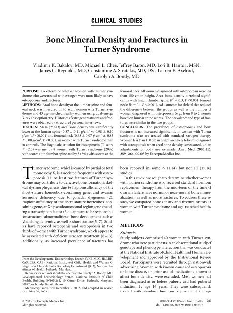
CLINICAL STUDIES<strong>Bone</strong> <strong>M<strong>in</strong>eral</strong> <strong>Density</strong> <strong>and</strong> <strong>Fractures</strong> <strong>in</strong><strong>Turner</strong> <strong>Syndrome</strong>Vladimir K. Bakalov, MD, Michael L. Chen, Jeffrey Baron, MD, Lori B. Hanton, MSN,James C. Reynolds, MD, Constant<strong>in</strong>e A. Stratakis, MD, DSc, Lauren E. Axelrod,Carolyn A. Bondy, MDPURPOSE: To determ<strong>in</strong>e whether women with <strong>Turner</strong> syndromewho were treated with estrogen were more likely to haveosteoporosis <strong>and</strong> fractures.METHODS: Areal bone density at the lumbar sp<strong>in</strong>e <strong>and</strong> femoralneck was measured <strong>in</strong> 40 adult women with <strong>Turner</strong> syndrome<strong>and</strong> 43 age-matched healthy women us<strong>in</strong>g dual-energyX-ray absorptiometry. Histories of estrogen treatment <strong>and</strong> fractureswere obta<strong>in</strong>ed by structured personal <strong>in</strong>terviews.RESULTS: Mean ( SD) areal bone density was significantlylower at the lumbar sp<strong>in</strong>e (0.87 0.11 g/cm 2 vs. 0.98 0.10g/cm 2 , P 0.001) <strong>and</strong> femoral neck (0.68 0.07 g/ cm 2 vs. 0.83 0.08 g/cm 2 , P 0.001) <strong>in</strong> women with <strong>Turner</strong> syndrome than<strong>in</strong> controls. The diagnostic criterion for osteoporosis (T-score2.5) was met by 8 women with <strong>Turner</strong> syndrome (20%)with scores at the lumbar sp<strong>in</strong>e <strong>and</strong> by 3 (8%) with scores at thefemoral neck. All women diagnosed with osteoporosis were lessthan 150 cm <strong>in</strong> height. Areal bone density correlated significantlywith height (lumbar sp<strong>in</strong>e: R 2 0.3, P 0.001; femoralneck: R 2 0.4, P 0.001). Adjustments for skeletal size reducedthe differences between the groups as well as the number ofwomen diagnosed with osteoporosis (e.g., from 8 to 2 womenbased on lumbar sp<strong>in</strong>e scores). The prevalence <strong>and</strong> type of fractureswere similar <strong>in</strong> the two groups.CONCLUSION: The prevalence of osteoporosis <strong>and</strong> bonefractures is not <strong>in</strong>creased significantly <strong>in</strong> women with <strong>Turner</strong>syndrome who are treated with st<strong>and</strong>ard estrogen therapy.Women less than 150 cm <strong>in</strong> height are likely to be misdiagnosedwith osteoporosis when areal bone density is measured, unlessadjustments for body size are made. Am J Med. 2003;115:259 –264. ©2003 by Excerpta Medica Inc.<strong>Turner</strong> syndrome, which is caused by partial or totalmonosomy X, is associated frequently with osteoporosis(1). At least two features of <strong>Turner</strong> syndromemay contribute to defective bone formation: skeletaldysmorphogenesis due to haplo<strong>in</strong>sufficiency of theshort-stature homeobox-conta<strong>in</strong><strong>in</strong>g gene, <strong>and</strong> ovarianhormone deficiency due to gonadal dysgenesis (2).Haplo<strong>in</strong>sufficiency of the short-stature homeobox-conta<strong>in</strong><strong>in</strong>ggene, an Xp pseudoautosomal region gene encod<strong>in</strong>ga transcription factor (3,4), appears to be responsiblefor structural abnormalities of bone development such asMadelung deformity, as well as short stature (5–7). Studieshave reported osteopenia <strong>and</strong> osteoporosis <strong>in</strong> twothirds of women with <strong>Turner</strong> syndrome, which appear tobe associated with deficient estrogen treatment (8 –13).Additionally, an <strong>in</strong>creased prevalence of fractures hasFrom the Developmental Endocr<strong>in</strong>ology Branch (VKB, MLC, JB, LBH,CAS, LEA, CAB), National Institute of Child Health; <strong>and</strong> Warren G.Magnuson Cl<strong>in</strong>ical Center Radiology Department (JCR), National Institutesof Health, Bethesda, Maryl<strong>and</strong>.Requests for repr<strong>in</strong>ts should be addressed to Carolyn A. Bondy, MD,Developmental Endocr<strong>in</strong>ology Branch, National Institute of ChildHealth, Build<strong>in</strong>g 10/10N262, 10 Center Drive, Bethesda, Maryl<strong>and</strong>20892, or bondyc@mail.nih.gov.Manuscript submitted December 3, 2002, <strong>and</strong> accepted <strong>in</strong> revisedform May 30, 2003.been reported <strong>in</strong> some (9,11,14) but not all (15,16)studies.In this study, we sought to determ<strong>in</strong>e whether womenwith <strong>Turner</strong> syndrome who received st<strong>and</strong>ard hormonereplacement therapy from the mid-teens or the time ofovarian failure have normal or near-normal bone m<strong>in</strong>eralization,as well as more fractures. To address these issues,we compared bone density <strong>and</strong> fracture history <strong>in</strong>women with <strong>Turner</strong> syndrome <strong>and</strong> age-matched healthywomen.METHODSSubjectsStudy subjects comprised 40 women with <strong>Turner</strong> syndromewho were participants <strong>in</strong> an observational study ofgenotype <strong>and</strong> phenotype <strong>in</strong>teraction that was conductedat the National Institute of Child Health <strong>and</strong> Human Development<strong>and</strong> approved by the Institutional ReviewBoard. Participants were recruited through nationwideadvertis<strong>in</strong>g. Women with known causes of osteoporosisor bone disease, or prior use of medications known toaffect bone density, were excluded. Most women hadbeen diagnosed at or before puberty <strong>and</strong> had pubertal<strong>in</strong>duction by age 16 years. They were subsequentlytreated with st<strong>and</strong>ard hormone replacement therapy© 2003 by Excerpta Medica Inc. 0002-9343/03/$–see front matter 259All rights reserved.doi:10.1016/S0002-9343(03)00364-4
<strong>Bone</strong> <strong>M<strong>in</strong>eral</strong> <strong>Density</strong> <strong>and</strong> <strong>Fractures</strong> <strong>in</strong> <strong>Turner</strong> <strong>Syndrome</strong>/Bakalov et al(about 50% were tak<strong>in</strong>g oral contraceptives conta<strong>in</strong><strong>in</strong>g20 to 35 g of eth<strong>in</strong>yl estradiol, <strong>and</strong> the rest were tak<strong>in</strong>g0.625 to 1.25 mg of conjugated estrogens <strong>in</strong> comb<strong>in</strong>ationwith cyclical or cont<strong>in</strong>uous progest<strong>in</strong>s). Two women whohad ovarian failure when they were about 25 years oldbegan hormone treatment at that time. Ten had beentreated with growth hormone for 1 to 6 years, when theywere between the ages of 6 <strong>and</strong> 15 years; <strong>and</strong> 2 had beentreated with ox<strong>and</strong>rolone before the age of 11 years. Onewoman was a current smoker, but smoked less than apack per week. All women had fewer than three to fouralcoholic dr<strong>in</strong>ks per week. Three patients were Hispanic<strong>and</strong> the rest were white. Karyotypes were determ<strong>in</strong>ed byG-b<strong>and</strong><strong>in</strong>g of 50 lymphocytes.Controls were 43 age-matched, healthy, premenopausalwomen recruited by the National Institutes ofHealth Normal Volunteer office. They had regular menstrualcycles; did not take medications conta<strong>in</strong><strong>in</strong>g estrogen,progesterone, or testosterone; did not smoke; <strong>and</strong>used alcohol occasionally <strong>in</strong> m<strong>in</strong>imal amounts. Threewere Asian, 6 were African American, <strong>and</strong> 34 were white.Histories concern<strong>in</strong>g menstruation, estrogen treatment,<strong>and</strong> fracture occurrence were obta<strong>in</strong>ed by structuredpersonal <strong>in</strong>terviews <strong>and</strong> questionnaires.Imag<strong>in</strong>gAreal bone density was measured us<strong>in</strong>g a dual-energy X-ray absorptiometer (Hologic QDR-4500A; Hologic, Inc.,Bedford, Massachusetts) with fan beam technology.Lumbar sp<strong>in</strong>e (anteroposterior <strong>and</strong> lateral, L2-L4) <strong>and</strong>proximal femur (hip) scans were performed accord<strong>in</strong>g tothe manufacturer’s procedures. Daily scans of an anthropomorphicsp<strong>in</strong>e phantom over a 6-month periodyielded a coefficient of variation of 0.36%. All scans werereviewed by experienced physicians to ensure that analyseswere correct <strong>and</strong> that measurements did not <strong>in</strong>cludeareas of vessel calcification, degenerative arthritis, oroverlap with the iliac crest or ribs. To obta<strong>in</strong> T- <strong>and</strong> Z-scores, bone density values were compared with normativedata for the anteroposterior lumbar sp<strong>in</strong>e (17), thewidth-adjusted lumbar sp<strong>in</strong>e (Hologic, Inc.), <strong>and</strong> thefemoral neck (18).Corrections of Areal <strong>Bone</strong> <strong>Density</strong>We corrected areal bone density values to adjust for differences<strong>in</strong> bone size. For lumbar sp<strong>in</strong>e data, we usedbone m<strong>in</strong>eral apparent density to derive bone volume,based on the area of the projected vertebral anteroposteriorimage (19). The formula assumes that the vertebralbody is a symmetric cube. Calculation of bone m<strong>in</strong>eralapparent density at the femoral neck is based on a similarassumption (20). Normative data for determ<strong>in</strong><strong>in</strong>g theseT- <strong>and</strong> Z-scores were provided by L. Joseph Melton III,MD (Department of Health Sciences Research, MayoCl<strong>in</strong>ic, Rochester, M<strong>in</strong>nesota) (21). Width-adjustedbone m<strong>in</strong>eral density (22) was also used to estimate thevolume of the vertebra, assumed to be an elliptical cyl<strong>in</strong>der,us<strong>in</strong>g the anteroposterior <strong>and</strong> lateral projected areas.Other adjustments were made us<strong>in</strong>g body surface area(23), which decreases the dependence of areal bone densitymeasurement on height (body size). This method assumesa proportionality of the bone size to the body sizethrough the measure of body surface area.Statistical AnalysisComparisons between groups were performed by onewayanalysis of variance with post hoc Bonferroni test.Where the distribution was not normal or the variancewas not equal, analysis of variance on ranks with theDunn test was used. Proportions were compared by Z-test with Yates correction.The associations of height, weight, age, <strong>and</strong> the diagnosisof <strong>Turner</strong> syndrome with areal bone density wereanalyzed by multiple forward stepwise regression analysis,<strong>and</strong> by best subset regression. As a criterion for thebest subset, the highest adjusted R 2 was used. All P values0.05 were considered statistically significant. Analyseswere performed us<strong>in</strong>g SigmaStat 2.03 software (J<strong>and</strong>elScientific, San Rafael, California).RESULTSWomen with <strong>Turner</strong> syndrome were similar <strong>in</strong> age tocontrols (mean [ SD], 34 11 years vs. 32 8 years,P 0.55). However, they were shorter (146 7 cm vs.164 6cm,P 0.0001) <strong>and</strong> had a higher body mass<strong>in</strong>dex (median [range], 26 [18 to 43] years vs. 23 [18-31]years, P 0.002). The karyotypes <strong>in</strong> women with <strong>Turner</strong>syndrome were similar to those reported previously (24).These women had timely diagnoses <strong>and</strong> reported a highcompliance rate with estrogen therapy; 39 of the 40women took hormone treatment consistently dur<strong>in</strong>g theentire prescribed period <strong>and</strong> 1 adhered to treatment for80% of the time. Less than one third had received growthhormone dur<strong>in</strong>g childhood, for average treatment durationof 4 years.Areal <strong>Bone</strong> <strong>Density</strong>Areal bone m<strong>in</strong>eral density values at the lumbar sp<strong>in</strong>e <strong>and</strong>femoral neck were lower <strong>in</strong> women with <strong>Turner</strong> syndromethan <strong>in</strong> controls (Table 1, Figure 1). However,l<strong>in</strong>ear regression analysis showed a significant positivecorrelation between lumbar <strong>and</strong> femoral areal bone density<strong>and</strong> height for all subjects (Figure 2). Accord<strong>in</strong>g to theareal bone density results, only women less than 150 cm<strong>in</strong> height met the criteria for osteoporosis (T-score2.5; Figure 2).Forward stepwise regression <strong>and</strong> best subset regressionanalyses revealed that height significantly affected arealbone density at the lumbar sp<strong>in</strong>e (adjusted R 2 0.26,P 0.001), whereas the diagnosis of <strong>Turner</strong> syndrome,260 September 2003 THE AMERICAN JOURNAL OF MEDICINE Volume 115
<strong>Bone</strong> <strong>M<strong>in</strong>eral</strong> <strong>Density</strong> <strong>and</strong> <strong>Fractures</strong> <strong>in</strong> <strong>Turner</strong> <strong>Syndrome</strong>/Bakalov et alTable 1. Transformations of Areal <strong>Bone</strong> <strong>Density</strong> to Adjust for <strong>Bone</strong> Size<strong>Turner</strong><strong>Syndrome</strong>(n 40)Control(n 43)DifferencebetweenGroupsP ValueNumber (%) or Mean SDLumbar sp<strong>in</strong>e*Anteroposterior areal bone density (g/cm 2 ) 0.87 0.11 0.98 0.10 12% 0.001Osteoporosis 8 (20) 0 0.003<strong>Bone</strong> m<strong>in</strong>eral apparent density (g/cm 3 ) 0.14 0.02 0.15 0.02 3% 0.18Osteoporosis 2 (5) † 2 (5) 1.0Lateral areal bone density (g/cm 2 ) 0.70 0.09 0.79 0.09 12% 0.001Osteoporosis 11 (28) 1 (2) 0.001Width-adjusted bone m<strong>in</strong>eral density (g/cm 3 ) 0.22 0.03 0.23 0.02 2% 0.43Osteoporosis 5 (12) 1 (2) 0.07Areal bone density/body surface area (g/cm 2 ) 0.94 0.12 1.00 0.10 7% 0.02Osteoporosis 2 (5) † 0 0.14Femoral neck*Areal bone density (g/cm 2 ) 0.68 0.07 0.83 0.08 18% 0.001Osteoporosis 3 (8) 0 0.07<strong>Bone</strong> m<strong>in</strong>eral apparent density (g/cm 3 ) 0.15 0.02 0.16 0.02 11% 0.001Osteoporosis 3 (8) 0 0.07Areal bone density/body surface area (g/cm 2 ) 0.73 0.1 0.83 0.1 11% 0.001Osteoporosis 0 1.0* Criteria for osteoporosis: T-score 2.5.† P 0.05 for comparison with areal bone density.age, <strong>and</strong> weight did not (P 0.05). The same type of analysesshowed that variation <strong>in</strong> femoral neck bone densitywas best predicted by the comb<strong>in</strong>ation of diagnosis of<strong>Turner</strong> syndrome, weight, <strong>and</strong> age (adjusted R 2 0.49),with the diagnosis of <strong>Turner</strong> syndrome (P 0.001) <strong>and</strong>weight (P 0.001) be<strong>in</strong>g significant contributors.Figure 1. Areal bone density of the lumbar sp<strong>in</strong>e (anteroposterior)<strong>and</strong> femoral neck <strong>in</strong> women with <strong>Turner</strong> syndrome(hatched boxes) <strong>and</strong> controls (gray boxes). The boxes representthe 25th <strong>and</strong> 75th percentiles, the bars represent the 10th <strong>and</strong>90th percentiles, <strong>and</strong> the dots represent the 5th <strong>and</strong> 95th percentiles.The horizontal l<strong>in</strong>e <strong>in</strong> the box is the median. P 0.005for both sites.Adjusted <strong>Bone</strong> <strong>Density</strong> MeasurementsVolumetric adjustments us<strong>in</strong>g bone m<strong>in</strong>eral apparentdensity <strong>and</strong> width-adjusted bone m<strong>in</strong>eral density elim<strong>in</strong>ateddifferences between groups for lumbar sp<strong>in</strong>e scores(Table 1, Figure 3). Body surface area adjustments elim<strong>in</strong>atedsignificant differences <strong>in</strong> lumbar sp<strong>in</strong>e Z-scoresbetween women with <strong>Turner</strong> syndrome <strong>and</strong> controls(Figure 3), although a small difference rema<strong>in</strong>ed forthe absolute value of the corrected areal bone density(Table 1).The volumetric transformation of femoral neck arealbone density values us<strong>in</strong>g bone apparent m<strong>in</strong>eral density<strong>and</strong> the body surface area correction reduced, but did notelim<strong>in</strong>ate, the differences between groups, although bothcorrections elim<strong>in</strong>ated the height dependency (Table 1).All normalization methods at both sites of measurementresulted <strong>in</strong> a reduction <strong>in</strong> the diagnosis of osteoporosis(Table 1), with no significant differences betweengroups.<strong>Bone</strong> <strong>Fractures</strong>The prevalence of fractures was similar <strong>in</strong> both groups,<strong>and</strong> most fractures <strong>in</strong>volved the appendicular skeleton(Table 2). A past history of fractures was not associatedwith differences <strong>in</strong> mean current bone density measurementsat the lumbar sp<strong>in</strong>e (0.90 0.10 g/cm 2 with historyof fracture vs. 0.90 0.12 g/cm 2 without history ofSeptember 2003 THE AMERICAN JOURNAL OF MEDICINE Volume 115 261
<strong>Bone</strong> <strong>M<strong>in</strong>eral</strong> <strong>Density</strong> <strong>and</strong> <strong>Fractures</strong> <strong>in</strong> <strong>Turner</strong> <strong>Syndrome</strong>/Bakalov et alFigure 2. Association of lumbar sp<strong>in</strong>e (A) <strong>and</strong> femoral neck(B) areal bone density T-scores with height <strong>in</strong> women with<strong>Turner</strong> syndrome (solid circles) <strong>and</strong> controls (open circles).The dashed l<strong>in</strong>e represents the T-score threshold (–2.5) for diagnosisof osteoporosis.fracture, P 0.95) or femoral neck (0.67 0.06 g/cm 2with history vs. 0.68 0.09 g/cm 2 without history, P 0.57) for women with <strong>Turner</strong> syndrome or controls (0.98 0.09 g/cm 2 vs. 1.03 0.09 g/cm 2 , P 0.20 for lumbarsp<strong>in</strong>e, <strong>and</strong> 0.83 0.09 g/cm 2 vs. 0.81 0.09 g/cm 2 , P 0.67 for femoral neck).DISCUSSIONIn our study of bone density <strong>in</strong> women with <strong>Turner</strong> syndromewho had undergone estrogen therapy <strong>and</strong> agematched,healthy controls, uncorrected areal bone densitymeasurements suggested that lumbar sp<strong>in</strong>e arealbone density was significantly lower, <strong>and</strong> osteoporosiswas significantly more common, <strong>in</strong> women with <strong>Turner</strong>syndrome. However, these values were <strong>in</strong>fluenced byheight <strong>and</strong> bone size, with the shortest women with<strong>Turner</strong> syndrome subject to misdiagnosis. Correction ofareal bone m<strong>in</strong>eral density data to adjust for bone sizeelim<strong>in</strong>ated the difference between groups, <strong>in</strong>clud<strong>in</strong>g thediagnosis of osteoporosis for most women with <strong>Turner</strong>syndrome. In contrast, adjustment for bone size reduced,but did not elim<strong>in</strong>ate, the between-group difference <strong>in</strong>bone m<strong>in</strong>eral density values for the femoral neck, suggest<strong>in</strong>gthat women with <strong>Turner</strong> syndrome may have reducedbone m<strong>in</strong>eralization at the femoral neck, despitest<strong>and</strong>ard estrogen treatment. F<strong>in</strong>ally, bone fractures werenot <strong>in</strong>creased <strong>in</strong> the women with <strong>Turner</strong> syndrome, suggest<strong>in</strong>gthat neither osteoporosis nor <strong>in</strong>creased fragility isa major problem <strong>in</strong> young adult women with <strong>Turner</strong> syndromewho are treated with estrogen.Some earlier studies have reported <strong>in</strong>creased fractures<strong>in</strong> patients with <strong>Turner</strong> syndrome. Us<strong>in</strong>g s<strong>in</strong>gle photonabsorptiometry, Ross et al (14) found that radial bonem<strong>in</strong>eral density was similar <strong>in</strong> girls with <strong>Turner</strong> syndromeaged 3 to 14 years <strong>and</strong> <strong>in</strong> height-matched controls(14). However, the <strong>in</strong>cidence of wrist fractures was <strong>in</strong>creased<strong>in</strong> children with <strong>Turner</strong> syndrome. This f<strong>in</strong>d<strong>in</strong>gmay be expla<strong>in</strong>ed partially by ascerta<strong>in</strong>ment bias due toobta<strong>in</strong><strong>in</strong>g <strong>in</strong>formation on <strong>Turner</strong> syndrome–relatedfractures by personal <strong>in</strong>terview, whereas control datawere obta<strong>in</strong>ed from medical records or registries. A Britishstudy of women who were similar <strong>in</strong> age to our patientsreported a similar fracture prevalence for <strong>Turner</strong>syndrome of about 50%, but a much lower rate ofabout 5% for controls (9). This low rate may reflect underreport<strong>in</strong>gfor the small control group <strong>in</strong> that study. Astudy based on Danish registry data reported an approximatelytwofold <strong>in</strong>creased <strong>in</strong>cidence of fractures <strong>in</strong> adultswith <strong>Turner</strong> syndrome (25). These data, which were collectedfor older adults almost 2 decades ago, most likely<strong>in</strong>cluded many women who had not been treated withestrogen <strong>and</strong> therefore suffered from osteoporosis relatedto prolonged hypogonadism. Another study evaluatedwomen (aged 15 to 57 years) with a cumulative total of770 years at risk, <strong>and</strong> found that the number of fracturesof the distal radius corresponded to the normal premenopausalrather than postmenopausal fracture <strong>in</strong>cidence(15). Thus, women with <strong>Turner</strong> syndrome who receive<strong>in</strong>adequate estrogen treatment may be at <strong>in</strong>creased risk ofosteoporotic fractures, whereas there is no conv<strong>in</strong>c<strong>in</strong>g evidencethat estrogen-treated women are at an <strong>in</strong>creasedrisk.Determ<strong>in</strong><strong>in</strong>g the prevalence of osteoporosis <strong>in</strong> <strong>Turner</strong>syndrome is complicated by confound<strong>in</strong>g factors of smallbone size <strong>and</strong> variability <strong>in</strong> estrogen treatment. Earlierstudies that used areal bone density without adjust<strong>in</strong>g forbone size reported reduced bone m<strong>in</strong>eral density(9,12,13,26,27). A recent Danish study that attempted toaddress these confound<strong>in</strong>g factors <strong>in</strong>itially reportedlower areal bone m<strong>in</strong>eral density measurements for thelumbar sp<strong>in</strong>e <strong>and</strong> femoral neck (24), similar to our f<strong>in</strong>d-262 September 2003 THE AMERICAN JOURNAL OF MEDICINE Volume 115
<strong>Bone</strong> <strong>M<strong>in</strong>eral</strong> <strong>Density</strong> <strong>and</strong> <strong>Fractures</strong> <strong>in</strong> <strong>Turner</strong> <strong>Syndrome</strong>/Bakalov et alFigure 3. Effect of volumetric transformations <strong>and</strong> body surface area adjustments on lumbar sp<strong>in</strong>e areal bone density Z-scores <strong>in</strong>women with <strong>Turner</strong> syndrome (hatched boxes) <strong>and</strong> controls (gray boxes). The boxes represent the 25th <strong>and</strong> 75th percentiles, the barsrepresent the 10th <strong>and</strong> 90th percentiles, <strong>and</strong> the dots represent the 5th <strong>and</strong> 95th percentiles. The horizontal l<strong>in</strong>e <strong>in</strong> the box is themedian. The asterisks (*) <strong>in</strong>dicate P 0.001 for comparison between women with <strong>Turner</strong> syndrome <strong>and</strong> controls. BMAD bonem<strong>in</strong>eral apparent density; WA/BMD width-adjusted bone m<strong>in</strong>eral density.Table 2. Characteristics of <strong>Fractures</strong> <strong>in</strong> Women with <strong>Turner</strong><strong>Syndrome</strong> <strong>and</strong> <strong>in</strong> ControlsCharacteristic<strong>Turner</strong><strong>Syndrome</strong>(n 40)Control(n 33) P ValueNumber (%) orMedian (Range)Age of first fracture 14 (3–34) 16 (4–38) 0.6(years)Women with at least 23 (58) 17 (52) 0.65one fracture episodeWomen with 1 8 (20) 5 (15) 0.6fracture episodeTotal fracture episodes (n 37) (n 25) 0.06Appendicular bone 30 (82) 21 (83) 0.9fractures<strong>Fractures</strong> dur<strong>in</strong>g sport 16 (43) 23 (71) 0.02or play<strong>Fractures</strong> dur<strong>in</strong>g daily 11 (31) 7 (21) 0.35activities<strong>Fractures</strong> dur<strong>in</strong>g car 5 (14) 0 0.03accidentOther fracture 4 (11) 9 (28) 0.07<strong>in</strong>gs. However, after volumetric corrections, the difference<strong>in</strong> femoral neck density disappeared, but the difference<strong>in</strong> lumbar sp<strong>in</strong>e density rema<strong>in</strong>ed (24). Theseauthors also found that <strong>in</strong> multivariate analysis, the diagnosisof <strong>Turner</strong> syndrome was not associated with lumbarsp<strong>in</strong>e or femoral neck bone m<strong>in</strong>eral density.The three forms of volumetric/bone size correctionthat we used— bone m<strong>in</strong>eral apparent density, width-adjustedbone m<strong>in</strong>eral density, <strong>and</strong> body surface area—elim<strong>in</strong>ated or reduced the difference between groups forlumbar sp<strong>in</strong>e areal bone density, result<strong>in</strong>g also <strong>in</strong> fewerdiagnoses of osteoporosis. Corrections for bone size <strong>in</strong>the case of the femoral neck reduced, but did not elim<strong>in</strong>ate,the difference <strong>in</strong> areal bone density between groups.These f<strong>in</strong>d<strong>in</strong>gs are consistent with the multiple regressionanalyses that showed height to be the only significantcontributor to variation <strong>in</strong> bone density at the lumbarsp<strong>in</strong>e, whereas the diagnosis of <strong>Turner</strong> syndrome wasfound to predict a reduction <strong>in</strong> bone density at the femoralneck, <strong>in</strong>dependently of height. The femoral neckconta<strong>in</strong>s a greater proportion of cortical bone comparedwith the lumbar sp<strong>in</strong>e, <strong>and</strong> evidence suggests that corticalbone is selectively reduced <strong>in</strong> <strong>Turner</strong> syndrome. For example,direct volumetric measurement of the radius us<strong>in</strong>gquantitative computer tomography showed thatyoung women with <strong>Turner</strong> syndrome, all of whom hadappropriate estrogen treatment, had reduced corticalbone content of the radius, whereas trabecular bone contentwas normal (28). In addition, it is notable that themean Z-scores for areal lumbar sp<strong>in</strong>e <strong>and</strong> femoral neckbone density <strong>in</strong> both groups fell below the normal meanprovided by the manufacturer (Figures 1 <strong>and</strong> 3), high-September 2003 THE AMERICAN JOURNAL OF MEDICINE Volume 115 263
<strong>Bone</strong> <strong>M<strong>in</strong>eral</strong> <strong>Density</strong> <strong>and</strong> <strong>Fractures</strong> <strong>in</strong> <strong>Turner</strong> <strong>Syndrome</strong>/Bakalov et allight<strong>in</strong>g the importance of a contemporaneous controlgroup for this type of study.In summary, it appears that women with <strong>Turner</strong> syndromewho have received st<strong>and</strong>ard hormone replacementdo not have significant reductions <strong>in</strong> bone m<strong>in</strong>eraldensity of the sp<strong>in</strong>e, <strong>and</strong> have only modest reductions atthe hip. Those who are less than 150 cm <strong>in</strong> height arelikely to be misdiagnosed with osteoporosis. The currentbone density does not seem to correlate with a history ofbone fractures.REFERENCES1. Bercu BB, Kramer SS, Bode HH. A useful radiologic sign for thediagnosis of <strong>Turner</strong>’s syndrome. Pediatrics. 1976;58:737–739.2. Rub<strong>in</strong> K. <strong>Turner</strong> syndrome <strong>and</strong> osteoporosis: mechanisms <strong>and</strong>prognosis. Pediatrics. 1998;102:481–485.3. Rao E, Weiss B, Fukami M, et al. Pseudoautosomal deletions encompass<strong>in</strong>ga novel homeobox gene cause growth failure <strong>in</strong> idiopathicshort-stature <strong>and</strong> <strong>Turner</strong> syndrome. Nat Genet. 1997;16:54 –63.4. Rao E, Blaschke RJ, March<strong>in</strong>i A, Niesler B, Burnett M, Rappold GA.The Leri-Weill <strong>and</strong> <strong>Turner</strong> syndrome homeobox gene SHOX encodesa cell-type specific transcriptional activator. Hum Mol Genet.2001;10:3083–3091.5. Clement-Jones M, Schiller S, Rao E, et al. The short-stature homeoboxgene SHOX is <strong>in</strong>volved <strong>in</strong> skeletal abnormalities <strong>in</strong> <strong>Turner</strong>syndrome. Hum Mol Genet.. 2000;9:695–702.6. Ogata T. SHOX: pseudoautosomal homeobox conta<strong>in</strong><strong>in</strong>g gene forshort-stature <strong>and</strong> dyschondrosteosis. Growth Horm IGF Res.. 1999;9:53–58 (suppl B).7. Ross JL, Scott C Jr, Marttila P, et al. Phenotypes associated withSHOX deficiency. J Cl<strong>in</strong> Endocr<strong>in</strong>ol Metab.. 2001;86:5674 –5680.8. Naeraa RW, Brixen K, Hansen RM, et al. Skeletal size <strong>and</strong> bonem<strong>in</strong>eral content <strong>in</strong> <strong>Turner</strong>’s syndrome: relation to karyotype, estrogentreatment, physical fitness, <strong>and</strong> bone turnover. Calcif Tissue Int.1991;49:77–83.9. Davies MC, Gulekli B, Jacobs HS. Osteoporosis <strong>in</strong> <strong>Turner</strong>’s syndrome<strong>and</strong> other forms of primary amenorrhoea. Cl<strong>in</strong> Endocr<strong>in</strong>ol(Oxf). 1995;43:741–746.10. Sylven L, Hagenfeldt K, R<strong>in</strong>gertz H. <strong>Bone</strong> m<strong>in</strong>eral density <strong>in</strong> middle-agedwomen with <strong>Turner</strong>’s syndrome. Eur J Endocr<strong>in</strong>ol. 1995;132:47–52.11. L<strong>and</strong><strong>in</strong>-Wilhelmsen K, Bryman I, W<strong>in</strong>dh M, Wilhelmsen L. Osteoporosis<strong>and</strong> fractures <strong>in</strong> <strong>Turner</strong> syndrome-importance of growthpromot<strong>in</strong>g <strong>and</strong> oestrogen therapy. Cl<strong>in</strong> Endocr<strong>in</strong>ol (Oxf). 1999;51:497–502.12. Benetti-P<strong>in</strong>to CL, Bedone A, Magna LA, Marques-Neto JF. Factorsassociated with the reduction of bone density <strong>in</strong> patients with gonadaldysgenesis. Fertil Steril. 2002;77:571–575.13. Costa AM, Lemos-Mar<strong>in</strong>i SH, Baptista MT, Morcillo AM, Maciel-Guerra AT, Guerra G Jr. <strong>Bone</strong> m<strong>in</strong>eralization <strong>in</strong> <strong>Turner</strong> syndrome:a transverse study of the determ<strong>in</strong>ant factors <strong>in</strong> 58 patients. J <strong>Bone</strong>M<strong>in</strong>er Metab. 2002;20:294 –297.14. Ross JL, Long LM, Feuillan P, Cassorla F, Cutler GB Jr. Normalbone density of the wrist <strong>and</strong> sp<strong>in</strong>e <strong>and</strong> <strong>in</strong>creased wrist fractures <strong>in</strong>girls with <strong>Turner</strong>’s syndrome. J Cl<strong>in</strong> Endocr<strong>in</strong>ol Metab. 1991;73:355–359.15. Smith MA, Wilson J, Price WH. <strong>Bone</strong> dem<strong>in</strong>eralization <strong>in</strong> patientswith <strong>Turner</strong>’s syndrome. J Med Genet. 1982;19:100 –103.16. Preger L, Ste<strong>in</strong>bach HL, Moskowitz P, Scully AL, Goldberg MB.Roentgenographic abnormalities <strong>in</strong> phenotypic females with gonadaldysgenesis. A comparison of chromat<strong>in</strong> positive patients <strong>and</strong>chromat<strong>in</strong> negative patients. Am J Roentgenol Radium Ther NuclMed. 1968;104:899 –910.17. Kelly TL. <strong>Bone</strong> m<strong>in</strong>eral reference database for American men <strong>and</strong>women. J <strong>Bone</strong> M<strong>in</strong>er Res. 1990;5:249 –260.18. Looker AC, Wahner HW, Dunn WL, et al. Proximal femur bonem<strong>in</strong>eral levels of US adults. Osteoporos Int. 1995;5:389 –409.19. Carter DR, Bouxse<strong>in</strong> ML, Marcus R. New approaches for <strong>in</strong>terpret<strong>in</strong>gprojected bone densitometry data. J <strong>Bone</strong> M<strong>in</strong>er Res. 1992;7:137–145.20. Cumm<strong>in</strong>gs SR, Marcus R, Palermo L, Ensrud KE, Genant HK. Doesestimat<strong>in</strong>g volumetric bone density of the femoral neck improvethe prediction of hip fracture? A prospective study. Study of Osteoporotic<strong>Fractures</strong> Research Group. J <strong>Bone</strong> M<strong>in</strong>er Res. 1994;9:1429 –1432.21. Melton LJ III, Khosla S, Achenbach SJ, O’Connor MK, O’FallonWM, Riggs BL. Effects of body size <strong>and</strong> skeletal site on the estimatedprevalence of osteoporosis <strong>in</strong> women <strong>and</strong> men. Osteoporos Int.2000;11:977–983.22. Blake GM. Measurement of bone density <strong>in</strong> the lumbar sp<strong>in</strong>e: thelateral sp<strong>in</strong>e scan. In: Dunitz M, ed. The Evaluation of Osteoporosis:Dual Energy X-ray Absorptiometry. 2nd ed. London: Dunitz; 1999:235–258.23. Pors Nielsen S, Kolthoff N, Barenholdt O, et al. Diagnosis of osteoporosisby planar bone densitometry: can body size be disregarded?Br J Radiol. 1998;71:934 –943.24. Gravholt CH, Lauridsen AL, Brixen K, Mosekilde L, HeickendorffL, Christiansen JS. Marked disproportionality <strong>in</strong> bone size <strong>and</strong>m<strong>in</strong>eral, <strong>and</strong> dist<strong>in</strong>ct abnormalities <strong>in</strong> bone markers <strong>and</strong> calcitropichormones <strong>in</strong> adult <strong>Turner</strong> syndrome: a cross-sectional study. J Cl<strong>in</strong>Endocr<strong>in</strong>ol Metab. 2002;87:2798 –2808.25. Gravholt CH, Juul S, Naeraa RW, Hansen J. Morbidity <strong>in</strong> <strong>Turner</strong>syndrome. J Cl<strong>in</strong> Epidemiol. 1998;51:147–158.26. Garden AS, Diver MJ, Fraser WD. Undiagnosed morbidity <strong>in</strong> adultwomen with <strong>Turner</strong>’s syndrome. Cl<strong>in</strong> Endocr<strong>in</strong>ol (Oxf). 1996;45:589 –593.27. Lanes R, Gunczler P, Esaa S, Mart<strong>in</strong>is R, Villaroel O, Weis<strong>in</strong>ger JR.Decreased bone mass despite long-term estrogen replacement therapy<strong>in</strong> young women with <strong>Turner</strong>’s syndrome <strong>and</strong> previously normalbone density. Fertil Steril. 1999;72:896 –899.28. Bechtold S, Rauch F, Noelle V, et al. Musculoskeletal analyses of theforearm <strong>in</strong> young women with <strong>Turner</strong> syndrome: a study us<strong>in</strong>gperipheral quantitative computed tomography. J Cl<strong>in</strong> Endocr<strong>in</strong>olMetab. 2001;86:5819 –5823.264 September 2003 THE AMERICAN JOURNAL OF MEDICINE Volume 115


