Zosteriform cutaneous metastasis from a breast ... - edigraphic.com
Zosteriform cutaneous metastasis from a breast ... - edigraphic.com
Zosteriform cutaneous metastasis from a breast ... - edigraphic.com
Create successful ePaper yourself
Turn your PDF publications into a flip-book with our unique Google optimized e-Paper software.
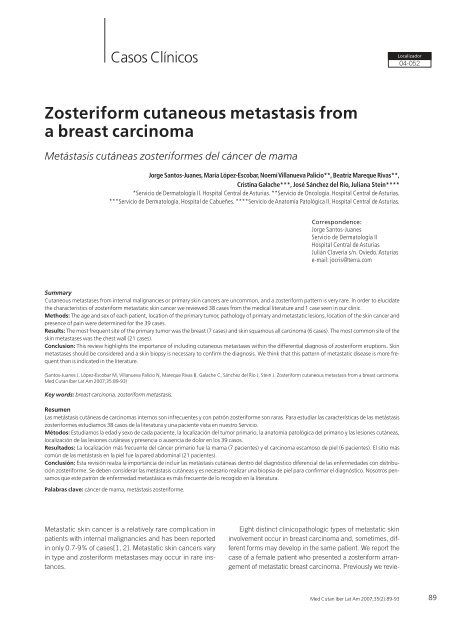
Casos ClínicosLocalizador04-052<strong>Zosteriform</strong> <strong>cutaneous</strong> <strong>metastasis</strong> <strong>from</strong>a <strong>breast</strong> carcinomaMetástasis cutáneas zosteriformes del cáncer de mamaJorge Santos-Juanes, María López-Escobar, Noemí Villanueva Palicio**, Beatriz Mareque Rivas**,Cristina Galache***, José Sánchez del Río, Juliana Stein*****Servicio de Dermatología II. Hospital Central de Asturias. **Servicio de Oncología. Hospital Central de Asturias.***Servicio de Dermatología. Hospital de Cabueñes. ****Servicio de Anatomía Patológica II. Hospital Central de Asturias.Correspondence:Jorge Santos-JuanesServicio de Dermatología IIHospital Central de AsturiasJulián Clavería s/n. Oviedo.Asturiase-mail: jocris@terra.<strong>com</strong>SummaryCutaneous metastases <strong>from</strong> internal malignancies or primary skin cancers are un<strong>com</strong>mon, and a zosteriform pattern is very rare. In order to elucidatethe characteristics of zosteriform metastatic skin cancer we reviewed 38 cases <strong>from</strong> the medical literature and 1 case seen in our clinic.Methods: The age and sex of each patient, location of the primary tumor, pathology of primary and metastatic lesions, location of the skin cancer andpresence of pain were determined for the 39 cases.Results: The most frequent site of the primary tumor was the <strong>breast</strong> (7 cases) and skin squamous all carcinoma (6 cases). The most <strong>com</strong>mon site of theskin metastases was the chest wall (21 cases).Conclusion: This review highlights the importance of including <strong>cutaneous</strong> metastases within the differential diagnosis of zosteriform eruptions. Skinmetastases should be considered and a skin biopsy is necessary to confirm the diagnosis. We think that this pattern of metastatic disease is more frequentthan is indicated in the literature.(Santos-Juanes J, López-Escobar M, Villanueva Palicio N, Mareque Rivas B, Galache C, Sánchez del Río J, Stein J. <strong>Zosteriform</strong> <strong>cutaneous</strong> <strong>metastasis</strong> <strong>from</strong> a <strong>breast</strong> carcinoma.Med Cutan Iber Lat Am 2007;35:89-93)Key words: <strong>breast</strong> carcinona, zosteriform <strong>metastasis</strong>.ResumenLas metástasis cutáneas de carcinomas internos son infrecuentes y con patrón zosteriforme son raras. Para estudiar las características de las metástasiszosteriformes estudiamos 38 casos de la literatura y una paciente vista en nuestro Servicio.Métodos: Estudiamos la edad y sexo de cada paciente, la localización del tumor primario, la anatomía patológica del primario y las lesiones cutáneas,localización de las lesiones cutáneas y presencia o ausencia de dolor en los 39 casos.Resultados: La localización más frecuente del cáncer primario fue la mama (7 pacientes) y el carcinoma escamoso de piel (6 pacientes). El sitio más<strong>com</strong>ún de las metástasis en la piel fue la pared abdominal (21 pacientes).Conclusión: Esta revisión realza la importancia de incluir las metástasis cutáneas dentro del diagnóstico diferencial de las enfermedades con distribuciónzosteriforme. Se deben considerar las metástasis cutáneas y es necesario realizar una biopsia de piel para confirmar el diagnóstico. Nosotros pensamosque este patrón de enfermedad metastásica es más frecuente de lo recogido en la literatura.Palabras clave: cáncer de mama, metástasis zosteriforme.Metastatic skin cancer is a relatively rare <strong>com</strong>plication inpatients with internal malignancies and has been reportedin only 0.7-9% of cases[1, 2]. Metastatic skin cancers varyin type and zosteriform metastases may occur in rare instances.Eight distinct clinicopathologic types of metastatic skininvolvement occur in <strong>breast</strong> carcinoma and, sometimes, differentforms may develop in the same patient. We report thecase of a female patient who presented a zosteriform arrangementof metastatic <strong>breast</strong> carcinoma. Previously we revie-Med Cutan Iber Lat Am 2007;35(2):89-93 89
Santos-Juanes J y cols. <strong>Zosteriform</strong> <strong>cutaneous</strong> <strong>metastasis</strong> <strong>from</strong> a <strong>breast</strong> carcinomaTabla 1. Cases of skin metastases with a zosteriform distribution reportedCase Ref. Year Sex Age Origin Pathology Location Pain1 20 1933 F 48 Left <strong>breast</strong> Nd Thorax n.d2 21 1957 M 83 Prostate Adenocarcinoma Left abdomen Yes3 21 1957 M 67 Prostate Nd Left thigh n.d4 22 1979 M 57 Right lung Adenocarcinoma Left thorax Yes5 11 1984 M 65 Skin Squamous Right arm n.d6 23 1986 M 61 Bladder Transitional cell c. Left flank Yes7 19 1987 F 63 Ovary Adenocarcinoma Right thorax Yes8 24 1988 M 65 Left lung Adenocarcinoma Left thorax Yes9 25 1988 F 70 Ovary Adenocarcinoma Left thigh n.d10 4 1991 F 43 Left <strong>breast</strong> Adenocarcinoma Left thorax Yes11 26 1991 F 64 Ovary Nd Abdomen n.d12 26 1991 F 53 Ovary Cystadenocarcinoma Abdomen Yes13 26 1991 F 69 Ovary Cystadenocarcinoma Abdomen No14 5 1992 F 48 Left <strong>breast</strong> Adenocarcinoma Right <strong>breast</strong> Yes15 7 1992 F 38 Colon Adenocarcinoma Right thigh Yes16 28 1994 M 67 Bladder Transitional cell c. Left thorax Yes17 29 1994 F 70 Breast Ductal carcinoma Right thorax No18 9 1995 F 29 Skin Melanoma Right thorax n.d19 7 1996 F 62 Uterus Adenocarcinoma Right thorax Yes20 15 1997 M 31 Skin Squamous Left thorax n.d21 6 1998 F 78 Left <strong>breast</strong> Ductal carcinoma Left thorax No22 14 1998 M 83 Skin Squamous Right thorax Yes23 8 1998 M 63 Skin Melanoma Right thorax Yes24 30 1999 M 65 Sigmoid colon Adenocarcinoma Left leg n.d25 10 2000 M 79 Skin Melanoma Left thorax No26 13 2000 M 56 Skin Squamous Left thorax Yes27 17 2000 M 71 Bronchus Adenocarcinoma Right face n.d28 18 2000 M 79 Colon Adenocarcinoma Left thorax n.d29 12 2001 F 72 Skin Squamous Right thigh Yes30 16 2001 M 53 Stomach Adenocarcinoma Left abdomen No31 16 2001 F 63 Stomach Adenocarcinoma Left thorax No32 16 2001 M 69 Lung Adenocarcinoma Right abdomen Yes33 31 2002 F 71 Skin Squamous Right abdomen n.d34 32 2003 F 54 Breast Adenocarcinoma Thorax No35 33 2003 F 59 Skin Melanoma Right thorax n.d36 34 2003 M 44 Rectal Adenocarcinoma Abdomen Yes37 35 2003 F n.d Pelvis renal Transitional cell c. Arm n.d38 36 2004 F 66 Bronchus Adenocarcinoma Right thigh No39 Our case 2002 F 71 Breast Ductal Left thorax NoN.d.: not disposable.wed the reported studies of dermatomal or zosteriformmetastases (table 1)[3-36].Skin metastases with zosteriform distribution <strong>from</strong> internalmalignancies or primary skin cancer are infrequent. Segmental<strong>metastasis</strong> was first reported in 1933 in a patient with <strong>breast</strong>cancer[20]. Up today cases have been reported with: Skincancer; melanoma [8-10, 33]; squamous cell carcinoma(SCC)[11-15, 31] and visceral tumors (22 cases)[16-19].Case reportA 71-year old woman was referred to our clinic in january2001 with a history of papules on the left side of her flanksince one month. She had not been previously affected withherpes zoster and had had a ductal carcinoma of the left <strong>breast</strong>two years previously. This was treated with radical mastectomy,lymphadenectomy, and irradiation. Adjuvant chemotherapyconsisting of cyclophosphamide, methotrexate,and fluouracil (six cycles) was given.The patient had been previously diagnosed in anotherhospital as having herpes zoster and had been treated withoral acyclovir (800 mg five times daily for 10 days). No improvementwas seen. No painful lesions or itching were referredto. Examination revealed groups of papules and nodules onthe left side of the surgical wound on the thorax and presenteda distribution consistent with left T dermatome (Figure 1). Askin biopsy revealed irregular islands of atypical epithelial cellsinfiltrating the superficial and deep dermis, consistent with90Med Cutan Iber Lat Am 2007;35(2):89-93
Santos-Juanes J y cols. <strong>Zosteriform</strong> <strong>cutaneous</strong> <strong>metastasis</strong> <strong>from</strong> a <strong>breast</strong> carcinomaFigure 3. The neoplastic cells are seen sticking to the wall.A broken vessel is seen (H/E stain; original magnificationx 100).Figure 1. Groups of linear, confluent papules and nodulesunder the amputed left <strong>breast</strong>.metastatic <strong>breast</strong> carcinoma. In some areas an “Indian file”arrangement of dermal nests can be seen (Figure 2). Lymphaticinfiltration by tumor cells was observed (Figure 3). A thoracic-abdominalscan showed lung <strong>metastasis</strong> and retroperitonealadenopathies. The patient began treatment withpaclitaxel and epiadriamicine and no improvement was seenand the ganglion and lung metastases continued to grow. Shedied one year later <strong>from</strong> brain <strong>metastasis</strong>.CommentEvery tumor may occasionally cause <strong>cutaneous</strong> metastases,but some do so more frequently. Although the incidenceof <strong>metastasis</strong> correlates well with the frequency of the primarymalignant tumours in both gender.Figure 2. Biopsy shows a band of atypical cells along thedermis (H/E stain; original magnification x 100).In our review of the literature (table 1) the primary sitesinvolved were the <strong>breast</strong> (7 cases) and skin squamous cellcarcinoma (6 cases each), the ovary and the lung (5 caseseach), melanoma and colon (4 cases each), prostate, bladderand stomach (2 cases each), and the uterus and renalpelvis (1 case). The skin metastases were located on thechest wall (21 cases), the abdominal wall (9 cases), the leg(6 cases), the arms (2 cases) and face (1 case). The chestwall was the most <strong>com</strong>mon site of metastatic skin lesionswhen the primary cancer was in the lung or the <strong>breast</strong>. Thehistology of the primary tumor was <strong>com</strong>patible with adenocarcinoma(20 cases), squamous cell carcinoma (6 cases),melanoma (4 cases), ductal carcinoma (3 cases), transitionalcell carcinoma (3 cases). Seventeen patients <strong>com</strong>plainedof spontaneous pain and were misdiagnosed and treatedfor herpes zoster.In our review of the literature, metastatic <strong>breast</strong> carcinomawas the most frequent in the general series and the most frequentlyreported in zosteriform pattern[16]. We wish toemphasize the high frequency in our revision of SCC (10.8%).Three of the SCC were in immunosupressed patients[11, 13,15]. It is well documented that SCC behave more aggressivelyin immunosupressed patients[13]. Although melanoma hasbeen reported to have the highest frequency of <strong>cutaneous</strong>metastases[37], we found only four cases of <strong>cutaneous</strong> melanomametastases with zosteriform distribution in the literature,which indicates the unusual nature of this particular manifestationof melanoma skin metastases.In males the highest prevalence of primary malignancywas SCC (22.2%) and lung carcinoma (22.2%). In femalesthe highest prevalence of primary malignancy was <strong>breast</strong>carcinoma (35%), followed by ovary carcinoma (25%).Med Cutan Iber Lat Am 2007;35(2):89-93 91
Santos-Juanes J y cols. <strong>Zosteriform</strong> <strong>cutaneous</strong> <strong>metastasis</strong> <strong>from</strong> a <strong>breast</strong> carcinomaThe mechanism of zosteriform distribution in metastaticskin cancer is still speculative. It has been suggested that itmight be due to: a) a Koebner-like reaction at the site of aprior herpes zoster infection; b) neural lymphatic spread viathe fenestrated vessels of the dorsal root ganglion; c) accidentalsurgical implantation, or d) perineural lymphatic spread[16,33]. Figure 3 (our case) shows the lymphatic vesselsfull of metastatic cells rupturing the vessel wall. This couldlend support to the second hypothesis.A dermatomal distribution does not necessarily indicateherpes zoster, because several <strong>cutaneous</strong> lesions have beendescribed by others: <strong>com</strong>edones, xantomatous changes, granulomaannulare, granulomas sarcoideos, tuberculoid granulomas,granulomatous vasculitis, tinea, acneiform eruption,furunculosis, contact dermatitis, nodular solar degeneration,pseudolymphoma, psoriasis, lichen planus, morphoea, lichenoidchronic graft versus host disease, eosinophilic dermatosis,acquired reactive perforating dermatosis[38]. Eventhough it is usually very easy to make a diagnosis of herpeszoster, a correct diagnosis can only be made by skin biopsy.Cutaneous metastases may represent the first sign ofinternal malignancy, or can represent the first indication ofrecurrence. These metastases are generally considered tobe a manifestation associated with a poor prognosis. Deathusually occurs within a few months (6.5 months), although afew patients have survived for several years[39]. Given thepoor prognosis which implies a <strong>cutaneous</strong> spread of internalcancer, we must remain alert and perform biopsies in lesionswith zosteriform patterns, which present atypical characteristics,both morphologically and developmentally.References1. Spencer PS, Helm TN. Skin metastases incancer patients. Cutis 1987;39:119-121.2. Schwartz RA. Cutaneous metastatic disease.J Am Acad Dermatol 1995;33:161-182.3. Weber FP. Bilateral thoracic zosteroid spreadingmarginate telangiectasia probably a varietyof carcinoma telangiectaticum. Br J Dermatol1993;45:418-23.4. Willians LR, Levine U, Kaugh YC. Cutaneousmalignancies mimicking herpes zoster. Int JDermatol 1991;30:432-4.5. Manteaux A, Cohen PR, Rapini RP. <strong>Zosteriform</strong>and epidermotropic <strong>metastasis</strong>: report of twocases. J Dermatol Surg Oncol 1992; 18:97-100.6. Cecchi R, Brunetti L, Bartoli L, Pavesi M,Giomi A. <strong>Zosteriform</strong> skin metastases <strong>from</strong><strong>breast</strong> carcinoma in association with herpeszoster. Int J Dermatol 1998;37:476-7.7. Heckmann M, Volkenande M, Lengyel ER,Schirren CG, Gizycki-Nienhaus BV. Cytologicaldiagnosis of zosteriform skin metastases inundiagnosed <strong>breast</strong> carcinoma. Br J Dermatol1996;135:502-3.8. North S. Recurrent malignant melanomapresenting with zosteriform metastases.Cutis 1998;62:143-46.9. Itin PH, Lautenschlager S, Buechner SA.<strong>Zosteriform</strong> metastases in melanoma. J AmAcad Dermatol 1995,32:854-57.10. Cuerda E, Sánchez de Paz F, Mansilla I, PozaO, Casellas M. Malignant melanoma withzosteriform metastases. Cutis 2000; 65:312-14.11. Buecker JW, Ratz JL. Cutaneous metastaticsquamous cell carcinoma in zosteriformdistribution. J Dermatol Surg Oncol 1984;10:718-20.12. Kato N, Aoyagi S, Sugawara H, Mayuzumi M.<strong>Zosteriform</strong> and epidermotropic metastaticprimary <strong>cutaneous</strong> squamous cell carcinoma.Am J Dermatopathol 2001; 23:216-20.13. Fearfield LA, Nelson M, Francis N, BunkerCB. Cutaneous squamous cell carcinomawith zosteriform metastases in a humanimmunodeficiency virus-infected patient. BrJ Dermatol 2000;142:573-4.14. Cuq-Viguier L, Viraben R. <strong>Zosteriform</strong><strong>cutaneous</strong> metastases <strong>from</strong> squamous cellcarcinoma of the stump of an amputatedarm. Clin Exp Dermatol 1998;23:116-8.15. Shafqat A, Viehman GE, Myers SA. Cutaneoussquamous cell carcinoma with zosteriform<strong>metastasis</strong> in a transplant recipient. J AmAcad Dermatol 1997;37:1008-9.16. Kikuchi Y, Matsuyama A, Nomura K. <strong>Zosteriform</strong>metastatic skin cancer: report of three casesand review of the literature. Dermatology2001;202:336-38.17. Bianchi L, Orlandi A, Carboni I, Costanzo A,Chimenti S. <strong>Zosteriform</strong> <strong>metastasis</strong> of occultbronchogenic carcinoma. Acta Derm Venereol2000;80:391-2.18. Ahmed I, Holley KJ, Charles Holmes R.<strong>Zosteriform</strong> <strong>metastasis</strong> of colonic carcinoma.Br J Dermatol 2000;142:182-3.19. Caroti A. Metastasi cutanee di a adenocarcinomapapilifero ovarico in sede di herpes zoster.Chron Dermatol 1987;18:769-73.20. Weber FP: bilateral thoracic zosteroid spreadingmarginate telangiectasia –probably variantof carcinoma telangiectatum. Br J Dermatol1933;45:418-23.21. Bluefarb SM, Walk S, Gecht M. Carcinoma ofprostate with zosteriform <strong>cutaneous</strong> lesions.Arch Dermatol 1957;76:402-6.22. Hodge SJ, Mackel S, Owen LG. <strong>Zosteriform</strong>inflamatory metastatic carcinoma. Int J Dermatol1979;18:142-45.23. Jaworsky C, Bergfeld WF. Metastatictransitional cell carcinoma mimicking zostersine herpete. Arch Dermatol 1985;122:1357-8.24. Matarasso SL, Rosen T. <strong>Zosteriform</strong> <strong>metastasis</strong>:case presentation and review of the literature.J Dermatol Surg Oncol 1988;14;774-8.25. Patsner B, Mann WJ, Chumas J, Loesch M.Herpetiform <strong>cutaneous</strong> metastases followingnegative second look laparatomy for ovarianadenocarcinoma. Arch Gynecol Obstet 1988;244:63-8.26. Merinsky O, Chaichik S, Inbar M. Skin<strong>metastasis</strong> of ovarian cancer. Report of threecases. Tumori 1991;77:268-70.27. Ferrer M, Borbujo J, García-Rubio B, Díaz R,Manzano R, Casado M. Metástasis cutáneazoniforme de carcinoma de colón. ActasDermo-Sif 1992;83:136-8.28. Ando K, Goto Y, Kato K, Murase T, Matsumoto Y,Ohashi M. <strong>Zosteriform</strong> inflamatory metastaticcarcinoma <strong>from</strong> transitional cell carcinomaof the renal pelvis. J Am Acad Dermatol1994;31:284-6.29. Suárez R, Martín F, López E, Núñez C,Nieto Y, Sánchez E. Metástasis cutáneaszosteriformes en carcinoma de mama.Una presentación poco habitual. ActasDermosifiliogr 1994;85:251-4.30. Maeda S, Hara H, Morishima T. <strong>Zosteriform</strong><strong>cutaneous</strong> metastases arising <strong>from</strong>adenocarcinoma of colon: diagnostic smear<strong>from</strong> <strong>cutaneous</strong> lesions. Acta Derm Venereol1999;79:90-1.31. Bauzá A, Redondo P, Idoate MA. Cutaneouszosteriform squamous cell carcinoma <strong>metastasis</strong>arising in an immuno<strong>com</strong>petent patient. ClinExp Dermatol 2002;27:199-201.32. Brasanac D, Boricic I, Todorovic V. Epidermotropicmetastases <strong>from</strong> <strong>breast</strong> carcinoma92Med Cutan Iber Lat Am 2007;35(2):89-93
Santos-Juanes J y cols. <strong>Zosteriform</strong> <strong>cutaneous</strong> <strong>metastasis</strong> <strong>from</strong> a <strong>breast</strong> carcinomashowing different clinical and histopathologicalfeatures on the trunk and on the scalpin a single patient. J Cutan Pathol 2003;30:641-6.33. Zalaudek I, Leinweber B, Richtig E, Smolle J,Hofmann-Wellenhof R. Cutaneous zosteriformmelanoma <strong>metastasis</strong> arising after herpeszoster infection: a case report and review ofthe literature. Mel Res 2003;13:635-9.34. Damin DC, Lazzaron AR, Tarta C, Cartel A,Rosito MA. Massive zosteriform <strong>cutaneous</strong><strong>metastasis</strong> <strong>from</strong> rectal carcinoma. TechColoproctol 2003;7:105-7.35. Lin CY, Lee CT, Huang JS, Chang LC. Transitionalcell carcinoma <strong>metastasis</strong> to arm skin <strong>from</strong>the renal pelvis. Chang Gung Med J 2003;26:525-9.36. LeSueur BW, Abraham RJ, DiCaudo DJ,O’Connor WJ. <strong>Zosteriform</strong> skin metastases.Int J Dermatol 2004;43:126-8.37. Lookigbill DP, Spangler N, Helm KF. Cutaneousmetastases in patients with metastatic carcinoma:a retrospective study of 4,020 patients. J AmAcad Dermatol 1993;29:228-36.38. Requena L. Kutzner H, Escalonilla P, Ortiz S,Schaller J, Rohwedder A. Cutaneous reactionsat sites of herpes zoster scars: and expandedspectrum. Br J Dermatol 1998; 138:161-168.39. Schoenlaub P, Sarraux A, Grosshans E, Heid E,Cribier B. Survie après metastases cutanées:etude de 200 cases. Ann Dermatol Venéreol2001;128:1310-50.Med Cutan Iber Lat Am 2007;35(2):89-93 93


