Study Guide RADIOLOGY - UMF
Study Guide RADIOLOGY - UMF
Study Guide RADIOLOGY - UMF
You also want an ePaper? Increase the reach of your titles
YUMPU automatically turns print PDFs into web optimized ePapers that Google loves.
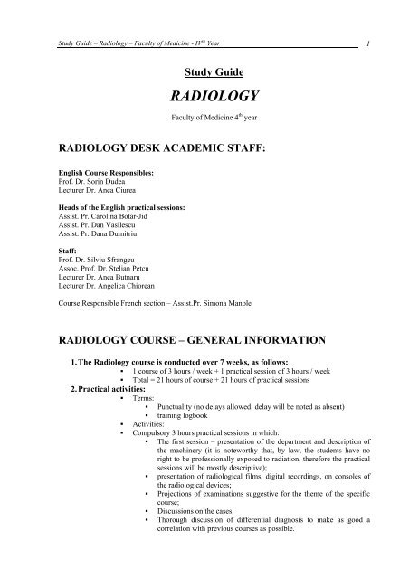
<strong>Study</strong> <strong>Guide</strong> – Radiology – Faculty of Medicine - IV th Year 1<strong>Study</strong> <strong>Guide</strong><strong>RADIOLOGY</strong>Faculty of Medicine 4 th year<strong>RADIOLOGY</strong> DESK ACADEMIC STAFF:English Course Responsibles:Prof. Dr. Sorin DudeaLecturer Dr. Anca CiureaHeads of the English practical sessions:Assist. Pr. Carolina Botar-JidAssist. Pr. Dan VasilescuAssist. Pr. Dana DumitriuStaff:Prof. Dr. Silviu SfrangeuAssoc. Prof. Dr. Stelian PetcuLecturer Dr. Anca ButnaruLecturer Dr. Angelica ChioreanCourse Responsible French section – Assist.Pr. Simona Manole<strong>RADIOLOGY</strong> COURSE – GENERAL INFORMATION1. The Radiology course is conducted over 7 weeks, as follows:• 1 course of 3 hours / week + 1 practical session of 3 hours / week• Total = 21 hours of course + 21 hours of practical sessions2. Practical activities:• Terms:• Punctuality (no delays allowed; delay will be noted as absent)• training logbook• Activities:• Compulsory 3 hours practical sessions in which:• The first session – presentation of the department and description ofthe machinery (it is noteworthy that, by law, the students have noright to be professionally exposed to radiation, therefore the practicalsessions will be mostly descriptive);• presentation of radiological films, digital recordings, on consoles ofthe radiological devices;• Projections of examinations suggestive for the theme of the specificcourse;• Discussions on the cases;• Thorough discussion of differential diagnosis to make as good acorrelation with previous courses as possible.
<strong>Study</strong> <strong>Guide</strong> – Radiology – Faculty of Medicine - IV th Year 23. Presence in the courses and practical sessions:• Up to 30% (2 courses) of absences are allowed in the courses. For more than2 absences, the student will not be admitted in the exam, losing one of thechances for sustaining it.• In the practical sessions, 100% presence is mandatory.4. Recovery of absences:• Up to 2 absences that are justified and oficially excused may be recoveredeither at the end of the module during recovery sessions organized by the deskor with another series, in another module. More than 2 absences, evenjustified and excused, mandate to retake the whole module.• Recovery sessions are taxed according to University regulations.5. Course Objectives:• General objectives:• Learning the physics basic concepts that underlie conventionalradiology and imaging equipment (nuclear physics, physics ofultrasound, MRI physics, etc.), biological effects of radiation and theprinciples of professional and population radiation protection.• Learning the basics of semiology related to each examinationtechnique (conventional and imaging), explaining the basic conceptsin obtaining images• Learning the indications and contraindications of the methods andalgorithms for examination, in order to reduce exposure to radiation;correlate the common and individual disease appearance inassessment of the whole body.• Specific objectives:• Recognition of the examination method.• Recognition of normal anatomical features and variants.• Recognition of the semiological elements and interpretation of theirsignificance.• Recognition and interpretation of disease evidence.• Develop a positive and differential diagnosis of a given disease.• Designing a radiology report.6. The objectives of practical sessions:o Knowledge on conventional radiological and imaging equipment;o Student awareness of the functional principle and how to obtain images for eachmethod separately;o Knowing the anatomical radiological elements in two- and three-dimensionalprojection;o Knowledge of semiology and the meaning of each semiological item in part;o Knowledge of pathology structured for each organ system;o How to formulate a radiology and imaging positive diagnosis associated withdifferential diagnosis;o Writing a radiology report.7. Examination in the end of the module:o The examination is held in the end of the module, in accordance with the student’sexamination regulation of the University.o Radiology and Medical Imaging desks hold a composite examination. The exam issustained before a board composed of teachers of the Departments of Radiology andMedical Imaging.o The exam has the following parts, with the corresponding weight in setting the finalscore:1. Theory - written - 50%2. practical test - 50% - consists of:a. interpretation of images - written - 30%b. clinical case - oral - 20%
<strong>Study</strong> <strong>Guide</strong> – Radiology – Faculty of Medicine - IV th Year 31. Theory examination consists of 50 multiple choice questions with simple complement,composed as follows:- 30 questions from the topics of Radiology- 20 questions from the topics of Medical Imaging- Test duration is 50 minutes- obtained score is multiplied by 0.2 to give the final mark of the written test.• The written test is eliminatory; students who do not get the minimum mark of5.00 fail and cannot attend the practical examination.• The papers are assessed immediately after the examination and the results areposted and announced to the students the same day, within the scheduledecided by the examination board.2. A. Image interpretation practical examination consists of viewing 10 images withpathological changes from the studied topics, as follows:- 6 images associated with the topics studied in Radiology- 4 images associated with the topics studied in Medical imaging• The test is written; students use a pre-printed answer sheet, and answer specific questions foreach image.• Length of the test is 15 minutes. The correct answer to each image is marked with 1 point; thesum of points represents the final score of the paper.• Depending on the number of students, they will be organized in 2 or 3 groups that will receiveeach distinct set of images for interpretation.• The correct answers will be presented to each group at the end of the examination.2. B. The oral clinical case examination will be held with a joint board composed of at least 2members, 1 teacher from the department of radiology and 1 teacher from the department of imaging.The student chooses an envelope that contains an image related to the examined topics. The Board willdiscuss with the student on the picture presented. The Board reserves the right to address the studentquestions from other studied topics.The examination is passed only if the practical test score (calculated according to the marks obtainedin examination 2.a. and 2.b.) is equal to or greater than 5.00.The final examination mark is calculated by averaging the scores of the 2 examination parts(theoretical and practical).In case of failure to pass the theoretical part, the student will retake the whole examination. In case offailure to pass the practical part, the student will retake only the practical examination, entirely.8. Prerequisites:o For students to be admitted to the IVth year Radiology examination, they should havepassed: Pathology and Pathophysiology9. Consultations:• Scheduled consultations will be displayed at the desk.10. References:D. Radulescu, Radiologie medicala, vol I si II, Ed. <strong>UMF</strong>, 1975D. Sutton, Textbook of Radiology and Imaging - Ed. Churchill Livingstone,2003S. Sfrangeu, M. Covalcic, Radiofizica si radiobiologie, Ed. Casa Cartii de Stiinta,1999S. Sfrangeu, M. Covalcic, R. Elefterescu, M. Vaida, Bazele Imagisticii Medicale, Ed.Polsib, 1995S. Sfrangeu, M. Covalcic,Hipertensiunea Pulmonara, Ed. <strong>UMF</strong>, 2001M. Buruian, Radiologie, Manual Practic, Ed. Imprimeria de Vest, Oradea, 1998M. Buruian, Tratat de Tomografie Computerizata, Ed. University Press, 2006C. Aldescu, Neuroradio Diagnostic, Vol. I si II, Ed. Junimea, 1987R. Badea, S. Dudea, PA Mircea,F. F. Stamatian, Tratat de Ultrasonografie Clinica,vol.I, Ed Medicala Buc. 2007R. Badea, S. Dudea, PA Mircea,D. Zdrenghea, Tratat de Ultrasonografie Clinica,vol.II, Ed Medicala Buc. 2004R. Badea, S. Dudea, PA Mircea, M. Stamate , Tratat de Ultrasonografie Clinica,vol.III, Ed Medicala Buc. 2008http://www.radiologyeducation.com
<strong>Study</strong> <strong>Guide</strong> – Radiology – Faculty of Medicine - IV th Year 4http://www.med.ufl.edu/medinfo/rademo/raintro.htmlhttp://www.martindalecenter.com./http://www.vh.org/Providers/Textbooks/ElectricGiNucs/GINucs.htmlhttp://www.jpnm.org/ http://www.jpnm.org/http://www.uhrad.com/http://www.netmedicine.com/xray/xr.htmhttp://www.med.univ-rennes1.fr/cerf/edicerf/index.htmlhttp://www.vh.org/Providers/TeachingFiles/MultimediaTeachingFiles.htmlhttp://www.uams.edu/CHRP/nuc_med.htmhttp://www.netmedicine.com/xray/xr.htmhttp://brighamrad.harvard.edu/education/online/Cardiac/Cardiacframe.htmlhttp://brighamrad.harvard.edu/education/online/Cardiac/Cardiacframe.htmlhttp://gamma.wustl.edu/home.htmlhttp://alpha.science.unitn.it/ ~ fisica1/raggi_x/siti/xraywwwserver/xray% 20wwwserver.htmhttp://home.earthlink.net/ ~ terrass / Radiography / medradhome.htmlhttp://www2.astate.edu/conhp/radsci/http://www.dip.ee.uct.ac.za/imageproc/medical/? http://www.medbioworld.com/MedBioWorld/ResourceLinks.aspx?http://www.geocities.com/Hollywood/Highrise/1713/mfile.htmlhttp://www.radiologist.com/http://www.radiologyweb.com/http://www.neuroimaging.theclinics.com/
<strong>Study</strong> <strong>Guide</strong> – Radiology – Faculty of Medicine - IV th Year 5TOPIC 1. PHYSICS OF X – RAYSContentsI. Definitions. Mechanisms for producing X-raysII. X-ray featuresIII. Effects of X-rayIV. Principles of radiological imaging genesis. Radiological image quality.V. Sample questionsEducational objectives• The student must know:o Structure of matter• Structure of atoms - Rutherford model• Atomic number: Z-electric charges• Mass Number: A-particles in the nucleus• Isotopes - same Z, A different• Bohr's model and assumptions:• Electrons move in circular orbits, stationary, without gain or loss ofenergy• The transition from one orbit to another is done by the emission orabsorption of energy in the form of quanta: E = hν (h = Planck's ct, ν= frequency of radiation emitted or absorbed)• Quantum numbers:• n-principal quantum number (layer number)• e-secondary quantum number• magnetic quantum number m• s - spin number• Pauli principle: in an atom no two electrons may have identical 4 quantumnumberso Wave-particle duality: DeBroglie principle (the greater the mass of a particle, thesmaller the wavelength of the associated wave):• E=hν E = hν• ν=c/λ ν = c / λ• E=mc 2 E = mc 2• λ=h/mc λ = h / mco The notion of radioactivity• Radionuclid - unstable nuclei that emit energy and / or particles tobecome stable• α decay (eg. helium - α particle emission massive ionization overshort distances)• β decay (positron emission)• Electromagnetic radiation-examples: light, infrared radiation, radiowaves, ultraviolet, X-rays and γo Definition of X-ray: electromagnetic radiation with energy between 150-200KeV andλ between 0.05 and 10 Å• Soft: λ = 1-5 Å, low energy (low penetration)• Hard: λ = 0.01 Å, high energy (high penetration)o The mechanisms of producing X-rays• Braking radiation: high speed incident electron is deflected whenpassing near the target nucleus X-ray energy of any value
<strong>Study</strong> <strong>Guide</strong> – Radiology – Faculty of Medicine - IV th Year 6• Characteristic (featured) X-ray: the incident electron strikes a targetatom's electron and it dislodges it from the orbit electronicrearrangement with heat and characteristic X-ray emission having Eand λ corresponding to the kinetic energy difference between twoenergy levels.o X-ray properties• Spread round (spherically) from the point of origin• are invisible and propagate in straight line at the speed of light• Their intensity decreases with the square of the distance• Their penetrability is inversely proportional to wavelength• Are absorbed and emitted in quanta characteristic of each chemicalelement (wave-particle duality photon)• Are absorbed by bodies they propagate through• Induce the effect of luminiscence (photoelectric), photochemical(photosensitive), ionizing and biological effects• are not deviated by electric or magnetic fields (no charge, no mass)• May suffer optical phenomena such as refraction, diffraction andpolarizationo Effects of X-rays:• Chemical• water radiolysis• Photochemical effect - breaking the link of the moleculeAgBr• Direct ionization (collision and braking)• Indirectly ionizing:• Compton scattering = energy transfer between aphoton (hard RX with high E) and an target electronfrom the environment RX with lower energyemitted• Thomson effect occurs when soft rays strike anelectron in the target environment, which is notdislodged from the orbit, it swings on the spot,emitting secondary X-ray with E = E of the incidentbeam• Photoelectric effect: complete transfer of theincident photon energy to target electron radiationemitted in the visible (light) spectrum• Materialization effect: electron + proton = neutron• Biological: Cell, tissue, somatic genetic feto-embryonic, cancero Radiological image formation - Principles• Geometric principles:• If X-ray source (focus) is a point then the image will be clear,with sharp margins• Conical beam shape always the radiological image will bea little larger and distorted compared to reality• Deformation is minimal in the range of the central beam andmaximal at the periphery of the image• Propagation in a straight line• Central beam perpendicular to the center of the structure /region examined• A plane of the examined region must be parallel to the film• Remember!• Projection of an object on the film plane is larger than theobject• Projection size increases with increasing distance between theobject and film
<strong>Study</strong> <strong>Guide</strong> – Radiology – Faculty of Medicine - IV th Year 7• Objects whose plane is oblique (non-parallel) related to thefilm appear distorted• If X-ray beam reaches the object surface tangentially sharp margin• Two overlapping objects located at different distances fromthe source and the film induce the effect of addition orsubtraction• The effect of parallax: if the object is stationary and thesource is moving to a sense object image is moving to theopposite directiono Radiological image quality depends on:• contrast quality• image definition: clarity, sharpnesso Factors that influence the contrast:• Kilovoltage (high low contrast)• Miliampers-condition the brightness of the image (high lowcontrast)• Film Quality• Developing the film• Examined Region - differences in absorbing incident radiation thatcrosses the target• X-ray absorption in a material body is directlyproportional to:• Atomic number (the greater - absorptionincreases)• Wavelength• The density of the object• The thickness of the object• Time for actiono Image definition (sharpness) depends on:• Sharpness of the contours (ideally – point sized source)• Radiographic blur:• Geometric (when the source is not point sized)• Kinetic induced by object movement during the exposure• Diffusion – due to secondary radiation (generated by usingsoft rays, controlled by antidiffusion grid)• Screen (due to screens used to amplify the image)• Resolving power - separate discernible line pairs / area unitSample questions for the written exam1. Z represents:a. Number of Neutronsb. Number of electronsc. mass Numberd. Quanta numberAnswer: b2. Which of the following statements are true:a. X-rays are electromagnetic radiation characterized by high energyand small wavelengthb. X-rays propagate in a straight line with speed of soundc. X-rays are characterized by the phenomenon of single wave- noparticle dualityd. X-rays can be diverted by a magnetic fielde. X-rays can not be polarizedAnswer: a
<strong>Study</strong> <strong>Guide</strong> – Radiology – Faculty of Medicine - IV th Year 83. Does not belong to the indirect effects of X-ray radiation:a. Thompson effectb. Compton scatteringc. Materializationd. The photochemical effecte. Photoelectric effectAnswer: d
<strong>Study</strong> <strong>Guide</strong> – Radiology – Faculty of Medicine - IV th Year 9TOPIC 2. THE ROENTGEN DEVICETable of ContentsI. Main components. Functioning principles.II. Dedicated types of X-ray devicesIII. Sample questionsEducational objectives• The student must know:o The structure of an X-ray device• X-ray tube - vacuum container with 2 electrodes:• Cathode (-) - producer of electrons: molybdenum, tungsten• Anode (+): tungsten• Electrical circuit: Low and high voltage• The command / control system = control panel• The reception device• Ways of receiving:• Fluoroscopy - the principle of fluorescence• Radiography - the principle of phosphorescence• Types of receptors:• Analogues• Digitalo The operating principle• Cathode is connected to a low voltage electrical circuit whose role isto heat the cathode filament (2700K). By heating electron cloudaround the cathode• The role of the high voltage power circuit is to speed up the cloud ofelectrons to the anode• The anode is composed of a material containing atoms with large Zthat facilitate interaction between the incident beam and electrons onthe orbits of target atoms to generate X-ray• Phenomena occur in a vacuum containing glass enclosure, with wallslined with lead and that has a small unleaded slot = emission windowof the tube.o Analog fluoroscopy• Receiver: Direct screen (Zn sulfide, cesium iodide)• Fluorescent principle: emission of light occurs as long as the atom issubjected to incident radiation• Transparency = white (rays reach the screen, light-emissiondue to photoelectric effect)• Opacity = black (rays do not reach the screen)• Light amplifiers• Transforming light signal into electrical signal• Transfer on the TV monitoro Analog radiography• The receiver has several components• Antidiffusion grid: lead blades of Pb, control secondaryradiation• Case that contains:• Intensifier screens
<strong>Study</strong> <strong>Guide</strong> – Radiology – Faculty of Medicine - IV th Year 10• Radiographic film covered with a fine film of silverbromide (photographic emulsion)• Phosphorescent principle: is based on the effect of photochemicaldissociation of the molecule of silver bromide (effect used inrevealing the film: Ag blackens the film)• Transparency = black (incident rays are not absorbed andreach the film box, causing dissociation of the molecule ofsilver bromide)• Opacity = white (incident rays are absorbed, do not get to thefilm)• X ray is invisible !!!o Digital receiver• Basic mechanism: scintillation detectors light analog-digitalconverter electrical impulsesdigital processing digital /analogue reconversion Image Display• Types of detectors:• Photosensitive phosphorus plates• Scintillation crystalso Dedicated X ray devices:• Mammography• Angiography• Device for dental radiography• OrthopantomographySample questions for the written exam of Radiology1. Which of the following statements is true:a. The cathode is connected to a high voltage circuitb. Radiography is based on photochemical effects of Rxc. The basis for the radiographic image is fluoroscopyd. Secondary radiation cannot be tackled with antidiffusion gridse. The anode usually contains atoms with low atomic numberAnswer: b
<strong>Study</strong> <strong>Guide</strong> – Radiology – Faculty of Medicine - IV th Year 11TOPIC 3. RADIATION BIOLOGY AND RADIATIONPROTECTIONTable of ContentsI. Radiation sources.II. Measurement systems. Notions of dosimetryIII. Types of RadiationIV. Major effects of ionizing radiation. Acute radiation disease.V. Prophylactic and curative RadioprotectionVI. Sample questionsEducational objectives:• The student must know:o Sources of radiation• : Natural:• Cosmic radiation• Terrestrial radiation (including buildings)• Organic radiation• Radon• Artificial• Medicalo Systems of measurement:• Roentgenologic and Radiobiology - SR• International - SIo Notions of dosimetry• Dose of radiation• Determining the number of ions produced in a mass of air by X or γradiation• SR: 1 Roentgen (R) = amount of X-ray that produces 2.1 x 10 10 ionpairs with the electric charge of 1 Franklin• SI: 1 coulomb / kg = 3876 R• Absorbed Dose• The ratio of absorbed energy and the mass of the radiated body• SR: Rad (Roentgen absorbed dose), 1 rad = 100 erg / g• SI: Gray (Gy) 1 Gy = 1 J / kg, 1 Gy = 100 rad, 1 rad = 10 mGy• Biological dose• Evaluate the biological effects• SR: 1 rem = biological effects induced by absorption of 1 rad• SI: Sievert, 1Sv = biological effects caused by absorption of 1 Gy• 1 Sv = 100 rem; 1 rem = 10 mSv• Please note:• The exposure of medical personnel kept under 0.05 Sv / yearfor whole body• The population exposure is maintained below 1mSv/year• Radioactivity• Assessing the speed of disintegration• SR: Curie-1Ci = 3.7x10 10 disintegration / second• SI: 1Bq Becquerel = 1 disintegration / second• 1 Ci = 37 GBq
<strong>Study</strong> <strong>Guide</strong> – Radiology – Faculty of Medicine - IV th Year 12o Types of Radiation• Natural natural radioactive background• External radiation: cosmic radiation (especially at high altitudes),terrestrial radiation and the one from construction / housing• Internal Radiation: air, water, food• Artificial:• Radiological• Therapyo Major effects of ionizing radiation - directly dependent on the dose, the cumulativeeffect over time:• Lethal• Mutagenic independent or transferable• Teratogenic• Carcinogenic effecto Acute radiation disease• State of illness that begins immediately or not later than a few weeks after amassive radiation• Minimal exposure dose :> 50 rad• Immediate onset at 400 rad• Clinical presentation:• Hematopoietic system: anemia, leuco-lymphopenia, bleeding• Digestive system: abdominal pain, diarrhea, dehydration, infections• Skin & mucosa: cutaneo-mucosal atrophy, epilation, loss offingernails, erythema, ulceration, stomatitis, epistaxis• Nervous System: Asthenia coma• Gonads: sterility• Cardiovascular system: hypotension, heart failure by myocardialdiseaseo Grouping critical organs in terms of irradiation (the first place: gonads, Hematopoieticsystem)o Medical radiation:• Useful• Useless• Techniques that irradiate too much: radiophotography• Indiscriminate examination• Technical defects - repeat examination• Wrong presumptive diagnosis / failure to examine the patientresulting in unnecessary additional examinations• Exaggerated demand from the population / administrative systemso Prophylactic and curative radioprotection• Prophylactic:• Physical shielding (screens, lead aprons, collars, goggles, gonadprotectors, etc.)• Chemical• Biological• curative treatment of radiation disease• Chemical - drugs• Biological - skin graftso Ways to achieve radiation protection:• Construction of the installation: filter, dome, collimator• Technique• Aprons, screens• Films, cases, amplifier screens• Legislative: reference levels, dose constraints, monitoringo Please note: the main rules of radioprotection• The important role of filtering and colimation of X-rays (diaphragms, grids)
<strong>Study</strong> <strong>Guide</strong> – Radiology – Faculty of Medicine - IV th Year 13• Reducing examinations with soft rays, generating secondary radiation,especially in children• Maintaining a high distance between tube and patient• Central beam perpendicular to the examined region and on film• Use protective screens and aprons• the shortest examination time• use as much as possible radiography and not fluoroscopy• Limit the number of exposuresSample question for the written exam of Radiology:Gray is the unit of measurement for:a. Dose of radiationb. Absorbed dosec. Biological dosed. RadioactivityAnswer: b
<strong>Study</strong> <strong>Guide</strong> – Radiology – Faculty of Medicine - IV th Year 14TOPIC 4. PHYSICAL PRINCIPLES OF ULTRASOUNDEXAMINATIONKnowing the physical principles underlying the operation of ultrasound examination technique isessential for understanding the diagnostic value of ultrasound applications and the limits of themethods.The student must know:o elementary notions of acoustics: sound source, sound wave, transmission media, theinteraction between wave and environment;o behavior of ultrasound in the human body: reflection, transmission, diffraction,refraction, absorption, attenuation, penetration;o obtaining and graphic representation of US information A, M mode, two-dimensional,real timeo ultrasound image: structure, resolution, format;o specific terminology in ultrasound examination;o Doppler effect - physical principles, application in medicine, information, obtained;o modalities of representing Doppler information: continuous, spectral, color, powerThe student will assist to the following practical demonstrations:o illustrate the types of ultrasound examinationso exemplify the terminology specific for ultrasonographyThe student will perform:o recognition of ultrasound techniqueso using specific terms for ultrasonographyPlease note:Ultrasonography is one of the morphological imaging examinations used in first intention to assess themajority of body organs and systems, because it is inexpensive, non-irradiating, easy to perform,quick, widely accessible and it provides results in real time. Ultrasound techniques are essential inbiliary pathology, cardiology, obstetrics and pathology of the peripheral veins.Sample questions for the written exam in radiology:In ultrasound the term interface defines the surface separating two media that differ interms of:a) thicknessb) molecular characteristicsc) histological structured) acoustic impedancee) electrical conductivityAnswer: dWithin the human body ultrasound waves are not attenuated by:a) airb) fluidc) fatty tissued) liver parenchymae) boneAnswer: b
<strong>Study</strong> <strong>Guide</strong> – Radiology – Faculty of Medicine - IV th Year 15TOPIC 5. PHYSICAL AND TECHNICAL BASES OFCOMPUTER TOMOGRAPHYTable of ContentsI. PrincipleII. Components of a computer tomography machine. Generations of CT scannersIII. The spiral acquisition systemIV. Terminology characteristic for computed tomography. Units of measurement. CTwindows.V. ArtifactsVI. Sample questionsEducational objectives• The student must know:o The operating principle• Individual measurements of X-ray attenuation along a single line throughspaceo CT scanner components• Gantry: tube, detectors, table• Command console - setting acquisition parameters• computero Basic components of a CT system• High voltage generator (150 kV, 500 mA)• Tubes• Cooling Systems• Filters• Beam collimators focusing the Xray beam (beam thickness)• emission colimation – sets up voxel size• reception colimation - conditioned by the size of the detectors• Detectors: convert residual radiation into electrical signal• Solid:• Cesium iodide crystals• High detection efficiency• Rapid wearing• Gas: Xenon• Sensitive to higher doses of residual Rx• works like an ionization chambero Generations of CT scanners• I: type translation - rotation (1 source, 1detector)• II: type translation - rotation with multiple detectors• III: Rotating-type (spinning both the source and the detectors)• IV: Rotating- fixed type (stationary detectors, rotating source)• Electron beam CT (electron flow system)o The spiral acquisition - defined by 3 main parameters• Volume scanned during a spiral scan• The number of full rotations during an acquisition• Pitch (p):• Definition (single slice spiral CT) = table movement (d)during a rotation reported to thickness (g) of the nominal slice(thickness of beam or beam width)
<strong>Study</strong> <strong>Guide</strong> – Radiology – Faculty of Medicine - IV th Year 16• d>g p >1 some spaces remain unscanned• d
<strong>Study</strong> <strong>Guide</strong> – Radiology – Faculty of Medicine - IV th Year 17• Level: 50 UH• brain Window• Width: 80-100 (-15 to 85 UH)• Level: 35 UHo CT artifacts• Due to malfunctioning or improper adjustment• Artifacts related to patient: metal, movement, breathing, pulsation• Contrast artifacts• Specific artifacts for spiral mode• Partial volume• Photopenia and noise• Motion artifacts• Beam hardening• window artifacts• Please note:• Partial volume effect• Second to setting of inadequate colimation• If slice thickness is too big, the average attenuationcoefficient of various elements does not allowindividualization of what is included in the slicethickness• beam hardening artifact• Occur when Xrays are crossing a dense structure likebone• Example: artifacts of the posterior fossa between bothrocky pyramids• Minimization: using a high kilovoltageSample questions for the written exam of Radiology1. Chose the false statement about X-ray beam colimationa. determine slice thicknessb. May be during emissionc. May be conditioned by the receiver apertured. The smaller it is the greater the radiation dosee. The smaller the better the resolutionAnswer: d2. Bone window:a. is a narrow windowb. is a large windowc. Its level is 0 UHd. Uses fewer shades of graye. The contrast is greatAnswer: b
<strong>Study</strong> <strong>Guide</strong> – Radiology – Faculty of Medicine - IV th Year 18TOPIC 6. PHYSICAL AND TECHNICAL BASES OFMAGNETIC RESONANCE IMAGINGTable of ContentsI. Basic principlesII. Fundamental sequencesIII. ArtifactsIV. ContraindicationsV. Sample questionsEducational objectives• The student must know:o The operating principle• Description of the steps to be followed in an MRI examination:• patient is placed in a magnet• radio wave is sent• patient emits a radio-frequency signal that is received and used toproduce the information• Image reconstructiono Basics:• Magnetization• Fundamental condition: the existence of an atom with magneticproperties ie odd number of nucleons• Within the human body the basis is the proton: H + (contained in H 2O)• Introduction of H + in an external magnetic field alignment of themagnetic moment with the direction of the vector of the externalmagnetic field:• Parallel (low energy proton population)• Antiparallel (high-energy proton population)• NMV = net magnetization vector• Each hydrogen nucleus describes an elliptical motion around its axis= motion of precession• Precession frequency is given by Larmor equation• Please note:• Protons have a + charge and present spin movement. Becauseof these characteristics they have their own magnetic field.• When placed in an external magnetic field they align with it,some parallel other antiparallel.• Protons move around magnetic field lines and a strongmagnetic field gives a higher frequency motion (relationshipdescribed by the equation of Larmor).• Parallel and antiparallel magnetic fields of the protons maycancel each other, but, as there are more parallel protons withlower energy levels, magnetic forces are not canceled remain.All these parallel protons join the force of their magneticfields along the external magnetic field direction (B0), sowhen the patient is placed within the MRI magnet, it has itsown magnetic field which is longitudinal and parallel to theexternal magnetic field of the MRI equipment. Because it islongitudinal it cannot be measured directly.
<strong>Study</strong> <strong>Guide</strong> – Radiology – Faculty of Medicine - IV th Year 19• Resonance• Appears when a radio frequency pulse close (or the same as) thetarget element precession frequency is applied.• Through resonance, some of the protons acquire energy passing on ahigher energy level (longitudinal magnetization drops)• Result of the resonance = transverse magnetization in phase• Radiofrequency pulse synchronizes proton movement; theyno longer move randomly - they are in phase. Protons aremoving in the same direction at the same time, therefore theirmagnetic vectors are added together in this direction,resulting in a magnetic vector pointing to where the protonmovements are directed (which is transverse to the basemagnetization vector) transverse magnetization.• Please note:• Radio frequency pulse causes a decrease in the longitudinalmagnetization and sets out a new transverse magnetization(NMV)• Transverse magnetization generates a signal received by aspecial antenna (electric power) and converted into digitalimage.• Relaxation times• When stopping the radiofrequency wave, NMV realigns with B0• Time to return to longitudinal magnetization = T1 or spin-latticerelaxation time (energy released in the environment)• time for loss of transverse magnetization = T2 or spin-spin relaxation,induced by interactions between individual neighboring atoms• Please note: the relaxation times are longer in liquids and shorter infat• Spatial location:• meaning what part of the body the signal comes from.• achieved by using a magnetic field having an intensity different fromthat of the patient, on each section in part• Why so? Precessional frequency of the proton depends on magneticfield strength. If this intensity is different from point to point in thepatient, then protons from different places move with differentfrequencies, thus the resulting MR signal also occurs with differentfrequencies, which can be used to determine the location of the signalat a certain point.• The contrast in MRI depends on:• Intrinsic parameters (tissue): relaxation times, proton density,diffusion coefficient• Extrinsic parameters (acquisition): pulse repetition time (TR), echotime (TE), the flip angle, inversion time (TI)o Fundamental sequence (weighted images)• T1 weighting• Conditioned by TR• Short TR: 400-600 ms• Short TE: 20 ms• Fluids = hyposignal (dark, black)• Fat = hypersignal (light-colored, white)• Abundant protein content (mucin) = hypersignal• Melanin = hypersignal• Subacute hematoma (methemoglobin) = hypersignal• T2 weighting• Conditioned by TE• Long TR: 2000 ms
<strong>Study</strong> <strong>Guide</strong> – Radiology – Faculty of Medicine - IV th Year 20• Long TE: 120 ms• Fluids = hypersignal• Fat = less intense signal compared to T1• Mucin = hyposignal• PD (proton density) weighting• Conditioned by the proton density• Characterized by long TR and short TEo MR Artifacts• Metallic - lack of signal (signal void) of large size, with peripheralenhancement• truncation = false bands or hyper - or hyposignal occurring where there is asharp transition in the signal - eg CSF-spinal cord (false appearance ofsyringomyelia)• Aliasing: object larger than FOV (field of view)• Chemical shift artifacts = band of hyposignal at the water-fat interface• Magnetic susceptibility: broad areas of hyposignal generated by theappearance of a magnetic field gradient at the interface of two structures withvery different magnetic susceptibility: ex air-watero Contraindications:• Absolute:• Cardiac pace-maker• Ferro-magnetic foreign body• Relative:• Pregnant women in the I trimester• Claustrophobia• Patients who are intubated, ventilatedSample Questions for the written exam of Radiology1. On standard sequences a subcutaneous lypoma appears:a. hypersignal T1b. black on T2c. hyposignal T1 and T2d. black on T1 and white on T2 weighted sequencese. It has an aspect of signal voidAnswer: a2. T1 weighting is:a. Conditioned by TRb. Conditioned by TEc. Proton density conditioned on the examined regiond. Characterized by long TR and TEe. Characterized by short TR and long TEAnswer: a3. The following statements are true, except one:a. Transverse magnetization can be measuredb. Longitudinal magnetization increases when a radio-frequency pulse isappliedc. Relaxation times are long for fluids compared to solid state.d. Adipose tissue has short relaxation timesAnswer: b
<strong>Study</strong> <strong>Guide</strong> – Radiology – Faculty of Medicine - IV th Year 21TOPIC 7. THE DIGITAL IMAGETable of ContentsI. Definitions.II. Image ProcessingIII. Image formatsIV. Sample questionsEducational objectives• The student must know:o Pixel definition• Two-dimensional picture element (monochrome square)o voxel definition• Volume element corresponding to a pixelo digital image definition• Picture which was digitized both in spatial coordinates (pixel-x, y, voxel - x,y, z) and in intensity• By digitization, the real scene is converted into an image composed of pixelsin the 2D space, respectively voxels in 3D, which have an associated grayvalue (usually ranging between 0 and 255) representing the intensity of thelight spot converted.o Image processing steps• Image acquisition• Preprocessing (improvement of primary acquisition):• noise removal• Isolation of the regions whose pixel structure indicate a similaritywith alphanumeric information required• Image Segmentation• Recognition and interpretation of data• Storage - storage of digital imageso Image formats• Binary images - the color of each pixel is expressed by a value (bit), 1 or 0. Ifit is monochrome the intensity of each pixel will be in the range of 0-255(usually)• True Color Image - the color of each pixel is expressed in terms of RGBintensities, red, green and blue• Image based on a variety of colors (Colormap) - each pixel value isinterpreted as an index into a table of RGB values• Common formats:• BMP• TIFF• GIF• JPEGo Image compression methods• Lossy compression – leads to decay of non-essential and redundantinformation (unpercievable by the human eye) at decompression• Lossless compression - does not lead to decay of information ondecompression, less used, low efficiency
<strong>Study</strong> <strong>Guide</strong> – Radiology – Faculty of Medicine - IV th Year 22Sample question for the written exam of Radiology:Which of the following statements on the digital image is false:a. The basic element of a two-dimensional image is voxelb. pixel is a monochrome square characterized by 2 coordinates: x, yc. The most used type of compression is lossy image compressiond. JPEG file extension format is one of the usual storage / transmission formats of digitalimagesAnswer:a
<strong>Study</strong> <strong>Guide</strong> – Radiology – Faculty of Medicine - IV th Year 23TOPICS 8-10 IMAGING OF THE MUSCULOSKELETALSYSTEMTopic 8. • Review of examination techniques (RX, ultrasound, CT, RM);• Radio-imaging anatomy of the musculoskeletal system;• elementary imaging semiology in musculoskeletal pathology.Topic 9. • Elementary imaging semiology in musculoskeletal pathology.• Basics of imaging infectious and tumoral musculoskeletal pathology.Topic 10. • Basics of imaging inflammatory joint disease.• Degenerative processes of the musculoskeletal system.The student must know:1. Methods of examination1.1. Radiography1.2. Plane Tomography1.3. Arthrography1.5. Computer tomography1.6. Magnetic resonance imaging1.7. Ultrasonography1.8. Nuclear Medicine2. Radiological anatomy2.1. Bone matrix2.2. Mineral salts2.3. Bone cells2.4. Compact bone2.5. Spongy bone2.6. Long bones2.7. Flat Bones2.8. Short bones2.9. Bone growth and modeling2.10. Assessment of bone maturation2.11. Imaging of joints3. Bone & joint semiology3.1. Changes in size- congenital: aplasia, hypoplasia, hyperplasia- acquired: anostosis, hipostosis, hiperostosis3.2. Changes of form: scoliostosis, oedostosis3.3 The changes of structure• Destructive:- Osteoporosis: acute, chronic, localized, regional, generalized- Osteolysis: circumscribed, diffuse- Osteonecrosis: septic, aseptic• Constructive:- Osteosclerosis: nontumoral, tumor- Periostosis: continuous, discontinuous, complex• Ossification and heterotopic calcification: bone spurs, osteophytesyndesmophyte, calcification in muscle, ligaments, dystrophic
<strong>Study</strong> <strong>Guide</strong> – Radiology – Faculty of Medicine - IV th Year 244.Inflammation of the musculoskeletal system4.1. Osteomyelitis:- Definition• Etiology• Route of infection• Location• Clinic• Pathogenesis• Radiological signs: in the period of onset, florid phase, during recovery• Particular clinical and imaging forms: bone whitlow, subacute osteomyelitis,Brodie abscess, chronic osteomyelitis4.2. Arthritis4.2.1. Bacterial arthritis - purulent: ways of infection imaging appearanceOsteoarticular tuberculosis: radiological appearance, particular forms (SpinaVentosa, caries sicca,)4.2.2.Arthritis in collagen diseases:- Rheumatoid arthritis- seronegative spondilarthropathy: ankylosing spondilytis5. Arthrosis (osteo-arthritis)1. Affecting the spine1. spondylosis2. disk arthrosis3. herniated disk: appearance on CT, MRI2. Hip joint arthrosis1. Knee arthrosis2. Arthritis in the hands6. Musculoskeletal system tumors6.1 Diagnostic imaging:Lesion detectionCharacterization of the lesionStaging6.2 Tumors of bone origin:1. Benign:Osteomaosteoid Osteoma2 Malignant:Osteosarcoma6.3 Tumors of cartilaginous origin:1. Benign:ChondromaOsteochondromaChondroblastoma2. Malignant:Chondrosarcoma6.4 Tumors of fibroblastic natureBenign:Fibrous cortical defectMalignant:Fibrosarcoma and malignant histiocytoma6.5 round cell tumorsEwing tumorMyeloma6.6 Vascular Tumors1 benign:Hemangioma6.7 tumors of uncertain origin
<strong>Study</strong> <strong>Guide</strong> – Radiology – Faculty of Medicine - IV th Year 251 benign:Giant cell tumorBone cyst6.8 metastases (osteolytic, osteosclerotic, mixed)Students will attend practical demonstrations of:• Images obtained by different techniques of examination• the anatomy of each imaging examination technique• images with incremental radiological and pathological changes• examinations of cases with different disease aimed at themes of semiology in the course, thepresentation of the characteristics, positive diagnostic criteria and differential diagnosis• identification of X-ray, ultrasound, CT and MRI of normal anatomical structures and theirdescription• identify on the presented images of elemental changes• Discussing cases with the group assistant or head of the courseStudents will need to:- Recognize the imaging technique and describe it- recognize the explored region.- identify the pathological appearance and described it in semiological terms: location, shape,size, contour, structure, relationships with neighboring organs and how it changes the functionof affected and neighboring organs- integrate the lesion in a syndrome by relating the radiological appearance to clinical andlaboratory data- formulate a positive diagnosis, differential diagnoses• recommend other useful diagnostic examinationsPlease note:• X-ray:o radiography is still the gold standard of investigation, being used as a method of firstintention in almost all musculoskeletal system disorders.o Bone radiographs should be performed in at least two perpendicular views AP and LL toavoid the phenomenon of addition.o Bone radiographs must include at least one joint to judge the position (alignment) of bone.o symmetrical regions are examined bilaterally in order to identify minimal changes duringthe onset of diseases.o Irradiated field should be as small as possible through beam collimation to avoidunnecessary irradiation.o Quality of the radiographs must be optimal. Machine settings are adapted to each regionand each patient so that on the radiograph one may identify both the bones and the softtissue fascial compartments and areolar structure of the subcutaneous cellular tissue.• MRI:o method of choice when examining osteonecrosis, bone marrow, soft tissue neoplasms.o particularly useful in examining the spine.o diagnosis of stress fractures.o Allows for the examination of intervertebral disks, tendons, menisci, cruciate ligaments ofthe knee, with a much better resolution than other methods.• Ultrasound:o use of the method to examine bone is limited.o indications: examining soft parts of the muscles, tendons, in real time.o diagnosis of hip dysplasia, especially before viewing the ossification nucleus of thefemoral head;
<strong>Study</strong> <strong>Guide</strong> – Radiology – Faculty of Medicine - IV th Year 26o examination of ligaments, tendons, menisci and fluid effusions.• CT:o due to X-ray attenuation, allows distinguishing between: muscle, fat, liquid, and tumors.o allows better visualization of complex anatomical regions and better differentiateparaosseous and periarticular soft tissues.o Is indicated in the diagnosis of bone and soft tissue tumors: origin, local extension, type,relationship, and metastases.o essential in bone fracture of the skull, pelvis and spine.Sample questions for the written exam:1. The Characteristic radiological sign of osteomyelitis is:a. osteochondromab. bone sequestrationc. osteoporosisd. hypoplasiae. scoliostosisanswer: B2. Radiological changes of arthrosis include the following, except:a. joint space narrowingb. bone cystsc. osteophyte (bone spurs )d. subchondral osteosclerosise. osteochondromaanswer:EReferences1. Grainger and Allison's. Diagnostic Radiology. 5 th ed. London: Churchill Livingstone,2008, Volume 2, Section V2. Stelian Petcu. Radiologie Imagistica medicala, Editura medicala universitara IuliuHatieganu, Cluj-napoca, 2001, pag. 59-204Useful web addresses:http://radiology.rsna.org/content/by/yearhttp://radiographics.rsna.org/
<strong>Study</strong> <strong>Guide</strong> – Radiology – Faculty of Medicine - IV th Year 27TOPIC 11: KIDNEY AND UPPER URINARY TRACTImaging techniques have a particularly important role in diagnosing disease of the urinary tract and inguiding therapeutic maneuvers. To understand the applications of imaging in urinary tract pathology itis necessary to review the basic concepts of anatomy, and the examination methods used. Theintroduction to imaging anatomical peculiarities of the urinary tract is followed by the presentation ofimaging semiology. The main excretory syndromes are discussed, highlighting the: indications of theimaging techniques as well as elements of positive and differential diagnosisThe student should know:• Elements of descriptive and topographic anatomy of the urinary system;• Methods of examination and radio-imaging anatomy specific to each of these methodso Radiological methods1. Simple renal radiography2. Intravenous pyelography (IVP)3. Plane tomography (seldom)4. Retrograde pyelography and uretero- pyelography5. Renal arteriographyo Imaging methods1. Ultrasonography2. Renal scintigraphy, SPECT, PET3. CT4. MRI• Indications, non-indications and contraindications of various imaging techniques in urinarytract disease• Elements of imaging semiology and urinary pathologyGeneral semiology. Renal disease states can be grouped into several "radio-imagingsyndromes", as follows:1. Changes in opacity:1.Increased (focal)1. Limestone (calcified) opacity (no contrast media –CM- given)1.stones2.calcification2. CM excess1.caverns2.diverticula3. Reflux2. Decreased (after CM administration)Gap (focal lack of filling)2. Amputation (focal)3. Dumb kidney (global absence of opacification)2. Renal function decay1. Absent2. Delayed1.Secretion2.Evacuation3. Urinary stasis1. Acute2. Chronic4. Hydronephrosis5. Small kidney syndrome1.Hypoxia2. Chronic stasis
<strong>Study</strong> <strong>Guide</strong> – Radiology – Faculty of Medicine - IV th Year 283. Chronic infectionsChronic pyelonephritisTuberculosis4. Nephrosclerosis5. Hypoplasia6. Large kidney syndrome1.Hyperplasia / hypertrophy2. Renal colic3. Acute renal infectionAcute pyelonephritisRenal and perirenal abscess4. Renal vein thrombosis5. Hydronephrosis6. Polycystic Kidney7. Tumors7. Renal mass (tumor) syndrome8. Malformations1.RenalUretericThe student will assist to the following practical demonstrations:• Intravenous pyelography: technique of the examination, setting the film in position, identifyspecific time sequences, their purpose;• Ultrasonography: indications, anatomy, utility;• Renal computer-tomography: technique of the examination, setting the film in position,identify specific time sequences, their purpose• Characteristic appearance, positive diagnostic criteria and algorithms to use imagingtechniques for:o Urinary stones, nephrocalcinosis, renal colic;o Acute and chronic urinary stasis;o Hydronephrosis and urinary tract obstructiono Hypoxic kidney, chronic pyelonephritis, urinary tuberculosiso Acute pyelonephritis, renal and perirenal abscess;o Polycystic kidney disease, renal simple cyst;o Tumors of the kidney and urinary tracto Renoureteric malformations.o Acute and chronic renal failure.The student will know how to do the following:• identify the type of imaging technique used;• the film in position;• anatomical recognition of the organs in the examined region;• identification and description of pathological changes in terminology specific for the imagingtechnique used;• classification of pathological changes in an imaging syndrome;• presumptive diagnosis, analysis of elements for differential diagnosis• formulation of personal observations in a description (report) that uses specific imaging terms.• Define the usefulness of imaging techniques in specific pathology, through a diagnosticalgorithm.
<strong>Study</strong> <strong>Guide</strong> – Radiology – Faculty of Medicine - IV th Year 29Please note:- Intravenous pyelography (IVP, urography), ultrasound and CT are the main methods ofmorphological exploration of the urinary system.- IVP and CT provide both morphological and functional information on the urinary system;- Preliminary assessment of the patient is required and strict observance of contraindications ismandatory;- Ultrasound provides only morphological information but has the advantage that isindependent of renal function and is not encumbered by the risk of contrast media;- In most of the upper urinary tract and kidney disorders (malformations, stones,hydronephrosis, infection, trauma, tumors, kidney failure) morphological imaging assessment isessential for establishing a proper diagnosis and setting an adequate therapeutic plan.Sample questions for the written exam of radiologyOn the 5 minutes IVP sequence one assesses:a. ;b. Renal secretion and evacuation function;c. Morphology of the ureters and bladder;d. Morphology of the pelvicalyceal system;e. Each of the arterial, parenchymal and venous transit time of contrast mediaAnswer: bSmall kidney syndrome is not produced by:a. Hypoxia;b. Nephrosclerosis;c. Acute pyelonephritis;d. Chronic stasis;e. HypoplasiaAnswer: c
<strong>Study</strong> <strong>Guide</strong> – Radiology – Faculty of Medicine - IV th Year 30TOPIC 12. RETROPERITONEUM AND PELVISComplex anatomy of the retroperitoneal and pelvic spaces includes elements of the digestive tract,urinary tract, vascular and nervous systems, genital apparatus and glandular system, thus explainingthe extensive pathology. Imaging diagnostic techniques will be chosen according to the pathologicalentity and complexity of disease and are represented by: classic X-ray, ultrasound examinations,computer tomography and magnetic resonance.The student should know:• Descriptive and sectional anatomy of the retroperitoneum;• Descriptive and sectional anatomy of the pelvis;• Basic physiology: digestive tract, urinary and vascular systems;• Pathology of the organs in these anatomical regions;• Principles of radiological image formation;• Physics and operating principles of imaging techniques;Course Content:A. The Retroperitoneum• Anatomical refresher of retroperitoneumo Contento Demarcationo Anatomical division• Imaging techniques used to investigate the retroperitoneumo Diagnostic principleso Imaging anatomy: ultrasound, CT, RM.• Retroperitoneal pathology to be presented:o Disease of adrenal glandso Lymph node pathologyo Collections of retroperitoneal space• The Adrenalso Anatomical refreshero Imaging anatomyo Nontumoral disease• Diffuse hyperplasia,• Gland calcification secondary to bleeding, cysts, tuberculosis.• Cysts: posthaemorrhagic, lymphangioma• Haemorrhageo Tumors• Benign: adenoma, myelolypoma, pheochromocytoma• Malignancy• Primary: carcinoma, neuroblastoma• Secondaryo Algorithm for imaging assessment• Lymph nodeso Node groupso Imaging anatomy: appearance and normal sizeo Imaging semiology of lymph node diseaseo Pathological node involvement:• Infectious• Autoimmune• Tumoral• Fluid collectionso Imaging appearance
<strong>Study</strong> <strong>Guide</strong> – Radiology – Faculty of Medicine - IV th Year 31o Locationo Determining the origin of collectiono Particular appearanceB. The Pelvis• Anatomical refresher of the pelviso Contento Demarcation• Imaging techniques used to investigate the pelviso Diagnostic principleso Imaging anatomy: radiographic, ultrasound (external and endocavitary), CT, RM.• Pelvic pathology to be presented:o Bladder pathologyo Genital pathology: prostate, uterus, ovaries• Bladdero Imaging anatomyo Direct and indirect cystography - technique, normal imageo Bladder Ultrasound• Ultrasound anatomy• Examination technique• Normal appearance• Measurement of bladder postvoiding residueo Imaging of nontumoral disease• Cystitis• Intravesical clot• Bladder stasis: compensated and decompensated stage• Bladder diverticula• Fighting bladder• Bladder stones• Vesico-ureteric reflux• Bladder involvement in neighborhood diseaseo Imaging of Tumors• Primary tumors: urothelioma, mesenchymal, other• Secondary neoplasm: extension from neighborhood malignancies, metastases• Prostateo Imaging techniques - anatomical aspects: cystography, ultrasound (external andendorectal), CT and RMo Anatomyo Ultrasound determination of prostate size and volumeo Benign prostate hypertrophyo Prostate carcinoma - aspects of ultrasound and RM• The uterus and ovarieso Imaging techniques - anatomical aspects: cystography, ultrasound (external andendovaginal), CT and RMo Uterine tumors• Uterine fibroids• Endometrial carcinomao Ovarian tumors• Cystic• Solid• Tumor relapse• Imaging evaluation of pelvic lymph nodes
<strong>Study</strong> <strong>Guide</strong> – Radiology – Faculty of Medicine - IV th Year 32Within hours of practical work students will be presented archive images on the issues presentedin the course.Students will be able, at the end of the course and practical work, to recognize the imagingtechniques used for diagnosis, recognize and describe semiologically the main pathological entities,indicate and follow the proper algorithm of investigation.Sample questions for the written exam of radiology:The examination of choice for retroperitoneal lymphadenopathy location is:a. Native MR;b. CT with contrast;c. Endocavitary ultrasound;d. Contrast enhanced MR;e. Transabdominal ultrasoundCorrect answer: bThe ultrasound appearance of bladder slime (sludge) is:a. Hypoechoic magma with liquid-liquid horizontal level, that changes its position while stirringthe patientb. Hyperechoic magma, which remains unchanged while stirring the patientc. Hypoechoic magma with constant bladder positiond. Hypoechoic magma with vertical liquid-liquid level, which changes its position while stirringthe patiente. Hyperechoic magma with hyperechoic liquid-liquid horizontal levelCorrect answer: a
<strong>Study</strong> <strong>Guide</strong> – Radiology – Faculty of Medicine - IV th Year 33TOPIC 13. IMAGING IN MEDICAL AND SURGICALEMERGENCYMedical and surgical emergencies are represented by a number of diseases and conditions involvingpractically all organs and systems. Their treatment requires a correct diagnosis in which imaging isextremely important. For a proper diagnosis is necessary to know the imaging examination methods,indications and limitations of each method separately and imaging appearance of various diseases.The student should know:Imaging issues in emergencies:Cranio-cerebral trauma: radiological examination, conventional CT examination, otherdiagnostic techniques (magnetic resonance, skull ultrasound, medicine nuclear, angiography) -indications, technique, positive and differential diagnosiso Fractures of the skull and face boneso Diffuse axonal lesiono Epidural hemorrhage,o Subdural Haemorrhageo Arachnoid Haemorrhageo Intraparenchymal Haemorrhageo Subdural HygromaImaging aspects of the spine in traumatic emergencies: Conventional radiologicalexamination, CT and MRI exams - indications, technique, positive and differential diagnosiso Spine fractureso The types of nervous involvementImaging of non - traumatic emergencies of the spine: radiological examination, CT andMRI exams - indications, technique, positive and differential diagnosiso Osteomyelitiso Discitiso Herniated discImaging in traumatic abdominal emergencies: Conventional radiological examination,imaging (ultrasound, CT and angiography) - indications, technique, positive and differential diagnosiso Pneumoperitoneumo Haemoperitoneumo Organ fractureImaging in non - traumatic abdominal emergencies: Conventional radiological examination,imaging (ultrasound, CT and angiography) - indications, technique, positive and differential diagnosiso Pneumoperitoneum / hydropneumoperitoneumo Ileuso Biliary colico Renal colicIn the practical works, the student will be presented the following:o Radiological appearance of fractures of the skull and facial boneso The appearance of intracranial bleeding on CT and MR – epidural, subdural, arachnoid,intraparenchymal bleedingo Radiological appearance of spine fractureso CT and RM appearance of spinal epidural hematoma, medullary compression and spinalsectiono Radiological appearance of vertebral osteomyelitiso Radiological and imaging appearance of discitiso Radiological and MR appearance of disc herniao Radiological and CT appearance of pneumoperitoneumo Radiological and CT appearance of intestinal occlusiono Imaging appearance of organ fractureso Radiology, CT and ultrasound in biliary colic
<strong>Study</strong> <strong>Guide</strong> – Radiology – Faculty of Medicine - IV th Year 34o Radiology, CT and ultrasound in renal colico Algorithms for emergency imaging examination in trauma of the skull, spine and abdomen.The student will be able to recognize the examination technique and pathological lesions,respectively:o Broken boneso Brain hemorrhageo Osteomyelitiso Discitiso Herniated disco Spinal compression / Sectiono Pneumoperitoneumo Ileuso Organ fractureo Gall stoneso NephrolithiasisSample questions for the written exam in radiology:1. Radiotransparent stones can be assessed by:a. Simple renal radiography (KUB)b. Ultrasoundc. Scintigraphyd. Magnetic resonancee. All the above methodsAnswer: b2. In case of acute abdomen, the first examination performed is:a. Simple abdominal radiographyb. Barium mealc. Intravenous urographyd. MRe. Contrast enhanced CTAnswer: a


