Cerebellum
Cerebellum
Cerebellum

You also want an ePaper? Increase the reach of your titles
YUMPU automatically turns print PDFs into web optimized ePapers that Google loves.
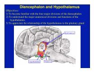
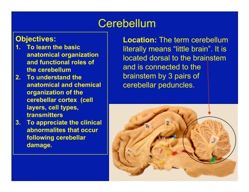
<strong>Cerebellum</strong>Objectives:1. To learn the basicanatomical organizationand functional roles ofthe cerebellum2. To understand theanatomical and chemicalorganization of thecerebellar cortex (celllayers, cell types,transmitters3. To appreciate the clinicalabnormalites that occurfollowing cerebellardamage.Location: The term cerebellumliterally means “little brain”. It islocated dorsal to the brainstemand is connected to thebrainstem by 3 pairs ofcerebellar peduncles.
Dorsal View of the <strong>Cerebellum</strong>
Functions- 3 major functional roles1. Coordination of Movement-the cerebellum controlsthe timing and pattern of muscle activation duringmovement.2. Maintenance of Equilibrium (in conjunction with thevestibular system)3. Regulation of Muscle Tone-modulates spinal cordand brain stem mechanisms involved in posturalcontrol.
Dysfunction: damage produces the following:• Ataxia- a disturbance that alters the direction andextent of voluntary movements; abnormal gait anduncoordinated movements• Dysmetria- altered range of motion (misjudge distance)• Intention Tremor-oscillating motion, especially of head,during movement• Vestibular signs-nystagmus, held tilt
Gross Anatomical Organization1. Internal Organization (similar to cerebral hemisphere)a. Cerebellar Cortex-surface gray matter;sulci & foliab. White Matter-internalc. Cerebellar Nuclei- three pairs located in white matter:FastigialInterpositusDentateCortexsulciWhite Matter
FastigialDentate
2. Cerebellar Lobes:A. Rostral Lobe= Spinocerebellum (paleocerebellum)-related to spinal cord, postural tone. Damage results inforelimb hyperextension and hindlimb hip flexionB. Caudal Lobe=cerebrocerebellum(neocerebellum)-damageresults in hypotonia,hypermetria & intentiontremor
C. FlocculonodularLobe=Vestibulocerebellumassociatedwith thevestibular system(eye movement, etc.);damage results indysequilibrium, widebased gait andnystagmus
3. Longitudinal ZonesA. Vermis- most medal portion of cerebellum;associated with the fastigial nucleus, concerned withregulation of muscle tone for posture and locomotion.B. Paravermis- intermediate part of the cerebellum,associated with the interpositus nucleus; participates inthe control of an evolving movement by utilizingproprioceptive sensory information generated by themovement itself to correct errors in the movementC. Hemisphere-the largest andmost lateral part of thecerebellum; associated with thedentate nucleus; influences theoutput to the motor cortex &permits fine delicateadjustments in muscle tone->skilled movementVermisParavermisHemisphere
Longitudinal Zones
HemisphereParavermisVermisParavermisHemisphereCerebellar NucleiLongitudinal Zones in Transverse Section
Cerebellar Peduncles (named by position)1. Caudal Cerebellar Peduncle- connects the cerebellumwith the medulla, contains afferent and efferent axons.2. Middle Cerebellar Peduncle- connects cerebellum withthe pons; contains only afferent axons from Pontine nuclei3. Rostral Cerebellar Peduncle-Connects cerebellum withthe midbrain; it is predominantly efferentaxons.Rostral Peduncle
Rostral and MiddleCerebellar Peduncles
Middle PeduncleTransverse Pontine Fibers
Cerebellar Cortex: the surface gray matter of the cerebellum.3 layers:1. Molecular Layer- most superficial, consisting of axonsof granule cells (parallel fibers) and dendrites of PCs2. Purkinje Cell Layer- middle layer consisting of a singlelayer of large neuronal cell bodies (Purkinje cells)3. Granule Cell Layer- deepest layer (next to white matter)consising of small neurons called granule cells
Cell Types and Afferent Fibers of the Cerebellar Cortex1. Purkinje Cells - the only output neuron from the cortexutilizes GABA to inhibit neurons in deep cerebellar nuclei2. Granule Cells- intrinsic cells of cerebellar cortex; useglutamate as an excitatory transmitter; excites Purkinjecells via axonal branches called “parallel fibers”3. Basket Cells- inhibitory interneuron;utilizes GABA to inhibit Purkinje cellsBasketCellGranule Cell
Cerebellar Purkinjecells in sagittalsectionand in transversesectionTransverse
GranulecellsGranule cellParallel Fibers
Basket cell axonforms a basket onPurkinje cell initialsegmentInhibits axonal firingBasket madeBy axon ofBasket cellBasket CellBasket
3. Climbing Fibers- axons arising from the olivary nucleus;use glutamate and aspartate to excite Purkinje cell andcerebellar nuclei neurons4. Mossy Fibers- all other axons that enter the cerebellum;excite granule cells (and neurons in cerebellar nuclei)
Major Cerebellar Inputs (axons entering the cerebellum):1. Climbing Fiber Inputs = Olivocerebellar fibers-- ariseexclusively from the olivary nucleus of the caudalmedulla; have a powerful excitatory effect on Purkinjecells upon which they synapse.OlivaryNucleusPyramidOlivo-CerebellarFibers
2. Mossy fiber Inputs:A. Vestibulocerebellar fibers--arise mainly from thevestibular nerve and vestibular nuclei; project to flocculonodularlobe and fastigial nucleus (coordinate head and eyemovement.B. Spinocerebellar Fibers- arise from spinal cord -->viaspinocerebellar tracts-->go to rostral lobe; makescerebellum aware ofongoing movementsvia proprioceptiveinput from musclespindles and jointreceptors.FVestibularNuclei8thNerveNodulusDorsal spinocerebellar Tr
C. Cerebropontocerebellar fibers--arise from pyramidalcells in the cerebral cortex, synapse on pontine nucleiwhich send their axons to the contralateral cerebellarcortex via pontocerebellar fibers (form middle peduncle)-Alerts cerebellum regarding anticipated movements.Ponto-CerebellarAxonAxon fromCerebralcortexNeuron inPontine NucleiMiddleCerebellarPeduncle
Major CerebellarOutputs (arise fromneurons in deepcerebellar nuclei):1. Fastigial NucleusProjections: (viacaudal peduncle)-->vestibular nuclei andreticular formation-->vestibulospinal &reticulospinal tractsinfluence spinalmotor neurons-->effect extensormuscles related tomaintaining postureand balance.Cerebellar Output (efferents)FastigialFastigialFrom Lab Guide, page 57
2. Interpositus NucleusProjections: (viarostral peduncle)-->goto red nucleus toinfluence rubrospinaltract--> correct errorsrelated to the grossmovements of theanimal3. Dentate NucleusProjections: (viarostral peduncle)-->projects to thalamus toinfluence the outputfrom the motor cortex-->makes delicateadjustments related tofine, skilledmovementsDentate
Clinical Abnormalities:Lesions of the cerebellum (damage to input, output orcortex) result in symptoms that occur because thecerebellum’s normal function is interrupted-->ataxia,dysmetria, intention tremor occur.Cerebellar disorders usually result from:1. Tumors (i.e., cerebellar cystic meningioma)2. Viral Infections (encephalitis; distemper)- this may occurin utero (feline panleukopenia virus)3. Heavy metal poisoning4. Genetic Disorders: cerebellar degeneration, i.e., Englishsheepdogs, Gordon setters, Kerry Blue Terriers, ArabianHorses, Yorkshire Pigs; autosomal recessive.
1. Small lesions produce no signs or onlytransient symptoms; small deficits arecompensated for by other parts of the brain2. Lesions of the cerebellar hemispheres result inloss of muscular coordination and jerkypuppet-like movements of the limbs on theipsilateral side (same side as lesion)3. Lesions of the vermis result in truncal tremorand gait ataxia (splayed stance and swaying ofthe body while walking)[signs]
Memories from my trip down south this past summer
SignSeen inAlabama
Outhouse in West Virginia