MRCPCH 1: Essential Questions in Paediatrics - PasTest
MRCPCH 1: Essential Questions in Paediatrics - PasTest
MRCPCH 1: Essential Questions in Paediatrics - PasTest
- No tags were found...
Create successful ePaper yourself
Turn your PDF publications into a flip-book with our unique Google optimized e-Paper software.
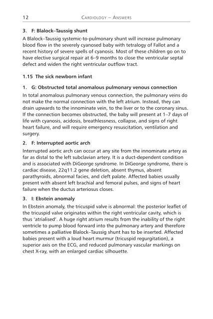
12CARDIOLOGY – ANSWERS3. F: Blalock–Taussig shuntA Blalock–Taussig systemic-to-pulmonary shunt will <strong>in</strong>crease pulmonaryblood flow <strong>in</strong> the severely cyanosed baby with tetralogy of Fallot and arecent history of severe spells of cyanosis. Most of these children go on tohave elective surgical repair at 6–9 months to close the ventricular septaldefect and widen the right ventricular outflow tract.1.15 The sick newborn <strong>in</strong>fant1. G: Obstructed total anomalous pulmonary venous connectionIn total anomalous pulmonary venous connection, the pulmonary ve<strong>in</strong>s donot make the normal connection with the left atrium. Instead, they candra<strong>in</strong> upwards to the <strong>in</strong>nom<strong>in</strong>ate ve<strong>in</strong>, to the liver or to the coronary s<strong>in</strong>us.If the connection becomes obstructed, the baby will present at 1–7 days oflife with cyanosis, acidosis, breathlessness, collapse, and signs of rightheart failure, and will require emergency resuscitation, ventilation andsurgery.2. F: Interrupted aortic archInterrupted aortic arch can occur at any site from the <strong>in</strong>nom<strong>in</strong>ate artery asfar as distal to the left subclavian artery. It is a duct-dependent conditionand is associated with DiGeorge syndrome. In DiGeorge syndrome, there iscardiac disease, 22q11.2 gene deletion, absent thymus, absentparathyroids, abnormal facies, and cleft palate. Affected babies usuallypresent with absent left brachial and femoral pulses, and signs of heartfailure when the ductus arteriosus closes.3. I: Ebste<strong>in</strong> anomalyIn Ebste<strong>in</strong> anomaly, the tricuspid valve is abnormal: the posterior leaflet ofthe tricuspid valve orig<strong>in</strong>ates with<strong>in</strong> the right ventricular cavity, which isthus ‘atrialised’. A huge right atrium results from the <strong>in</strong>ability of the rightventricle to pump blood forward <strong>in</strong>to the pulmonary artery and thereforesometimes a palliative Blalock–Taussig shunt has to be <strong>in</strong>serted. Affectedbabies present with a loud heart murmur (tricuspid regurgitation), asuperior axis on the ECG, and reduced pulmonary vascular mark<strong>in</strong>gs onchest X-ray, with an enlarged cardiac silhouette.


