06 Transdermal Patches - Student.pdf - haspi
06 Transdermal Patches - Student.pdf - haspi
06 Transdermal Patches - Student.pdf - haspi
- No tags were found...

Create successful ePaper yourself
Turn your PDF publications into a flip-book with our unique Google optimized e-Paper software.
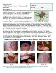
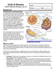
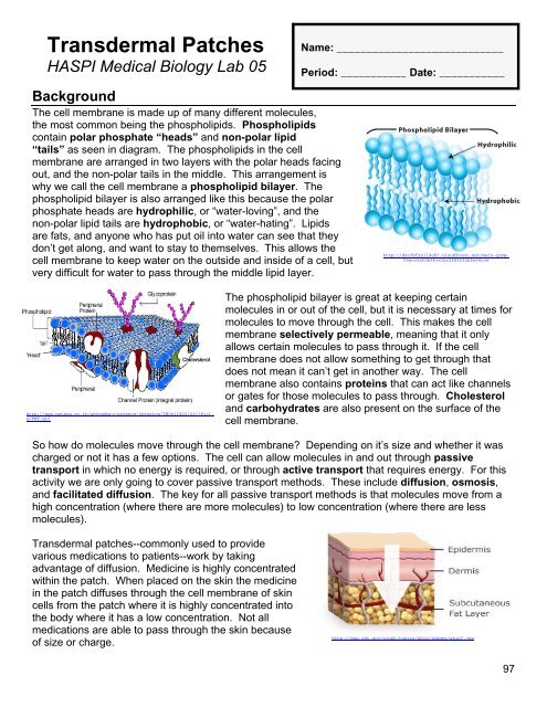
<strong>Transdermal</strong> <strong>Patches</strong>HASPI Medical Biology Lab 05Name: ____________________________Period: ___________ Date: ___________BackgroundThe cell membrane is made up of many different molecules,the most common being the phospholipids. Phospholipidscontain polar phosphate “heads” and non-polar lipid“tails” as seen in diagram. The phospholipids in the cellmembrane are arranged in two layers with the polar heads facingout, and the non-polar tails in the middle. This arrangement iswhy we call the cell membrane a phospholipid bilayer. Thephospholipid bilayer is also arranged like this because the polarphosphate heads are hydrophilic, or “water-loving”, and thenon-polar lipid tails are hydrophobic, or “water-hating”. Lipidsare fats, and anyone who has put oil into water can see that theydon’t get along, and want to stay to themselves. This allows thecell membrane to keep water on the outside and inside of a cell, butvery difficult for water to pass through the middle lipid layer.http://d2o7bfz2il9cb7.cloudfront.net/main-qimg-59ecc5a50b981e1bc2f85632b5e94ccehttp://www.patana.ac.th/secondary/science/ibtopics/IBCell%20(01)/Pic1.4/FMT.gifThe phospholipid bilayer is great at keeping certainmolecules in or out of the cell, but it is necessary at times formolecules to move through the cell. This makes the cellmembrane selectively permeable, meaning that it onlyallows certain molecules to pass through it. If the cellmembrane does not allow something to get through thatdoes not mean it can’t get in another way. The cellmembrane also contains proteins that can act like channelsor gates for those molecules to pass through. Cholesteroland carbohydrates are also present on the surface of thecell membrane.So how do molecules move through the cell membrane? Depending on it’s size and whether it wascharged or not it has a few options. The cell can allow molecules in and out through passivetransport in which no energy is required, or through active transport that requires energy. For thisactivity we are only going to cover passive transport methods. These include diffusion, osmosis,and facilitated diffusion. The key for all passive transport methods is that molecules move from ahigh concentration (where there are more molecules) to low concentration (where there are lessmolecules).<strong>Transdermal</strong> patches--commonly used to providevarious medications to patients--work by takingadvantage of diffusion. Medicine is highly concentratedwithin the patch. When placed on the skin the medicinein the patch diffuses through the cell membrane of skincells from the patch where it is highly concentrated intothe body where it has a low concentration. Not allmedications are able to pass through the skin becauseof size or charge.http://www.cdc.gov/niosh/topics/skin/images/skin7.jpg97
Name: _________________________________________ Date: ____________ Period: __________Passive TransportDiffusionMovement of molecules from high to lowconcentration(no energy required)Examples: In the digestive system nutrients areabsorbed through the small intestine, and in therespiratory system gases are exchanged in thelungs through diffusion.http://dickinsonn.ism-online.org/files/2009/11/626px-Scheme_simple_diffusion_in_cell_membrane-en_svg.pngOsmosisThe diffusion of waterExample: The kidneys are responsible forregulating the amount of water lost from the body.http://www.glogster.com/media/2/11/19/88/11198812.jpgFacilitated DiffusionMolecules cross the cell membrane with helpfrom transport proteinsExample: The movement of glucose into the redblood cells occurs through a transport protein.http://www.williamsclass.com/SeventhScienceWork/ImagesCellBricks/facilitatedDiffusion.jpgMaterialsDialysis tubingStringFilter paperMedicineCytosol solutionPlastic pipettePaper towelsBeaker (250 mL minimum)WaterProcedureThe goal of this lab will be to simulate and observe how a transdermal patch works to allowmedication to diffuse into the body.1. Fill the beaker half way with water. Soak the dialysis tubing in the water and rub to open.2. Using the string, tie a knot in one end of the tube tightly to ensure there is no leakage.3. Using a dropper, fill your dialysis tube approximately 2/3 full with cytosol solution.4. Hold onto the filled tubing and try to squeeze as much air out of the open end as possible.5. Use string to tie off the open end of the dialysis tube tightly to make sure there is no leakage.Dry off the dialysis tubing and lay on a paper towel. This will represent our simulated skin.6. Record initial observations of the simulated skin in Data Table 1.7. Stack three of the filter paper sheets together on top of the simulated skin to represent thepatch that will hold the medicine.8. Using the medicine dropper place one drop of medicine at a time on the patch until it issaturated. DO NOT saturate it too much or it will run from the patch onto the simulated skin. Ifthis happens wipe off the simulated skin as soon as possible.9. Observe the inside of the simulated skin at 5-minute intervals and record in Data Table 1.Adapted from Neo/Sci <strong>Transdermal</strong> Drug Delivery Lab Investigation 2011 98
Name: _________________________________________ Date: ____________ Period: __________AnalysisTime(min)Initial510152025Data Table 1Cytosol AppearanceOn the picture to theright, draw in andlabel thetransdermal patchand the medicinemolecules as theydiffused through thesimulated skin.Analysis Questions - on a separate sheet of paper complete the following1. Why was dialysis tubing used as the model for a cell membrane?2. What do you think a change in color simulates as the “transdermal drug” enters the cytosol?3. What do you think would happen if the simulated skin was left out over night?4. What are two characteristics of molecules that would not allow them to pass through the cellmembrane?5. Why do you think the water did not move into the transdermal patch?6. Summarize in one paragraph the results of this laboratory investigation.7. Name two functions of the cell membrane.8. The cell membrane contains _______ molecules that are embedded in the lipid bilayer.9. Draw a picture of a phospholipid and label the phosphate head, lipid tail, polar end, non-polarend, hydrophobic end, and hydrophilic end.10. What does it mean for a cell membrane to be selectively permeable?11. On the image, identify the:a. lipid bilayerb. protein channelc. carbohydrate chainsd. cholesterol112. In your own words, define diffusion.413. In your own words, define osmosis.14. In your own words, define facilitated diffusion.15. Do some research on transdermal drugs. List 3 transdermal drug names, the medication inthe patch, and its use.16. CONCLUSION: In 1-2 paragraphs summarize the procedure and results of this lab.23Adapted from Neo/Sci <strong>Transdermal</strong> Drug Delivery Lab Investigation 2011 99
Name: _________________________________________ Date: ____________ Period: __________Adapted from Neo/Sci <strong>Transdermal</strong> Drug Delivery Lab Investigation 2011 100