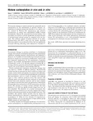
494 D. M. Williams and othersFigure 1 Structure of the 14-3-3ζ dimer and ClustalW2 alignments of 14-3-3 C-termini with in silico predictions of their intrinsically disordered and orderedregions based on their primary amino acid sequences(A) Front (left-hand panel) and top (right-hand panel) view of 14-3-3ζ illustrating the dimeric structure of 14-3-3 proteins (PDB code: 1A38 [57]), viewed using PyMOL (http://www.pymol.org).Each monomer consists of nine α-helices, arranged in an antiparallel fashion. The amphipathic binding groove, composed of a hydrophobic side and polar side, is indicated in black. The sidechains of Lys 49 ,Arg 56 ,Arg 60 and Arg 127 form a basic cluster at the bottom of the amphipathic binding groove. (B) ClustalW2 [26] alignments of 14-3-3 C-termini with disorder-promoting aminoacids shaded grey. Top panel: comparison of the amino acid sequences of the synthesized peptide used in the present study (peptide) and the seven human 14-3-3 isoforms (η, γ , σ , β, θ, ε and ζ )aligned from the ninth α-helix (overline). The amino acid sequence of 14-3-3ζ is boxed. Bottom panel: comparison of the C-terminal sequences of 14-3-3ζ from a range of evolutionarily diversespecies highlighting the high degree of sequence conservation and the inherent disorder within this region (boxed). (C) PONDR score [27–29] of 14-3-3ζ where ordered and disordered regions ofthe protein are signified by a score of less than or more than 0.5 respectively. The thick black line on the 0.5 line indicates that this region was resolved in the crystal structure of the protein despite itbeing predicted to be disordered.ordered conformation, the end of which is located near a basiccluster formed by the three arginine residues, Arg 56 and Arg 60of helix 3, and Arg 127 of helix 5, which are all located withinthe polar face of the amphipathic binding groove [11]. Bindingof the C-terminus to this basic cluster is proposed to result inocclusion of the binding groove and hence regulation of ligandbinding [11–14]. In the crystal structures of 14-3-3 proteins, theC-terminal region is not resolved, implying that this region isconformationally mobile. In a significant development in 14-3-3 cellular function, the chaperone ability of 14-3-3ζ has beendescribed, whereby it prevents target proteins from amorphouslyaggregating under conditions of stress [15]. In doing so, 14-3-3ζprevents the aggregation and precipitation of thermally aggregatedtarget proteins in a manner akin to that of sHsps (small heatshockproteins), a family of unrelated intracellular molecularchaperones. A common structural feature of mammalian sHsps isthe marked intrinsic flexibility, polarity and unstructured natureof their extreme C-terminus, properties which are independent ofthe domain core of the oligomeric and heterogeneous protein[16–18]. The C-terminal extension in mammalian sHsps playsan important role in maintaining the solubility of the proteinon its own and when it is in complex with aggregation-pronetarget proteins [19]. From the discussion above, 14-3-3 proteinsmay also have a flexible C-terminal extension which providessimilar stability to it and the complex 14-3-3ζ forms with targetproteins.The purpose of the present study was to understand furtherthe structure and function of the C-terminus of 14-3-3ζ and,in particular, its role in the chaperone action of 14-3-3ζ .Accordingly, we have undertaken spectroscopic and biophysicalcharacterization of the full-length protein and a truncated formfrom which the final 15 C-terminal amino acids were absent,i.e. 15C 14-3-3ζ . It is concluded that, like sHsps, 14-3-3ζ has ashort C-terminal extension that is flexible, polar and unstructured.Its function is to stabilize and solubilize the protein, and it has nodirect role in its chaperone action. Furthermore, we investigatedthe role of the binding groove of 14-3-3ζ in its chaperone action. Itis concluded that the function of the binding groove for chaperoneactivity is not analogous to its role in phosphoserine peptidebinding.MATERIALS AND METHODSMaterialsAll reagents used were of analytical grade and were obtainedfrom Sigma–Aldrich unless otherwise specified. Unless otherwisestated, all solutions were prepared in 50 mM phosphate buffercontaining, 100 mM sodium chloride and 0.05% sodium azide,pH 7.4. Prior to use, all pre-existing aggregates were removed byfiltration using a 0.22 μM Minisart syringe filter (Sartorius). Allprotein concentrations were determined by their absorbance atc○ The Authors <strong>Journal</strong> compilation c○ 2011 <strong>Biochemical</strong> Society
Chaperone action of 14-3-3ζ 495280 nm and calculated based on a subunit molecular mass. TheR18 peptide was generated on an Applied Biosystem model 430Asynthesizer by standard Merrifield solid-phase synthesis protocolsand t-butoxycarbonyl chemistry.Generation of 15C 14-3-3ζ constructsThe 15C C-terminally truncated 14-3-3ζ constructs weregenerated using the Stratagene QuikChange ® site-directedmutagenesis kit following the manufacturer’s protocol. TheWT (wild-type) 14-3-3ζ constructs in the pPRoEX vectorwere used as templates for mutagenesis to obtain 15Ctruncatedplasmids that could be used in bacterial expression.The primers used for the PCR to exchange Asp 231 fora stop codon (underlined) were the 5 ′ -GACATTGTGGA-CGTCGTAAACCCAAGGAGACGAAG-3 ′ oligonucleotide andits reverse complimentary strand. Successful mutagenesis wasverified using DNA sequencing.Expression and purification of WT and 15C 14-3-3ζRecombinant 14-3-3ζ –His 6 fusion proteins were expressed underthe control of the trc promoter in Escherichia coli BL21(DE3)-Codon Plus-RIL cells and subsequently purified using Ni-NTA (Ni 2 + -nitrilotriacetate) column chromatography (Qiagen).Following purification of the 14-3-3ζ –His 6 fusion protein,cleavage of the His 6 tag was achieved using the tobacco etchvirus protease, expressed under tac control in E. coli BL21(DE3)-Codon Plus-RIPL cells. After which, the cleavage products werepurified using Ni-NTA column chromatography (Qiagen).1 H-NMR spectroscopyPeptide (biotin-SGSGDTQGDEAEAGEGGEN-OH) (Mimotopes)and protein samples (0.1 mM, 700 μl) were prepared in20 mM phosphate buffer, 10% 2 H 2 O/90% 1 H 2 O and 0.05%sodium azide (pH 7.4) in 5 mm P-535 NMR tubes (Wilmad-Labglass, Quantum Scientific). All 1 H-NMR experiments wereacquired at 600 MHz, with a sweep width of 7002 Hz at25 ◦ C, using a Varian Inova-600 NMR spectrometer equippedwith a pulsed-field gradient 5 mm probe. All spectral chemicalshifts were referenced to the residual water resonance at 4.81p.p.m. Through-bond (scalar) connectivities were obtained fromTOCSY (total correlation spectroscopy) experiments with aspin-lock period of 60 ms [20]. Through-space connectivitieswere obtained for the peptide using ROESY (rotating-frameOverhauser enhancement spectroscopy) experiments with amixing time of 100 ms [21]. All spectra were processed usingVnmrJ (version 2.2d) software and visualized in Sparky 3(http://www.cgl.ucsf.edu/home/sparky/).Yeast two-hybrid analysis: galactosidase liquid assayThe strain EGY48 containing the pSH18-34 lacZ reporter plasmid(MATa, trp1, his3, ura3, lexAops-LEU2) of Saccharomycescerevisiae was used for yeast two-hybrid interaction trap analysis.It was transformed with a bait plasmid encoding either the DNAbindingdomain alone (vector), Raf-1, 14-3-3ζ or a Bad-fusionprotein. Both WT Bad and the S136A Bad mutant were used. Aplasmid encoding either a 14-3-3ζ WT or C-terminally truncated(15C) fusion protein was also co-transformed into these cellsalong with the lacZ reporter plasmid using the lithium acetatemethod [23]. Three individual colonies were randomly selectedfor each experiment, and the assay was performed as describedpreviously by Zhang et al. [23] and Truong et al. [14]. Cellswere grown in glucose minimal medium and then transferredto galactose minimal medium to induce the expression of 14-3-3ζ . The interaction between those fusion proteins reconstitutesa bipartite transcription factor that allows for the expressionof β-galactosidase. The level of β-galactosidase activity thusindicates the amount of interaction between the two proteins.β-Galactosidase units were calculated by D 550 /(D 550 ×volumeof cells×time in minutes). The relative β-galactosidase unitswere estimated based on the following equation: β-galactosidaseunit of test samples/β-galactosidase unit of WT 14-3-3ζ homodimers.Activities were obtained from at least three separateexperiments, each performed in triplicate and results presentedare means + − S.D.SEC (size-exclusion chromatography)–MALLS (multi-angle laserlight scattering)SEC–MALLS was performed using an S200HR 10/30 Superdexcolumn (GE Healthcare) connected to a DAWN EOS multianglelaser light-scattering detector (Wyatt Technology). Samples(100 μl) were loaded on to the column at 10 mg/ml and elutedwith 50 mM phosphate buffer containing 100 mM NaCl and 2 mMEDTA (pH 7.4) at a flow rate of 0.2 ml/min.CD spectroscopyThe far-UV CD spectra of WT and truncated 14-3-3ζproteins (7.2 μM, 200 μl) were recorded on a Jasco J-815spectropolarimeter over a wavelength range of 185–250 nm witha scan rate of 100 nm/min and a band-width of 1 nm. Alldata presented are the average of three accumulations obtainedin 20 mM phosphate buffer (pH 7.4) and high-voltage tensionrecords below 600 V. Protein thermostability CD profiles wererecorded over a temperature range of 25–75 ◦ Cat5 ◦ Cintervals.The temperature ramp rate was set at 2 ◦ C/min and once the desiredtemperature was reached, all solutions were equilibrated for 3 minprior to data collection.DLS (dynamic light scattering)For both WT and 15C 14-3-3ζ (7.2 μM, 500 μl), timeresolvedDLS analysis was performed at 25 ◦ C and 37 ◦ Cusinga Zetasizer Nano-ZS (Malvern Instruments). Every 20 min (forup to 24 h), the particle diameter-intensity distribution andmean hydrodynamic diameter were determined from 13 acquiredcorrellograms using the program CONTIN [24] and the methodof cumulants [25] respectively, via the attached DispersionTechnology Software (Malvern Instruments).Intrinsic tryptophan fluorescenceThe tryptophan fluorescence of WT and 15C 14-3-3ζ(7.2 μM, 2 ml) was measured using a Cary Eclipse fluorescencespectrophotometer equipped with a Peltier temperature controller(Varian). The temperature of the cell block was monitoredbetween 37 and 80 ◦ C, and measurements were collected at 1 ◦ Cintervals. For all recordings, the excitation wavelength was set to295 nm, fluorescence emission was monitored between 300 and400 nm, and the excitation and emission slit-widths were set to5nm.c○ The Authors <strong>Journal</strong> compilation c○ 2011 <strong>Biochemical</strong> Society