Full Text PDF - Biochemical Journal
Full Text PDF - Biochemical Journal
Full Text PDF - Biochemical Journal

You also want an ePaper? Increase the reach of your titles
YUMPU automatically turns print PDFs into web optimized ePapers that Google loves.
www.biochemj.orgBiochem. J. (2011) 437, 493–503 (Printed in Great Britain) doi:10.1042/BJ20102178 493NMR spectroscopy of 14-3-3ζ reveals a flexible C-terminal extension:differentiation of the chaperone and phosphoserine-binding activities of14-3-3ζDanielle M. WILLIAMS*†, Heath ECROYD*‡, Katy L. GOODWIN*†, Huanqin DAI§, Haian FU‖, Joanna M. WOODCOCK†,Lixin ZHANG§ 1 and John A. CARVER* 1*School of Chemistry and Physics, The University of Adelaide, Adelaide, SA 5005, Australia, †Centre for Cancer Biology, SA Pathology, Adelaide, SA 5000, Australia, ‡School ofBiological Sciences, University of Wollongong, NSW 2522, Australia, §Chinese Academy of Sciences Key Laboratory of Pathogenic Microbiology and Immunology, Institute ofMicrobiology, Chinese Academy of Sciences, Beijing 100190, China, and ‖Department of Pharmacology and Emory Chemical Biology Discovery Center, Emory University, Atlanta, GA30322, U.S.A.Intracellular 14-3-3 proteins bind to many proteins, via aspecific phosphoserine motif, regulating diverse cellular tasksincluding cell signalling and disease progression. The 14-3-3ζ isoform is a molecular chaperone, preventing the stressinducedaggregation of target proteins in a manner comparablewith that of the unrelated sHsps (small heat-shock proteins).1H-NMR spectroscopy revealed the presence of a flexible andunstructured C-terminal extension, 12 amino acids in length,which protrudes from the domain core of 14-3-3ζ and issimilar in structure and length to the C-terminal extensionof mammalian sHsps. The extension stabilizes 14-3-3ζ , buthas no direct role in chaperone action. Lys 49 is an importantfunctional residue within the ligand-binding groove of 14-3-3ζ with K49E 14-3-3ζ exhibiting markedly reduced bindingINTRODUCTIONIn mammals, the 14-3-3 family of ∼30 kDa acidic proteinsconsists of seven isoforms (β, γ , ε, ζ , η, σ and τ) whichare widely expressed in all tissues and organs. For example,they account for ∼1% of the total soluble brain protein [1–3].The different 14-3-3 isoforms form homo- and hetero-dimers thatpossess enhanced affinity to ligands containing phosphorylatedserine or threonine residues within a specific sequence motif[4,5]. To date, more than 200 target proteins have been identifiedfor 14-3-3 [6]. Depending on the nature of its target protein,the binding to 14-3-3 affects multiple signalling pathwayswhich determine cell fate and organ development. For example,14-3-3 association controls Raf signalling fidelity, neutralizesBad-mediated apoptosis, and couples histone H3 with H4 tocreate a histone code for transcriptional elongation [2,3,7,8].Through these highly regulated interactions, 14-3-3 proteinsgovern diverse physiological processes and cellular status.Furthermore, 14-3-3 proteins have been implicated in numerousdisease states, including cancers and neurodegenerative diseasessuch as Alzheimer’s disease, Parkinson’s disease and ataxia.Various 14-3-3 isoforms are found to be associated with theintracellular deposits that are characteristic of Parkinson’s diseaseand Alzheimer’s disease. Furthermore, α-synuclein, the principalcomponent of Lewy body deposits in Parkinson’s disease,interacts with 14-3-3 and, because of this, both proteins have beenimplicated in the pathology of Parkinson’s disease [1–3]. In theto phosphorylated and non-phosphorylated ligands. The R18peptide binds to the binding groove of 14-3-3ζ with high affinityand also reduces the interaction of 14-3-3ζ ligands. However,neither the K49E mutation nor the presence of the R18 peptideaffected the chaperone activity of 14-3-3ζ , implying that theC-terminal extension and binding groove of 14-3-3ζ do notmediate interaction with target proteins during chaperone action.Other region(s) in 14-3-3ζ are most likely to be involved, i.e.the protein’s chaperone and phosphoserine-binding activities arefunctionally and structurally separated.Key words: biophysical characterization, C-terminal flexibility,molecular chaperone, 14-3-3 protein, protein aggregation,protein–protein interaction.same vein, 14-3-3 proteins have been implicated in Huntington’sdisease via interaction with the polyglutamine-containing proteinhuntingtin [9,10].X-ray crystallographic studies have shown that each 14-3-3monomer consists of a bundle of nine α-helices organized inan antiparallel fashion, forming a central amphipathic bindinggroove which is highly conserved across the 14-3-3 isoforms.The hydrophobic surface of this groove is formed from helices7 and 9, with the opposing polar face consisting of basic andpolar side chains of helices 3 and 5 (Figure 1A). The homoandhetero-dimerization of 14-3-3 isomers occurs through N-terminal interactions of the monomers resulting in the twoamphipathic grooves in each dimer combining to generatea40Å(1Å= 0.1 nm) wide central channel [11] (Figure 1A). Inthe case of the 14-3-3ζ isoform, the majority of its ligands interactwith the binding groove via a conserved basic cluster (Lys 49 ,Arg 56and Arg 127 ). Other residues along the polar and hydrophobic facesof the groove stabilize the ligand [3].While amino acid sequences throughout the seven human14-3-3 isoforms are generally highly conserved, particularlywithin the amphipathic binding groove, maximum sequencediversity is found in their extreme C-termini (encompassing, onaverage, the last 18 amino acids). Despite this diversity, the C-termini of all 14-3-3 isoforms share the characteristics of beingacidic and contain a large number of amino acid residues which aredisorder-promoting (Figure 1B, top panel). It has been proposedthat the C-terminus of 14-3-3ζ (Asp 231 –Asn 245 ) adopts a poorly<strong>Biochemical</strong> <strong>Journal</strong>Abbreviations used: ADH, alcohol dehydrogenase; DLS, dynamic light scattering; DTT, dithiothreitol; α-LA, α-lactalbumin; MALLS, multi-angle laser lightscattering; Ni-NTA, Ni 2 + -nitrilotriacetate; PDI, polydispersity index; ROESY, rotating-frame Overhauser enhancement spectroscopy; SEC, size-exclusionchromatography; sHsp, small heat-shock protein; TOCSY, total correlation spectroscopy; WT, wild-type.1 Correspondence may be addressed to either of these authors (email lzhang03@gmail.com or john.carver@adelaide.edu.au).c○ The Authors <strong>Journal</strong> compilation c○ 2011 <strong>Biochemical</strong> Society
494 D. M. Williams and othersFigure 1 Structure of the 14-3-3ζ dimer and ClustalW2 alignments of 14-3-3 C-termini with in silico predictions of their intrinsically disordered and orderedregions based on their primary amino acid sequences(A) Front (left-hand panel) and top (right-hand panel) view of 14-3-3ζ illustrating the dimeric structure of 14-3-3 proteins (PDB code: 1A38 [57]), viewed using PyMOL (http://www.pymol.org).Each monomer consists of nine α-helices, arranged in an antiparallel fashion. The amphipathic binding groove, composed of a hydrophobic side and polar side, is indicated in black. The sidechains of Lys 49 ,Arg 56 ,Arg 60 and Arg 127 form a basic cluster at the bottom of the amphipathic binding groove. (B) ClustalW2 [26] alignments of 14-3-3 C-termini with disorder-promoting aminoacids shaded grey. Top panel: comparison of the amino acid sequences of the synthesized peptide used in the present study (peptide) and the seven human 14-3-3 isoforms (η, γ , σ , β, θ, ε and ζ )aligned from the ninth α-helix (overline). The amino acid sequence of 14-3-3ζ is boxed. Bottom panel: comparison of the C-terminal sequences of 14-3-3ζ from a range of evolutionarily diversespecies highlighting the high degree of sequence conservation and the inherent disorder within this region (boxed). (C) PONDR score [27–29] of 14-3-3ζ where ordered and disordered regions ofthe protein are signified by a score of less than or more than 0.5 respectively. The thick black line on the 0.5 line indicates that this region was resolved in the crystal structure of the protein despite itbeing predicted to be disordered.ordered conformation, the end of which is located near a basiccluster formed by the three arginine residues, Arg 56 and Arg 60of helix 3, and Arg 127 of helix 5, which are all located withinthe polar face of the amphipathic binding groove [11]. Bindingof the C-terminus to this basic cluster is proposed to result inocclusion of the binding groove and hence regulation of ligandbinding [11–14]. In the crystal structures of 14-3-3 proteins, theC-terminal region is not resolved, implying that this region isconformationally mobile. In a significant development in 14-3-3 cellular function, the chaperone ability of 14-3-3ζ has beendescribed, whereby it prevents target proteins from amorphouslyaggregating under conditions of stress [15]. In doing so, 14-3-3ζprevents the aggregation and precipitation of thermally aggregatedtarget proteins in a manner akin to that of sHsps (small heatshockproteins), a family of unrelated intracellular molecularchaperones. A common structural feature of mammalian sHsps isthe marked intrinsic flexibility, polarity and unstructured natureof their extreme C-terminus, properties which are independent ofthe domain core of the oligomeric and heterogeneous protein[16–18]. The C-terminal extension in mammalian sHsps playsan important role in maintaining the solubility of the proteinon its own and when it is in complex with aggregation-pronetarget proteins [19]. From the discussion above, 14-3-3 proteinsmay also have a flexible C-terminal extension which providessimilar stability to it and the complex 14-3-3ζ forms with targetproteins.The purpose of the present study was to understand furtherthe structure and function of the C-terminus of 14-3-3ζ and,in particular, its role in the chaperone action of 14-3-3ζ .Accordingly, we have undertaken spectroscopic and biophysicalcharacterization of the full-length protein and a truncated formfrom which the final 15 C-terminal amino acids were absent,i.e. 15C 14-3-3ζ . It is concluded that, like sHsps, 14-3-3ζ has ashort C-terminal extension that is flexible, polar and unstructured.Its function is to stabilize and solubilize the protein, and it has nodirect role in its chaperone action. Furthermore, we investigatedthe role of the binding groove of 14-3-3ζ in its chaperone action. Itis concluded that the function of the binding groove for chaperoneactivity is not analogous to its role in phosphoserine peptidebinding.MATERIALS AND METHODSMaterialsAll reagents used were of analytical grade and were obtainedfrom Sigma–Aldrich unless otherwise specified. Unless otherwisestated, all solutions were prepared in 50 mM phosphate buffercontaining, 100 mM sodium chloride and 0.05% sodium azide,pH 7.4. Prior to use, all pre-existing aggregates were removed byfiltration using a 0.22 μM Minisart syringe filter (Sartorius). Allprotein concentrations were determined by their absorbance atc○ The Authors <strong>Journal</strong> compilation c○ 2011 <strong>Biochemical</strong> Society
Chaperone action of 14-3-3ζ 495280 nm and calculated based on a subunit molecular mass. TheR18 peptide was generated on an Applied Biosystem model 430Asynthesizer by standard Merrifield solid-phase synthesis protocolsand t-butoxycarbonyl chemistry.Generation of 15C 14-3-3ζ constructsThe 15C C-terminally truncated 14-3-3ζ constructs weregenerated using the Stratagene QuikChange ® site-directedmutagenesis kit following the manufacturer’s protocol. TheWT (wild-type) 14-3-3ζ constructs in the pPRoEX vectorwere used as templates for mutagenesis to obtain 15Ctruncatedplasmids that could be used in bacterial expression.The primers used for the PCR to exchange Asp 231 fora stop codon (underlined) were the 5 ′ -GACATTGTGGA-CGTCGTAAACCCAAGGAGACGAAG-3 ′ oligonucleotide andits reverse complimentary strand. Successful mutagenesis wasverified using DNA sequencing.Expression and purification of WT and 15C 14-3-3ζRecombinant 14-3-3ζ –His 6 fusion proteins were expressed underthe control of the trc promoter in Escherichia coli BL21(DE3)-Codon Plus-RIL cells and subsequently purified using Ni-NTA (Ni 2 + -nitrilotriacetate) column chromatography (Qiagen).Following purification of the 14-3-3ζ –His 6 fusion protein,cleavage of the His 6 tag was achieved using the tobacco etchvirus protease, expressed under tac control in E. coli BL21(DE3)-Codon Plus-RIPL cells. After which, the cleavage products werepurified using Ni-NTA column chromatography (Qiagen).1 H-NMR spectroscopyPeptide (biotin-SGSGDTQGDEAEAGEGGEN-OH) (Mimotopes)and protein samples (0.1 mM, 700 μl) were prepared in20 mM phosphate buffer, 10% 2 H 2 O/90% 1 H 2 O and 0.05%sodium azide (pH 7.4) in 5 mm P-535 NMR tubes (Wilmad-Labglass, Quantum Scientific). All 1 H-NMR experiments wereacquired at 600 MHz, with a sweep width of 7002 Hz at25 ◦ C, using a Varian Inova-600 NMR spectrometer equippedwith a pulsed-field gradient 5 mm probe. All spectral chemicalshifts were referenced to the residual water resonance at 4.81p.p.m. Through-bond (scalar) connectivities were obtained fromTOCSY (total correlation spectroscopy) experiments with aspin-lock period of 60 ms [20]. Through-space connectivitieswere obtained for the peptide using ROESY (rotating-frameOverhauser enhancement spectroscopy) experiments with amixing time of 100 ms [21]. All spectra were processed usingVnmrJ (version 2.2d) software and visualized in Sparky 3(http://www.cgl.ucsf.edu/home/sparky/).Yeast two-hybrid analysis: galactosidase liquid assayThe strain EGY48 containing the pSH18-34 lacZ reporter plasmid(MATa, trp1, his3, ura3, lexAops-LEU2) of Saccharomycescerevisiae was used for yeast two-hybrid interaction trap analysis.It was transformed with a bait plasmid encoding either the DNAbindingdomain alone (vector), Raf-1, 14-3-3ζ or a Bad-fusionprotein. Both WT Bad and the S136A Bad mutant were used. Aplasmid encoding either a 14-3-3ζ WT or C-terminally truncated(15C) fusion protein was also co-transformed into these cellsalong with the lacZ reporter plasmid using the lithium acetatemethod [23]. Three individual colonies were randomly selectedfor each experiment, and the assay was performed as describedpreviously by Zhang et al. [23] and Truong et al. [14]. Cellswere grown in glucose minimal medium and then transferredto galactose minimal medium to induce the expression of 14-3-3ζ . The interaction between those fusion proteins reconstitutesa bipartite transcription factor that allows for the expressionof β-galactosidase. The level of β-galactosidase activity thusindicates the amount of interaction between the two proteins.β-Galactosidase units were calculated by D 550 /(D 550 ×volumeof cells×time in minutes). The relative β-galactosidase unitswere estimated based on the following equation: β-galactosidaseunit of test samples/β-galactosidase unit of WT 14-3-3ζ homodimers.Activities were obtained from at least three separateexperiments, each performed in triplicate and results presentedare means + − S.D.SEC (size-exclusion chromatography)–MALLS (multi-angle laserlight scattering)SEC–MALLS was performed using an S200HR 10/30 Superdexcolumn (GE Healthcare) connected to a DAWN EOS multianglelaser light-scattering detector (Wyatt Technology). Samples(100 μl) were loaded on to the column at 10 mg/ml and elutedwith 50 mM phosphate buffer containing 100 mM NaCl and 2 mMEDTA (pH 7.4) at a flow rate of 0.2 ml/min.CD spectroscopyThe far-UV CD spectra of WT and truncated 14-3-3ζproteins (7.2 μM, 200 μl) were recorded on a Jasco J-815spectropolarimeter over a wavelength range of 185–250 nm witha scan rate of 100 nm/min and a band-width of 1 nm. Alldata presented are the average of three accumulations obtainedin 20 mM phosphate buffer (pH 7.4) and high-voltage tensionrecords below 600 V. Protein thermostability CD profiles wererecorded over a temperature range of 25–75 ◦ Cat5 ◦ Cintervals.The temperature ramp rate was set at 2 ◦ C/min and once the desiredtemperature was reached, all solutions were equilibrated for 3 minprior to data collection.DLS (dynamic light scattering)For both WT and 15C 14-3-3ζ (7.2 μM, 500 μl), timeresolvedDLS analysis was performed at 25 ◦ C and 37 ◦ Cusinga Zetasizer Nano-ZS (Malvern Instruments). Every 20 min (forup to 24 h), the particle diameter-intensity distribution andmean hydrodynamic diameter were determined from 13 acquiredcorrellograms using the program CONTIN [24] and the methodof cumulants [25] respectively, via the attached DispersionTechnology Software (Malvern Instruments).Intrinsic tryptophan fluorescenceThe tryptophan fluorescence of WT and 15C 14-3-3ζ(7.2 μM, 2 ml) was measured using a Cary Eclipse fluorescencespectrophotometer equipped with a Peltier temperature controller(Varian). The temperature of the cell block was monitoredbetween 37 and 80 ◦ C, and measurements were collected at 1 ◦ Cintervals. For all recordings, the excitation wavelength was set to295 nm, fluorescence emission was monitored between 300 and400 nm, and the excitation and emission slit-widths were set to5nm.c○ The Authors <strong>Journal</strong> compilation c○ 2011 <strong>Biochemical</strong> Society
496 D. M. Williams and othersLight scattering thermostability assaysThe thermal stability of the 14-3-3ζ proteins (7.2 μM, 2 ml)in 50 mM phosphate buffer containing 100 mM NaCl and2 mM EDTA (pH 7.4) was monitored by measuring the lightscattering of the sample at 360 nm using a Cary 5000 UV–visiblespectrophotometer equipped with a Peltier temperature controller(Varian). The absorbance of protein solutions was read at 1 ◦ Cintervals. The solutions were stirred for 1 min at each temperatureprior to recording the light scattering. The time from temperatureramping to absorbance reading at each temperature was 2.5 min.Urea denaturationA stock solution of urea (8 M) was prepared volumetrically in5 mM phosphate buffer (pH 7.4). Using a Hamilton Microlabapparatus (Taylor Scientific), the urea stock was systematicallydiluted into 5 mM phosphate buffer (pH 7.4) to produce 68aliquots, each with a final volume of 800 μl. A 100 μl aliquot of14-3-3ζ was added to each 800 μl aliquot to yield a final proteinconcentration of 2 μM and a final urea concentration range of0–7 M. The protein denaturation mixtures were equilibrated atroom temperature (20 ◦ C) for 3.5 h. The extent of unfolding forboth WT and 15C 14-3-3ζ was assessed by acquiring the far-UVCD spectrum using a Jasco J-810 CD spectopolarimeter, and thesignal was integrated between 215 and 230 nm. All data presentedare the average of three accumulations and high-voltage tensionrecords below 600 V.Chaperone assaysα-LA (α-lactalbumin; 140 μM), ADH (alcohol dehydrogenase;14 μM), insulin (44 μM) and lysozyme (18 μM), in a finalvolume of 200 μl, were incubated in phosphate buffer (50 mM)containing NaCl (100 mM) and EDTA (2 mM) at 37 ◦ C (pH 7.4),in the presence and absence of WT and 15C 14-3-3ζ (1:0–2molar equivalents). In the case of α-LA, insulin and lysozyme,aggregation was initiated by the addition of DTT (dithiothreitol;final concentration 1 mM) just prior to incubation. Assayswere conducted in 96-well plates (Interpath Services) with theaggregation and change in light scattering at 340 nm for eachsample being measured and recorded in a Fluorostar Optimaplatereader (BMG Labtechnologies). Experiments containingK49E 14-3-3ζ or the R18 peptide were carried out under thesame conditions. When ADH was used as the target protein,the temperature was maintained at 42 ◦ C. The chaperone assayswith insulin in the presence of Mg 2 + or spermine (at molar ratiosof 0–50 and 0–2.0 respectively to 14-3-3ζ ) were conducted asdescribed above, but with no EDTA present. In all cases, thequoted molar ratios refer to the moles of target protein monomerto moles of 14-3-3ζ monomer.RESULTSThe C-terminus of 14-3-3ζ is highly conserved between species14-3-3 proteins are a family of highly conserved proteins.However, as shown in Figure 1(B) (top panel), when the sevenhuman 14-3-3 isoforms are aligned [26] there is little sequencesimilarity between them after the ninth α-helix.However,theselast 14–25 amino acids share a high degree of disorder-promotingand acidic residues (e.g. glycine and glutamate in 14-3-3ζ ).When the C-terminal amino acids of 14-3-3ζ are aligned acrossevolutionarily diverse species (Figure 1B, bottom panel) [26],there is a very high degree of sequence similarity, suggestingthat this region plays an important role in the protein which hasbeen conserved during evolution. Furthermore, based on its aminoacid sequence, the extreme C-terminus of 14-3-3ζ is predicted bythe PONDR [27–29] (Figure 1C) and FoldIndex [30] (results notshown) algorithms to be the most disordered region of the protein.NMR identification of a flexible C-terminal extension in 14-3-3ζDespite the relatively large mass of the 14-3-3ζ dimer (∼54 kDa),the one-dimensional 1 H-NMR spectrum of 14-3-3ζ containswell-resolved relatively narrow resonances (results not shown),indicating that a portion(s) of the protein possesses inherentconformational mobility which is independent of the bulkof the 14-3-3ζ dimer. To identify these flexible regions, aseries of two-dimensional 1 H- 1 H-NMR spectra were acquiredfor the full-length 14-3-3ζ protein, a C-terminally truncatedform of 14-3-3ζ (15C) in which the last 15 C-terminalamino acids were absent, and a synthesized peptide (biotin-SGSGDTQGDEAEAGEGGEN-OH) corresponding to the last 15C-terminal amino acids of 14-3-3ζ with an additional four aminoacids (SGSG) and a biotin moiety at its N-terminus. Figure 2(A)shows the N-H to α-CH, β-CH and γ -CH region of the TOCSYspectra for the C-terminal peptide (top panel) and full-length 14-3-3ζ (bottom panel) obtained at 25 ◦ C. Cross-peaks in these spectraarise from through-bond (scalar) connectivities. There is extensivecoincidence of cross-peaks between the spectra of the C-terminalpeptide and the full-length 14-3-3ζ . By contrast, the C-terminaldeletionmutant did not give rise to well-resolved cross-peaks inthe TOCSY spectrum (results not shown). Assignments of thecross-peaks arising from full-length 14-3-3ζ and the C-terminalpeptide were achieved from ROESY spectra via the sequentialassignment procedure [31], in particular utilizing cross-peaksfrom the NH proton of residue i + 1totheα-CH proton(s) of itspreceding residue (i). Taken together these results indicated thatthe strongest cross-peaks in the two-dimensional NMR spectraof full-length 14-3-3ζ were attributable to the 12 amino acidresidues at the extreme C-terminus of the 14-3-3ζ dimer (i.e.from Gly 234 to Asn 245 ), with no cross-peaks being observed forresidues preceding Gly 234 . Also, when the α-CH chemical-shiftvalues arising from full-length 14-3-3ζ were compared with thoseof random coil values [32], no significant deviation was found(Figure 2B). The X-ray crystal structure of 14-3-3ζ indicatesthat Trp 228 is the first amino acid following the end of the ninthα-helix of 14-3-3ζ [11]. From this, it can be inferred that theamino acids from Trp 228 to Gln 233 (inclusive) form a ‘hinge’ whichhas decreased conformational flexibility, spanning the regionbetween the domain core of the protein and the 12 amino acids(Gly 234 –Asn 245 ) at the extreme C-terminus of 14-3-3ζ which havesignificantly enhanced flexibility.The significant overlap of cross-peaks in the TOCSYspectra of 14-3-3ζ from Gly 234 to Asn 245 with the peptidecorresponding to the last 16 amino acids of the protein,the close similarity of α-CH chemical shifts to randomcoil values [32] and the presence of strong sequentialNH i + 1 to α-CH i nuclear Overhauser effects show thatthe C-terminus of 14-3-3ζ has an extended conformation withlittle or no preferred secondary structure, and a much greater degreeof conformational flexibility than the rest of the protein. Thus14-3-3ζ has a flexible C-terminal extension encompassing its last12 amino acids that is directly comparable with the C-terminalextension of mammalian sHsps in terms of its polar nature andconformational flexibility.Role of the C-terminus in maintaining the gross conformation of14-3-3ζSince the C-terminal extension of sHsps plays an important role inthe stability and function of the protein [16–19], an investigationc○ The Authors <strong>Journal</strong> compilation c○ 2011 <strong>Biochemical</strong> Society
Chaperone action of 14-3-3ζ 497Figure 2Two-dimensional 1 H- 1 H TOCSY NMR spectra of WT 14-3-3ζ and a synthesized peptide corresponding to its C-terminus(A) Two-dimensional 1 H- 1 H TOCSY spectrum at 25 ◦ C of the NH to α-CH, β-CH, γ -CH region of the synthesized peptide corresponding to the C-terminus of 14-3-3ζ (top panel) and WT 14-3-3ζ(bottom panel). (B) The difference in α-CH 1 H chemical shifts of the C-terminal amino acids in 14-3-3ζ from random coil values [32].was undertaken to ascertain whether the C-terminal extension in14-3-3ζ has a similar role.A yeast two-hybrid assay assessed the interaction in vivo ofWT and 15C 14-3-3ζ with various ligands, i.e. WT 14-3-3ζ andtwo proteins (Raf-1 and Bad) that are well-characterized ligandsof 14-3-3 proteins [5] (Figure 3A). As expected, WT 14-3-3ζinteracted well with itself to form a homodimer. Furthermore,WT 14-3-3ζ formed a heterodimer with 15C 14-3-3ζ ,andC-terminal truncation had no effect on the ability of 14-3-3ζ tobind to either Raf-1 or Bad. Although the phosphorylation statusof Bad at Ser 136 is a significant regulator of 14-3-3ζ binding, therewas some phosphorylation-independent binding to this ligand (i.e.to S136A Bad) by 15C 14-3-3ζ , which was not evident for WT14-3-3ζ . This slight difference may be due to the absence of theC-terminal extension in 15C 14-3-3ζ and is consistent withthe proposed role of the extension in negatively regulating ligandbinding to 14-3-3ζ [11–14].MALLS coupled with SEC, and DLS were used to determinewhether truncation of 14-3-3ζ affected its dimerization(Figures 3B and 3D). SEC–MALLS (Figure 3B) indicated thatthe average masses of WT (top panel) and 15C (bottom panel)14-3-3ζ were 53.8 + − 5.9 kDa and 49.0 + − 0.5 kDa respectively, ingood agreement with the predicted dimer masses (55.5 kDa and52.6 kDa respectively). Each protein was also homogeneous, asindicated by their PDI (polydispersity index), which is quantifiedusing the ratio between the molar mass of each protein averaged byweight (M w ) and number (M n ). The PDI (i.e. M w /M n )ofWTand15C 14-3-3ζ was 1.061 + − 0.127 and 1.003 + − 0.020 respectively.Far-UV CD spectroscopy examined the effect of removingthe C-terminal extension of 14-3-3ζ on the overall secondarystructure of the protein (Figure 3C). The CD spectrum of WT14-3-3ζ was comparable with that reported previously [23].Truncation of 14-3-3ζ resulted in a slight decrease in themean residue elipticity at 208 and 222 nm relative to the WTprotein, implying reduced α-helical content for the truncatedmutant relative to the WT protein. As our NMR studies indicatethat the C-terminal extension of 14-3-3ζ adopts a disorderedsolvent-exposed structure in solution with no preferred secondarystructure, the decreased CD minima observed for truncated 14-3-3ζ relative to WT suggest that this extension stabilizes the domaincore and α-helical content of the 14-3-3ζ dimer.DLS studies were undertaken to measure the hydrodynamicdiameter and degree of heterogeneity of WT and 15C 14-3-3ζin solution, as a means to determine whether truncation of14-3-3ζ affected its overall size and stability with time and temperature.As shown in Figure 3(D) (left-hand panel), the diameterof WT 14-3-3ζ was stable over time, with a symmetrical distributioncurve centred at 6.05 + − 0.50 nm at 25 ◦ C. A very similar diameterwas observed at 37 ◦ C (6.27 + − 0.18 nm) which did not alterwith time. The diameter of 15C 14-3-3ζ at 25 ◦ C was not significantlydifferent from that of the WT protein (7.21 + − 1.05 nm)and did not vary with time (Figure 3D, right-hand panel).However, at 37 ◦ C, there was a time-dependency in the size of15C 14-3-3ζ with its diameter increasing from 5.90 + − 1.22 nmto 14.18 + − 0.56 nm over 20 min of incubation, and then to33.29 nm after 1360 min of incubation. Thus the truncatedmutant has a tendency to aggregate at physiological temperature(Figure 3D, right-hand panel).Thus, on the basis of the yeast two-hybrid studies, SEC,MALLS, CD and DLS analysis, it is concluded that althoughtruncation of 14-3-3ζ does not affect the ability of the protein toform dimers at 25 ◦ C, the C-terminal extension is important inmaintaining the overall shape, secondary structure and dimericstate of the protein at 37 ◦ C.Importance of the C-terminus in maintaining the thermo- anddenaturant-stability of 14-3-3ζThe propensity of 15C 14-3-3ζ to thermal aggregation relativeto WT 14-3-3ζ , as indicated by DLS, led us to investigatec○ The Authors <strong>Journal</strong> compilation c○ 2011 <strong>Biochemical</strong> Society
498 D. M. Williams and othersFigure 3Yeast two-hybrid assays, SEC coupled with MALLS, far-UV CD spectra and DLS of WT 14-3-3ζ and 15C 14-3-3ζ(A) The ability of WT and 15C 14-3-3ζ to interact with the bait proteins Raf-1, Bad and S136A Bad in a yeast two-hybrid assay. Triplicate samples were used for this experiment and error barsindicate S.D. The results shown are representative of three individual experiments. (B) SEC coupled with MALLS illustrating the increase of 15C 14-3-3ζ ’s elution volume and reduction in massrelative to WT 14-3-3ζ . The continuous line represents UV absorption at 280 nm and circles represent the molecular mass of each protein obtained as a function of their elution volume. (C) Thefar-UV CD spectra of WT (continuous line) and 15C (broken line) 14-3-3ζ illustrating the decreased α-helical structure of the truncated protein at 37 ◦ C, as denoted by the decreased minima at208 and 222 nm relative to that of WT 14-3-3ζ .(D) DLS profiles of WT (left-hand panel) and 15C (right-hand panel) 14-3-3ζ at 25 ◦ C and 37 ◦ C, where the eluted particle size distribution isshown by number and demonstrates the propensity of 15C 14-3-3ζ to aggregate with time at 37 ◦ C.the involvement of the C-terminal extension of 14-3-3ζ in itsthermostability in more detail. The alteration in structure of fulllengthand truncated 14-3-3ζ as a function of temperature (37–80 ◦ C) was assessed by intrinsic fluorescence of the tryptophanresidues (at positions 59 and 228). The wavelength of maximumtryptophan fluorescence emission (340 nm) was the same for boththe WT and truncated protein and, over the temperature rangetested, the emission profile did not undergo a red or blue shiftfor either protein (results not shown). Thus, over the temperaturerange examined, removal of the C-terminal extension of 14-3-3ζ does not significantly alter the environment of the tryptophanresidues within the protein.The change in light scattering at 360 nm, as a functionof temperature for WT and 15C 14-3-3ζ , was measured(Figure 4A). Initially both proteins were relatively stabledisplaying no significant alteration in light-scattering propensitybetween 37 ◦ C and 56 ◦ C. At 56 ◦ C, the 15C 14-3-3ζ solutionunderwent a marked increase in turbidity indicating significantaggregation of the protein. A second increase in turbidity occurredat 74 ◦ C. The WT protein, on the other hand, displayed nochange in light scattering until 60 ◦ C after which there was aslight increase. The precipitation profile for WT 14-3-3ζ was alsobiphasic with a substantial increase in precipitation at 82 ◦ C.The light-scattering data suggest that C-terminal truncation of14-3-3ζ decreases its thermostability and hence its C-terminalextension contributes to the stability of the protein. In agreementwith this, the far-UV CD-melting profiles also indicated thatthe truncated protein had decreased thermostability relative to theWT protein, as quantified by a decrease in α-helical content ofboth proteins at 222 nm with increasing temperature due tounfolding and aggregation (Figure 4B). These data indicated that15C 14-3-3ζ began to lose α-helical content at 32 ◦ C, whereasthis did not occur until approximately 37 ◦ C for the WT protein.The melting points of the 15C and WT proteins determinedfrom the CD data were 56 ◦ C and 60 ◦ C respectively, and weredirectly comparable with the temperatures at which aggregationwas observed by light scattering (Figure 4A). These data are alsoconsistent with the DLS data in Figure 3(D) which highlight thec○ The Authors <strong>Journal</strong> compilation c○ 2011 <strong>Biochemical</strong> Society
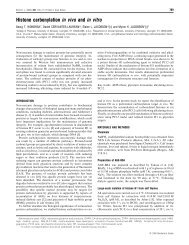
Chaperone action of 14-3-3ζ 499Figure 4 Light scattering and far-UV CD profiles of WT and 15C 14-3-3ζproteins as a function of temperature or chemical denaturant(A) The change in light scattering of WT (continuous line) and 15C (broken line) 14-3-3ζat 360 nm as a function of temperature between 37 and 100 ◦ C. (B) The change in molarellipticity of WT (continuous line) and 15C (broken line) 14-3-3ζ at 222 nm as a function oftemperature between 25 and 75 ◦ C. The melting curve of 15C relative to WT 14-3-3ζ illustratesa decrease from 60 ◦ Cto56 ◦ C at which the mid-point of protein denaturation/unfolding occursdue to C-terminal truncation. (C) The integrated far-UV CD signal of WT (×) and15C (∇)14-3-3ζ between 215 and 230 nm indicative of protein unfolding under denaturing conditionsfrom 0 to 7 M urea. Curves are fitted to an equation describing the mid-point of denaturation.C-terminal truncation of 14-3-3ζ reduces the half-point of unfolding by 0.33 M urea relative tothe full-length protein.greater tendency for 15C 14-3-3ζ to aggregate compared withthe full-length protein.Urea denaturation curves of WT and 15C 14-3-3ζ at 25 ◦ Cgave half-points of unfolding at 4.08 + − 0.03 and 3.75 + − 0.07 Murea respectively (Figure 4C). Thus truncation of the C-terminalextension led to decreased stability of 14-3-3ζ to denaturant. It istherefore concluded that the C-terminal extension of 14-3-3ζ hasa role in maintaining the structural integrity of the domain core ofthe protein.The effect of the C-terminal extension of 14-3-3ζ on its chaperoneabilityPrevious studies by Yano et al. [15] showed that 14-3-3ζ iscapable of preventing the amorphous aggregation of citratesynthase induced by mild heat stress at 45 ◦ C, and thus 14-3-3ζ has chaperone activity. We confirmed this finding (resultsnot shown) and extended it further by testing whether 14-3-3ζ prevented amorphous target protein aggregation followingchemical or reduction stress. We also investigated whether theC-terminus of 14-3-3ζ was important for its chaperone activity.The amorphous aggregation of insulin can be induced by theaddition of DTT, which reduces the intramolecular disulfidebonds, leading to the amorphous aggregation of the insulinFigure 5 The chaperone ability of WT and 15C 14-3-3ζ against theamorphous aggregation of target proteins(A) Top panels: the DTT-induced precipitation of insulin (44 μM) in the presence of WT (left-handpanel) and 15C (right-hand panel) 14-3-3ζ at 1.0:0.5–2.0 molar ratios of insulin/14-3-3ζ .Bottom panels: the percentage protection achieved by WT 14-3-3ζ against the amorphousaggregation of reduced insulin at 1.0:0.5–2.0 molar ratios of insulin/WT (left-hand panel). Acomparison of the chaperone ability of WT and 15C 14-3-3ζ against the aggregation of insulinusing the percentage protection afforded at a 1:1 molar ratio of insulin/14-3-3ζ (right-handpanel). (B) The percentage protection achieved by WT 14-3-3ζ against the chemically inducedaggregation of ADH (14 μM) at 1.0:0.5–2.0 molar ratios of ADH/WT (left-hand panel). Acomparison of the chaperone ability of WT and 15C 14-3-3ζ against the aggregation ofADH using the percentage protection afforded at a 1:1 molar ratio of ADH/14-3-3ζ (right-handpanel). (C) DTT-induced precipitation of αLA (140 μM) in the absence () and presence ofWT () and15C () 14-3-3ζ at 1:1 molar ratios of α-LA/14-3-3ζ (left-hand panel). TheDTT-induced precipitation of lysozyme (18 μM) in the absence () and presence of WT ()and 15C (∇) 14-3-3ζ at 1:1 molar ratios of lysozyme/14-3-3ζ (right-hand panel). For eachtarget protein, the assay was conducted in triplicate, repeated a minimum of three times, andthe data used to calculate the percentage protection afforded by either WT or 15C 14-3-3ζ .Results are presented as means + −S.E.M. In all cases, the percentage protection was calculatedonce protein aggregation had reached a plateau.B-chain. In the absence of 14-3-3ζ , this resulted in an increasein light scattering 10 min after the addition of DTT. The lightscattering due to the precipitation of insulin B-chain reached amaximum after 40 min. Both the truncated and full-length formsof 14-3-3ζ suppressed the aggregation and precipitation of insulinin a concentration-dependent manner (Figure 5A, top panels). At a1:1 molar ratio of insulin/WT 14-3-3ζ , the precipitation of insulinwas suppressed by 46 + − 5% (Figure 5A, bottom left-hand panel).At this ratio, the truncated protein had similar chaperone-likeactivity (41 + − 12%) in this assay (Figure 5A, bottom right-handpanel).c○ The Authors <strong>Journal</strong> compilation c○ 2011 <strong>Biochemical</strong> Society
500 D. M. Williams and othersSimilar results were also obtained when ADH was used asthe target protein (Figure 5B). The amorphous aggregation ofADH can be induced by chelation of its Zn 2 + ion using EDTA.In the absence of 14-3-3ζ , precipitation of ADH commenced60 min after incubation with EDTA, reaching a maximum after300 min (results not shown). As seen for insulin, both full-lengthand truncated forms of 14-3-3ζ suppressed the increase in lightscattering associated with ADH precipitation in a concentrationdependentmanner such that, at a 1:1 molar ratio of ADH/WT14-3-3ζ , aggregation was suppressed by 33 + − 11% (Figure 5B,left-hand panel). No significant differences were observed in thechaperone ability between the full-length and truncated proteinin this assay (Figure 5B, right-hand panel). No high-molecularmasscomplexes were observed by SEC of the soluble fraction atthe completion of either of these amorphous aggregation assays,suggesting that the interaction between 14-3-3ζ and its targetproteins is transient (results not shown).The chaperone ability of 14-3-3ζ to inhibit the aggregation ofreduced insulin was also tested in the presence of excess Mg 2 +and the polyamine spermine (at 0–50 and 0–2.0 molar equivalentsrespectively). Both species interact electrostatically and stronglywith the negatively charged C-terminal extension of 14-3-3ζ [33].Under both sets of conditions, there was no statistical differencebetween the chaperone ability of 14-3-3ζ in the presence andabsence of Mg 2 + and spermine (results not shown). Thus thesedata are consistent with the lack of direct involvement of theC-terminal extension in the chaperone action of 14-3-3ζ .When reduced α-LA was used as the target protein, truncatedand full-length 14-3-3ζ were ineffective chaperones (Figure 5C,left-hand panel). In the absence of 14-3-3ζ , α-LA aggregationinduced by reduction caused an increase in light scattering atapproximately 50 min which reached a plateau after 120 min.Truncated 14-3-3ζ (at a 1:1 molar ratio to α-LA) increased thelag phase of α-LA aggregation to 104 + − 7 min, as compared withwhen the full-length protein was present, in which case the lagphase was 84 + − 3 min. However, both forms of 14-3-3ζ causedenhanced precipitation (Figure 5C, left-hand panel). Analysis ofthe precipitates after completion of the experiment by SDS/PAGErevealed the presence of both 14-3-3ζ and α-LA (results notshown), i.e. the chaperone action of 14-3-3ζ with reduced α-LAled to complexation and co-precipitation. The chaperone ability ofWT and 15C 14-3-3ζ was also tested against the DTT-inducedaggregation of lysozyme, a protein that is structurally related toα-LA [34]. Interestingly, the light-scattering profile of reducedlysozyme was slightly enhanced by the presence of either WT or15C 14-3-3ζ , i.e. neither protein prevented reduced lysozymefrom aggregating and precipitating (Figure 5C, right-hand panel).After completion of this assay, analysis of the supernatants byanalytical SEC showed a decrease in the amount of 14-3-3ζ insolution when incubated in the presence of lysozyme (results notshown). Thus both WT and 15C 14-3-3ζ interact with reducedlysozyme in a manner similar to that with reduced α-LA, resultingin co-precipitation of both proteins from solution.To gain insight into whether the amphipathic binding groove of14-3-3ζ is involved in chaperone action, the chaperone activityof K49E 14-3-3ζ and of the WT protein in the presence of theR18 peptide was investigated. Charge reversal at Lys 49 (i.e. inK49E) gives rise to a protein that binds phosphorylated and nonphosphorylatedligands with significantly reduced affinity [23].The R18 peptide binds to the amphipathic binding groove of 14-3-3ζ with high affinity (K d = 80 nM), preventing ligand interactions,e.g. with ExoS and Raf [35,36]. However, the K49E mutation andthe presence of the R18 peptide did not affect the chaperoneactivity of 14-3-3ζ (Figure 6). Therefore it is unlikely that thebinding groove of 14-3-3ζ , particularly its hydrophilic side, isFigure 6 The chaperone ability of WT and K49E 14-3-3ζ against theamorphous aggregation of insulin and ADHThe percentage protection afforded against the DTT-induced aggregation of insulin (44 μM)and chemically induced aggregation of ADH (14 μM) by WT and K49E 14-3-3ζ at a 1:1 molarratio of target protein/14-3-3ζ . The percentage protection afforded against the DTT-inducedaggregation of insulin (44 μM) by WT 14-3-3ζ in the presence and absence of the R18 peptideat a 20:1:2 molar ratio of R18/insulin/WT 14-3-3ζ . For each target protein, the assay wasconducted in triplicate, repeated a minimum of three times and the data used to calculate thepercentage protection afforded by either K49E 14-3-3ζ or WT 14-3-3ζ in the presence andabsence of the R18 peptide. Results are presented as means + − S.D. The percentage protectionwas calculated once protein aggregation had reached a plateau.involved in the interaction with target proteins during chaperoneaction.DISCUSSIONConsistent with folding prediction algorithms, the NMR dataindicate that the last 12 C-terminal amino acids of 14-3-3ζ(Gly 234 –Asp 245 ) are solvent-exposed and exhibit flexibility thatis independent of the domain core of the protein, while adoptingno preferred secondary structure. This structural motif is akinto the C-terminal extension in mammalian sHsps (e.g. αA- andαB-crystallin) which is similar in length to that in 14-3-3ζand is also unstructured, flexible and highly dynamic in nature[17,18,37,38]. Indeed, the C-terminal extension in both proteinshas characteristics that are comparable with those of an isolatedpeptide of the same sequence in solution [39]. Therefore it isconcluded that 14-3-3ζ , and by analogy all other 14-3-3 proteins,have a flexible C-terminal extension.The inherent flexibility of the C-terminal extension of 14-3-3ζ and its structural similarity to that of sHsps, along with itsconservation across species, indicate that the C-terminal extensionof 14-3-3ζ may play an important role in the function andstability of the protein. Indeed, Silhan et al. [12], Obsilovaet al. [13] and Truong et al. [14] proposed that, in the absenceof a ligand, the C-terminus of 14-3-3ζ utilizes its negativecharges to occupy the peptide-binding groove of the protein andthereby regulates binding of ligands, particularly phosphorylatedones, to the binding groove. Our NMR results indicating thatthe C-terminal extension is flexible are not inconsistent withthis proposal. The extension could interact transiently with thepeptide-binding groove and give rise to the fluorescence responsesbetween the two regions that are observed by Obsil and coworkers[12,13]. The much shorter timescale of the fluorescencecompared with the NMR experiments is compatible with thisproposal. Furthermore, the well-conserved ‘hinge’ region, Trp 228 –Gln 233 (Figure 1B), which is not mobile enough to be observedby NMR spectroscopy but is not part of the last helix [11], mayc○ The Authors <strong>Journal</strong> compilation c○ 2011 <strong>Biochemical</strong> Society
Chaperone action of 14-3-3ζ 501occupy the binding groove principally via its negatively chargedresidue Asp 231 .Using an in vivo yeast two-hybrid assay and a range of in vitrospectroscopic and biophysical techniques, the structural andfunctional roles of the C-terminal extension was investigated bycomparing the full-length protein with a C-terminally truncatedform (15C 14-3-3ζ ). Truncation did not affect the ability of14-3-3ζ to form dimers in vitro or in vivo, or to dimerize withWT 14-3-3ζ . In yeast two-hybrid assays, C-terminal truncationalso had no effect on the ability of WT and 15C 14-3-3ζto bind to the phosphorylated ligands Raf and Bad. However,although C-terminal truncation of 14-3-3ζ did not significantlyalter the average diameter of the dimer at 25 ◦ C, at physiologicaltemperature, the truncated protein aggregated progressively withtime. C-terminal truncation also slightly decreased the α-helicalcontent of 14-3-3ζ . Thus the C-terminal extension of 14-3-3ζis important in maintaining the overall structural integrity ofthe protein without directly being involved in dimer formation.Consistent with this, C-terminal truncation resulted in a significantdecrease in the thermostability of the protein, and enhanced itssusceptibility to denaturant. Obsil and co-workers [12,13] alsoconcluded that the C-terminal extension stabilizes 14-3-3ζ in theabsence of a binding ligand. Finally, similarly to 14-3-3ζ ,theCterminalextension in mammalian sHsps also plays a vital role inthe overall thermostability, solubility and structure of the proteinand the large and heterogeneous complexes they form with targetproteins during chaperone action [16,17,19,38].The chaperone activity of the full-length and truncated 14-3-3ζ proteins was investigated using a variety of target proteinsundergoing amorphous aggregation induced by heat or reduction.In comparison with the sHsp αB-crystallin, 14-3-3ζ was a 4-foldpoorer chaperone in preventing the aggregation of reduced insulin[40,41]. In sHsps, truncation or site-specific mutations within theC-terminal extension result in an alteration in chaperone action[19,42–44]. In contrast, removal of this region in 14-3-3ζ hadlittle effect on its chaperone activity against the aggregating targetproteins insulin and ADH. Likewise, binding of positively chargesspecies (Mg 2 + and spermine) to the C-terminal extension of 14-3-3ζ did not affect its chaperone activity. With reduced α-LA,removal of the C-terminal extension of 14-3-3ζ ledtoadelayinonset of aggregation of α-LA compared with the situation for WT14-3-3ζ . However, in both cases, co-precipitation of α-LA and 14-3-3ζ occurred. With reduced lysozyme, the presence of WT and15C 14-3-3ζ also increased the precipitation of both proteins at37 ◦ C, but to a smaller degree relative to that of α-LA, and withno alteration in the onset of aggregation. A naturally occurringmutant of αB-crystallin, R120G, is associated with desmin-relatedmyopathy [45]. R120G αB-crystallin has a destabilized structure[46] and also causes enhanced precipitation of itself and reducedα-LA [46,47], which possibly arises from greater exposure of thechaperone-binding site(s) in R120G αB-crystallin compared withthe WT protein. Similar behaviour may be occurring with 14-3-3ζ whereby interaction with reduced α-LA leads to formation ofthe 14-3-3ζ /α-LA complex which is not stable in solution due togreater exposed hydrophobicity.Charge reversal mutants of the conserved basic residues onthe polar side of the binding groove of 14-3-3ζ , in particularK49E, result in a drastic reduction of 14-3-3ζ ligand affinitypresumably due to electrostatic repulsion, indicating that theconserved basic residues within the ligand-binding groove of 14-3-3ζ are important for ligand interaction [23]. However, basedon the very similar chaperone activity of K49E and WT 14-3-3ζ ,and the lack of effect of the R18 peptide, a species that has avidaffinity for the binding groove, on the chaperone activity of 14-3-3ζ , it seems unlikely that the amphipathic binding groove of14-3-3ζ (or at least its hydrophilic side) is a major determinantin chaperone action. The C-terminal extension of 14-3-3ζ alsoappears to not be involved in the interaction with target proteinsduring chaperone action. Thus other region(s) may encompass thechaperone-binding sites(s) of 14-3-3ζ , possibly on the exterior ofthe protein. Examination of the crystal structure of 14-3-3ζ [11]does not reveal any obvious regions of clustered hydrophobicityon its surface that may be potential chaperone-binding sites.Potentially, the hydrophobic side of the binding groove may be afactor in regulating chaperone activity, e.g. the conserved residuesLeu 172 and Leu 220 in helices 7 and 9. Future studies will explorethis possibility via site-directed mutagenesis.No high-molecular-mass complexes were isolated between 14-3-3ζ and its target proteins, suggesting an interaction which istransient in nature, similar to the interaction observed betweenmammalian sHsps and some target proteins under mild stressconditions [40,41]. Despite the similarity in mechanism betweenthe interaction of target proteins with 14-3-3ζ and sHsps, theC-terminal extension of 14-3-3ζ is not required to maintainthe solubility of the 14-3-3ζ –target protein complex, nor does itsremoval affect chaperone activity. The difference in dependencybetween 14-3-3ζ and sHsps on their C-terminal extension duringchaperone action may be due to 14-3-3ζ not undergoing subunitexchange as part of a large heterogeneous complex, as is the casefor mammalian sHsps. For the latter, subunit exchange is linkedto sHsp chaperone action [48] and may intimately involve theC-terminal extension [49]. The inability of 14-3-3ζ to preventthe aggregation of the related reduced target proteins α-LA andlysozyme is indicative of the selective nature of the chaperoneaction of 14-3-3ζ and the need to utilize a range of target proteinsto explore its chaperone action. Variation in chaperone abilitywith target proteins is also observed with sHsps and is related tofactors such as the rate of target protein aggregation, the nature ofthe intermediate state of the target protein, the mass of the targetprotein and the type of target protein aggregation [38,40,41,50–53]. Similar factors are most likely to be important in determiningthe efficiency of 14-3-3ζ chaperone action.Our understanding of the function of 14-3-3 proteins asmolecular chaperones is in its infancy. It has previously beenestablished that 14-3-3ζ has a regulatory role in the formationof intracellular tau filaments [54,55] as occurs in Alzheimer’sdisease, and also in the aggregation of polyglutamine proteins,such as ataxin-1 and huntingtin [9,10,56]. In the case of tau, onemode of 14-3-3ζ interaction is dependent on tau phosphoryation[54], implying that 14-3-3ζ interacts with tau via its amphipathicbinding groove. The present study and previous studies [15]demonstrate a different role for 14-3-3ζ as a molecular chaperonein preventing the aggregation of partially folded target proteins.The results of the present study indicate that the amphipathicgroove may not be required for this interaction. The implicationis that 14-3-3ζ has diverse roles in protein misfolding andaggregation and strengthens the notion of the involvementof 14-3-3 proteins in neurodegenerative diseases such asAlzheimer’s disease, Parkinson’s disease and Huntington’sdisease.AUTHOR CONTRIBUTIONDanielle Williams designed and performed most of the experiments and, with John Carver,wrote the paper. John Carver also devised the overall experimental concepts and content ofthe paper. Huanqin Dai, Haian Fu, Lixin Zhang and Katy Goodwin conducted the remainingexperiments and undertook a critical review of the manuscript. Heath Ecroyd and JoannaWoodcock provided assistance with, and design of, the experiments along with a criticalreview of the paper.c○ The Authors <strong>Journal</strong> compilation c○ 2011 <strong>Biochemical</strong> Society
502 D. M. Williams and othersACKNOWLEDGEMENTSWe acknowledge the technical support of Phil Clements (University of Adelaide), Dr KeithShearwin (University of Adelaide) and Alexander Buell (University of Cambridge). The CDspectra were acquired at the Biophysical Characterisation Facility, Adelaide, Australia.FUNDINGThis work was supported, in part, by the China Important National Science and TechnologySpecific Projects [grant number 2008ZX09401-05 (to L.Z.)] and the Australian ResearchCouncil (to J.C.). L.Z. was an awardee of the Hundred Talents Program. H.E. was supportedby an Australian National Health and Medical Research Council Peter Doherty BiomedicalFellowship. D.W. was the recipient of an Australian Postgraduate Award and was partiallyfunded by the Daphne Elliott Bursary, AUGC/RC Heddle Award, George Murray Scholarshipand an Australian Bicentennial Scholarship .REFERENCES1 Aitken, A. (2006) 14-3-3 proteins: a historic overview. Semin. Cancer Biol. 16, 162–1722 Dougherty, M. K. and Morrison, D. K. (2004) Unlocking the code of 14-3-3. J. Cell Sci.117, 1875–18843 Fu, H., Subramanian, R. R. and Masters, S. C. (2000) 14-3-3 proteins: structure, function,and regulation. Annu. Rev. Pharmacol. Toxicol. 40, 617–6474 Muslin, A. J., Tanner, J. W., Allen, P. M. and Shaw, A. S. (1996) Interaction of 14-3-3 withsignaling proteins is mediated by the recognition of phosphoserine. Cell 84, 889–8975 Yaffe, M. B., Rittinger, K., Volinia, S., Caron, P. R., Aitken, A., Leffers, H., Gamblin, S. J.,Smerdon, S. J. and Cantley, L. C. (1997) The structural basis for 14-3-3:phosphopeptidebinding specificity. Cell 91, 961–9716 Rubio, M. P., Geraghty, K. M., Wong, B. H. C., Wood, N. T., Campbell, D. G., Morrice, N.and Mackintosh, C. (2004) 14-3-3-affinity purification of over 200 humanphosphoproteins reveals new links to regulation of cellular metabolism, proliferation andtrafficking. Biochem. J. 379, 395–4087 Zhang, L., Chen, J. and Fu, H. (1999) Suppression of apoptosis signal-regulating kinase1-induced cell death by 14-3-3 proteins. Proc. Natl. Acad. Sci. U.S.A. 96, 8511–85158 Zippo, A., Serafini, R., Rocchigiani, M., Pennacchini, S., Krepelova, A. and Oliviero, S.(2009) Histone crosstalk between H3S10ph and H4K16ac generates a histone code thatmediates transcription elongation. Cell 138, 1122–11369 Waelter, S., Boeddrich, A., Lurz, R., Scherzinger, E., Lueder, G., Lehrach, H. and Wanker,E. E. (2001) Accumulation of mutant huntingtin fragments in aggresome-like inclusionbodies as a result of insufficient protein degradation. Mol. Biol. Cell 12, 1393–40710 Omi, K., Hachiya, N. S., Tanaka, M., Tokunaga, K. and Kaneko, K. (2008) 14-3-3ζ isindispensable for aggregate formation of polyglutamine-expanded huntingtin protein.Neurosci. Lett. 431, 45–5011 Liu, D., Bienkowska, J., Petosa, C., Collier, R. J., Fu, H. and Liddington, R. (1995) Crystalstructure of the ζ isoform of the 14-3-3 protein. Nature 376, 191–19412 Silhan, J., Obsilova, V., Vecer, J., Herman, P., Sulc, M., Teisinger, J. and Obsil, T. (2004)14-3-3 protein C-terminal stretch occupies ligand binding groove and is displaced byphosphopeptide binding. J. Biol. Chem. 279, 49113–4911913 Obsilova, V., Herman, P., Vecer, J., Sulc, M., Teisinger, J. and Obsil, T. (2004) 14-3-3ζC-terminal stretch changes its conformation upon ligand binding and phosphorylation atThr 232 . J. Biol. Chem. 279, 4531–454014 Truong, A. B., Masters, S. C., Yang, H. and Fu, H. (2002) Role of the 14-3-3 C-terminalloop in ligand interaction. Proteins 49, 321–32515 Yano, M., Nakamuta, S., Wu, X., Okumura, Y. and Kido, H. (2006) A novel function of14-3-3 protein: 14-3-3ζ is a heat-shock-related molecular chaperone that dissolvesthermal-aggregated proteins. Mol. Biol. Cell 17, 4769–477916 Carver, J. A. (1999) Probing the structure and interactions of crystallin proteins by NMRspectroscopy. Prog. Retinal Eye Res. 18, 431–46217 Carver, J. A. and Lindner, R. A. (1998) NMR spectroscopy of α-crystallin. Insights intothe structure, interactions and chaperone action of small heat-shock proteins. Int. J. Biol.Macromol. 22, 197–20918 Carver, J. A., Aquilina, J. A., Truscott, R. J. W. and Ralston, G. B. (1992) Identification by1 H NMR spectroscopy of flexible C-terminal extensions in bovine lens α-crystallin. FEBSLett. 311, 143–14919 Treweek, T. M., Ecroyd, H., Williams, D. M., Meehan, S., Carver, J. A. and Walker, M. J.(2007) Site-directed mutations in the C-terminal extension of human αB-crystallin affectschaperone function and blocks amyloid fibril formation. PLoS ONE 2, e104620 Bax, A. and Davis, D. G. (1985) MLEV-17-based two-dimensional homonuclearmagnetization transfer spectroscopy. J. Magn. Reson. 65, 355–36021 Kessler, H., Griesinger, C., Kerssebaum, R., Wagner, K. and Ernst, R. R. (1987) Separationof cross-relaxation and J cross-peaks in 2D rotating-frame NMR spectroscopy. J. Am.Chem. Soc. 109, 607–60922 Reference deleted23 Zhang, L., Wang, H., Liu, D., Liddington, R. and Fu, H. (1997) Raf-1 kinase andexoenzyme S interact with 14-3-3ζ through a common site involving lysine 49. J. Biol.Chem. 272, 13717–1372424 Provencher, S. W. (1982) CONTIN: a general purpose constrained regularization programfor inverting noisy linear algebraic and integral equations. Comput. Phys. Commun. 27,229–24225 Koppel, D. E. (1972) Analysis of macromolecular polydispersity in intensity correlationspectroscopy: the method of cumulants. J. Chem. Phys. 57, 4814–482026 Larkin, M. A., Blackshields, G., Brown, N. P., Chenna, R., McGettigan, P. A., McWilliam,H., Valentin, F., Wallace, I. M., Wilm, A., Lopez, R. et al. (2007) Clustal W and Clustal Xversion 2.0. Bioinformatics 23, 2947–294827 Romero, P., Obradovic, Z., Li, X., Garner, E. C., Brown, C. J. and Dunker, A. K. (2001)Sequence complexity of disordered protein. Proteins 42, 38–4828 Li, X., Romero, P., Rani, M., Dunker, A. K. and Obradovic, Z. (1999) Predicting proteindisorder for N-, C-, and internal regions. Genome Inform. Ser. Workshop Genome Inform.10, 30–4029 Romero, P., Obradovic, Z. and Dunker, K. (1997) Sequence data analysis for longdisordered regions prediction in the calcineurin family. Genome Inform. Ser. WorkshopGenome Inform. 8, 110–12430 Prilusky, J., Felder, C. E., Zeev-Ben-Mordehai, T., Rydberg, E. H., Man, O., Beckmann, J.S., Silman, I. and Sussman, J. L. (2005) FoldIndex: a simple tool to predict whether agiven protein sequence is intrinsically unfolded. Bioinformatics 21, 3435–343831 Wüthrich, K. (1986) NMR of Proteins and Nucleic Acids, Wiley, New York32 Wishart, D. S., Bigam, C. G., Holm, A., Hodges, R. S. and Sykes, B. D. (1995) 1 H, 13 Cand15 N random coil NMR chemical shifts of the common amino acids. I. Investigations ofnearest-neighbor effects. J. Biomol. NMR 5, 67–8133 Visconti, S., Camoni, L., Marra, M. and Aducci, P. (2008) Role of the 14-3-3 C-terminalregion in the interaction with the plasma membrane H + -ATPase. Plant Cell Physiol. 49,1887–189734 Kumagai, I., Takeda, S. and Miura, K. (1992) Functional conversion of the homologousproteins α-lactalbumin and lysozyme by exon exchange. Proc. Natl. Acad. Sci. U.S.A. 89,5887–589135 Masters, S. C. and Fu, H. (2001) 14-3-3 proteins mediate an essential anti-apoptoticsignal. J. Biol. Chem. 276, 45193–4520036 Wang, B., Yang, H., Liu, Y. C., Jelinek, T., Zhang, L., Ruoslahti, E. and Fu, H. (1999)Isolation of high-affinity peptide antagonists of 14-3-3 proteins by phage display.Biochemistry 38, 12499–1250437 Treweek, T. M., Rekas, A., Walker, M. J. and Carver, J. A. (2010) A quantitative NMRspectroscopic examination of the flexibility of the C-terminal extensions of the molecularchaperones, αA- and αB-crystallin. Exp. Eye Res. 91, 691–69938 Treweek, T. M., Morris, A. M. and Carver, J. A. (2003) Intracellular protein unfolding andaggregation: the role of small heat-shock chaperone proteins. Aust. J. Chem. 56,357–36739 Esposito, G., Viglino, P., Fogolari, F., Gaestel, M. and Carver, J. A. (1998) Selective NMRexperiments on macromolecules: implementation and analysis of QUIET-NOESY. J.Magn. Reson. 132, 204–21340 Ecroyd, H. and Carver, J. A. (2008) The effect of small molecules in modulating thechaperone activity of αB-crystallin against ordered and disordered protein aggregation.FEBS J. 275, 935–94741 Ecroyd, H., Meehan, S., Horwitz, J., Aquilina, J. A., Benesch, J. L., Robinson, C. V.,Macphee, C. E. and Carver, J. A. (2007) Mimicking phosphorylation of αB-crystallinaffects its chaperone activity. Biochem. J. 401, 129–14142 Lindner, R. A., Carver, J. A., Ehrnsperger, M., Buchner, J., Esposito, G., Behlke, J., Lutsch,G., Kotlyarov, A. and Gaestel, M. (2000) Mouse Hsp25, a small shock protein. The role ofits C-terminal extension in oligomerization and chaperone action. Eur. J. Biochem. 267,1923–193243 Morris, A. M., Treweek, T. M., Aquilina, J. A., Carver, J. A. and Walker, M. J. (2008)Glutamic acid residues in the C-terminal extension of small heat shock protein 25 arecritical for structural and functional integrity. FEBS J. 275, 5885–589844 Smulders, R. H.P. H., Carver, J. A., Lindner, R. A., Van Boekel, M. A., Bloemendal, H. andDe Jong, W. W. (1996) Immobilization of the C-terminal extension of bovine αA-crystallinreduces chaperone-like activity. J. Biol. Chem. 271, 29060–2906645 Vicart, P., Caron, A., Guicheney, P., Li, Z., Prevost, M. C., Faure, A., Chateau, D., Chapon,F., Tome, F., Dupret, J. M. et al. (1998) A missense mutation in the αB-crystallinchaperone gene causes a desmin-related myopathy. Nat. Genet. 20, 92–9546 Treweek, T. M., Rekas, A., Lindner, R. A., Walker, M. J., Aquilina, J. A., Robinson, C. V.,Horwitz, J., Perng, M. D., Quinlan, R. A. and Carver, J. A. (2005) R120G αB-crystallinpromotes the unfolding of reduced α-lactalbumin and is inherently unstable. FEBS J.272, 711–724c○ The Authors <strong>Journal</strong> compilation c○ 2011 <strong>Biochemical</strong> Society
Chaperone action of 14-3-3ζ 50347 Bova, M. P., Yaron, O., Huang, Q., Ding, L., Haley, D. A., Stewart, P. L. and Horwitz, J.(1999) Mutation R120G in αB-crystallin, which is linked to a desmin-related myopathy,results in an irregular structure and defective chaperone-like function. Proc. Natl. Acad.Sci. U.S.A. 96, 6137–614248 Bova, M. P., Ding, L.-L., Horwitz, J. and Fung, B. K.-K. (1997) Subunit exchange ofαA-crystallin. J. Biol. Chem. 272, 29511–2951749 Laganowsky, A., Benesch, J. L., Landau, M., Ding, L., Sawaya, M. R., Cascio, D., Huang,Q., Robinson, C. V., Horwitz, J. and Eisenberg, D. (2010) Crystal structures of truncatedαA andαB crystallins reveal structural mechanisms of polydispersity important for eyelens function. Protein Sci. 19, 1031–104350 Carver, J. A., Lindner, R. A., Lyon, C., Canet, D., Hernandez, H., Dobson, C. M. andRedfield, C. (2002) The interaction of the molecular chaperone α-crystallin with unfoldingα-lactalbumin: a structural and kinetic spectroscopic study. J. Mol. Biol. 318, 815–82751 Carver, J. A., Rekas, A., Thorn, D. C. and Wilson, M. R. (2003) Small heat-shock proteinsand clusterin: intra- and extracellular molecular chaperones with a common mechanismof action and function? IUBMB Life 55, 661–66852 Lindner, R. A., Kapur, A., Mariani, M., Titmuss, S. J. and Carver, J. A. (1998) Structuralalterations of α-crystallin during its chaperone action. Eur. J. Biochem. 258, 170–18353 Ecroyd, H. and Carver, J. A. (2009) Crystallin proteins and amyloid fibrils. Cell. Mol. LifeSci. 66, 62–8154 Sadik, G., Tanaka, T., Kato, K., Yamamori, H., Nessa, B. N., Morihara, T. and Takeda, M.(2009) Phosphorylation of tau at Ser 214 mediates its interaction with 14-3-3 protein:implications for the mechanism of tau aggregation. J. Neurochem. 108, 33–4355 Hernandez, F., Cuadros, R. and Avila, J. (2004) 14-3-3ζ protein favours the formation ofhuman tau fibrillar polymers. Neurosci. Lett. 357, 143–14656 Chen, H.-K., Fernandez-Funez, P., Acevedo, S. F., Lam, Y. C., Kaytor, M. D., Fernandez, M.H., Aitken, A., Skoulakis, E. M. C., Orr, H. T., Botas, J. and Zoghbi, H. Y. (2003) Interactionof Akt-phosphorylated ataxin-1 with 14-3-3 mediates neurodegeneration inspinocerebellar ataxia type-1. Cell 113, 457–46857 Petosa, C., Masters, S. C., Bankston, L. A., Pohl, J., Wang, B., Fu, H. and Liddington, R.C. (1998) 14-3-3ζ binds a phosphorylated Raf peptide and an unphosphorylated peptidevia its conserved amphipathic groove. J. Biol. Chem. 273, 16305–16310Received 4 January 2011/19 April 2011; accepted 10 May 2011Published as BJ Immediate Publication 10 May 2011, doi:10.1042/BJ20102178c○ The Authors <strong>Journal</strong> compilation c○ 2011 <strong>Biochemical</strong> Society