Helicobacter Pylori Ag - Linear
Helicobacter Pylori Ag - Linear
Helicobacter Pylori Ag - Linear

Create successful ePaper yourself
Turn your PDF publications into a flip-book with our unique Google optimized e-Paper software.
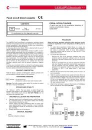
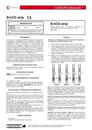
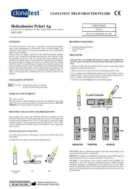
CLONATEST HELICOBACTER PYLORI<strong>Helicobacter</strong> <strong>Pylori</strong> <strong>Ag</strong>A rapid test for the qualitative detection of <strong>Helicobacter</strong> pylori in faeces.ONE STEPCOD CT4245120 TestFor in vitro diagnostic use onlySUMMARYThe <strong>Linear</strong> <strong>Helicobacter</strong> pylori <strong>Ag</strong> is a qualitative immunochromatographicassay for the determination of <strong>Helicobacter</strong> pylori in faeces samples. Themembrane is pre-coated with monoclonal antibodies, on the test band region,against H. pylori antigens.During testing, the sample is allowed to react with the coloured conjugate(anti-H. pylori monoclonal antibodies-red polystyrene micro spheres) whichwas pre-dried on the test strip. The mixture then moves upward on themembrane by capillary action. As the sample flows through the test membrane,the coloured particles migrate. In the case of a positive result the specificantibodies present on the membrane will capture the coloured conjugate. Themixture continues to move across the membrane to the immobilized antibodyplaced in the control band region, a green coloured band always appears. Thepresence of this green band serves as 1) verification that sufficient volume isadded, 2) that proper flow is obtained and 3) as an internal control for thereagents.PACKAGING CONTENTSMATERIALS REQUIRED---Specimen collection containerDisposable glovesTimer.PROCEDUREAllow the tests, stool samples and controls to reach to room temperature(15-30ºC) prior to testing. Do not open pouches until ready to perform theassay.1. Proceed to shake the stool collection tube in order to assure good sampledispersion. Cut the end of the top (3).2. Remove the H. pylori <strong>Ag</strong> device from its sealed bag just before using.3. Use a separate stool collection tube and device for each sample or control.Dispense exactly 5 drops or 150 µL into the circular window marked with anarrow, avoiding to add solid particles with the liquid (4).4.- Read the result at 10 minutes (the coloured bands appear).REF CT 42451 20 helicobacter pylori <strong>Ag</strong> cassettes20 Stool collection tubes with bufferSTORAGE AND STABILITYStore at 2-30ºC.The test cassette is stable through the expiration date printed on the sealedpouch. The test cassette must remain in the sealed pouch until use. DO NOTFREEZE. Do not use beyond the expiration date.SPECIMEN COLLECTION AND PREPARATIONH. pylori CassetteControl region Result region(3) (4)SAdd 5 dropsStool samples (not watery and diarrhoeal) should be collected in cleancontainers and the assay should be done right after collection. The samples canbe stored in the refrigerator (2-4 ºC) for 1-2 days prior to testing. For longerstorage, maximum 1 year, the specimen must be kept frozen at –20ºC. In thiscase, the sample will be totally thawed, and brought to room temperaturebefore testing.Specimen preparation (see illustration):(1) Use the stick to pick up a little sample. Close the tube with the diluent andstool sample.(2) Shake the tube in order to assure good sample dispersion.GREENREDREDNEGATIVE POSITIVE INVALIDStool sampleStool Collection (1) (2)TubeNEGATIVE: Only one GREEN band appears across the central window in thesite marked with the letter C (control line).POSITIVE: In addition to the GREEN control band, another RED band (testline) also appears in the site marked with the letter T (result line).INVALID: A total absence of the control coloured band regardless theappearance or not of the result line. Insufficient specimen volume, incorrectprocedural techniques or deterioration of the reagents are the most likelyreasons for control line failure. Review the procedure and repeat the test with anew test. If the problem persists, discontinue using the test kit and contact youlocal distributor.QUALITY SYSTEM CERTIFIEDISO 9001 ISO 13485LINEAR CHEMICALS S.L.08390 Montgat, SPAIN (EU)
CLONATEST HELICOBACTER PYLORIQUALITY CONTROLInternal procedural controls are included in the test. A GREEN line appearingin the control region (C) is the internal procedural control. It confirms sufficientspecimen volume and correct procedural technique. A clear background is aninternal negative background control. If the test is working properly, thebackground in the result area should be clear and not interfere with the abilityto read the result.CLINICAL SIGNIFICANCEREFERENCES1. Cutler AF. Testing for <strong>Helicobacter</strong> pylori in clinical practice. Am j.Med. 1996; 100:35S-41S2. Soll, AH. Pathogenesis of pectic ulcer and implications for theraphy.New England J. Med. (1990), 322: 909-16.3. Martin J. Blaser. <strong>Helicobacter</strong> pylori and gastric diseases. BMJ; 316:1507-1510 (1998).<strong>Helicobacter</strong> pylori (H. pylori) is a spiral-shaped bacterium that is found in thegastric mucous layer or adherent to the epithelial lining of the stomach. H.pylori causes more than 90% of duodenal ulcers and up to 80% of gastriculcers.The importance of <strong>Helicobacter</strong> pylori testing has increased greatly since thestrong correlation between the presence of bacteria and confirmedgastrointestinal diseases (stomach and duodenum) like gastritis, peptic ulcerdisease and gastric carcinoma.ANALYTICAL PERFORMANCESENSITIVITYDetection limit: A culture of H. pylori bacteria was sonicated, centrifuged andits protein concentration was determined. This reference antigen preparation ofH pylori was diluted in the PBS-BSA buffer and tested in accordance with thekit instructions. The detection limit of <strong>Helicobacter</strong> pylori is 4-8 ng/mL.SPECIFICITYThe evaluation was conducted comparing the results obtained using the<strong>Helicobacter</strong> pylori <strong>Ag</strong> test to another available commercial ELISA assay.The detection of <strong>Helicobacter</strong> pylori showed 95% of concordance with thecommercial ELISA assay.The antibodies used to elaborate the H. pylori <strong>Ag</strong> recognise epitopes present inthe antigen found in stool of patients, as well as in preparations from thebacteria cultures in vitro. Sonicated <strong>Helicobacter</strong> pylori extract from differentcommercial samples reacts with H. pylori <strong>Ag</strong>.The possibility for interference by human anti-mouse antibodies (HAMA) orhigh levels of RF in the stools sample, has not evaluated. Some stool samplescould produce control lines with a light red colour.NOTESThe intensity of the red coloured band in the result line region (T) will varydepending on the concentration of antigens in the specimen. However, neitherthe quantitative value, nor the rate of increase in antigens can be determined bythis qualitative test.PRECAUTIONSFor professional in vitro diagnostic use only.Do not use after expiration date.All the specimens should be considered potentially hazardous and handled inthe same manner as an infectious agent.The tests should be discarded in a proper biohazard container after testing.LIMITATIONS1. The test must be carried out within 2 hours of opening the sealed bag.2. An excess of sample could cause wrong results (brown bands appear).Dilute the sample with the buffer and repeat the test.3. Some stool samples can decrease the intensity of the control green line.4. This test provides a presumptive diagnosis of <strong>Helicobacter</strong> pyloriinfections. A confirmed infection diagnosis should only be made by aphysician after all clinical and laboratory findings have been evaluatedmust be based in the correlation of the results with further clinicalobservations.CT4245-2/0601R1.ingQUALITY SYSTEM CERTIFIEDISO 9001 ISO 13485LINEAR CHEMICALS S.L.08390 Montgat, SPAIN (EU)