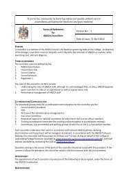
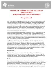
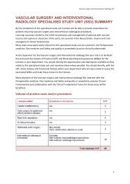
84 <strong>Australasian</strong> <strong>Anaesthesia</strong> <strong>2011</strong>Jet Ventilation <strong>and</strong> <strong>Anaesthesia</strong> 85It is possible that supraglottic jet ventilation delivered through an LMA could provide adequate oxygenation ina critically obstructed airway. 41It is often very useful in a less urgent setting where a difficult airway has been anticipated. For example, theprophylactic use of PTTJV to ensure oxygenation during an anticipated difficult fibreoptic intubation or while teachingthe use of flexible fibreoptic intubation has been described. 42-43 The sight of the catheter just beyond the vocalcords has been used as a marker during difficult laryngoscopy in situations where the anatomy may be distorted[44]. It has been the author’s practice to place a trans-tracheal cannula under local anaesthesia <strong>and</strong> light sedationprior to commencement of anaesthesia or airway manipulation in situations where major difficulties are anticipated.Laryngeal surgeryHFJV is most frequently used during surgery on or around the larynx. Having no conduit or only a very small conduitthrough the larynx offers optimum conditions for the surgical intervention while the airway is shared between thesurgeon <strong>and</strong> the anaesthetist.General anaesthesia during these procedures is normally provided using a total intravenous (TIVA) method <strong>and</strong>a muscle relaxant. Topical analgesia of the vallecula, epiglottis, laryngeal inlet <strong>and</strong> the vocal cords before theintroduction of the rigid laryngoscope is a very useful adjunct to blunt airway reflexes.In this setting HFJV may be achieved using a supraglottic or a subglottic jet delivery. Subglottic jet delivery mayin turn be via a catheter passed trough the laryneal inlet (translaryngeal) from above or via a trans-tracheal catheteror cannula (PTTJV).Supraglottic jet ventilationSupraglottic jet ventilation is done through an operating laryngoscope while the tip of the scope is aligned with thelaryngeal inlet. Jet delivery is done either with an injector clamped into the main lumen of the scope or through adedicated channel built into the wall of the scope. A technique delivering both a low frequency <strong>and</strong> a high frequencyjet stream through dedicated channels built into a modified laryngoscope has also been reported 45 <strong>and</strong> is termed“combined frequency jet ventilation”.The main advantage of supraglottic jet ventilation is that view <strong>and</strong> access to the larynx is unhindered. However,entrainment of air is large <strong>and</strong> accurate monitoring of FiO2, airway pressure <strong>and</strong> expired CO2 is difficult. Furthermore,the large airflow not only causes a ripple movement of mucus membranes <strong>and</strong> vocal cords but can also blow blood,smoke or tissue debris down the trachea. There has been a concern expressed of tumour implantation in moredistal airways. Finally, this technique may not be suitable in the presence of a compromised glottic aperture causedby stenosis, tumour or paralysed vocal cords. 6,46The proper alignment of the rigid laryngoscope is very important as a misdirected jet may cause gastric insufflation<strong>and</strong> a vagal reaction.Subglottic methodsThe value of subglottic jet ventilation without a large tracheal tube placed through the larynx obscuring most of itfrom view <strong>and</strong> restricting room needed for surgical manipulation has long been appreciated (Figure 3). A range oftubes, catheters <strong>and</strong> cannulae had been reported in the literature for delivery of translaryngeal subglottic jetventilation. 47-56The use of flexible catheters for subglottic HFJV is by far the most popular choice for endolaryngeal intervention.Whipping of the tip of a catheter during jet ventilation is a concern as it can cause trauma to the tracheal mucosa,leading to the spread of pressurized air into the mediastinum, lungs <strong>and</strong> soft tissues of the neck <strong>and</strong> upper chestarea. Jet induced movement of the catheter tip is reduced or eliminated by having multiple openings at the tip,using a stiffer material <strong>and</strong> placing a minimum length of catheter inside the airway.Figure 3. The view <strong>and</strong> workspace available with a shielded 7Fr (2.33 mm O.D) catheter through the larynxof a child <strong>and</strong> a 5 mm (O.D) catheter through an adult larynx.A cage design at the tip of the catheter to keep the catheter central inside the trachea, was first describedby Benjamin <strong>and</strong> Gronow. 50 A catheter with modified cage design <strong>and</strong> a second channel designed by Hunsaker(Mon- Jet TM tube Xomed-Treace, Jacksonville, FL, USA) is commonly used now. 54With the introduction of CO2 laser for endolaryngeal interventions, flammability of the jet conduits became anissue. Long metal cannulae, various coverings for the catheters like metal foils <strong>and</strong> specially designed tapes thatcould be soaked in water were used. The Hunsaker device has a fluoroplastic coating resistant to laser beams.Recently use of a new device named the ‘Jockjet ‘ tube system has been described. 56 The design has the jetflow encased in a segment of 4 mm teflon tube <strong>and</strong> a trailing double lumen catheter to measure distal airwaypressure <strong>and</strong> to sample gas for capnography.HFJV has been successfully employed for airway management <strong>and</strong> anaesthesia in neonates, infants <strong>and</strong>children. 57-58During subglottic ventilation one must ensure a clear expiratory pathway before the first breath is delivered. Thisshould be managed as described below in the section on PTTJV. The technique of subglottic HFJV using a cathetermay avoid tracheostomy or laryngectomy during endoscopic treatment <strong>and</strong> resection of various glottic <strong>and</strong> subglottictumours. It has also been used successfully to deliver ventilation during tracheostomy, tracheoplasty <strong>and</strong> trachealresection. 59Percutaneous trans-tracheal jet ventilationWhile less common than a “trans-laryngeal” catheter approach, PTTJV is often used for endolaryngeal interventionsas the technique offers the best view <strong>and</strong> working space with no tubes at all in or around the laryngeal inlet(Figure 4)Figure 4. A narrow laryngeal inlet that may not have enough room for even the smallest catheter,precluding safe translaryngeal ventilation.The percutaneous transtracheal catheter is introduced using local anaesthesia <strong>and</strong> tested while the patient is awake.After induction of general anaesthesia using TIVA an oropharyngeal or nasopharyngeal airway is inserted <strong>and</strong> theneck extended before HFJV is delivered. Driving pressure is increased once the expiratory outlet is confirmedpatent. Jet ventilation is stopped transiently while the airway is taken out <strong>and</strong> the operating laryngoscope ispositioned in place. (figure 5)There are several series of case reports describing the usefulness of this technique <strong>and</strong> a low incidence ofcomplications. 60-65 Recently, a prospective audit of successful management of 50 cases with severe airwaycompromise <strong>and</strong> stridor has been reported [65]. It should be stressed that there is a learning curve <strong>and</strong> all reportsstrongly recommend that HFJV should be monitored closely <strong>and</strong> conducted in presence of experienced operators.Figure 5. PTTJV for endolaryngeal surgery
86 <strong>Australasian</strong> <strong>Anaesthesia</strong> <strong>2011</strong>Jet Ventilation <strong>and</strong> <strong>Anaesthesia</strong> 87Other indications for the use of HFJVHFJV has several applications in areas other than in laryngeal surgery. A subglottic technique is usually employedfor this purpose.Minimum movement with ventilation:The movements of thoracic <strong>and</strong> abdominal viscera during HFJV are much smaller than those seen during tidalventilation. This produces a relatively immobile target that is ideal for precision therapeutic interventions.Extracorporeal Shock Wave Lithotripsy (ESWL) uses focused, high intensity, acoustic pulses to break up stonesinside the kidney with minimal collateral damage. Most current lithotriptors have smaller focal zones with theadvantage of minimizing damage to surrounding soft tissues. Unfortunately, movement of the diaphragm duringspontaneous or intermittent positive pressure ventilation causes movement of the kidney <strong>and</strong> therefore the stonein <strong>and</strong> out of the acoustic ‘energy cone’ with every respiratory cycle. The shockwaves applied while the stones areout of the target zone have no effect on fragmentation leading to higher failure rates, incomplete treatment, <strong>and</strong>the need for retreatment. Energy from such ‘stray’ shock waves can also cause injury to surrounding kidney tissues.Subglottic HFJV has been used during ESWL with great success. 66-67 A simplified technique of HFJV through asupraglottic airway such as a laryngeal mask airway is certainly simple <strong>and</strong> easy for this outpatient procedure. 68For similar reasons, HFJV has also been reported as a ventilation technique for stereotactic irradiation ofmetastasis in the lungs, cardiac radiofrequency ablation <strong>and</strong> percutaneous radiofrequency ablation of hepatictumours. 69-71Thoracic surgery:The low peak tracheal <strong>and</strong> transpulmonary pressures <strong>and</strong> low mean airway pressures (relaxed lungs) during HFJVmake it very useful for thoracic surgery. It has been used in conjunction with a wide range of thoracic procedures.(Table 2)Table 2. Some thoracic procedures in which HFJV has been reported as a useful ventilation technique<strong>Anaesthesia</strong> for open thoracic surgery 59,72-82One lung ventilationPartial lung ventilation during lung resectionStaple excision of subcentimeter lung nodulesBronchial repairTransthoracic oesophagectomyTAA with tracheal <strong>and</strong> right bronchial compressionOff pump single lung transplantRepair of coarctationAnterior approach scoliosis surgeryCarinal resection / TracheoplastyBronchial stentingRepair of congenital tracheal stenosis<strong>Anaesthesia</strong> for closed chest procedures 83-87Endobronchial laser treatment for tumoursTransthoracic endoscopic sympathectomyHFJV through the FOB for identifying segmental planesLung lavageInterventional fibreoptic bronchoscopyUninterrupted oxygenation during airway manoeuvres:The use of jet ventilation through an airway exchange device is an option that provides a way to deliver oxygeninto the lungs during procedures like a trial of extubation or the re-positioning of an endotracheal tube. 53,88 In thissetting, the narrow internal diameter <strong>and</strong> rather long length of some exchange catheters can result in volume lossdue to compression 89 <strong>and</strong> yet can also be dangerous when used in conjunction with an endotracheal tube withsmall internal diameter (restricted expiratory outlet) or when the tip of the catheter is inside a snugly fitting distalairway (risk of barotrauma). 88 The delivery of jet ventilation through the suction-biopsy channel of a flexible fibreopticbronchoscope has been shown to provide adequate ventilation. 90Common problems seen with HFJVHypercarbia is well documented <strong>and</strong> is more usually a problem with obese patients.Trans-tracheal cannulae have been found to be a major independent risk factor for complications in a 10 yearreview of different ventilation strategies in endoscopic laryngeal surgery. 91 Obstruction of trans-tracheal cannulaecan occur in up to 20% of cases 92 even with custom made, kink-resistant varieties. Care must be taken duringinsertion of the cannula to aim in a 45 degree angle caudad direction as soon as the trachea is entered. In somepatients with restricted space between the larynx <strong>and</strong> the jaw, a lower point of puncture at the cricotracheal junctionor even between the upper rings of the trachea may help to get the angle right.Difficulty with CO2 measurement is encountered at times with side stream CO2 monitoring as the sampling linetends to get blocked by respiratory secretions <strong>and</strong> blood. The use of preoperative anticholinergics, periodic suctioning<strong>and</strong> the use of a three way tap in the CO2 sampling line providing a route to unblock it are helpful.Serious complications associated with HFJV that have been reported are summarized in table 3.9, 45, 60- 65, 93-94Table 3. Summary of reported complications associated with HFJVExhalation difficultiesSurgical emphysema in the neck <strong>and</strong> chestPneumothoraxPneumomediastinumOccasional hypoxia (hypoventilation)Necrotising tracheo bronchitisHaemodynamic instabilityBenign gastric distensionCONCLUSIONJet ventilation during anaesthesia has been practiced for over forty years but is not widely practiced as a techniqueof choice mainly due to lack of exposure. Surveys on the practice show that most complications are associatedwith the use of manual jet ventilation at direct pipeline pressure without monitoring. HFJV delivered with automaticjet ventilators is safer but is only available in a few centres [97,98]. All reports concur that the essential factors forsuccessful HFJV are use of automatic jet ventilators, close monitoring <strong>and</strong> expert supervision.ACKNOWLEDGEMENTSThe author is thankful to Dr Simon Morphett, Staff Specialist, Department of <strong>Anaesthesia</strong> at the Royal HobartHospital, Hobart, Tasmania for editing the manuscript <strong>and</strong> J. Hanuszewicz from the Teaching <strong>and</strong> Research SupportUnit of University of Tasmania for help with the illustrations.