Molecular and Phenotypic Associations in the Open Angle Glaucomas
Molecular and Phenotypic Associations in the Open Angle Glaucomas
Molecular and Phenotypic Associations in the Open Angle Glaucomas

You also want an ePaper? Increase the reach of your titles
YUMPU automatically turns print PDFs into web optimized ePapers that Google loves.
<strong>Molecular</strong> <strong>and</strong> <strong>Phenotypic</strong> <strong>Associations</strong> <strong>in</strong><strong>the</strong> <strong>Open</strong> <strong>Angle</strong> <strong>Glaucomas</strong>.Alex<strong>and</strong>er W. HewittA <strong>the</strong>sis submitted for <strong>the</strong> degree of Doctor of Philosophy,Department of Ophthalmology, Faculty of Health Science,Fl<strong>in</strong>ders University of South Australia, 2008
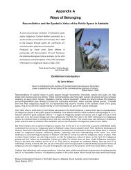
Front PieceHaplotype Block Structure <strong>in</strong> <strong>the</strong> HapMap CEU population of <strong>the</strong> novel putativeglaucoma locus on Xp25. Image based on <strong>the</strong> work of Dr Ben Fry, colours representallelic variants <strong>and</strong> <strong>the</strong> z-offset emphasises <strong>the</strong> transition between blocks (see page 197).ii
I certify that this <strong>the</strong>sis does not <strong>in</strong>corporate without acknowledgment any materialpreviously submitted for a degree or diploma <strong>in</strong> any university; <strong>and</strong> that to <strong>the</strong> best of myknowledge <strong>and</strong> belief it does not conta<strong>in</strong> any material previously published or written byano<strong>the</strong>r person except where due reference is made <strong>in</strong> <strong>the</strong> text.iii
Table of Contents:Front Piece...........................................................................................................................iiSummary .............................................................................................................................vAcknowledgments.............................................................................................................viiChapter 1 – INTRODUCTION: The significance <strong>and</strong> pathoaetiology of <strong>the</strong>glaucomas............................................................................................................................1Chapter 2 – MYOCILIN GLAUCOMA: Dissect<strong>in</strong>g <strong>the</strong> genotype-phenotypecorrelations of myocil<strong>in</strong> glaucoma. ...................................................................................34Chapter 3 – GLAUCOMA AS A SYSTEMIC DISEASE..............................................108Chapter 4 – BLINDING GLAUCOMA: Biometric associations of severe glaucoma. ..132Chapter 5 – GLAUCOMA GENETIC ASSOCIATIONS: Prob<strong>in</strong>g for geneticpredispositions <strong>in</strong> <strong>Open</strong> <strong>Angle</strong> Glaucoma.......................................................................152Chapter 6 – USING TWINS TO DISSECT THE GLAUCOMA PHENOTYPE...........207Chapter 7 – CONCLUSION : Dimensional complexities <strong>in</strong> a molecularly <strong>and</strong>phenotypically heterogeneous disease.............................................................................224Appendices ......................................................................................................................246Bibliography....................................................................................................................274iv
SummaryGlaucoma is <strong>the</strong> commonest cause for irreversible optic neuropathy worldwide.Be<strong>in</strong>g a complex heterogeneous disease, Primary <strong>Open</strong> <strong>Angle</strong> Glaucoma (OAG) islikely to manifest due to <strong>the</strong> collision of germ-l<strong>in</strong>e, somatic, environmental <strong>and</strong>stochastic factors. This <strong>the</strong>sis explores both <strong>the</strong> phenotypic features <strong>and</strong> geneticmechanisms of <strong>the</strong> glaucomatous process.Investigation of <strong>the</strong> myocil<strong>in</strong> gene, which has been unequivocally associated withOAG, demonstrated firm genotype-phenotype correlations. Possibly reflect<strong>in</strong>g <strong>the</strong>association between myocil<strong>in</strong>-related glaucoma <strong>and</strong> elevated <strong>in</strong>traocular pressure,myocil<strong>in</strong> mutation carriers were found to have a lower prevalence of optic dischaemorrhages, compared to <strong>in</strong>dividuals with non-myocil<strong>in</strong> OAG. No structuraldifferences of <strong>the</strong> optic nerve head were identified <strong>in</strong> young people known to carrymyocil<strong>in</strong> mutations, but who do not have manifest glaucoma.At <strong>the</strong> phenotypic level <strong>the</strong> role of OAG as a systemic disease <strong>and</strong> <strong>the</strong> biometricassociations of advanced OAG were <strong>in</strong>vestigated. Us<strong>in</strong>g mortality data from over27,000 people of whom 741 were known to have OAG, adjusted for gender <strong>and</strong> ageat death, we identified a statistically significant association between death due toischaemic heart disease <strong>and</strong> OAG. In a separate study <strong>in</strong>vestigat<strong>in</strong>g <strong>the</strong> systemicassociations of OAG <strong>in</strong> 1,700 patients, a past history of migra<strong>in</strong>e or presence ofa<strong>the</strong>rosclerosis was identified as be<strong>in</strong>g more common <strong>in</strong> patients with familial formsof OAG compared to people with sporadic disease. Biometric <strong>in</strong>vestigation ofpatients who had def<strong>in</strong>itive end-stage glaucomatous visual field loss, confirmed thatcentral corneal thickness was a significant risk factor for disease progression.Automatic optic disc imag<strong>in</strong>g, which was performed on a subset of this end-stagecohort, revealed that <strong>the</strong> Stratus optical coherence tomography ret<strong>in</strong>al nerve fibrev
layer clock hour scan was most sensitive <strong>in</strong> detect<strong>in</strong>g advanced disease. Thesef<strong>in</strong>d<strong>in</strong>gs may have important ramifications on phenotype-based screen<strong>in</strong>g programs.At <strong>the</strong> genotypic level, <strong>the</strong> Asp658Gly variant <strong>in</strong> <strong>the</strong> W<strong>in</strong>ged Doma<strong>in</strong> 40- repeat 36gene was found, <strong>in</strong> a relatively small case-control study, to be a neutral variant <strong>in</strong> <strong>the</strong>Australian population <strong>and</strong> meta-analysis of <strong>the</strong> common opt<strong>in</strong>eur<strong>in</strong> Met98Lys,variant confirmed that its association with OAG, although weak, is highlystatistically significant. Replicat<strong>in</strong>g previous work, two nonsynonymous variants <strong>in</strong>exon 1 of lysyl oxidase–like 1 (Arg141Leu;Gly153Asp) were found to be stronglyassociated with pseudoexfoliative glaucoma. After validat<strong>in</strong>g a novel method ofgenome-wide association us<strong>in</strong>g equimolar DNA pools, where we were easily able toidentify a strong association between markers at <strong>the</strong> complement factor H locus <strong>and</strong>age-related macular degeneration, genetic risk variants for OAG on chromosomes3q21, 6p25, 14q13 <strong>and</strong> Xq25 were found. None<strong>the</strong>less, fur<strong>the</strong>r work is requiredbefore <strong>the</strong> association of variants at <strong>the</strong> novel OAG loci are def<strong>in</strong>itively proven.Accurate phenotypic descriptions, when compiled with relevant genetic <strong>in</strong>formationshould enhance cl<strong>in</strong>icians’ underst<strong>and</strong><strong>in</strong>g of <strong>the</strong> specific natural history of an<strong>in</strong>dividual patient's disease. Ongo<strong>in</strong>g work <strong>in</strong>vestigat<strong>in</strong>g <strong>the</strong> cl<strong>in</strong>ical natural history<strong>and</strong> outcome to available <strong>the</strong>rapy is required to correlate specific disease-caus<strong>in</strong>gvariants with <strong>the</strong> phenotype, <strong>the</strong>reby bridg<strong>in</strong>g <strong>the</strong> cl<strong>in</strong>ician to <strong>the</strong> laboratory.vi
AcknowledgmentsThis body of knowledge is dedicated to Meggy who shared <strong>in</strong> <strong>the</strong> joys of discovery.Marry<strong>in</strong>g <strong>the</strong> cl<strong>in</strong>ician to <strong>the</strong> laboratory is an important endeavour, <strong>and</strong> I havecerta<strong>in</strong>ly been fortunate to partake <strong>in</strong> <strong>the</strong> transfer of cl<strong>in</strong>ical questions generated at<strong>the</strong> slitlamp to <strong>the</strong> laboratory bench. The work that contributed to this <strong>the</strong>sis hasprovided <strong>the</strong> unique prospect of cl<strong>in</strong>ically phenotyp<strong>in</strong>g patients, collect<strong>in</strong>g specimensfrom <strong>the</strong>m, work<strong>in</strong>g <strong>in</strong> <strong>the</strong> laboratory <strong>and</strong> <strong>the</strong>n return<strong>in</strong>g to <strong>the</strong> cl<strong>in</strong>ic with usefulmolecular results. I am grateful to <strong>the</strong> many patients <strong>and</strong> study participants who arerepresented <strong>in</strong> every facet of this work.It has been an immense privilege to have <strong>the</strong> opportunity to learn from Australia’sprom<strong>in</strong>ent ophthalmic geneticists. Associate Professors Jamie Craig <strong>and</strong> DavidMackey have been extremely supportive. Their refresh<strong>in</strong>g approach to cl<strong>in</strong>icalsciencediffers positively; such that <strong>the</strong>y complement each o<strong>the</strong>r well. They havecerta<strong>in</strong>ly re<strong>in</strong>forced to me that many of <strong>the</strong> most pert<strong>in</strong>ent questions relat<strong>in</strong>g to eyehealth arise <strong>in</strong> <strong>the</strong> cl<strong>in</strong>ic. Ano<strong>the</strong>r important axiom that I learnt from <strong>the</strong>m is thatcl<strong>in</strong>ical research should be focused so as to ensure a translatable outcome. Nodoctorial c<strong>and</strong>idate could seek more enthusiastic, driven or scientifically astutesupervisors. I look forward to ongo<strong>in</strong>g work with <strong>the</strong>m.In today’s medical research environment, more than ever, significant contributionsfur<strong>the</strong>r<strong>in</strong>g <strong>the</strong> underst<strong>and</strong><strong>in</strong>g <strong>in</strong>to any discipl<strong>in</strong>e are be<strong>in</strong>g facilitated by coord<strong>in</strong>atedteamwork. The time of solo authored scientific works present<strong>in</strong>g major f<strong>in</strong>d<strong>in</strong>gs hasprobably passed (see Nature. 2007 450:1165). Research <strong>and</strong> travel <strong>in</strong> different areasof medical-science ensured that many people contributed to this treatise. To reflectvii
this, I endeavoured <strong>in</strong> <strong>the</strong> body of this <strong>the</strong>sis to use <strong>the</strong> plural first person pronoun“we” ra<strong>the</strong>r than <strong>the</strong> s<strong>in</strong>gular form.My <strong>in</strong>itial <strong>in</strong>terest <strong>in</strong> glaucoma was spawned by Dr Richard Cooper, who is certa<strong>in</strong>lyone of <strong>the</strong> most astute cl<strong>in</strong>icians I have met. For example, two years prior to <strong>the</strong>publication of Estermann <strong>and</strong> colleagues (J Ocul Pharmacol Ther. 2006; 22:62-67),Richard mentioned his observation that donepezil lowers <strong>in</strong>traocular pressure. Be<strong>in</strong>ga modern day Priestly Smith, Richard’s openness about <strong>the</strong> complexity of <strong>the</strong>glaucomas is humbl<strong>in</strong>g.Over <strong>the</strong> course of this doctorial c<strong>and</strong>idature I have had <strong>the</strong> opportunity to work <strong>in</strong>many cl<strong>in</strong>ical <strong>and</strong> research Departments. I am <strong>in</strong>debted to many people at <strong>the</strong>Cl<strong>in</strong>ical Genetics Unit at <strong>the</strong> Centre for Eye Research Australia, University ofMelbourne <strong>and</strong> <strong>the</strong> Royal Victorian Eye <strong>and</strong> Ear Hospital where I spent my first yearof study, <strong>in</strong> particular Lisa Kearns, as well as Drs Sonya Bennett, Johan Poulson, <strong>and</strong>Jon Ruddle. Maree R<strong>in</strong>g from <strong>the</strong> Department of Ophthalmology at <strong>the</strong> University ofTasmania performed much of <strong>the</strong> background genealogy <strong>in</strong>cluded <strong>in</strong> Chapter 2 <strong>and</strong>Chapter 3. I am also very appreciative of <strong>the</strong> constructive, grammatical commentsprovided by Lori Bonertz on <strong>the</strong> manuscripts aris<strong>in</strong>g from this <strong>the</strong>sis. Manyadditional people contributed over <strong>the</strong> previous decade to <strong>the</strong> phenotyp<strong>in</strong>g <strong>and</strong>recruitment of participants for <strong>the</strong> Glaucoma Inheritance Study <strong>in</strong> Tasmania <strong>and</strong> <strong>the</strong>Tw<strong>in</strong>s Eye Study <strong>in</strong> Tasmania. In particular I am grateful to Drs Ca<strong>the</strong>r<strong>in</strong>e Green <strong>and</strong>Johnny Wu.I am also grateful for <strong>the</strong> support provided by many people from <strong>the</strong> Department ofOphthalmology at Fl<strong>in</strong>ders University <strong>and</strong> Fl<strong>in</strong>ders Medical Centre, where my f<strong>in</strong>alviii
years of study were spent, specifically Tania Straga, David Dimasi, Amy McMellon,Tor<strong>in</strong> Clack, Sarah Sibson <strong>and</strong> Drs Richard Mills, L<strong>in</strong>gjun Ma, Shiwani Sharma <strong>and</strong>Kathryn Burdon. The organisational <strong>and</strong> adm<strong>in</strong>istrative support provided by DebSullivan, Joyce Moore, Lyn Hard<strong>in</strong>g, Sue Harris as well as by Professors KerynWilliams <strong>and</strong> Douglas Coster has also been <strong>in</strong>valuable. It has been <strong>in</strong>spir<strong>in</strong>g to workalong side one of Australia’s foremost cl<strong>in</strong>ical photographers, Angela Chappell, whophotographed many of <strong>the</strong> patients which contributed to <strong>the</strong> studies outl<strong>in</strong>ed <strong>in</strong>Chapter 4.I am <strong>in</strong> awe of <strong>the</strong> quality of work produced by <strong>the</strong> Genetic Epidemiology Unit of <strong>the</strong>Queensl<strong>and</strong> Institute of Medical Research (QIMR) headed by Professor NicholasMart<strong>in</strong>. The opportunity to learn from this team, specifically Megan Campbell,Anjali Henders <strong>and</strong> Drs Grant Montgomery, <strong>and</strong> Gu Zhu, was <strong>in</strong>valuable. I am also<strong>in</strong>debted to Dr Stuart Macgregor <strong>and</strong> Professor Peter Visscher who analysed <strong>the</strong> datagenerated for <strong>the</strong> genome wide association described <strong>in</strong> Chapter 5.The Blue Mounta<strong>in</strong>s Eye Study (BMES) is cemented <strong>in</strong> its reputation as one ofAustralia’s greatest contributions to ophthalmic research. I am particularly thankfulto Professor Paul Mitchell <strong>and</strong> Associate Professor Jie J<strong>in</strong> Wang who allowed accessto BMES samples used <strong>in</strong> Chapter 5 as well as <strong>the</strong> protocol sheets which formed <strong>the</strong>basis for <strong>the</strong> questionnaire adm<strong>in</strong>istered <strong>in</strong> Chapter 3.Dr Tim Chataway <strong>and</strong> Amy McCormick from <strong>the</strong> Department of Human Physiologyat Fl<strong>in</strong>ders University <strong>in</strong>troduced me to <strong>the</strong> evolv<strong>in</strong>g world of proteomics. TarunKakaday assisted with f<strong>in</strong>ite element modell<strong>in</strong>g of proteomic biomarkers forglaucoma.ix
Be<strong>in</strong>g <strong>in</strong>volved <strong>in</strong> <strong>the</strong> establishment of <strong>the</strong> Myocil<strong>in</strong> gene screen<strong>in</strong>g service possiblyrepresents <strong>the</strong> most positive, cl<strong>in</strong>ically relevant outcome undertaken dur<strong>in</strong>g my PhDc<strong>and</strong>idature. As such, I am particularly appreciative for <strong>the</strong> work undertaken byAssociate Professor Pamela Sykes <strong>and</strong> Drs Scott Grist <strong>and</strong> Andrew Dubowsky from<strong>the</strong> Department of Genetic Pathology at Fl<strong>in</strong>ders Medical Centre <strong>in</strong> facilitat<strong>in</strong>g thisservice.Dr Pat Toohey was fundamental <strong>in</strong> <strong>the</strong> website development, which was <strong>the</strong> primarytranslation of <strong>the</strong> research described <strong>in</strong> Chapter 2. The haplotype analysis ofThr377Met myocil<strong>in</strong> families, presented <strong>in</strong> Chapter 2, was k<strong>in</strong>dly performed <strong>in</strong> <strong>the</strong>laboratory of Associate Professor Mary Wirtz. Paul Sanfilippo assisted with much of<strong>the</strong> disease cod<strong>in</strong>g as described <strong>in</strong> Chapter 3. Dr John L<strong>in</strong>acre <strong>and</strong> AssociateProfessor Konrad Pesudovs provided assistance with <strong>the</strong> WINSTEPS programm<strong>in</strong>gutilized <strong>in</strong> Chapter 6.Much of <strong>the</strong> content of <strong>the</strong> last section of Chapter 6 arose from discussions withnumerous ophthalmic experts <strong>in</strong>clud<strong>in</strong>g: Drs Wido Budde; John F<strong>in</strong>gert; Paul Foster;David Garway-Heath; Ca<strong>the</strong>r<strong>in</strong>e Green; Christopher Hammond; William Morgan;<strong>and</strong> Professors Wallace Alward; Sohan Hayreh; Jost Jonas; Paul Kaufman; NeilMiller; Nancy Newman; Harry Quigley; John Samples; <strong>and</strong> George Spaeth. It wasthoroughly enjoyable to openly discuss broad topics, rang<strong>in</strong>g for example from <strong>the</strong>architecture of <strong>the</strong> optic nerve head or means to develop a unify<strong>in</strong>g <strong>the</strong>ory forglaucomas, to <strong>the</strong> possible demise of Tasmanian Gondwanian forests. I also enjoyedimmensely <strong>the</strong> discussion with Professor Don Melrose from <strong>the</strong> Department ofx
Theoretical Physics at <strong>the</strong> University of Sydney regard<strong>in</strong>g <strong>the</strong> dimensionality ofbiological systems <strong>in</strong>troduced briefly <strong>in</strong> <strong>the</strong> conclud<strong>in</strong>g chapter.Fund<strong>in</strong>g for this work has been obta<strong>in</strong>ed from many sources <strong>and</strong> it has certa<strong>in</strong>ly beena privilege to be paid for undertak<strong>in</strong>g such an enjoyable hobby. There can be nobetter community endorsement for research than <strong>the</strong> provision of funds to junior<strong>in</strong>vestigators. F<strong>in</strong>ancially I have been supported by a Medical PostgraduateScholarship from <strong>the</strong> National Health <strong>and</strong> Medical Research Council (NHMRC) <strong>and</strong>a travel award from <strong>the</strong> NHMRC allowed me to undertake <strong>the</strong> laboratory work at <strong>the</strong>QIMR which contributed predom<strong>in</strong>ately to Chapter 5. The Australia-Ch<strong>in</strong>a SpecialFund<strong>in</strong>g scheme of <strong>the</strong> International Science L<strong>in</strong>kages programme allowed travel <strong>the</strong>Zhongshan Ophthalmic Centre, Guangzhou, People’s Republic of Ch<strong>in</strong>a, to visit <strong>the</strong>excit<strong>in</strong>g Ophthalmic tw<strong>in</strong> project established by Associate Professor M<strong>in</strong>gguang He.Travel fellowships from <strong>the</strong> Association for Research <strong>in</strong> Vision <strong>and</strong> Ophthalmology(ARVO) <strong>and</strong> <strong>the</strong> S<strong>in</strong>gapore Eye Research Institute/ARVO permitted some of <strong>the</strong>results from this <strong>the</strong>sis to be presented. The majority of this work has also beensusta<strong>in</strong>ed by project grants from <strong>the</strong> NHMRC, <strong>the</strong> Ophthalmic Research Institute ofAustralia, <strong>the</strong> American Health Assistance Foundation as well as an NHMRCEnabl<strong>in</strong>g Grant.xi
Manuscripts published dur<strong>in</strong>g <strong>the</strong> Doctoral c<strong>and</strong>idature (those marked with anasterisk contributed directly to this <strong>the</strong>sis):1. Dimasi DP, Hewitt AW, Green CM, Mackey DA, Craig JE. Lack of associationof p53 polymorphisms <strong>and</strong> haplotypes <strong>in</strong> high <strong>and</strong> normal tension open angleglaucoma. Journal of Medical Genetics. 2005; 42:e55.2. Toh TY, Liew SHM, MacK<strong>in</strong>non JR, Hewitt AW, Poulsen JL, Spector TD,Gilbert CE, Hammond CJ, Mackey DA. High heritability of Central CornealThickness: The Tw<strong>in</strong> Eye Study. Investigative Ophthalmic Visual Sciences. 2005;46: 3718-3722.3. Hewitt AW, <strong>and</strong> Burdon KP. The relative contribution of <strong>the</strong> X chromosome to<strong>the</strong> eye. Ophthalmic Genetics. 2005; 26: 191-193.4. Hewitt AW, Jeganathan VS, Kidd J, Pesudovs K, Verma N. Is visual functionpreserved <strong>in</strong> patients receiv<strong>in</strong>g photodynamic <strong>the</strong>rapy for age related maculardegeneration? Graefes Archives of Cl<strong>in</strong>ical <strong>and</strong> Experimental Ophthalmology. 2006;244: 972-977.5. Hewitt AW <strong>and</strong> Cooper RL. Glaucoma nosology. Cl<strong>in</strong>ical & ExperimentalOphthalmology. 2006; 34 (1): 94.6. Hewitt AW, Bennett SL, Dimasi DP, Craig JE, Mackey DA. A Myocil<strong>in</strong>Gln368Stop homozygote does not exhibit a more severe glaucoma phenotype thanheterozygous cases. American Journal of Ophthalmology. 2006; 141 (2): 402-403.*7. Hewitt AW, Traill A, Cooper RL, Morgan JE, Mackey DA. Tools for opticcup:disc measurement. Cl<strong>in</strong>ical & Experimental Ophthalmology. 2006; 34 (2): 288-289.8. Wu J, Hewitt AW, Green CM, R<strong>in</strong>g MA, McCartney PJ, Craig JE, Mackey DA.Disease severity of familial glaucoma compared with sporadic glaucoma. Archivesof Ophthalmology, 2006; 124: 950-954.9. Hewitt AW, Craig JE, Mackey DA. Complex genetics of complex traits: <strong>the</strong> caseof primary open-angle glaucoma. Cl<strong>in</strong>ical <strong>and</strong> Experimental Ophthalmology. 2006;34(4): 472-484.*10. Hewitt AW, Dimasi DP, Mackey DA, Craig JE. The WDR-36 D658G mutationis not a glaucoma-caus<strong>in</strong>g variant <strong>in</strong> Australia. American Journal of Ophthalmology.2006; 142: 324-325.*11. Craig JE, Hewitt AW, Dimasi DP, Toomes C, Howel N, Cohn AC, Mackey DA.The role of <strong>the</strong> Opt<strong>in</strong>eur<strong>in</strong> Met98Lys variant <strong>in</strong> hereditary optic neuropathies. BritishJournal of Ophthalmology. 2006; 90:1420-1424.*12. Hewitt AW, MacK<strong>in</strong>non JR, Elder JE, Giubilato A, Craig JE, Mackey DA. Riskof familial transmission of <strong>in</strong>fantile glaucoma <strong>in</strong> Australia. Ophthalmic Genetics.2006; 27(3) 93-97.13. Hewitt AW, Bennett SL, F<strong>in</strong>gert JH, Cooper RL, Stone EM, Craig JE, MackeyDA. The optic nerve head <strong>in</strong> Myocil<strong>in</strong> glaucoma. Investigative Ophthalmic VisualSciences. 2007; 48: 238-243.*14. Hewitt AW, Bennett SL, Richards JE, Booth AP, Inglehearn C, Rashida A,Stone EM, Craig JE, Mackey DA. Myocil<strong>in</strong> Gly252Arg mutation <strong>and</strong> glaucoma of<strong>in</strong>termediate severity <strong>in</strong> Caucasians. Archives of Ophthalmology. 2007; 125:98-104.*xii
15. Bennett SL, Hewitt AW, Poulsen JL, Kearns LS, Morgan JE, Craig JE, MackeyDA. Screen<strong>in</strong>g for glaucomatous disc changes prior to diagnosis of glaucoma <strong>in</strong>myocil<strong>in</strong> pedigrees. Archives of Ophthalmology. 2007; 125: 112-116.*16. Cohn AC, Toomes C, Potter C, Towns KV, Hewitt AW, Inglehearn CF, CraigJE, Mackey DA. Autosomal Dom<strong>in</strong>ant Optic Atrophy: a review of penetrance <strong>and</strong>expressivity <strong>in</strong> patients with OPA1 mutations. American Journal of Ophthalmology.2007; 143: 656-662.17. Hewitt AW, Gaudette D, All<strong>in</strong>gham RR, Järvelä I, Kitsos G, Krishnadas SR,Petersen MB, Richards JE, Sundaresan P, Wiggs JL, Mackey DA, Wirtz MK.Investigation for founder effects of <strong>the</strong> Thr377Met Myocil<strong>in</strong> mutation <strong>in</strong> glaucomafamilies from differ<strong>in</strong>g ethnic backgrounds. <strong>Molecular</strong> Vision. 2007; 13: 487-492.*18. Hewitt AW, Poulsen JP, Alward WL, Bennett SL, Budde WM, Cooper RL,Craig JE, F<strong>in</strong>gert JH, Foster PJ, Garway-Heath DG, Green CM, Hammond CJ,Hayreh SS, Jonas JB, Kaufman PL, Miller N, Morgan WH, Newman NJ, QuigleyHA, Samples JR, Spaeth GL, Pesudovs K, Mackey DA. Heritable features of <strong>the</strong>optic nerve head: A novel tw<strong>in</strong> method for determ<strong>in</strong><strong>in</strong>g genetic significance.Investigative Ophthalmic Visual Sciences. 2007; 48: 2469-2475.*19. Dimasi DP, Hewitt AW, Straga T, Pater J, MacK<strong>in</strong>non JR, Elder JE, Casey T,Mackey DA, Craig JE. The prevalence of CYP1B1 mutations <strong>in</strong> Australian patientswith primary congenital glaucoma. Cl<strong>in</strong>ical Genetics. 2007; 72: 255-260.20. Hewitt AW, Kearns LS, Jamieson R, Williamson K, van Heyn<strong>in</strong>gen V, MackeyDA. PAX6 mutations may be association with high myopia. Ophthalmic Genetics.2007; 28: 179-182.21. Hewitt AW. Genetic diseases of <strong>the</strong> optic nerve head: from embryogenesis topathogenesis. Expert Review <strong>in</strong> Ophthalmology. 2007; 2 (5): 769-777.22. Hewitt AW, Mackey DA, Craig JE. Myocil<strong>in</strong> allele-specific glaucomaphenotype database. Human Mutation. 2008; 29: 207-211.*23. Zhu G, Hewitt AW, Ruddle JB, Kearns LS, Brown SA, MacK<strong>in</strong>non JR, CraigJE, Hammond CJ, Mart<strong>in</strong> NG, Mackey DA. Genetic dissection of myopia: Evidencefor l<strong>in</strong>kage of ocular axial length to chromosome 5q. Ophthalmology. 2008; 115:1053-1057.24. Hewitt AW†, Sharma S†, Burdon KP, Wang JJ, Dimasi DP, Mackey DA,Mitchell P, Craig JE. Ancestral LOXL1 variants are associated withpseudoexfoliation <strong>in</strong> Caucasian Australians but with markedly lower penetrance than<strong>in</strong> Nordic people. Human <strong>Molecular</strong> Genetics. 2008; 17: 710-716. Onl<strong>in</strong>e Nov 232007,† Equal contribution by authors.*25. Green CM, Kearns LS, Wu J, Barbour JM, Wilk<strong>in</strong>son RM, R<strong>in</strong>g MA, Craig JE,Wong TL, Hewitt AW, Mackey DA. How significant is a family history ofglaucoma? Experience from <strong>the</strong> Glaucoma Inheritance Study <strong>in</strong> Tasmania (GIST).Cl<strong>in</strong>ical <strong>and</strong> Experimental Ophthalmology. 2007; 35: 739-799.26. Hewitt AW, Sanfilippo P, R<strong>in</strong>g MA, Craig JE, Mackey DA. Mortality <strong>in</strong>Primary <strong>Open</strong> <strong>Angle</strong> Glaucoma: Two cupped discs <strong>and</strong> funeral. In Press Eye.*27. Sherw<strong>in</strong> J, Hewitt AW, Ruddle JB, Mackey DA. The role of genetic isolates <strong>in</strong>Ophthalmic disease. Ophthalmic Genetics. 2008; 29: 149-161.28. Mackey DA, Green CM, Craig JE, Hewitt AW. The pathogenesis of <strong>the</strong>glaucomas: Nature versus nature. Cl<strong>in</strong>ical <strong>and</strong> Experimental Ophthalmology. 2008;36: 297.xiii
Chapter 1 – INTRODUCTION: The significance <strong>and</strong>pathoaetiology of <strong>the</strong> glaucomas.The glaucomas are <strong>the</strong> pr<strong>in</strong>cipal cause for optic nerve degeneration <strong>and</strong> one of <strong>the</strong>lead<strong>in</strong>g causes for irreversible bl<strong>in</strong>dness worldwide (Quigley 1996; Resnikoff et al.,2004). They are a heterogeneous group of disorders, of which primary open-angleglaucoma (OAG) is <strong>the</strong> most common subset. Although <strong>the</strong> def<strong>in</strong>ition of OAG hasnot been consistent across studies, it is generally referred to as a progressiveexcavation of <strong>the</strong> optic disc with correspond<strong>in</strong>g loss of visual field (Foster et al.,2002). OAG is often, but not <strong>in</strong>variably, associated with an elevated <strong>in</strong>traocularpressure (IOP) (Hollows <strong>and</strong> Graham, 1966). In over 20% of cases, IOP elevation isabsent <strong>and</strong> a diagnosis of normal tension glaucoma (NTG) can be made (Hollows<strong>and</strong> Graham, 1966; Kamal <strong>and</strong> Hitch<strong>in</strong>gs, 1998). Although it may be erroneous tosub-classify OAG as high-tension glaucoma (HTG) on <strong>the</strong> basis of an IOP greaterthan 21mmHg, <strong>the</strong>re is evidence that even <strong>in</strong> NTG <strong>the</strong>rapeutic lower<strong>in</strong>g of IOP mayslow fur<strong>the</strong>r loss of visual field (van der Valk et al., 2005). Fur<strong>the</strong>r to this, both <strong>the</strong>NTG <strong>and</strong> HTG groups could be fur<strong>the</strong>r subcategorized (e.g. a NTG cohort maycomprise people with pr<strong>in</strong>cipally vascular risk factors for OAG, neurodegenerativeOAG, or misclassified HTG, i.e. people with erroneously low applanation IOPread<strong>in</strong>gs). It is difficult to diagnose OAG <strong>in</strong> <strong>the</strong> early stages because of <strong>the</strong> subtletyof its cl<strong>in</strong>ical features (Figure 1-1).1
Figure 1-1Spectrum of glaucomatous disease. (A) Optic nerve photography: small central cup<strong>in</strong> healthy eye; enlargement of cup <strong>and</strong> loss of neuroret<strong>in</strong>al rim <strong>in</strong> glaucomatous eye.(B) Confocal scann<strong>in</strong>g laser ophthalmoscopy: neuroret<strong>in</strong>al rim area with<strong>in</strong> normallimits (ticks) <strong>in</strong> healthy eyes, but reduced <strong>in</strong> glaucomatous eyes (crosses). (C) Opticalcoherence tomography: cross section of optic disc display<strong>in</strong>g deep large cup <strong>in</strong>glaucomatous eye with th<strong>in</strong> neuroret<strong>in</strong>al rim (red). (D) Optical coherencetomography: ret<strong>in</strong>al nerve fibre layer thickness <strong>in</strong> each sector is normal (black l<strong>in</strong>ewith<strong>in</strong> green region) <strong>in</strong> healthy eyes, yet is markedly reduced <strong>in</strong> advanced glaucoma(black l<strong>in</strong>e dipp<strong>in</strong>g <strong>in</strong>to red region). (E) St<strong>and</strong>ard automated perimetry: normal bl<strong>in</strong>dspot <strong>in</strong> healthy eyes, with progressive loss of sight <strong>in</strong> advanc<strong>in</strong>g glaucoma severity.2
Large population-based epidemiological studies have revealed that <strong>the</strong> prevalence ofOAG <strong>in</strong> Australia is between 2.3 <strong>and</strong> 4.4 % <strong>in</strong> people aged greater than 49 years(Mitchell et al., 1996; Wensor et al., 1998). Def<strong>in</strong>ite OAG <strong>in</strong> Caucasians aged morethan 40 years, has an overall 5-year <strong>in</strong>cidence between 0.5% <strong>and</strong> 0.62%, with <strong>the</strong><strong>in</strong>cidence <strong>in</strong>creas<strong>in</strong>g with age (de Voogd et al., 2005; Mukesh et al., 2002).Alarm<strong>in</strong>gly, <strong>in</strong> <strong>the</strong> general community more than half of <strong>the</strong> people with OAGrema<strong>in</strong> undiagnosed – a statistic that has not improved over <strong>the</strong> past 40 years,provid<strong>in</strong>g fur<strong>the</strong>r support for <strong>the</strong> notion that current screen<strong>in</strong>g algorithms are fail<strong>in</strong>g(Hollows <strong>and</strong> Graham, 1966; Mitchell et al., 1996; Wensor et al., 1998).To date, ophthalmic-based screen<strong>in</strong>g systems for OAG have specifically<strong>in</strong>corporated assessment of <strong>the</strong> optic disc, IOP measurement <strong>and</strong> <strong>in</strong>vestigation forvisual field deficit. Given <strong>the</strong> high likelihood of miss<strong>in</strong>g <strong>in</strong>cident disease <strong>and</strong> <strong>the</strong> factthat many people are repeatedly reviewed unnecessarily, such methods are not costeffectivefor a community (Tuck <strong>and</strong> Crick, 1997). Strategies to elim<strong>in</strong>ate <strong>the</strong>bl<strong>in</strong>d<strong>in</strong>g toll of glaucoma must be aimed at identify<strong>in</strong>g at-risk <strong>in</strong>dividuals. Thecorollary of this is that a screen<strong>in</strong>g regimen must be highly sensitive <strong>and</strong> specific soas to only detect potentially serious disease not pseudo-disease (Harris 2005).Because OAG is <strong>in</strong>itially asymptomatic, effective screen<strong>in</strong>g techniques shouldidentify people with no obvious signs or symptoms of <strong>the</strong> disease, allow<strong>in</strong>g earlydiagnosis <strong>and</strong> management.Glaucoma is a model disease for evaluation of genetic screen<strong>in</strong>g <strong>in</strong> a complexdisease. The evidence for success of OAG treatment, <strong>the</strong> ma<strong>in</strong>stay of which is IOPreduction, is exp<strong>and</strong><strong>in</strong>g (van der Valk et al., 2005). Increased cl<strong>in</strong>ical screen<strong>in</strong>g ofgenetically at-risk <strong>in</strong>dividuals would allow early <strong>the</strong>rapeutic <strong>in</strong>tervention prior to <strong>the</strong>3
loss of visual function. Despite OAG be<strong>in</strong>g identified as a cl<strong>in</strong>ical entity almost assoon as <strong>the</strong> ophthalmoscope was developed, <strong>the</strong> precise pathogenesis rema<strong>in</strong>s elusive(von Graefe 1857). Our current underst<strong>and</strong><strong>in</strong>g of <strong>the</strong> disease mechanisms at <strong>the</strong>molecular level is relatively poor.The field of genetics is pivotal <strong>in</strong> underst<strong>and</strong><strong>in</strong>g underly<strong>in</strong>g molecular mechanisms<strong>and</strong> pathways. The advances <strong>in</strong> methods for genetic screen<strong>in</strong>g cont<strong>in</strong>ually add to <strong>the</strong>cl<strong>in</strong>ician’s diagnostic armoury. It must be recognized that some <strong>in</strong>dividuals have amisplaced fear that draconian <strong>in</strong>tervention is required when a genetic predispositionis recognized however; this is generally not <strong>the</strong> case. For example, <strong>the</strong> identificationof <strong>the</strong> genetic predisposition to phenylketonuria has allowed thous<strong>and</strong>s of at-risk<strong>in</strong>dividuals to avoid dietary stressors, with a subsequence avoidance of mentalretardation (Lenke <strong>and</strong> Levy, 1980).This chapter will summarise <strong>the</strong> current underst<strong>and</strong><strong>in</strong>g of <strong>the</strong> genetics of OAG,clearly del<strong>in</strong>eat<strong>in</strong>g it as a complex trait, <strong>and</strong> <strong>the</strong>n briefly explore potential avenuesfor future breakthroughs. This review will not discuss developmental or congenitalglaucoma for which much genetic progress has been made (Mackey <strong>and</strong> Craig, 2003;Sarfarazi et al., 2003).Current underst<strong>and</strong><strong>in</strong>gs of <strong>the</strong> genetics of OAG:Prior to embark<strong>in</strong>g on a full-scale probe for <strong>the</strong> genes <strong>in</strong>volved <strong>in</strong> OAG, it isnecessary to first consider <strong>the</strong> evidence support<strong>in</strong>g <strong>the</strong> fact that glaucoma is a“genetic disease.” Over <strong>the</strong> past century <strong>the</strong>re has been a paradigm shift <strong>in</strong> <strong>the</strong>underst<strong>and</strong><strong>in</strong>g of <strong>the</strong> <strong>in</strong>heritance of OAG. In 1927 it was stated that “cases of4
hereditary glaucoma, though by no means unknown, are yet relatively rare”(James1927). This example was followed by <strong>the</strong> 1932 publication of Julia Bell’s Treasuryof Human Inheritance, which conta<strong>in</strong>s a large section on <strong>the</strong> <strong>in</strong>heritance of glaucoma(Bell 1932). In it she noted that “… certa<strong>in</strong>ly relatively few good pedigrees of <strong>the</strong>condition have ever been published.”Family history has now been revealed to be one of <strong>the</strong> most important risk factors forOAG development (Tielsch et al., 1994). The Glaucoma Inheritance Study <strong>in</strong>Tasmania (GIST) found a positive family history is found <strong>in</strong> over 50% of cases ofglaucoma (Green et al., 2007). Fur<strong>the</strong>rmore, <strong>the</strong> screen<strong>in</strong>g of relatives has beenproven to be a successful strategy for OAG case detection (Miller <strong>and</strong> Paterson,1962; Vernon 1991). Investigators from <strong>the</strong> Rotterdam Eye Study <strong>in</strong>vestigated <strong>the</strong>familial aggregation of OAG by exam<strong>in</strong><strong>in</strong>g not only first-degree relatives ofglaucoma cases identified through <strong>the</strong>ir prevalence study but also a matched set ofcontrols (Wolfs et al., 1998). Wolfs <strong>and</strong> colleagues found that first-degree relativesof OAG patients had a 22% risk of develop<strong>in</strong>g glaucoma <strong>in</strong> comparison to 2.3% <strong>in</strong><strong>the</strong> relatives of controls, imply<strong>in</strong>g a 10 fold <strong>in</strong>creased relative risk of <strong>the</strong> disease <strong>in</strong>first degree relatives of affected patients compared with <strong>the</strong> general population(Wolfs et al., 1998). Although this study was rigorously conducted it could, however,underestimate <strong>the</strong> genetic component of glaucoma, especially if <strong>the</strong> children ofglaucoma cases were too young to manifest <strong>the</strong> disease. There is often a poorknowledge of glaucoma family history (McNaught et al., 2000). While it is clear thatmany diseases have a tendency to run <strong>in</strong> families, it may be difficult to dissect outwhe<strong>the</strong>r this is due to familial shar<strong>in</strong>g of a similar environment, or to similarity <strong>in</strong>genetic predisposition. For example, it has been shown that attend<strong>in</strong>g medical school5
aggregates <strong>in</strong> families, as probably does a preference for eat<strong>in</strong>g vegemite on toast(McGuff<strong>in</strong> <strong>and</strong> Huckle, 1990).Racial differences <strong>in</strong> prevalence of OAG exist. The prevalence <strong>in</strong> Africans isestimated to be six times as high, <strong>in</strong> certa<strong>in</strong> age groups, as that <strong>in</strong> Caucasians(Buhrmann et al., 2000; Ntim-Amponsah et al., 2004; Racette et al., 2003). Thef<strong>in</strong>d<strong>in</strong>g of a similar greater prevalence <strong>in</strong> Africans <strong>and</strong> African-Americans lessens <strong>the</strong>likelihood that such differences are primarily due to external societal or environmentspecificconfounders (Tielsch et al., 1991). The differ<strong>in</strong>g genetic composition ofAfrican-Americans compared to Caucasian-Americans may account for <strong>the</strong>difference <strong>in</strong> OAG prevalence. Interest<strong>in</strong>gly, OAG is thought to be extremely rare <strong>in</strong>Australian Aborig<strong>in</strong>als (Hollows 1980; Mann 1966).Fur<strong>the</strong>r evidence for a genetic basis of OAG stems from tw<strong>in</strong>s studies. Theophthalmic literature is peppered by case descriptions of identical tw<strong>in</strong>s concordantfor OAG <strong>and</strong> NTG (Gedda et al., 1970; Ofner <strong>and</strong> Samples, 1992; Teikari et al.,1987). In a large series by Gottfredsdottir <strong>and</strong> colleagues, OAG was found to besignificantly more concordant <strong>in</strong> monozygotic tw<strong>in</strong> pairs (98.0%) than <strong>the</strong>ir spouses(70.2%)(Gottfredsdottir et al., 1999).A fundamental genetic paradigm for OAG is also supplemented by <strong>the</strong> fact that somenon-human animal species also develop heritable forms of OAG (Gelatt et al.,1998a). Inherited spontaneous OAG has been identified <strong>in</strong> rhesus monkeys (Macacamulatta) <strong>and</strong> both autosomal recessive <strong>and</strong> dom<strong>in</strong>ant OAG is present <strong>in</strong> dog breeds(<strong>in</strong> particular <strong>the</strong> beagle <strong>and</strong> m<strong>in</strong>iature poodle)(Gelatt et al., 1998a).6
In <strong>the</strong> majority of OAG cases it is likely that more than one genetic predisposition isrequired to manifest disease, <strong>and</strong> it is generally well accepted now that OAG is acomplex trait. S<strong>in</strong>ce <strong>the</strong> first description of a heritable form of OAG by Benedict <strong>in</strong>1842, a number of genetic loci have been reported <strong>and</strong> a smaller number of geneshave been implicated or identified (Table 1-1) (Benedict 1842).Table 1-1Identified primary open-angle glaucoma loci.Loci OMIM Gene Location Initial l<strong>in</strong>kage / gene identify<strong>in</strong>g studyTypicalPhenotypeGLC1A 601652 myocil<strong>in</strong> 1q23-25 (Sheffield et al., 1993; Stone et al., 1997) JOAG / HTGGLC1B 606689 2cen-q13 (Stoilova et al., 1996) NTG / HTGGLC1C 601682 3q21-24 (Wirtz et al., 1997) HTGGLC1D 602429 8q23 (Trifan et al., 1998) NTG / HTGGLC1E 602432 opt<strong>in</strong>eur<strong>in</strong> 10p15-14 (Rezaie et al., 2002; Sarfarazi et al., 1998) NTGGLC1F 603383 7q35-q36 (Wirtz et al., 1999) HTGGLC1G 609669 WDR-36 5q21-35* (Monemi et al., 2005; Samples et al., 2004) NTG / HTGGLC1H 611276 2p16.3-p15 (Suriyapperuma et al., 2007) NSGLC1I 609745 15q11-13 (All<strong>in</strong>gham et al., 2005b) NTG / HTGGLC1J 608695 9q22 (Wiggs et al., 2004) JOAGGLC1K 608696 20p12 (Wiggs et al., 2004) JOAGGLC1L 137750 3p22-p21 (Baird et al., 2005a) HTGGLC1M 610535 5q22.1-q32 (Pang et al., 2006) JOAGGLC1N 611274 15q22-q24 (Wang et al., 2006) JOAG* Locus may conta<strong>in</strong> more than 1 glaucoma associated geneAbbreviations: HTG, high-tension glaucoma; NTG, normal tension glaucoma;JOAG, juvenile onset glaucoma; NS, not specified.7
Myocil<strong>in</strong> Glaucoma:The 1997 discovery of <strong>the</strong> myocil<strong>in</strong> gene (MYOC) has significantly impacted uponmany OAG families (Stone et al., 1997). The MYOC gene (formerly referred to as<strong>the</strong> trabecular meshwork-<strong>in</strong>duced glucocorticoid response prote<strong>in</strong> or TIGR) wasmapped to 1q where <strong>the</strong> locus for <strong>the</strong> juvenile form of OAG had previously beenidentified (GLC1A)(Sheffield et al., 1993; Stone et al., 1997).MYOC encodes a predicted 504 am<strong>in</strong>o acid polypeptide <strong>and</strong> conta<strong>in</strong>s two majordoma<strong>in</strong>s, an N-term<strong>in</strong>al myos<strong>in</strong>-like doma<strong>in</strong> <strong>and</strong> a C-term<strong>in</strong>al olfactomed<strong>in</strong>-likedoma<strong>in</strong>.(Green <strong>and</strong> Kle<strong>in</strong>, 2002) The encod<strong>in</strong>g region is divided <strong>in</strong>to three exons, ofwhich <strong>the</strong> majority of <strong>the</strong> disease-caus<strong>in</strong>g variations are clustered <strong>in</strong> <strong>the</strong> olfactomed<strong>in</strong>homology doma<strong>in</strong> of <strong>the</strong> third exon. The structure of <strong>the</strong> MYOC prote<strong>in</strong> has beenwell conserved through evolution (Mukhopadhyay et al., 2002).Although MYOC is found ubiquitously <strong>in</strong> <strong>the</strong> eye, it is also expressed <strong>in</strong> manyextraocular tissues, suggest<strong>in</strong>g that it may not have an eye-specific function (F<strong>in</strong>gertet al., 2002; Karali et al., 2000). However, it is <strong>in</strong> <strong>the</strong> trabecular meshwork (TM)where <strong>the</strong> primary consequences of MYOC dysfunction are found (Jacobson et al.,2001). In <strong>the</strong> TM, MYOC has been revealed to pr<strong>in</strong>cipally <strong>in</strong>teract with optimed<strong>in</strong>,an olfactomed<strong>in</strong>-related prote<strong>in</strong> (Torrado et al., 2002), as well as b<strong>in</strong>d<strong>in</strong>g with flot<strong>in</strong>-1, a lipid raft prote<strong>in</strong> (Joe et al., 2005). Genes <strong>in</strong>teract<strong>in</strong>g with MYOC are potentiallygood c<strong>and</strong>idate genes for future OAG <strong>in</strong>vestigation or <strong>the</strong>rapeutic <strong>in</strong>tervention.Despite numerous descriptions of nonsense <strong>and</strong> premature term<strong>in</strong>ation mutations,haplo<strong>in</strong>sufficiency of <strong>the</strong> MYOC prote<strong>in</strong> appears unlikely to be <strong>the</strong> primary diseasecaus<strong>in</strong>gmechanism (Wiggs <strong>and</strong> Vollrath, 2001). Cell expression studies compar<strong>in</strong>g8
mutant to normal MYOC secretion levels suggest that OAG develops ei<strong>the</strong>r becauseof <strong>in</strong>sufficient or compromised MYOC secretion from TM cells due to congestion of<strong>the</strong> TM secretory pathway (Jacobson et al., 2001). The work of Liu <strong>and</strong> Vollrath,which demonstrated that mutant forms of <strong>the</strong> MYOC prote<strong>in</strong> are misfolded <strong>and</strong>aggregate <strong>in</strong> <strong>the</strong> endoplasmic reticulum, also provided weight to a ga<strong>in</strong>–of–functiondisease model (Liu <strong>and</strong> Vollrath, 2004). A model of disease causation pr<strong>in</strong>cipallythrough reduced Triton solubility of MYOC was fur<strong>the</strong>r supported by <strong>the</strong> f<strong>in</strong>d<strong>in</strong>g thatglaucoma was not <strong>in</strong>duced through genetically <strong>in</strong>creas<strong>in</strong>g or decreas<strong>in</strong>g normalMYOC expression (Gould et al., 2004). Interest<strong>in</strong>gly, people who are homozygousfor MYOC mutations do not seem to manifest severe disease <strong>in</strong>dicat<strong>in</strong>g a novel modeof <strong>in</strong>heritance (Hewitt et al., 2006a; Morissette et al., 1998).Substantial evidence now exists to suggest that approximately one <strong>in</strong> 30 unselectedOAG patients has a MYOC mutation (F<strong>in</strong>gert et al., 1999). To date more than 40disease-associated mutations <strong>in</strong> MYOC have been identified (F<strong>in</strong>gert et al., 2002),with <strong>the</strong> Gln368STOP mutation <strong>the</strong> most common <strong>in</strong>dividual glaucoma caus<strong>in</strong>gvariant worldwide (F<strong>in</strong>gert et al., 1999). It has been revealed that <strong>the</strong> majority ofpatients with this specific mutation have descended from a s<strong>in</strong>gle ancestorharbour<strong>in</strong>g <strong>the</strong> MYOC Gln368STOP (Baird et al., 2003; Faucher et al., 2002). Thesecond most common MYOC mutation identified <strong>in</strong> Australia, which is also foundworldwide, is <strong>the</strong> Thr377Met mutation (F<strong>in</strong>gert et al., 1999; Mackey et al., 2003).The cl<strong>in</strong>ical pattern of MYOC glaucoma reflects <strong>the</strong> specific underly<strong>in</strong>g mutation.MYOC is traditionally thought of as hav<strong>in</strong>g a HTG phenotype, with some mutations(such as Pro370Leu) caus<strong>in</strong>g severe juvenile-onset OAG (Alward et al., 1998). Thedistribution of age <strong>and</strong> maximum recorded IOP for Australian patients with both <strong>the</strong>9
Gln368STOP <strong>and</strong> Thr377Met MYOC mutations is similar to descriptions of o<strong>the</strong>rpedigrees with <strong>the</strong>se mutations (All<strong>in</strong>gham et al., 1998; Alward et al., 1998; Craig etal., 2001; Graul et al., 2002; Mackey et al., 2003; Puska et al., 2005; Shimizu et al.,2000). A stepwise decrease <strong>in</strong> <strong>the</strong> mean age at diagnosis across Australian patientswith <strong>the</strong> Gln368STOP, Thr377Met <strong>and</strong> Pro370Leu MYOC mutations is mirrored by areciprocal <strong>in</strong>crease <strong>in</strong> maximum recorded IOP (Figure 1-2)( Hewitt et al. 2006b). It isclear that <strong>the</strong> specific MYOC mutation can be <strong>in</strong>ferred by <strong>in</strong>dividual cl<strong>in</strong>ical features(genotype-phenotype correlation).Currently it is not cost-effective to conduct population-based screen<strong>in</strong>g for MYOCmutations (Aldred et al., 2004). However, <strong>the</strong> efficacy for genetic screen<strong>in</strong>g will<strong>in</strong>crease when conduct<strong>in</strong>g comprehensive comb<strong>in</strong>ed screen of many OAG genes <strong>and</strong>as <strong>the</strong> cost of genetic tests decreases markedly due to technological advances. An <strong>in</strong>depth<strong>in</strong>vestigation of <strong>the</strong> genotypic <strong>and</strong> phenotypic association of MYOC-relatedglaucoma is undertaken <strong>in</strong> Chapter 2.10
Figure 1-2The stepwise decrease <strong>in</strong> mean age (A) at diagnosis across Australian patients with<strong>the</strong> Gln368STOP, Thr377Met <strong>and</strong> Pro370Leu Myocil<strong>in</strong> mutations, with a reciprocal<strong>in</strong>crease <strong>in</strong> maximum recorded <strong>in</strong>traocular pressure (B) <strong>and</strong> proportion requir<strong>in</strong>gfilter<strong>in</strong>g surgery (C) (from Hewitt et al. 2006b).11
Opt<strong>in</strong>eur<strong>in</strong> Glaucoma:The second OAG gene identified was <strong>the</strong> opt<strong>in</strong>eur<strong>in</strong> (OPTN) gene at <strong>the</strong> GLC1Elocus (Rezaie et al., 2002). The GLC1E locus was <strong>in</strong>itially mapped from a largeBritish pedigree with autosomal dom<strong>in</strong>ant NTG (Sarfarazi et al., 1998). Referr<strong>in</strong>g to“optic neuropathy-<strong>in</strong>duc<strong>in</strong>g,” OPTN is located on <strong>the</strong> short arm of chromosome 10<strong>and</strong> encodes a 147 am<strong>in</strong>o acid polypeptide (Rezaie et al., 2002). OPTN has a pivotalrole <strong>in</strong> exocytosis as well as Golgi ribbon formation <strong>and</strong> is potentially <strong>in</strong>volved with<strong>the</strong> FAS-lig<strong>and</strong> as well as <strong>the</strong> tumour necrosis factor-α (TNF-α) apoptotic pathways(Sahlender et al., 2005; Sarfarazi <strong>and</strong> Rezaie, 2003). Although OPTN has beendemonstrated to be up-regulated after exposure to TNF-α <strong>and</strong> dexamethasone(Vittitow <strong>and</strong> Borras, 2002), its response to elevated IOP rema<strong>in</strong>s controversial(Kamphuis <strong>and</strong> Schneemann, 2003; Vittitow <strong>and</strong> Borras, 2002).Mutations <strong>in</strong> OPTN account for approximately 16.7% of familial OAG from an NTG<strong>in</strong>dex case, however are only likely to constituted approximately 0.1% of unselectedOAG cases (Alward et al., 2003; Aung et al., 2003; Rezaie et al., 2002; Wiggs et al.,2003). The most common OPTN disease-caus<strong>in</strong>g variant is Glu50Lys (Rezaie et al.,2002). Individuals with this mutation develop aggressive NTG <strong>and</strong> have a lower ageat diagnosis (mean ± SD: 40.8 ± 11.0 years) <strong>and</strong> greater need for trabeculectomycompared to o<strong>the</strong>r non-OPTN NTG cases (Aung et al., 2005). The cl<strong>in</strong>icalimportance of many o<strong>the</strong>r OPTN variants (<strong>in</strong> particular Met98Lys) rema<strong>in</strong>controversial (Alward et al., 2003; Aung et al., 2003; Fuse et al., 2004; Jansson et al.,2005; Leung et al., 2003; Rezaie et al., 2002; Tang et al., 2003; Umeda et al., 2004;Weisschuh et al., 2005; Wiggs et al., 2003; Willoughby et al., 2004). In a Japanesecohort Funayama <strong>and</strong> colleagues found that OAG patients were more likely thancontrol subjects to have both <strong>the</strong> TNF-α/-863A change with <strong>the</strong> OPTN Met98Lys12
variant (Funayama et al., 2004). In support of a NTG-modify<strong>in</strong>g variant, Melki et al.reported that <strong>the</strong> Met98Lys substitution may be associated with a lower IOP at <strong>the</strong>time of diagnosis <strong>and</strong> may even modify MYOC glaucoma (Melki et al., 2003a).Additional Glaucoma Genes:Investigat<strong>in</strong>g <strong>the</strong> genetics of pedigrees with diseases with late age of onset isdifficult. The parents of OAG cases are often deceased, whilst <strong>the</strong> patients’ childrenare frequently too young to manifest disease. Confound<strong>in</strong>g this fur<strong>the</strong>r is <strong>the</strong> fact thatOAG can be discordant <strong>in</strong> time, differ<strong>in</strong>g <strong>in</strong> age of onset for some related cases, <strong>and</strong><strong>the</strong>re is often also considerable overlap between glaucoma families (Sack et al.,1996). Despite <strong>the</strong>se issues, large pedigrees have been genetically l<strong>in</strong>ked <strong>and</strong>numerous loci have been identified (Table 1-1), although not all have been replicated<strong>in</strong> later studies.Dur<strong>in</strong>g <strong>the</strong> first month of my doctorial c<strong>and</strong>idature, evidence was providedimplicat<strong>in</strong>g <strong>the</strong> WD repeat-conta<strong>in</strong><strong>in</strong>g prote<strong>in</strong> 36 (WDR36) gene <strong>in</strong> caus<strong>in</strong>g OAG(Monemi et al., 2005). To date this f<strong>in</strong>d<strong>in</strong>g has not been fully replicated <strong>in</strong> <strong>the</strong>published literature; however, one prelim<strong>in</strong>ary study has not supported this f<strong>in</strong>d<strong>in</strong>g(All<strong>in</strong>gham et al., 2005a). In addition, <strong>the</strong> orig<strong>in</strong>al family that provided <strong>the</strong> <strong>in</strong>itial <strong>and</strong>only evidence of l<strong>in</strong>kage to <strong>the</strong> GLC1G locus has not been found to conta<strong>in</strong> cod<strong>in</strong>gregion mutation <strong>in</strong> WDR-36 segregat<strong>in</strong>g with <strong>the</strong> disease phenotype (Kramer et al.,2006). Fur<strong>the</strong>r discussion <strong>and</strong> <strong>in</strong>vestigation of <strong>the</strong> WDR36 gene is performed <strong>in</strong> <strong>the</strong>first section of Chapter 5. Researchers must be cautious about herald<strong>in</strong>g noveldisease-caus<strong>in</strong>g genes until such time as confirmatory replicate studies are reported.13
A number of OAG loci have cytogenetic support <strong>in</strong> <strong>the</strong> published literature. Cases ofcongenital glaucoma due to cytogenetic derangement at <strong>the</strong> GLC1B (Mu et al.,1984), GLC1C (Allderdice et al., 1975; Kondo et al., 1979), GLC1D (Cohn et al.,2005), <strong>and</strong> GLC1F loci have been described (Kato et al., 2001; Speleman et al.,2000). It is certa<strong>in</strong>ly possible that mildly deleterious mutations cause OAG, whilstmore significant rearrangement of <strong>the</strong>se underly<strong>in</strong>g genes cause a markedly moresevere disease phenotype (such as congenital onset glaucoma). It is also <strong>in</strong>terest<strong>in</strong>gthat <strong>the</strong> GLC1F locus is <strong>in</strong> close proximity to (but does not seem to overlap) a locusfor Pigment Dispersion Syndrome (GPDS1)(Andersen et al., 1997; Anderson et al.,2002).In 2000, Wiggs <strong>and</strong> colleagues reported a genome-wide scan for OAG us<strong>in</strong>g a sibpair multipo<strong>in</strong>t analysis (Wiggs et al., 2000). This study identified suggestive l<strong>in</strong>kageto a region near <strong>the</strong> GLC1B locus on chromosome 2 <strong>and</strong> at loci on chromosomes 14,17 <strong>and</strong> 19 (Wiggs et al., 2000). Follow<strong>in</strong>g this, a genome-wide scan of OAG familiesof African descent highlighted causative gene regions on chromosomes 2q <strong>and</strong> 10p(Nemesure et al., 2003). The 10p locus implicated by this study did not <strong>in</strong>clude <strong>the</strong>OPTN gene (Nemesure et al., 2003). Recently, a novel OAG locus on <strong>the</strong> short armof chromosome 3 was proposed, us<strong>in</strong>g a genome-wide scan, as dom<strong>in</strong>antly<strong>in</strong>dependently segregat<strong>in</strong>g <strong>in</strong> a large Australian family with some affected memberscarry<strong>in</strong>g <strong>the</strong> Gln368STOP MYOC mutation (Baird et al., 2005a).It is noteworthy that many of <strong>the</strong> implicated OAG loci have been identified from <strong>the</strong>same cl<strong>in</strong>ic base (e.g. GLC1C, GLC1F, GLC1G)(Samples et al., 2004; Wirtz et al.,1997; Wirtz et al., 1999). The upshot of such a f<strong>in</strong>d<strong>in</strong>g is that ei<strong>the</strong>r few research14
groups have <strong>the</strong> facilities for genomic work or that fur<strong>the</strong>r <strong>in</strong>formative familiesrema<strong>in</strong> to be identified <strong>in</strong> o<strong>the</strong>r geographic regions.OAG <strong>and</strong> Genetic Association studies:Numerous genetic association studies for OAG have been conducted. Many of <strong>the</strong>sestudies have had conflict<strong>in</strong>g results or have not been replicated. When review<strong>in</strong>g thisever <strong>in</strong>creas<strong>in</strong>g list of gene alleles studied <strong>in</strong> OAG (Table 1-2), it is important to notethat such tabulation of this data only facilitates crude comparison. These studiesoften <strong>in</strong>clude different racial groups or subtypes of OAG (e.g. HTG versus NTG) <strong>and</strong>some suffer from <strong>in</strong>adequate power<strong>in</strong>g or poorly characterized <strong>and</strong> matched controlgroups. Frequently, different alleles with<strong>in</strong> <strong>the</strong> same gene have been studied, mak<strong>in</strong>gdirect comparison of <strong>the</strong> literature difficult. Should a specific haplotype be revealedto be associated with <strong>the</strong> disease it is also important to consider that this f<strong>in</strong>d<strong>in</strong>g mayrepresent a type one error. Alternatively it may have occurred as <strong>the</strong> result of l<strong>in</strong>kagedisequilibrium (LD) or <strong>the</strong> <strong>in</strong>fluence of neighbour<strong>in</strong>g genes. LD is <strong>the</strong> tendency ofalleles to be <strong>in</strong>herited toge<strong>the</strong>r, ra<strong>the</strong>r than would be expected given <strong>the</strong>ir knownfrequency <strong>in</strong> a population <strong>and</strong> <strong>the</strong> recomb<strong>in</strong>ation fraction between <strong>the</strong> loci. Forconciseness, studies <strong>in</strong>vestigat<strong>in</strong>g <strong>the</strong> association between various blood groups <strong>and</strong>OAG have been omitted from Table 1-2. Failure to replicate a genetic associationmay occur due to locus or allele heterogeneity, as well as under-power<strong>in</strong>g a study toaccount for LD.15
Table 1-2Conflict<strong>in</strong>g evidence for gene-disease <strong>in</strong>teraction <strong>in</strong> primary open-angle glaucoma.Gene / AlleleGenBankAccessionNo.LocationGSTM1 NM_000561 1p13Positive / Support<strong>in</strong>gStudies(Juronen et al., 2000; Yildirim etal., 2005)Negative / Non-replicat<strong>in</strong>gStudies(Jansson et al., 2003)MTHFR NM_005957 1p36 (Junemann et al., 2005) -MYOC.mt1 NM_000261 1q24(Alward et al., 2002; Fan et al., 2004;(Colomb et al., 2001; PolanskyOzgul et al., 2005; Sjostr<strong>and</strong> et al.,et al., 2003)2002)REN NM_000537 1q32 - (Hashizume et al., 2005)AGT NM_000029 1q42 - (Hashizume et al., 2005)ACP1 NM_177554 2p25 (Abecia et al., 1996) -AGTR1 NM_000685 3q21 - (Hashizume et al., 2005)TF AH010951 3q21 - (Abecia et al., 1996)OPA1 NM_015560 3q28(Aung et al., 2002b; Aung et al.,2002a; Powell et al., 2003)(Woo et al., 2004)B2AR NM_000024 5q32 - (Gungor et al., 2003)GLO1 NM_006708 6p21 - (Abecia et al., 1996)CDKN1A NM_000389 6p21 (Tsai et al., 2004) -TAP1/2 NM_000593 6p21 (L<strong>in</strong> et al., 2004) -TNFα NM_000594 6p21(Funayama et al., 2004; L<strong>in</strong> etal., 2003)-EDN1 NM_001955 6p24 - (Logan et al., 2005)NOS3 NM_000603 7q36 (Logan et al., 2005) (L<strong>in</strong> et al., 2005)IGF2 NM_000612 11p15 (Tsai et al., 2003) -GSTP1 NM_000852 11q13 -(Juronen et al., 2000; Yildirim et al.,2005)CMA1 NM_001836 14q11 - (Hashizume et al., 2005)TP53 NM_000546 17p13(L<strong>in</strong> et al., 2002; Ress<strong>in</strong>iotis et (Acharya et al., 2002; Dimasi et al.,al., 2004a)2005)ACE NM_000789 17q23 -(Bunce et al., 2005; Hashizume et al.,2005; Ozkur et al., 2004)MPO NM_000250 17q23 - (L<strong>in</strong> et al., 2005)APOE NM_000041 19q13(Cop<strong>in</strong> et al., 2002; Fan et al.,2005; Junemann et al., 2004;Mabuchi et al., 2005; Vickers etal., 2002)(Ress<strong>in</strong>iotis et al., 2004c; Ress<strong>in</strong>iotis etal., 2004b)(Juronen et al., 2000; Yildirim et al.,2005)GSTT1 NM_000853 22q11 -AGTR2 NM_000686 Xq22 (Hashizume et al., 2005) -16
If it is challeng<strong>in</strong>g enough to replicate causative disease loci <strong>in</strong> OAG, it seems moredifficult to replicate a positive f<strong>in</strong>d<strong>in</strong>g for a predispos<strong>in</strong>g genetic risk allele. A part ofthis problem is that molecular pathways have been used to work backwards to agenetic predisposition. As <strong>in</strong> <strong>the</strong> case of nitric oxide synthase 3 (NOS3), differentexpression patterns were found <strong>in</strong> <strong>the</strong> aqueous humour of glaucomatous patientscompared to matched patients (L<strong>in</strong> et al., 2005; Logan et al., 2005). However, whennucleotide polymorphisms that had been proven to be functionally important <strong>in</strong> <strong>the</strong>NOS3 gene were <strong>in</strong>vestigated, conflict<strong>in</strong>g results were obta<strong>in</strong>ed (L<strong>in</strong> et al., 2005;Logan et al., 2005). Such negative associations may reflect <strong>the</strong> fact that <strong>the</strong> <strong>in</strong>itialhypo<strong>the</strong>sis was based on a substance important <strong>in</strong> <strong>the</strong> down-stream pathogeneticpathway. The premise that because ret<strong>in</strong>al ganglion cell death <strong>in</strong> glaucoma occursthrough apoptosis, any pro-apoptotic allele <strong>in</strong> <strong>the</strong> respective cascade should be foundmore commonly <strong>in</strong> OAG cases whilst not unreasonable, may not prove to be <strong>the</strong>case.Animal models for OAG have also identified genes <strong>in</strong>volved <strong>in</strong> glaucoma <strong>and</strong>susceptibility to optic neurodegeneration. Through a series of back <strong>and</strong> <strong>in</strong>tercrosses,mutations <strong>in</strong> <strong>the</strong> glycoprote<strong>in</strong> NMB (GPNMB) gene on <strong>the</strong> telomeric region of <strong>the</strong>long arm of chromosome 7 were found to cause pigmentary glaucoma <strong>in</strong> DBA/2Jmice (Anderson et al., 2002). Follow-up studies <strong>in</strong> this same animal glaucoma modelhave found that deficiency of <strong>the</strong> pro-apoptotic BCL2 associated X prote<strong>in</strong> gene slowret<strong>in</strong>al ganglion cell death <strong>and</strong> that neurodegeneration can be prevented by high-doseradiation with bone marrow transfer (Anderson et al., 2005; Libby et al., 2005).Pathogenetic pathways that may be <strong>in</strong>tr<strong>in</strong>sically <strong>in</strong>volved <strong>in</strong> animal models need to17
e <strong>in</strong>vestigated <strong>in</strong> human cohorts prior to advocat<strong>in</strong>g <strong>the</strong>ir adoption <strong>in</strong> populationbasedscreen<strong>in</strong>g platforms or targeted <strong>the</strong>rapy.The genetics of complex traits: Is glaucoma lagg<strong>in</strong>g?Complex disorders lack a simple Mendelian mode of <strong>in</strong>heritance, <strong>and</strong> <strong>the</strong>refore as<strong>in</strong>gle underly<strong>in</strong>g susceptibility gene cannot be assumed. Disease expression <strong>in</strong> OAGcases most likely <strong>in</strong>volves more than one gene, of which some may display<strong>in</strong>complete penetrance or variable expressivity. Never<strong>the</strong>less, <strong>the</strong> ‘holy grail’ ofgenetic research <strong>in</strong>to complex traits, is <strong>the</strong> identification a s<strong>in</strong>gle locus of large effect.The human genome conta<strong>in</strong>s approximately three billion nucleotides <strong>and</strong> close to30,000 genes (International Human Genome Sequenc<strong>in</strong>g Consortium 2004).However, caution must be ascribed when review<strong>in</strong>g this figure. Analogous toMat<strong>the</strong>w Fl<strong>in</strong>ders (1774-1814) produc<strong>in</strong>g his “General chart of Terra Australis orAustralia” <strong>in</strong> 1804, we now appreciate that this first mapp<strong>in</strong>g neglected much of ourcoastl<strong>in</strong>e, <strong>in</strong>clud<strong>in</strong>g many bays <strong>and</strong> <strong>in</strong>lets. Similarly some 200 years later, despite <strong>the</strong>‘complete genome mapp<strong>in</strong>g’, many functionally significant regions are stillunknown.In <strong>the</strong> human genome, sequence variation <strong>in</strong>clude: s<strong>in</strong>gle nucleotide polymorphisms(SNPs); <strong>in</strong>sertions or deletions of a few nucleotides; <strong>and</strong> variation <strong>in</strong> <strong>the</strong> repeatnumber of a motif (micro-satellites) (Nowotny et al., 2001). Previous familial <strong>and</strong>sib-pair l<strong>in</strong>kage studies have relied pr<strong>in</strong>cipally on micro-satellite markers. However,SNPs are more abundant <strong>and</strong> densely distributed than micro-satellites, occurr<strong>in</strong>gapproximately every 1,000 basepairs along <strong>the</strong> human genome, <strong>and</strong> thus mak<strong>in</strong>g<strong>the</strong>m more suited to high-resolution genotyp<strong>in</strong>g (Nowotny et al., 2001). SNPs can18
occur <strong>in</strong> gene cod<strong>in</strong>g regions as well as <strong>the</strong> <strong>in</strong>terven<strong>in</strong>g regions (<strong>in</strong>trons). To dateconsiderable disease-gene research has focused on <strong>the</strong> cod<strong>in</strong>g (exons) <strong>and</strong>, to a lesserextent, <strong>the</strong> promoter regions of genes. However, recent evidence suggests that<strong>in</strong>tronic regions are subject to stronger selective constra<strong>in</strong>t <strong>and</strong> thus, may befunctionally more important than previously presumed (Andolfatto 2005). Obscur<strong>in</strong>g<strong>the</strong> clarity of underst<strong>and</strong><strong>in</strong>g <strong>in</strong> gene function fur<strong>the</strong>r, is <strong>the</strong> fact that remarkably fewgenes are found <strong>in</strong> <strong>the</strong> human genome compared to o<strong>the</strong>r species (InternationalHuman Genome Sequenc<strong>in</strong>g Consortium 2004). It is now clear that many genes canproduce more than one prote<strong>in</strong> (through alternate splic<strong>in</strong>g) <strong>and</strong> that different prote<strong>in</strong>saris<strong>in</strong>g from <strong>the</strong> same gene can have dramatically different functional roles (Zhang etal., 2005).The allelic architecture for almost all common diseases is still be<strong>in</strong>g uncovered.Generally, <strong>the</strong> disease-caus<strong>in</strong>g variants (or mutations) <strong>in</strong> <strong>the</strong> population can vary <strong>in</strong>prevalence (be<strong>in</strong>g ei<strong>the</strong>r rare or common) whilst <strong>the</strong> effect exerted by <strong>the</strong> specificallele can also differ <strong>in</strong> magnitude (small to large effect) (Figure 1-3). Given <strong>the</strong>paucity of gene identification <strong>in</strong> complex traits, it is clear that common genes of largeeffect are extraord<strong>in</strong>ary <strong>and</strong> do not account for such diseases (Chakravarti 1999).Consider<strong>in</strong>g extreme cases, if rare alleles account for <strong>the</strong> prevalence of commondisease it is likely that affected <strong>in</strong>dividuals have mutations at only one of manypossible disease loci (as may be <strong>the</strong> case for cardiovascular disorders (Williams etal., 2004)). Conversely if <strong>the</strong> alleles are common <strong>in</strong> <strong>the</strong> population, <strong>the</strong>n people withdisease have mutations at multiple loci simultaneously (as may occur <strong>in</strong> colorectaladenomas (Fearnhead et al., 2004)). Given that rare Mendelian diseases have manyrare variants, it is reasonable to postulate that common diseases may also have manyrare variants. If multiple common genes are <strong>in</strong>volved <strong>in</strong> a common complex disease,19
it is crucial to determ<strong>in</strong>e whe<strong>the</strong>r a sole mutation at any particular gene is sufficient<strong>and</strong> necessary to cause disease (Chakravarti 1999). Given this possibility, dismissalof any gene proposed to be associated with OAG is difficult.Figure 1-3Allelic architecture of genetic diseases.20
If strong epistasis prevails, a mutation may be necessary for a particular phenotype(Chakravarti 1999). Epistasis classically assumes that genes do not act alone, butra<strong>the</strong>r that particular genotypes or environmental factors formulate gene expression.However, gene-gene <strong>in</strong>teractions are likely to be multifaceted such that despite onebeneficial gene-gene allele be<strong>in</strong>g present, disease may manifest by a separate gene<strong>in</strong>teraction that is ‘endorsed’ by a separate detrimental allele. Stochasticenvironmental factors are also likely to have a marked <strong>in</strong>fluence <strong>in</strong> diseaseexpression. Post-translational modification is <strong>the</strong> proteolytic cleavage follow<strong>in</strong>gDNA replication. Many prote<strong>in</strong>s are syn<strong>the</strong>sized as <strong>in</strong>active precursors that areactivated under physiological conditions by limited proteolysis (such asmethylation). Although gene-environment <strong>in</strong>teractions can be modelled, <strong>the</strong>detection of precise environmental stressors (especially <strong>in</strong> OAG) has been difficult(Potter 2001). It is clearly a formidable task to cleanly dissect <strong>the</strong> underly<strong>in</strong>gmechanisms for many complex diseases, <strong>in</strong> which <strong>the</strong>re are likely to be severalgenetic <strong>and</strong> environmental factors <strong>in</strong>volved <strong>in</strong> <strong>the</strong> pathophysiology.The Bottleneck of Glaucoma Genetics:Many potential avenues exist for untw<strong>in</strong><strong>in</strong>g <strong>the</strong> complex genetics of OAG. It is likelythat adopt<strong>in</strong>g a comb<strong>in</strong>ation of approaches <strong>and</strong> pursu<strong>in</strong>g many genetic methods willallow <strong>the</strong> bottleneck of glaucoma genetics to be broken.Search<strong>in</strong>g for fur<strong>the</strong>r pedigrees around <strong>the</strong> world has had limited success. OAG geneidentification by conventional l<strong>in</strong>kage analysis has been greatly complicated byphenocopy or <strong>in</strong>tra-pedigree genetic heterogeneity (Craig et al., 2001; Sack et al.,1996). These issues of phenocopy, where a disease <strong>in</strong> separate patients seems21
cl<strong>in</strong>ically identical, yet is found to have a different aetiology; <strong>and</strong> variableexpressivity, where <strong>the</strong> same gene mutation causes a variety of phenotypic effects, isnot unique to glaucoma, let alone <strong>the</strong> eye. For example prior to recent molecularwork, fungi were pr<strong>in</strong>cipally characterised by <strong>the</strong>ir morphological appearance.However, an <strong>in</strong>creased underst<strong>and</strong><strong>in</strong>g of <strong>the</strong>ir underly<strong>in</strong>g gene sequences hasrevealed that despite some fungal species be<strong>in</strong>g genetically similar <strong>the</strong>y haveremarkably different structures (analogous to phenotypic heterogeneity or variableexpressivity). Conversely some taxa which have similar appearance are actuallygenetically very different (analogous to genotypic heterogeneity) (Figure 1-4).Compound<strong>in</strong>g <strong>the</strong> issues of phenocopy <strong>and</strong> variable expressivity is <strong>the</strong> <strong>in</strong>itialobscurity of cl<strong>in</strong>ical diagnosis <strong>in</strong> early OAG. The determ<strong>in</strong>ation of l<strong>in</strong>kage ispr<strong>in</strong>cipally a statistical process <strong>and</strong> uncerta<strong>in</strong>ties <strong>in</strong>troduced about cl<strong>in</strong>ical statussignificantly reduce <strong>the</strong> power of any such studies. Despite this, pedigree l<strong>in</strong>kagestudies have good power for detect<strong>in</strong>g uncommon genes of major effect (as was <strong>the</strong>case for MYOC <strong>and</strong> OPTN glaucoma).22
Figure 1-4Phenocopy <strong>and</strong> phenotypic heterogeneity (variable expressivity) between asexualfungus species. In this phylogenetic tree, which was generated us<strong>in</strong>g <strong>the</strong> CLUSTALW program <strong>and</strong> data on <strong>the</strong> 28S ribosomal RNA gene from <strong>the</strong> NCBI taxonomydatabase, it is clear that several species of <strong>the</strong> Sporidesmium genus arephylogenetically distant (e.g. S. australiens <strong>and</strong> S. tropicale), despite be<strong>in</strong>gmorphologically similar (phenocopy). Conversely, o<strong>the</strong>r taxa (e.g. Venturiahanl<strong>in</strong>iana <strong>and</strong> Repetophragma goidanichii) are morphologically different, yet aresimilar genetically (represent<strong>in</strong>g phenotypic heterogeneity or variable expressivity).In this neighbour-jo<strong>in</strong><strong>in</strong>g plot, l<strong>in</strong>e distances represent <strong>the</strong> relative degree ofsimilarity, with <strong>the</strong> shorter <strong>the</strong> l<strong>in</strong>e be<strong>in</strong>g more similar. Based on <strong>the</strong> work of Shenoyet al. (Shenoy et al., 2006).23
Previous l<strong>in</strong>kage of OAG pedigrees <strong>and</strong> sibl<strong>in</strong>g pairs us<strong>in</strong>g micro-satellite markersmay have had a high false negative result, be<strong>in</strong>g <strong>in</strong>sufficient to detect true loci. Withimproved knowledge of <strong>the</strong> SNP variation across <strong>the</strong> genome, it may be beneficial tore<strong>in</strong>vestigate <strong>the</strong>se pedigrees us<strong>in</strong>g SNP-based platforms. The high SNP densityallows loci to be def<strong>in</strong>ed more precisely (John et al., 2004). A major technicalobstacle for genome-wide SNP <strong>in</strong>vestigations, high through-put diploid PCRamplification, has recently been overcome through a highly multiplexed microarraygenotyp<strong>in</strong>g system (Syvanen 2005). None<strong>the</strong>less, <strong>the</strong> difficulty <strong>in</strong> selection of mostadvantageous markers <strong>and</strong> <strong>the</strong> high cost are drawbacks. Additionally <strong>in</strong> any SNPselection it is important to know <strong>the</strong> allele frequencies <strong>in</strong> <strong>the</strong> population, someth<strong>in</strong>gwhich <strong>the</strong> HapMap project has addressed (Altshuler et al., 2005). L<strong>in</strong>kage studiesprovide a partial scaffold for association studies. Through identify<strong>in</strong>g chromosomalregions of <strong>in</strong>terest, l<strong>in</strong>kage studies substantially reduce <strong>the</strong> resources required forgene-mapp<strong>in</strong>g association.Whilst <strong>the</strong> high age at functional impairment or manifestation of OAG <strong>in</strong>creases <strong>the</strong>difficulty <strong>in</strong> perform<strong>in</strong>g a pedigree l<strong>in</strong>kage analysis, it may improve <strong>the</strong> power of anassociation study (through conserved LD regions). From <strong>the</strong> evolutionary viewpo<strong>in</strong>t,diseases with a late onset of functional impairment may not have conferred anegative selection pressure. Thus, founder haplotypes, as has been found <strong>in</strong> MYOC,may exist (Baird et al., 2003). Recomb<strong>in</strong>ation hotspots are widespread <strong>and</strong> accountfor LD structure across <strong>the</strong> genome. The efficacy of f<strong>in</strong>e mapp<strong>in</strong>g can be improvedthrough adopt<strong>in</strong>g a tagged-SNP approach (Figure 1-5). In underst<strong>and</strong><strong>in</strong>g <strong>the</strong> majorityof common variations <strong>in</strong> <strong>the</strong> genome, choos<strong>in</strong>g non-redundant sets of SNPs (i.e.SNPs not <strong>in</strong> LD) offers considerable efficiency without loss of study power. In this24
way genomic regions can be tested for association without requir<strong>in</strong>g <strong>the</strong> discovery of<strong>the</strong> exact functional variant (Mach<strong>in</strong>i et al., 2005).Figure 1-5To identify which variants or SNPs (top of figure) a person carries, one can ei<strong>the</strong>rgenotype all <strong>the</strong> SNPs (i.e. 1 through to 5), or alternatively tagg<strong>in</strong>g SNPs can beselected. Here it can be seen that every person who has an “A” nucleotide at SNP 1,also has a “G” <strong>and</strong> “A” nucleotide at SNP positions 2 <strong>and</strong> 4 respectively. Hence,<strong>the</strong>se SNPs are <strong>in</strong> complete l<strong>in</strong>kage disequilibrium. Fur<strong>the</strong>r to this, knowledge ofSNP 1 <strong>and</strong> 3 allows <strong>the</strong> nucleotide at SNP 5 to be determ<strong>in</strong>ed. Hence, for Person 5 apicture of <strong>the</strong>ir genomic sequence at this locus can be identified through onlygenotyp<strong>in</strong>g SNPs 1 <strong>and</strong> 3.SNP-based strategies for complex disease have had recent success. Identification of as<strong>in</strong>gle risk allele for age-related macular degeneration (AMD) <strong>in</strong> <strong>the</strong> complementfactor H gene was achieved through a focused f<strong>in</strong>e SNP mapp<strong>in</strong>g (Edwards et al.,2005; Kle<strong>in</strong> et al., 2005). Given that subjects with severe disease were <strong>in</strong>cluded <strong>in</strong><strong>the</strong>se case-control association studies, <strong>the</strong>reby <strong>in</strong>creas<strong>in</strong>g <strong>the</strong> power to detect an25
important allele, it is very likely that <strong>the</strong> prevalence of that allele <strong>in</strong> unselected AMDhas been overestimated.The <strong>in</strong>tegration of expression <strong>and</strong> o<strong>the</strong>r functional data with genetic associationstudies greatly enhances <strong>the</strong> power of such <strong>in</strong>vestigations. The identification ofsuitable c<strong>and</strong>idate genes can be performed by gene expression <strong>and</strong> <strong>in</strong>teractionthrough gene array experiments. For example MYOC was <strong>in</strong>itially implicated throughTM dexamethasone <strong>in</strong>duction studies (Stone et al., 1997). As discussed by Stone,focused direct sequenc<strong>in</strong>g of <strong>the</strong> functionally important regions of bio<strong>in</strong>fomaticallydeterm<strong>in</strong>ed c<strong>and</strong>idate genes should <strong>in</strong>crease <strong>the</strong> yield for identify<strong>in</strong>g fundamentaldisease alleles (Stone 2003).As discussed above, chromosomal breakpo<strong>in</strong>ts at GLC1 (OAG) loci have beenimplicated <strong>in</strong> severe glaucoma pathogenesis. Given that <strong>the</strong>re is a proven track recordof ocular disease gene identification (e.g. PAX6, PITX2 <strong>and</strong> FOXC1) throughchromosomal <strong>in</strong>vestigations fur<strong>the</strong>r review of such methods is warranted <strong>in</strong> OAG(Cohn et al., 2005). However, it must be acknowledged that chromosomalrearrangement may not account for <strong>the</strong> haphazard segregation of complex diseases.Break<strong>in</strong>g down or “splitt<strong>in</strong>g” <strong>the</strong> OAG phenotype <strong>in</strong>to its constitutional anatomicalor pathophysiological components is ano<strong>the</strong>r means for progress. Much evidenceexists for “success of method” <strong>in</strong> us<strong>in</strong>g a SNP-based approached for identify<strong>in</strong>gimportant quantitative trait loci (QTL) (Hugot et al., 2001). As an aside, maximumvalue from <strong>the</strong> genotype data is obta<strong>in</strong>ed through ensur<strong>in</strong>g that a QTL approach canalso be comb<strong>in</strong>ed with or performed follow<strong>in</strong>g an association study. The study of<strong>in</strong>termediate phenotypes can be more powerful than simply ascerta<strong>in</strong><strong>in</strong>g whe<strong>the</strong>r26
disease is present or absent. Such a method for progress has been implemented forIOP <strong>and</strong> cup-to-disc ratios (Charlesworth et al., 2005).Risk <strong>in</strong>dicators of OAG correlate highly <strong>in</strong> families. The Beaver Dam Eye Study(BDES) found that optic nerve parameters (pr<strong>in</strong>cipally vertical cup-to-disc diameterratios) <strong>and</strong> IOP are more strongly correlated <strong>in</strong> sibl<strong>in</strong>gs than <strong>in</strong> cous<strong>in</strong>s (Kle<strong>in</strong> et al.,2004). Us<strong>in</strong>g a comm<strong>in</strong>gl<strong>in</strong>g analysis on data from <strong>the</strong> Blue Mounta<strong>in</strong>s Eye Study itwas suggested that a major gene accounts for approximately 18% of <strong>the</strong> variance ofIOP (Viswanathan et al., 2004). A genome-wide sib-pair l<strong>in</strong>kage study by <strong>the</strong> BDES<strong>in</strong>vestigators found two potential l<strong>in</strong>kage regions on chromosomes 6 <strong>and</strong> 13 (Duggalet al., 2005). Us<strong>in</strong>g a micro-satellite genome-wide multipo<strong>in</strong>t variance-componentsl<strong>in</strong>kage analysis <strong>in</strong> a well <strong>in</strong>vestigated Australian MYOC pedigree, Charlesworth etal. recently revealed significant l<strong>in</strong>kage of IOP to <strong>the</strong> long arm of chromosome 10,whilst suggestive l<strong>in</strong>kage for vertical cup-to-disc ratio on <strong>the</strong> short arm ofchromosome 1 (Charlesworth et al., 2005). Naturally however, such results requirereplication.Tw<strong>in</strong> studies are a major tool <strong>in</strong> determ<strong>in</strong><strong>in</strong>g heritability <strong>and</strong> identify<strong>in</strong>g diseasecaus<strong>in</strong>ggenes. Tw<strong>in</strong>s studies allow for well-controlled association studies, as well as<strong>the</strong> study of <strong>the</strong> genetic versus environmental contribution to traits. When referr<strong>in</strong>g totw<strong>in</strong> studies many people assume <strong>the</strong> case of ei<strong>the</strong>r two identical tw<strong>in</strong>s, one with <strong>the</strong>disease/trait <strong>and</strong> one without <strong>the</strong> disease/trait <strong>and</strong> conclude that environmentalfactors are important, alternatively many consider two identical tw<strong>in</strong>s reared apart (arare <strong>and</strong> unusual situation) where <strong>the</strong>y develop <strong>the</strong> same disease/trait (often at <strong>the</strong>same time) <strong>and</strong> conclude that <strong>the</strong> cause is likely to be genetic. Although helpful,<strong>the</strong>se cases are rare. A classic tw<strong>in</strong> study ensures a sophisticated analysis of <strong>the</strong>27
variation between a large collection of identical (monozygotic, MZ) tw<strong>in</strong> sets <strong>and</strong> <strong>the</strong>variation of a similar number of non-identical (dizygotic, DZ) tw<strong>in</strong> sets. MZ tw<strong>in</strong>shave <strong>the</strong> same genes <strong>and</strong> a similar early environment, whilst DZ tw<strong>in</strong>s share a similarenvironment but have on average only half of <strong>the</strong>ir genes <strong>in</strong> common. Therefore anygreater similarity between MZ tw<strong>in</strong>s compared to DZ tw<strong>in</strong>s is due to this extra gene‘shar<strong>in</strong>g.’ Comparison between <strong>the</strong> covariance of MZ <strong>and</strong> DZ tw<strong>in</strong> pairs allowsestimation of <strong>the</strong> genetic <strong>and</strong> environmental contributions to <strong>the</strong> trait <strong>in</strong> question.This can be broken down <strong>in</strong>to dom<strong>in</strong>ant versus additive genetic components <strong>and</strong>shared versus non-shared environmental elements (Figure 1-6).Once <strong>the</strong> components of a trait have been modelled, <strong>and</strong> <strong>the</strong> importance of geneticeffects on human differences has been determ<strong>in</strong>ed, it is <strong>the</strong>n possible to elucidate <strong>the</strong>precise location of <strong>the</strong>se genes. Gene identification is performed through us<strong>in</strong>gdiscordant sibl<strong>in</strong>g-pair analysis of <strong>the</strong> DZ tw<strong>in</strong>s. There are also several examples ofwhere tw<strong>in</strong> studies have been used to confirm disease caus<strong>in</strong>g genes (Nyholt et al.,2005; Zhu et al., 1999; Zhu et al., 2004). One of <strong>the</strong> most pert<strong>in</strong>ent cases was arecent study <strong>in</strong>vestigat<strong>in</strong>g <strong>the</strong> genetics of eye colour, which found that up to 74% of<strong>the</strong> normal variation <strong>in</strong> eye colour liability is due to a QTL <strong>in</strong> <strong>the</strong> OCA2 gene (Zhu etal., 2004). The OCA2 gene has been previously implicated <strong>in</strong> caus<strong>in</strong>g oculocutaneousalb<strong>in</strong>ism (R<strong>in</strong>chik et al., 1993). Tw<strong>in</strong>s Eye Studies have found a high heritability ofcentral corneal thickness (Toh et al., 2005), optic disc cup area (Poulsen JL, et al.IOVS 2005; 46: ARVO E-abstract 1092) <strong>and</strong> IOP (MacK<strong>in</strong>non J, et al. IOVS 2004;45: ARVO E-abstract 4390).28
Figure 1-6Path model for univariate analysis of a tw<strong>in</strong> study. Observed phenotype on tw<strong>in</strong> 1<strong>and</strong> tw<strong>in</strong> 2 are represented as squares, latent factors <strong>in</strong> circles. A = additive genetic<strong>in</strong>fluence, E = unique environmental <strong>in</strong>fluence, C = common environmental<strong>in</strong>fluence. Regression coefficients are shown <strong>in</strong> lower case, a = additive genetic, c =common environment, e = unique environment. Both variables A correlate by afactor of 1.0 <strong>in</strong> MZ tw<strong>in</strong>s <strong>and</strong> by 0.5 <strong>in</strong> DZ tw<strong>in</strong>s (i.e. DZ tw<strong>in</strong>s share half <strong>the</strong>iradditive genes). Dom<strong>in</strong>ant genetic <strong>in</strong>fluences can be substituted for commonenvironmental <strong>in</strong>fluences <strong>and</strong> correlate by a factor 1.0 <strong>in</strong> MZ tw<strong>in</strong> or 0.25 <strong>in</strong> DZtw<strong>in</strong>s. Given that <strong>the</strong> common environment is shared, C correlate exactly between<strong>the</strong> sibl<strong>in</strong>gs.29
Animal models offer ano<strong>the</strong>r pr<strong>in</strong>cipal means for dissect<strong>in</strong>g complex traits. Asdiscussed previously <strong>the</strong>re has already been some relative success with pigmentaryglaucoma. However, whilst animal models are promis<strong>in</strong>g <strong>the</strong>y must also beapproached vigilantly <strong>and</strong> <strong>the</strong>ir applicability to human disease must be ascerta<strong>in</strong>ed.Unfortunately, despite mutations <strong>in</strong> <strong>the</strong> GPNMB gene be<strong>in</strong>g found to causepigmentary glaucoma <strong>in</strong> mice, no mutations were found <strong>in</strong> <strong>the</strong> cod<strong>in</strong>g regions <strong>in</strong>human cases of <strong>in</strong>herited pigment dispersion glaucoma (Anderson et al., 2002). Todate no genes important <strong>in</strong> spontaneous OAG development <strong>in</strong> <strong>the</strong> can<strong>in</strong>e, or rhesusmonkey have been identified (Gelatt et al., 1998a).Translational Research - from <strong>the</strong> bench to <strong>the</strong> slitlamp:Gaps between <strong>the</strong> translation of current general genetic medical research <strong>in</strong>to <strong>the</strong>cl<strong>in</strong>ical sett<strong>in</strong>g are widespread. Whilst <strong>the</strong> aetiology for many common preventablecomplex diseases rema<strong>in</strong> to be unravelled, it is less useful to allocate researchresources for <strong>the</strong> genetic detection of untreatable or non-preventable diseases at <strong>the</strong>demise of treatable ones. Glaucoma is a model genetic disease to <strong>in</strong>vestigate. Inshort, what does OAG genetics mean to <strong>the</strong> treat<strong>in</strong>g ophthalmologist? It will provide<strong>the</strong> ability to detect <strong>and</strong> treat a disease with potentially bl<strong>in</strong>d<strong>in</strong>g consequences asearly as possible.Prior to <strong>the</strong> <strong>in</strong>corporation of genetic tests <strong>in</strong>to <strong>the</strong> diagnostic algorithm, it is clearlyessential to determ<strong>in</strong>e <strong>the</strong> significance of a disease gene variant. Along withdifferentiat<strong>in</strong>g disease mutations from normal genetic variability between<strong>in</strong>dividuals, underst<strong>and</strong><strong>in</strong>g <strong>the</strong> cl<strong>in</strong>ical implications of a specific genotype iselemental. Calculat<strong>in</strong>g <strong>the</strong> pathogenetic probability of sequence variation byreview<strong>in</strong>g <strong>the</strong> alteration <strong>in</strong> gene function or prote<strong>in</strong> structure is only feasible <strong>in</strong> genes30
which have been unequivocally statistically implicated <strong>in</strong> disease causation (Stone2003). A strong foundation of cl<strong>in</strong>ical research is essential prior to amalgamation ofgenetic counsell<strong>in</strong>g <strong>and</strong> predictive molecular test<strong>in</strong>g. Underst<strong>and</strong><strong>in</strong>g <strong>the</strong> sensitivity,spectrum, prevalence <strong>and</strong> penetrance of gene sequence changes ensures that patientscan be adequately <strong>in</strong>formed of <strong>the</strong> likely implications of carry<strong>in</strong>g such a change.Community-based longitud<strong>in</strong>al studies, which <strong>in</strong>corporate both molecular <strong>and</strong>environmental components, are required.Once <strong>the</strong> relative implication of a specific gene mutation has been recognised it isnecessary to ensure that any gene-screen<strong>in</strong>g platforms are focused <strong>and</strong> efficient. InMendelian diseases it has been well demonstrated that disease mutations areunevenly distributed, such that molecular screen<strong>in</strong>g can be ref<strong>in</strong>ed to detect morethan half of <strong>the</strong> cl<strong>in</strong>ically important variants with only one tenth <strong>the</strong> effort (Stone2003).In <strong>the</strong> cl<strong>in</strong>ic, detect<strong>in</strong>g those who are at risk, or equally importantly, those who donot require <strong>in</strong>creased cl<strong>in</strong>ical surveillance would be cost-beneficial <strong>and</strong> allow <strong>the</strong>streaml<strong>in</strong><strong>in</strong>g of f<strong>in</strong>ite resources. Once an OAG patient is identified as hav<strong>in</strong>g adisease-caus<strong>in</strong>g gene mutation(s), all of <strong>the</strong>ir first-degree relatives (children, parents<strong>and</strong> sibl<strong>in</strong>gs) can be tested for <strong>the</strong> same mutation(s) (Figure 1-7). If <strong>the</strong>y carry <strong>the</strong>mutation(s) <strong>the</strong>n <strong>the</strong>y are followed closely for early cl<strong>in</strong>ical signs of glaucoma, <strong>and</strong><strong>the</strong>ir first-degree relatives are also tested. Thus, mutation test<strong>in</strong>g moves out <strong>in</strong> astepwise direction from <strong>the</strong> <strong>in</strong>dex case until all <strong>the</strong> (distant) relatives who harbour <strong>the</strong>mutation(s) are identified. This process is known as cascade screen<strong>in</strong>g, <strong>and</strong> has beensuccessfully applied <strong>in</strong> cancer genetics.31
Figure 1-7A schematic of <strong>the</strong> cascade genetic screen<strong>in</strong>g cycle. Note that <strong>the</strong> first-degreerelatives of <strong>the</strong> non-mutation carriers do not require <strong>in</strong>creased surveillance ormolecular exam<strong>in</strong>ation.Although <strong>the</strong> <strong>in</strong>corporation of cascade screen<strong>in</strong>g <strong>in</strong>to cl<strong>in</strong>ical practice woulddramatically reduce <strong>the</strong> cost of ‘unnecessary cl<strong>in</strong>ical screen<strong>in</strong>g,’ an evidence base forscreen<strong>in</strong>g regimens is required. We have evaluated <strong>the</strong> perceptions of familymembers <strong>in</strong>volved <strong>in</strong> cascade genetic screen<strong>in</strong>g for MYOC glaucoma <strong>and</strong> found <strong>the</strong>mto be generally positive (Healey et al., 2004). Predictive glaucoma test<strong>in</strong>g <strong>in</strong>appropriate circumstances is acceptable to OAG patients <strong>and</strong> <strong>the</strong>ir family (Healey etal., 2004).32
Conclusion <strong>and</strong> sett<strong>in</strong>g of this <strong>the</strong>sis:In summary, glaucoma is an ideal disease for genetic <strong>in</strong>vestigation. ‘Geneticmechanisms’ have been unequivocally l<strong>in</strong>ked to <strong>the</strong> disease process. Populationbasedcl<strong>in</strong>ical screen<strong>in</strong>g currently misses at least 50% of cases <strong>in</strong>clud<strong>in</strong>g some withadvanced disease. When OAG is detected early <strong>and</strong> appropriate <strong>the</strong>rapeutic<strong>in</strong>tervention is <strong>in</strong>itiated, bl<strong>in</strong>dness from glaucoma is preventable. The primarypremise of this <strong>the</strong>sis is that glaucoma is a complex heterogeneous disease <strong>and</strong> thatan improved underst<strong>and</strong><strong>in</strong>g of its molecular pathogenesis will have important cl<strong>in</strong>icalramifications.This <strong>the</strong>sis has been written such that it could be read <strong>in</strong> its entirety or <strong>in</strong> discreteportions, such that <strong>the</strong> subsection of each chapter is preceded by a brief <strong>in</strong>troduction.As a consequence, abbreviations used throughout <strong>the</strong> <strong>the</strong>sis appear <strong>in</strong> full upon firstuse <strong>in</strong> each subsection. With <strong>the</strong> rapid dissem<strong>in</strong>ation of knowledge, predom<strong>in</strong>atelydue to <strong>in</strong>ternet peer-review <strong>and</strong> pre-pr<strong>in</strong>t onl<strong>in</strong>e publication, <strong>the</strong> role of <strong>the</strong>ses assignificant learned journals will cont<strong>in</strong>ue to dim<strong>in</strong>ish. This is particularly true <strong>in</strong> <strong>the</strong>genetics arena where <strong>the</strong> lag between novel gene discovery <strong>and</strong> report<strong>in</strong>g isshorten<strong>in</strong>g. Never<strong>the</strong>less, <strong>the</strong> role of a major dissertation as a resource forexperimental methodology cannot be undervalued <strong>and</strong> as a consequence asubstantive appendix has been <strong>in</strong>cluded. It is likely that <strong>the</strong> best academic purposefor this collection of short stories is as a time capsule, whereby <strong>the</strong> genetic <strong>and</strong>phenotypic methods, as well as scientific vogue at <strong>the</strong> time of submission can besecurely archived.33