VIRUS-MEMB NE INTERACTIONS spectroscopic studies
VIRUS-MEMB NE INTERACTIONS spectroscopic studies
VIRUS-MEMB NE INTERACTIONS spectroscopic studies

Create successful ePaper yourself
Turn your PDF publications into a flip-book with our unique Google optimized e-Paper software.
<strong>VIRUS</strong>-<strong>MEMB</strong>RA<strong>NE</strong> <strong>INTERACTIONS</strong><strong>spectroscopic</strong> <strong>studies</strong>
Promotor: dr. T.J. Schaafsma, hoogleraar in de moleculaire fysicaCo-promotor: dr. M.A. Hemminga, universitair hoofddocent
K.P. DATEMA<strong>VIRUS</strong>-<strong>MEMB</strong><strong>NE</strong> <strong>INTERACTIONS</strong><strong>spectroscopic</strong> <strong>studies</strong>PROEFSCHRIFTter verkrijging van de graad vandoctor in de landbouwwetenschappen,op gezag van de rector magnificus,dr. C.C. Oosterlee,in het openbaar te verdedigenop dinsdag 8 september 1987des namiddags te vier uur in de aulavan de Landbouwuniversiteit te Wageningen.
aan Irenaen mijn ouders
VOORWOORDIn dit proefschrif wordt verslag gedaan van een 'onderzoek, dat isuitgevoerd van 1983 tot 1987 bij de vakgroep moleculaire fysica. Veleleden van de vakgroep waren hierbij betrokken, waarvan ik met name devolgende personen wil noemen: Prof. Dr. T.J. Schaafsma was mijnpromotor. Het onderzoek is uitgevoerd binnen de "virusgroep", waarvanmijn co-promotor, Marcus Hemminga, werkgroepleider is. Ruud Spruijt enCor Wolfs zijn nauw betrokken geweest bij dit onderzoek als biochemischanalysten.Tijdens dit onderzoek heeft onze werkgroep nauw samengewerkt metandere laboratoria, zowel binnen als buiten Wageningen. In Wageningenzijn experimenten gedaan in samenwerking met Dick Verduin (Virologie)en Ton Visser (Biochemie), buiten Wageningen in samenwerking met DerekMarsh (M. Planck Institut fiir Biophysikalische Chemie, Gottingen),Anthony Watts (University of Oxford) en Myer Bloom (University ofBritish Columbia, Vancouver).Ik bedank allen hartelijk voor hun bijdrage.Klaas Pieter Datemajuni 1987
CONTENTSGE<strong>NE</strong>RAL INTRODUCTIONPlant virusesBacteriophage '~13Optical spectroscopyMagnetic resonanceOutline of this thesisEFFECT OF PLANT VWUSES ON <strong>MEMB</strong>RA<strong>NE</strong>SInteraction of Plant Viruses and viral Coat Proteinswith Mixed ~odel Membranes (Biochemistry, in press)EFFECT OF THE <strong>MEMB</strong>RA<strong>NE</strong> ON BACTERIOPHAGE MI3 COAT PROTEINTime-Resolved Tryptophan Anisotropy Investigation ofBacteriophage MI3 Coat Protein in Micelles and MixedBilayers (Biochemistry, in press)Dynamic Properties of MI3 Coat Protein in Mixed Bilayers.A Deuterium NMR Study of the Exchangeable Proton SitesEFFECT OF BACTERIOPHAGE MI3 COAT PROTEIN ON THE <strong>MEMB</strong>RA<strong>NE</strong>Spin Label ESR of Bacteriophage MI3 Coat Protein Incorporationinto Mixed Lipid Bilayers (Biochemistry, in press)Deuterium NMR Investigation of Bacteriophage MI3 CoatProtein in DMPC Liposomes Using Palmitic Acid as a ProbeSUMMARIZING DISCUSSIONSAMENVATTINGABBREVIATIONSCURRICULUM VITAE
CHAPTER 1GE<strong>NE</strong>RAL INTRODUCTION
1 GE<strong>NE</strong>RAL INTRODUCI'IONFor an animal, a plant or bacterium, a cell constitutes the fundamentalunit of structure and function. For a virus, a cell providesthe machinery to make the components for the assembly of new viruses.The genetic information, specific for a particular virus, is presentin the nucleotide sequence of the nucleic acid. In spite of this,a virus can not reproduce itself independently; it must penetrate ahost cell and use its metabolic systems. After penetration, the hostcell produces the necessary viral components, encoded by the viralnucleic acid. The new virus particles are assembled and leave the hostto repeat the reproductive cycle in another host cell.Most bacterial and plant viruses are non-enveloped viruses, whichconsist of single- or double-stranded ribonucleic (RNA) or desoxyribonucleicacid (DNA), mainly encapsidated by several identical proteinmolecules. Many animal viruses are more complicated; the nucleoproteinparticle is surrounded by a lipid bilayer, forming membrane-envelopedviruses. A variety of virus particles is shown in Fig.1.With respect to the early events in infection, enveloped virusesare distinctly different from non-enveloped viruses due to the presenceof the surrounding membrane. The virus membrane or envelope, hasthree functions: it protects the nucleoprotein during the extracellularstage of the reproductive cycle, it is responsible for penetrationof the nucleoprotein into the host and it is involved in the assemblyof new virus particles in the infected cell.The general features of the infection mechanism of some envelopedviruses are known (Helenius et al., 1980; White et al., 1983; Crowell& Lonberg-Holm, 1986). These viruses enter the host after attachmentof its membrane spike glycoproteins to the cell membrane. Penetrationof the nucleoprotein particle occurs either after endocytosis of thevirus or directly by passage through the plasma membrane (Fig.2). In
poxvirusiridovirusherpesvirus odenovirus papavoviridoe parvovirusdno viruses-1 poramyxovirus orthomyxovirus comnavkus orenovirus leuhovirus~mv@bunyomwerareovirus picornaviridoe rhobdovi~s togaviridae supergroup-Corbwiruses100 nmrno virusesFigure 1. Schematic structure of various viruses.case of adsorptive endocytosis the endosome is formed by invaginationof the cell membrane, surrounding the attached virus.Non-enveloped viruses may follow a similar endocytotic pathway, buteventually the virus particles have to pass the membrane. These latterprocesses are poorly understood for both animal and plant viruses.Even for the well-characterized bacterial virus M13 the moleculardetails of the infection mechanism, have not been completely elucidated.1.1 PLANT <strong>VIRUS</strong>ESStructure of plant viruses. The majority of the plant viruses con-
I(A) Fusion at the plasma membraneFigure 2. Pathways for entry - of enveloped viruses. Inpathway A the viral membrane fuses with the plasmamembrane and releases the nucleoprotein in thecytoplasm. In pathway B the entire virus particle entersby endocytosis through coated pits into acidic endosomes,where fusion occurs between the viral and the hostmembrane. (From White et al., 1983).sist of single-stranded RNA of positive sense, surrounded by a proteincoat (Kaper, 1975; Francki, 1985). The protein coat may be rod-shaped,e.g, tobacco mosaic virus (TMV) or spherical, e.g. cowpea chloroticmottle virus (CCMV), brome mosaic virus (BMV) and southern bean mosaicvirus (SBMV). The protein coat of CCMV, BMV and SBMV is a T=3 icosahedronthat consists of 180 coat protein molecules with M=19,400,20,300 and 28,218, respectively. The rod-shaped protein coat of TMVconsists of numerous copies of identical coat protein molecules withM=17,400. The detailed molecular structure of TMV (Bloomer et al.,1978; De Wit, 1978) and a number of spherical plant viruses, like CCMV(Verduin, 1978; Vriend, 1983) and SBMV (Harrison et al., 1978; 1980;Abad-Zapatero et al., 1980, Rossmann, 1984), has been investigated. In
Figure 3. (a) Coat protein packing in the TMV proteindisk (below) and in the virus (above). Schematicdiagrams of the two-layer (34 coat protein) disk andpart of the virus helix.(b) Side views of sectionsthrough the central axis of the disk and the the virusto show the RNA location in the virus and the diameterof the aggregates (From Lomonossof & Wilson, 1985).the virus particle protein-RNA and protein-protein interactions occur.In TMV, besides the electrostatic interaction of the RNA with helicalparts of the coat protein molecules, the strong hydrophobic proteinproteininteractions dictate the particle structure (Fig.3, Lauffer,1975; Bloomer et al., 1978; Lomonossoff & Wilson, 1985). In sphericalviruses, like CCMV, similar hydrophobic intersubunit bonds stabilizethe particle. The positively charged N-termini of the coat protein areimportant because of electrostatic interaction with the negativelycharged RNA, that occurs during the assembly of the virus particle.Recently, a model for CCMV assembly has been proposed (Vriend et al.,1986).
I) virus entry / 1Figure 4. General features of the reproductive cycle ofplant viruses. Several stages are indicated: virus entry(1) dissociation or uncoating (2) replication andtranslation (3) assembly of new virus particles (4) andtransport from cell to cell (5).Plant virus infection. In the reproductive cycle of a plant virusseveral steps can be distinguished: initial interactions (binding andpenetration), disassembly or uncoating, replication and translation,assembly of virus particles and transport from cell to cell (Fig.4).In contrast to their structure, little is known about the early eventsin plant virus infection (Wilson, 1985). Possible initial interactionsare attachment of the virus to the host cell, similar to the attachmentof bacteriophage MI3 to Escherichia @, or direct penetrationvia local, transient wounding of the plasma membrane (Burgess et al.,
Lipid BFigure 5. Hypothetical model for plant virus disassemblyand membrane passage. The nucleic acid is released inthe cytoplasm, while the coat proteins are incorporatedin the plasma membrane. Co-transport of ions, like ~a2+,can provide the free energy to drive penetration anddisassembly (From Durham, 1978).1973a,b; Kassanis et al., 1977).Also, about the mechanism of plant virus dissociationlittle is known. Various host cell components may be involved.Components, that were reported to affect plant virus dissociation are:the cell wall (Gaard & De Zoeten, 1979; De Zoeten, 1981), the plasmamembrane (Kiho & Shimomura, 1976; Kiho et al., 1979a,b; Watts et al.,1981; Watts & King, 1984; Hull & Maule, 1985), lipids (Kiho & Abe,1980; Banerjee et al., 1981a,b; Abdel-Salam et al. 1982) and ribosomes(Wilson, 1984).Models for plant virus infection. Several models describing theinitial plant virus-cell interaction have been proposed.Already in 1963 Caspar speculated about the role of the hydrophobicportion of the host membranes in uncoating of viral RNA (Caspar,1963). The observation that divalent cations are important factors inplant virus assembly (Durham et al., 1977) and evidence that coat proteinof the bacterial virus MI3 spans the cytoplasmic bilayer of E.
- coli during infection (Marvin & Wachtel, 1975), led Durham to proposea model for plant virus infection (Fig.5). In this model the coatprotein subunits become integral membrane proteins of the plasmamembrane, stabilized by hydrophobic lipid-protein interactions (Durham,(1978).Recently, Wilson proposed that destabilized TMV and SBMV nucleocap-sids disassemble as a consequence of translation of the viral RNA bythe ribosomes of the host cell, i.e. a co-translational disassembly(Wilson, 1985). Evidence that this mechanism of uncoating of viralnucleic acid occurs & has been presented recently (Shaw et al.,1986). The model does not discriminate between membrane passage, inwhich(the lipids of) the host membranes take part, or penetration ofthe cell through membrane lesions.1.2 BACTERIOPHAGE MI3Structure of M13. The filamentous bacteriophage MI3 consists of acircular, single stranded DNA molecule of 6407 nucleotides (VanWezenbeek et al., 1980), protected by a tubular protein coat ofapproximately 2700 subunits of coat protein molecules (Marvin & Hohn,1969; Kaper, 1975; Denhardt, 1975; Ray, 1977; Rasched & Oberer, 1986).The virus contains 12% DNA and 88% coat protein by weight. MI3 has adiameter of 6 nm and a length of about 870 nm.98% of the protein coat is formed by the so-called B-protein (thebulk or major coat protein), M=5,240, that is encoded by gene 8(Marvin & Hohn, 1969; Van Wezenbeek et al., 1980). Spectroscopic <strong>studies</strong>have shown that the secondary structure of the B-protein in MI3particles is entirely a-helix (Van Asbeck et al., 1969; Day, 1966;1969). The N-terminal end is located at the outside of the particleand the C-terminal end at the inside, interacting with DNA (Marvin &Wachtel, 1975).
+H3~- la-~lu-~ly-Asp-Asp-Pro-Ala-Lys-Ala-a-Phe-Asn-Ser-Leu-Gln-Ala-Ser-Ala-Thr-Gluacidicdomain-Tyr-Ile-Gly-Tyr-Ala-TRP-Ala-Met-Val-Val-Val-Ile-Val-Gly-Ala-Thr-Ile-Gly-Ile-Lyshydrophobicdonlain-Let]-Phe-Lys-Lys-Phe-Thr-Ser-Lys-Ala-Ser-COO-basic domainFigure 6, The amino acid sequence of the major (gene-8product) MI3 coat protein. Three domains are indicated:an acidic N-terminus of 20 residues, a hydrophobicdomain of 20 residues and a basic C-terminus of 10 residues.The single tryptophan, used for fluorescence anisotropydecay measurements is at position 26 (After Becket al., 1978; Van Wezenbeek et al., 1980).The amino acid sequence of the major coat protein has beenestablished (Nakashima & Koningsberg, 1974; Beck et al., 1978; VanWezenbeek et al., 1980). It has 50 a$ino acid residues (Fig.6): anacidic domain of 20 residues at the N-terminus, a hydrophobic, centraldomain of 20 residues and a basic domain of 10 residues at the C-terminus. In the major coat protein of the related bacteriophages fdand fl the asparagine residue at position 12 is substituted by an asparticacid residue (Beck et al., 1978; Van Wezenbeek et al., 1980). Furthermore,the protein coat contains four or five molecules of A-protein(the adsorption or assembly protein), M=42,600, which are located atone end of the filamentous virus particle (Fig.7, Marco, 1975; Goldsmith& Konigsberg, 1977; Woolford et al., 1977) and two additional minorcapsid protein molecules, designated C- and D-protein, M=3,500 andM=11,500, respectively (Simons et al., 1979).Reproductive cycle of M13. The rate of adsorption of MI3 to thetarget g, @ cell is limited solely by diffusion and requires noactivation energy (Tzagoloff & Pratt, 1964). For the initial interac-
A proteinC protein- B protein - D proteinFigure 7. Schematic representation of the positions ofstructural proteins in bacteriophage MI3 (From Websteret al., 1981).tion with the F-pilus of the host cell the A-protein, located at onetip of the M13 particle, is required (Pratt et .al., 1969; Henry &Pratt, 1969). The mechanism of penetration is such, that both DNA andprotein of the virus enter the cell (Trenkner et al., 1967). In fact,the major coat protein migrates into the plasma membrane and becomesan integral membrane protein (Smilowitz, 1974; Wickner, 1975; 1976;Chang et al., 1979), while DNA replication takes place in thecytoplasm of the host cell (Fig.8). The newly synthesized, circularDNA is associated with gene-5 protein, forming a linear complex (Prattet al., 1974).At the same time new coat protein is synthesized in the cytoplasmas a water soluble precursor of the major coat protein or procoat. Theprocoat has an extra 23 amino acid (leader) sequence at the N-terminus(Konings et al., 1975; Sugimoto et al., 1977; Model et al., 1979). Itis proposed that the procoat is also inserted into the plasma membraneas a double, U-shaped transmembrane protein as a result of theelectric membrane potential (Fig.9, Smilowitz et al., 1972; Pratt etal., 1974; Wickner et al., 1978; Date et al., 1980). A leader peptidaseoutside the cytoplasmic membrane cleaves the double transmembraneprocoat to mature, single transmembrane coat protein (Chang et al.,1978a; Kuhn et al., 1986).During the membrane-associated assembly of new virions gene-5 pro-
Figure 8. Schematic illustration of some features of thereproductive cycle of bacteriophage M13. (a) Virusattachment by means of a minor coat protein, the A-protein, (b) DNA entry in the cytoplasm, while the major(gene-8 product) coat protein, the B-protein, is incorporatedin the cytoplasmic membrane and (c) membraneboundassembly of new virus particles, without lysis ofthe host cell (From Marvin & Wachtel, 1975).tein is removed from the DNA and replaced by the major (gene-8) coatprotein. Concomittantly, the nucleoprotein particle is extruding fromthe plasma membrane into the exterior of the bacterium. Both newlysynthesized coat protein, and parental coat protein are used for the
CYTOPLASMFigure 9. . Insertion of the MI3 procoat, which has anadditional 23 amino acid (leader) sequence into theplasma membrane. After insertion in a U-shape the procoatis cleaved to mature coat protein by a leader peptidase(From Kuhn. et al., 1986).assembly. The virus is released without lysis, i.e. killing of the &.-coli cell (Hoffmann-Berling & Maze, 1964).Structure and dynamics of the coat protein in the virus and membranemod ell in^ systems. The structure and dynamics of coat protein of M13and the related fd in the virus particle, micelles and vesicles havebeen subject of a number of biophysical investigations, to be reviewedbelow.In the virus particle the coat protein secondary structure wasdetermined to be entirely a-helix (Van Asbeck, 1969; Day, 1969) withthe helical axis approximately parallel to the filament axis, whilethe DNA backbone is completely disordered (Cross & Opella, 1981; Crosset al., 1983). Refinement of its structure and position by one and twodimensional NMR indicates that the amino acid residues 40-45 are in asomewhat distorted a-helix, (Cross & Opella, 1985), that the helicalaxis of residues 28-32 is tilted away with respect to the filamentaxis by 20°, the axis of residue 33 to 39 by 7' and the axis of residue40-48 by 14' (Valentine et 1 1985), in agreement with the
slightly slewed helix found from diffraction <strong>studies</strong> (Marvin et al.,1974; Banner et al., 1981). The coat protein exhibits rapid reorien-tation on the s timescale only at the terminal residues at the N-terminus, while the C-terminus is immobile on the timescales atthe inside of the' assembled virus particles (Valentine et al., 1985;Colnago et al., 1987, cf. Fig.7). The motions of the aromatic residlieTrp-26 are slow on the s timescale, but the Phe-11, Phe-42,Phe-45 and Tyr-21, Tyr-24 undergo 180' flips about the CdCy bond axisat a rate higher than 10'~ s-l, as determined from 2 ~ , 13c and 15~ NMR(Gall et al., 1982; Opella et al., 1987).In SDS micelles the coat protein is dimeric (Makino et al., 1975).The protein secondary structure is 50% a-helix (25 residues) and 30%0-structure (15 residues) as determined from CD (Nozaki et al., 1976;1978), while laser Raman yields slightly more a-helix and less 0-structure (Williams et al., 1984). The hydrophobic core (20 residues)is predominantly @-structure (Chamberlain et al., 1978). The terminiare a-helix in micelles (Nozaki et al., 1976; 1978). The backbone ofthe hydrophobic core, which is buried within the micelle, and part ofthe hydrophylic domains are rigid in SDS and DOC micelles, i.e. theirrotational-correlation time equals that of the micelle as observed by13c NMR. In contrast, at the extreme ends Ala-1 and Ala-49 are movingmore rapidly (Cross & Opella, 1980; Henry et al., 1986a). Hydrogen-exchange rates of the protein in SDS micelles indicate that the N-terminal region up to Asp-12 is well exposed to proton-exchange, aswell as Ala-49 at the C-terminus, while the residues 41-46 are muchless accessible, possibly due to some hydrogen-bonded structure (Henryet al., 1986b).In model membranes the coat protein has partly a-helix and 8-structrure or almost entirely 8-structure,depending on the lipid com-position of the membrane, the L/P ratio and the preparation method(Nozaki et al., 1976; 1978; Williams & Dunker, 1977; Chamberlain etal., 1978). Also a-helix structure has been suggested (Valentine et
al., 1985), although this paper presents no direct experimental evi-dence for such a structure. The membrane-bound form of the protein hasfour mobile sites at the N-terminus while the protein backbone isis immobile and does not undergo rapid rotation within the bilayer(Leo et al., 1987; Bogusky et al., 1987). MI3 coat protein (L/P = 30)has been found to increase the acyl chain order parameter signifi-cantly, while havi~g only a small effect on the rate of acyl chainrotation on the nariosecond timescale, as seen by fluorescence ani-sotropy measurements of parinaric acid (Wolber & Hudson, 1982).Quenching of the single Trp-26 of the coat protein by parinaric acidin the bilayer has been found to be very efficient, demonstrating theabsence of extensive protein aggregation (Kimelman et al., 1979).In such artificially formed phosphoLipid vesicles it has been shownthat the coat protein spans the membrane, but it is still possiblethat it has a U-shape .in such an orientation and that both the N-the C-terminusandare located at the same side of the membrane (Wickner,1976; Chamberlain et al., 1978; Bayer & Feigenson, 1985). In thecytoplasmic membrane of E. plJ antibody- (Wickner, 1975) and tritium-labeling (Ohkawa & Webster, 1981) experiments have shown that the N-terminus is exposed at the outside, while the C-terminusis exposed atthe cytoplasmic side. The asymmetric orientation of the coat protein- in - vivo is also indicated by oriented incorporation in inve~tedvesicles after synthesis of the coat protein in an & system(Chang et al., 1979).1.3 OPTICAL SPECTROSCOPYOptical spectroscopy offers an excellent means to investigate proteinstructure and dynamics in membrane modelling systems, such asmicelles and small unilamellar vesicles (SUVs). Systems containinglarge structures are much less suitable due to light scattering. The
techniques to be considered here are circular dichroism and time-resolved fluorescence spectroscopy.Circular dichroism (CD). CD spectroscopy in the far UV can be usedto determine the secondary structure of the polypeptide chain. Forthis purpose the CD spectrum of the protein is compared to a set ofreference spectra, derived from proteins containing known amounts ofa-helix, 8-structure and other structure. Several basis sets ofreference spectra exist (Greenfield & Fasman, 1969; Saxena &Wetlaufer, 1971; Chen et al., 1974; Chang et al., 1978b; Hennessey &Johnson, 1981). One example is given in Fig.10 (Greenfield & Fasman,1969).The CD spectrum of a protein at any wavelength between 190 and 250nm can be expressed in terms of a basis set asin which Otot(A), Oa(A), OS(A) and Brc(A) are the mean residue ellipticities,8, at wavelength A of the protein of which the structure isto be analyzed, the reference a-helix, @-structure and other structurewith fractions fa, fS and frc, respectively. Each f has a value between0 and 1, and the sum of the fractions is 1.The method of secondary structure determination from CD has beentested for several proteins of which the three dimensional structurehas been determined by X-ray diffraction. The accuracy of the determinationdepends on the amount of secondary structure, such that moreaccurate results are obtained for ,proteins with a high degree of a- or8-structure (Greenfield & Fasman, 1969).It is possible to improve the accuracy of the determination of thesecondary structure analysis for large proteins, using a variety ofmethods (Chen et al., 1974; Hennessey & Johnson, 1981; Mao et al.,1982; Mao & Wallace, 1984: Wallace & Mao, 1984; Wallace & Teeters,
Random coil-Wavelength (nm)Figure 10. Circular Dichroism (CD) spectra of a polypeptide,poly-1-lysine in various conformations (From Greenfield &Fasman, 1969).1987). For the relatively small MI3 coat protein, studied in this thesis,these methods are not relevant.CD and absorption spectra of membrane proteins can be distorted byoptical artifacts, like light scattering due to the size of thevesicle and hypochromism due to protein aggregation, as is the case invirus particles (Mao & Wallace, 1984, Wallace & Mao, 1984; Wallace &Teeters, 1987). Hypochromism is minimized if the protein is incorporatedat high lipid to protein ratio in SUVs, making correction ofthe spectra unnecessary (Mao & Wallace, 1984).Time-resolved fluorescence spectroscopy. Time-resolved fluorescenceanisotropy measurements can provide detailed information about the
orientation ofpolarization directionLemissiondirectiondetection of pmllelpolarized emission IllIi90° rotatable 1polarizerIII/ Y//'detection of perpendicularpolarized emmission !,Figure 11. Experimental set-up for a time-resolved anisotropymeasurement. The polarization of the excitationpulse is indicated, as well as the parallel, 111, andperpendicular, 11, detection directions.dynamical properties of the fluorescent probe and its environmentduring the lifetime of the excited singlet state, Lifetimes offluorescent probes range from picoseconds to hundreds of nanoseconds(Zannoni et al., 1983), which makes the technique suitable for a realtimeinvestigation of the dynamics of proteins in solution (whole proteinrotation, segmental protein mobility, amino acid side chainmotion), of lipids in membranes (restricted acyl chain rotation) andof integral membrane proteins in micelles or membranes (protein backbonemobility, side chain motion).In a time-resolved fluorescence anisotropy measurement (Fig.11) ashort (several picoseconds), monochromatic and polarized laser pulseexcites the probe molecules to a higher energy level. Next, the
isotropic distributionpdarized fotos'hlection polarized fluorescence bock to isotropic distributionlight pulseby brownian rotationFigure 12. Principle of fluorescence anisotropy decaymeasurements of isotropically tumbling probe molecules(fluorophores). At t=O a polarized light pulse excitesthe probe molecules, that have their excitation dipolemoments parallel to the pulse. In the following timeinterval the fluorescence anisotropy decays to theequilibrium value by rotation of the probes. The anisotropyreaches zero, when the distribution of the probemolecules has become isotropic again.emission from the fluorophore is measured at a higher wavelength as afunction of time. The intensity components parallel and perpendicularto the polarization of the excitation pulse are measured by timecorrelatedsingle photon counting (for experimental description, see:O'Connor & Phillips, 1984). From the two decays, Il((t) and IL(t)(Fig.ll), the total fluorescence is obtained by
Figure 13. Illustration of the anisotropy decay, r(t),in some specific cases: isotropically tumbling probemolecules with (a) TF > I$, resulting in r(t) = 0 at alltimes, with (c) TF 1: I$, resulting in a time-dependentanisotropy, containing information on the molecularmotion, I$ and (d) anisotropically tumbling probe molecules,like probe molecules ordered in one direction inbilayers, with TF 1: I$ resulting in a time-dependentanisotropy, containing information on the molecularmotion and r(w) = constant # 0, containing informationon the orientational order.in which S(t) is the convolution product of the undistorted totalfluorescence, s(t), and the pulse response, P(t). The fluorescencelifetime(s), TF, characterize(s) the decay of the emitted radiation,S(t) (Zannoni et al., 1983). The anisotropy is defined byin which ill(t) and il(t) are the (deconvoluted) parallel and perpen-dicular polarized components derived from the experimental Ill(t) and
Il(t) components. The fluorescence anisotropy, r(t), contains informationon the molecular motion if TF, is of the same order of magnitudeas the molecular motion, characterized by 4 (Zannoni et al.,1983). This is made clear in Fig.12 and 13.If TF > 4, the rapid molecular motion has already reduced thenon-equilibrium distribution of the fluorophores to zero beforefluorescence can be observed (Fig.12 and 13b).If, however, TF = 4, the fluorescence anisotropy decay is relatedto the molecular motion(s) of the fluorophore (Fig.12 and 13c). If thecondition rF 1 4 is satisfied, the acquisition of information on themolecular motion from the fluorescence anisotropy decay requires theoreticalanalysis. The analysis can be on the basis of a particular modelof which the choice depends on the system under investigation (Kinositaet al., 1977; 1982; Lipari & Szabo, 1980; 1981; Zannoni et al., 1983;Cundall & Dale, 1983; Dale, 1983; Van der Meer et al., 1984; Beecham &Brand, 1985).For fluorophores in isotropic liquids, illustrated in Fig.12 and13c, every orientation is equally probable and the anisotropy willfinally decay to zero, r(m) = 0; the system will reach itsequilibrium. Information on the molecular motion is obtained fromanalysis of the fluorescence anisotropy decay.In vesicle bilayers that consist of lipid molecules, which areordered in one direction, but highly disordered in the plane perpendicularto the normal of the bilayer, the molecular distribution ofthe fluorophore is anisotropic, although the vesicle is isotropic as awhole. The molecular anisotropy remains unaffected since the rotationof small unilamellar vesicles is too slow (Burnell et al., 1980) onthe nanosecond timescale of the fluorescence anisotropy experiment. Asa consequence, in the long time limit, t -t 00, r(t) does not decay to
zero, but to a constant value, r(w), depending on the orientationalorder of the fluorophore (Fig.13d). Therefore, apart from informationon the molecular motion from the decay, also information on theorientational order is available from r(m). The degree of orientationalconstraint to the fluorescence anisotropy is related to thesecond rank, orientational order parameter, S, by (Heyn, 1979; Jaehnig,1979)1.4 MAG<strong>NE</strong>TIC RESONANCEIn this section an outline of the magnetic resonance techniques, asapplied in chapter 2-4, is presented. In particular, attention will bepaid to the information which can be obtained from magnetic resonanceabout virus-membrane interactions.In large anisotropic structures, such as viruses and membranes,solid state nuclear magnetic resonance (NMR) techniques are mostappropriate. High resolution NMR is less suitable, because of theincomplete motional averaging of orientation-dependent tensor interactions,e.g. dipole-dipole, quadrupolar and chemical shift anisotropyinteraction. Spin-label ESR suffers from the same complication. Thelatter technique is only suitable by choosing spin labels withappropriate motional characteristics. The techniques to be consideredhere are spin-label electron spin resonance (ESR), phosphorus NMR anddeuterium NMR.Spin label ESR. Measurements of nitroxide spin labelled phospholi-pid molecules in membranes are sensitive to molecular motion at thetimescale determined by the 14~ hyperfine splitting anisotropy of thenitroxide free radical group, rc = h/(AZZ-Axx) = 3.10-~ s.
Spin label ESR <strong>studies</strong> of model membranes containing protein werefirst performed in 1973 by Jost et al.. After incorporation ofcytochrome C oxidase they observed a second component, apart from thesharp three line spectrum, typical for bilayers in the fluid phase(Jost et al., 1973).Other integral membrane proteins reconstituted in liquid-crystallinephase bilayers, mhibit a similar second component, arising frommotionally-restricted lipid molecules (Marsh, 1981; Marsh & Watts,1982; Griffith et al., 1982; Devaux, 1983; Devaux & Seigneuret, 1985).In addition, the , preferences of several phospholipids, differing onlyin the headgroup, have been summarized for these proteins (Marsh,1985). Whether a well-resolved second component is observed dependsprimarily on the 2Amax value of the spin label, but also on the rateof exchange between, the bulk and motionally-restricted sites. A twositeexchange model is commenly used for the interpretation (Brotheruset al., 1981; Marsh, 1985).The inherent high sensitivity of ESR as compared to NMR makes spinlabel ESR particularly well suitable in case of small sample quantities.In comparison, deuterium NMR requires up to a 1,000 times morematerial than spin label ESR.Phosphorus NMR. The phosphorus (31~) nucleus in the phosphate ..ofthe headgroup of the phospholipid molecule provides an excellentintrinsic membrane probe in the study of virus membrane interactions.31~ (I=%) has a natural abundance of loo%, so that isotope labellingis not required for 31~ NMR spectroscopy.The 31~ NMR spectrum, in our case recorded at 121.48 MHz in amagnetic field of 7.05 T, is determined by the chemical shift ani-sotropy (CSA) and the phosphorus-proton (31~-1~) dipolar interactions,
Figure 14. Theoretical 3 1 ~ NMR spectra of a phospholipidmolecule in a membrane: (a) static, (b) rapid, axialrotation ground an axis at one fixed angle (c) or withina cone. (After Smith, 1984).in which HZ. HCSA and HDIp are the contributions of the Zeemaninteraction, the chemical shift anisotropy interaction and the 31~-1~dipole-dipole interaction to the spin Hamiltonian, HTOT, respectively.The chemical shift anisotropy Hamiltonian of 31~ in phospholipidsdescribed by a tensor interaction (Seelig, 1978), caused by reductionof the local magnetic field through shielding of electrons surroundingthe 31~ nucleus. This interaction depends on the magnetic fieldstrength. The dipolar Hamiltonian, on the other hand, is independentof the external magnetic field and results in a spectral broadening of1-5 kHz, depending on the motional freedom of the phosphate group andthe distance to the nearest methylene groups (Seelig, 1978). Thevarious phospholipids in a solid are very similar with respect totheir principal axis CSA tensor values, all, a22 and a33. The valuesof a static phosphodiester are typically all = -80 ppm, a22 = -20 ppmand 033 = 110 ppm(Seelig, 1978). Therefore, at 7.05 T, the edges of atypical powder pattern are separated by 190 x 121.48 = 23 kHz(Fig.14a).31~ NMR spectra of multilamellar or large unilamellar vesicles (r >1,000 nm), however, are axially symmetric (Burnell et al., 1980) witha typical width of 50 ppm, corresponding to 6 kHz at 7.05 T (Fig.14~).The axial symmetry is caused by partial motional averaging, that isis
limited by the amplitude of the angular excursion during the averagingand the angle between the axis of rotation and one of the principalcomponents of the CSA tensor (Seelig, 1978; Seelig & Seelig, 1980;Smith, 1984). Therefore, at 7.05 T powder pattern 3 1 ~ NMRdominated by a residual CSAspectra areinteraction, which is broadened by dipolarinteractions, which are partly reduced as well. The residual CSA, Au =011 - 01 , can be determined from the edges of the spectrum (Seelig,1978), but this will involve some error, that depends on the magnitudeof the (homogeneous) line broadening of the line width of the indivi-dual components in the powder pattern (Seelig, 1978; Rance & Byrd,1983; Smith, 1984; Dietrich & Trahms, 1987). The 31~-1~ linebroadening can be removed by proton decoupling (Gally et al., 1975;Cullis et al., 1976; Niederberger 81 Seelig, 1976; Kohler & Klein,1976; Griffin, 1976) to minimize this type of error in the deter-mination of A@. The experimental aspects of solid state 3 1 ~ NMR,required for membranes, have been described (Rance & Byrd, 1983;Ellena et al., 1986).For small unilamellar vesicles (r < 100 nm) of bilayers in theliquid-crystalline phase, rapid vesicle tumbling and lateral diffusionof the lipid molecules within the plane of the bilayer average out the3 1 ~ CSA and the 31~-1~ dipolar interactions (Seelig, 1978; Hemminga &Cullis, 1982). Under these conditions the 3 1 ~ NMRasspectrum consists ofan isotropic peak of only a few Hz width (Burnell et al., 1980).For lipids in a hexagonal phase, the lineshape of the spectrum isreversed and its width reduced by a factor of 2 in comparison with abilayer spectrum, due to an additional averaging mode in the hexagonalcylinders (see, e.g.: Cullis & De Kruijff, 1976; 1979).In a biological membrane, the lipids are predominantly organized ina bilayer. Therefore, membrane proteins have generally been incorporatedin model membranes of phospholipid bilayer systems. However, the lipidstructure can be influenced by the presence of membrane proteins andpossible roles of lipid polymorphism in biological membranes have been
suggested (De Kruijff et al., 1985). 31~ NMR is quite suitable toinvestigate the polymorphic phase behaviour of lipids in the presenceof proteins.Deuterium NMR. Presently, synthesis of phospholipids, that areselectively deuterated at the headgroup positions (Gally et al., 1981;Seelig & Borle, 1983; Tamm & Seelig, 1983; Sixl & Watts, 1982; 1983;1985; Sixl et al., 1984) or at acyl chain positions (Jacobs &Oldfield, 1981; Davis, 1983; Smith & Oldfield, 1984; Bloom & Smith,1985), and production of membrane proteins, that contain specificallydeuterated amino acid residues (Keniry et al., 1986; Opella et al.,1987), is routinely performed. Using selectively deuterated lipid andprotein probes, lipid-protein interactions have been studied and thespecific location of the interaction at the headgroup for severalextrinsic proteins (Seelig & Seelig, 1980; Gally et al., 1981; Seelig &Borle, 1983; Tamm & Seelig, 1983; Sixl & Watts, 1982; 1983; 1985; Sixlet al., 1984) and at the acyl chain for several intrinsic proteinsSmith, 1985) has been determined.The deuterium (2~) nucleus is a spin 1=1 particle and has a nonzeroelectric quadrupole moment, eQ. The spin Hamiltonian of a 2~nucleus in a high magnetic field is described byin which HZ, HQ, HCSA and HDIp are the contribution of the Zeemaninteraction, the quadrupolar interaction, the chemical shift anisotropyinteraction and the dipole-dipole interaction, respectively.The quadrupolar interaction of maximally 250 kHz for 2~ in a C-2~bond (Davis, 1983) is much smaller than the Zeeman interaction in amagnetic field of 7.05 T. On the other hand, the quadrupolar interactionis much larger than the dipolar and chemical shift anisotropyinteractions. Therefore, the quadrupolar interaction can be treated as
x allpossible8Figure 15. Origin of the deuterium NMR powder spectrumof membranes. 9 is the angle between the applied magneticfield, HZ, and the axis of motional averaging(dashed). The parameter D, describes the splitting inthe powder pattern, which corresponds to the Wq for 9 =90° (From Smith, 1985).a first order perturbation on the Zeeman interaction,The electric quadrupolar Hamiltonian is described by a interactiontensor, described by two unique parameters:where lvyyl d )vxX) d )v,I and 0 d n d 1, and Vxx, Vyy and V, are thequadrupolar interaction components, and 1) the asymmetry parameter.In practice a 2~ NMR spectrum arises from randomly orientedsamples, so that a powder pattern lineshape is obtained. A charac-
I I I I I-50 -25 0 25 50frequency (kHz)Figure 16. Simulated deuterium NMR spectra with anasymmmetry parameter, 0 = 0.5 and 0 (From Davis, 1983).teristic spectrum is shown in Fig.15.For rapid axial motion (T~ < lov5 s) of the probe molecule, the 2~NMR spectrum is averaged to an axial symmetric powder patternlineshape in which o=O with a quadrupolar splitting, defined as theseparation of the singularities at the 90" orientation. This can beexpressed in terms of the orientational order parameter for the C-2~bond,
in which SC-+ = .The line shape of the 2~ NMR spectrum of a lipid probe is extremelysensitive to the phase transition of the membrane, because of the dramaticchanges in molecular order and motion. The spectral parameters(Fig.16, AVq, q) can therefore be used to study the effects of temperatureand incorporation of protein molecules in membranes.1.5 OUTLI<strong>NE</strong> OF TIIIS THESISAs a first step towards the elucidation of the molecular mechanismsof non-enveloped virus infection, the interaction between viruses andviral coat proteins and model membranes has been investigated in thisthesis. In addition ,,to several plant viruses, a bacterial virus MI3has been chosen for comparison. A number of advanced <strong>spectroscopic</strong>methods have been chosen to determine the origin of the interactions.Chapter 2 deals with the effect of plant viruses on membranes. Thenature of interaction (hydrophobic or electrostatic) between modelmembranes and several plant viruses and their coat proteins has beeninvestigated to mimic uncoating and determine the role of the lipidcomponent of the host plasma membrane in the early events of plantvirus infection.TMV, CCMV, BMV and SBMV and their coat proteins have been employedin combination with negatively charged, neutral and positively chargedsmall unilamellar vesicles (SUVs). Various electrostatic and hydrophobicinteractions were observed. The experiments showed that plantvirus coat protein never incorporates in membranes as integralmembrane protein. In addition it was found that the exposed hydrophobicprotein domains are able to destabilize membranes and inducemembrane fusion.Chapter 3 deals with the effect of the lipids in the membrane onbacteriophage M13 coat protein.
Time-resolved fluorescence and anisotropy measurements (section3.1) of the single tryptophan in the coat protein have been carriedout at various temperatures and L/P ratios in membranes of mixedlipids, giving information on the order and dynamics of the proteintryptophan and its environment. The results show a constant order overthe whole temperature range studied, indicating protein-protein aggregation.The 2~ NMR measurements (section 3.2) of the exchangeable sites onMI3 coat protein in membranes of similar mixed bilayers indicate thatthe backbone of the coat protein is highly ordered and that the backboneorder is unaffected by the rapid lipid motions in liquid-crystallinemembranes, in agreement with the protein-protein aggregation concludedfrom the time-resolved fluorescence anisotropy measurements.Chapter 4 deals with the effect of bacteriophage MI3 coat proteinon the lipids of the membrane in which it is incorporated.From spin label ESR results (section 4.1), obtained with a varietyof different spin-labelled phospholipids in M13 coat proteincontainingmembranes, it is concluded that the presence of the coatprotein introduces a second, motionally-restricted component. The twocomponentspectra have been used to investigate the preference of thedifferent spin-labelled phospholipids for the coat protein. A highpreference is found for cardiolipin, a negatively-charged lipid in thetarget E. coli membrane.From 2~ NMR measurements (section 4.2) of selectively 2~ labelledpalmitic acid in DMPC membranes with various incorporation levels ofMI3 coat protein the order and dynamics along the acyl chains in themembranes have been studied. No second component is seen in the 2~ NMRspectra in presence of coat protein. The results of both techniquesagree with a two site exchange for the fatty acid molecules (at a rateof lo7 Hz) between the sites in the bulk of the membrane and themotionally-restricted sites.
The concluding chapter 5 discusses and summarizes the observedinteractions between the viruses, their coat proteins and the modelmembranes. Also the possible biological relevance of the observedinteractions is indicated.REFERENCESAbad-Zapatero, C., Abdel-Meguid, S.S., Johnson, J.E., Leslie, A.G.W.,Rayment, I., Rossmann, M.G., Suck, D., & Tsukihara, T. (1980) Nature286, 33-39.~bdz~alam, A., White, J.A., 81 Sehgal, O.P. (1982) Phytopath. Z. 105,334-336.Banerjee, S., Vandenbranden, M., & Ruysschaert, J.M. (1981a) Biochim.Biophys. Acta 646, 360-364.Banerjee, S., Vandenbranden, M., & Ruysschaert, J.M. (1981b) FEES Lett.133, 221-224.~anner, D.W., Nave, C., & Marvin, D.A. (1981) Nature 289, 814-816.Bayer, R., & Feigenson, G.W. (1985) Biochim. Biophys. Acta 815,369-379.Beck, E., Sommer, R., Auerswald, E.A., Kurz, Ch., Zink, B., Osterburg,G., Schaller, H., Sugimoto, K., Sugisaki, H., Okamoto, T., &Takanami, M. (1978) Nucl. Acids Res. 5, 4495-4503.Beechem, J.M., & Brand, L. (1985) Ann. Rev. Biochem. 54, 43-71.Bloom, M., & Smith, I.C.P. (1985) in Progress in Protein-LipidInteractions (Watts, A., & Depont, J.J.H.H.M., Eds.), Chapt. 2,pp.61-88, Elsevier Science Publishers, New York, NY.Bloomer, A.C., Champness, J.N., Bricogne, G., Staden, R., & Klug, A.(1978) Nature 276, 362-368.Bogusky, M.J., Schiksnis, R.A., Leo, G.C., & Opella, S.J. (1987) J.Magn. Reson. 72, 186-190.Brotherus, J.R., Griffith, O.H., Brotherus, M.O., Jost, P.C.,Silvius, J.R., & Hokin, L.E. (1981) Biochemistry 20, 5261-5267.Burgess, J., Motoyoshi, F., & Fleming, E.N. (1973a) Planta 111,199-208.Burgess, J., Motoyoshi, F., & Fleming. E.N. (1973b) Planta 112,323-332.Burnell, E.E., Cullis, P.R., & De Kruijff, B. (1980) Biochim. Biophys.Acta 603, 63-69.Caspar, D.L.D. (1963) Adv. Protein Chem. 18, 87-121.Chamberlain, B.K., Nozaki, Y., Tanford, C. & Webster, R.E. (1978)Biochim. Biophys. Acta 510, 18-37.
Chang, C.T., Wu, C.-S.C., & Yang, J.T. (1978a) Anal. Biochem. 91, 13-31.Chang, C., Blobel, G., & Model, P. (1978b) Proc. Natl. Acad. Sci. USA75, 361-365.hang, C., Model, P., & Blobel, G. (1979) Proc. Natl. Acad. Sci. USA76, 1251-1256.Chen, Y.-H., Yang, J.T., & Chan, K.H. (1974) Biochemistry 13,3350-3359.Colnago, L.A., Valentine, K.G., & Opella, S.J. (1987) Biochemistry 26,847-854.Cross, T.A., & Opella, S.J. (1980) Biochem. Biophys. Res. Comm. 92,478-484.Cross, T.A., & Opella, S.J. (1981) Biochemistry 20, 290-297.Cross, T.A., Tsang, P., & Opella, S.J. (1983) Biochemistry 22,721-726.Cross, T.A., & Opella, S.J. (1985) J. Mol. Biol. 182, 367-381.Crowell, R.L., & Lonberg-Holm, K. (1986) Virus Attachment and Entryinto Cells, Am. Soc. Microbiol., Washington, D.C.Cullis, P.R., & De Kruijff, B. (1976) Biochim. Biophys. Acta 436,523-540.Cullis, P.R., De Kruijff, B., & Richards, ROE. (1976) Biochim.Biophys. Acta 426, 433-446.Cullis, P.R., & De Kruijff, B. (1979) Biochim. Biophys. Acta 559,399-420.Cundall, R.B., & Dale, R.E. (1983) Time-Resolved FluorescenceSpectroscopy in Biochemistry and Biolopy, Plenum Press, New York,NY.Dale, R.E. (1983) in Time-Resolved Fluorescence Spectroscopy inBiochemistry and Biology, Plenum Press, New York, NY (Cundall,R.B., and & Dale, R.E., Eds.) pp. 555-604.Date, T., Goodman, J.M., & Wickner, W.T. (1980) Proc. Natl. Acad. Sci.USA 77, 4669-4673 (1980).Davis, J.H. (1983) Biochim. Biophys. Acta 737, 177.Day, L.A. (1966) J. Mol. Biol. 15, 395-398; (1969) 33, 265-297.De Kruijff, B., Cullis, P.R., Verkleij, A.J., Hope, M.J., VanEchteld, C.J.A., Taraschi, T.F., Van Hoogevest, P., Killian, J.A.,Rietveld, A,, & Van der Steen, A.T.M. (1985) in Progress inLipid-Protein Interactions (Watts, A. & De Pont, J.J.H.H.M., Eds.)pp.89-142, Elsevier, NY.Denhardt, D.T. (1975) CRC Crit. Rev. Microbiol. 4, 161-223.Devaux, P.F. (1983) in Biological Mapnetic Resonance (Berliner, L.J.,& Reuben, J., Eds.), vol. 5, pp.183-299, Plenum Press, NY.Devaux, P.F., & Seigneuret, M. (1985) Biochim. Biophys. Acta 822,63-125.De Wit, J.L. (1978) Thesis Wageningen Agricultural University.De Zoeten, G.A. (1981) in Plant Diseases and Vectors (Maramorosch, K.,& Harris, K.F., Eds.) pp. 221-239, Academic Press, NY.Dietrich, R. & Trahms, L. (1987) J. Magn. Reson. 71, 337-341.Durham, A.C.H., Hendry, D.A., & Von Wechmar, M.B. (1977) Virolop~ 77,524-533.32
Durham, A.C.H. (1978) Biomedicine 28, 307-314.Ellena, J.F., Pates, R.D., & Brown, M.F. (1986) Biochemistry 25,3742-3748.Francki, R.I.B. (1985) The Viruses, Plenum Press, NY.Gaard, G., & De Zoeten, G.A. (1979) Virology 96, 21-31.Gall, C.M., Cross, T.A., DiVerdi, J.A., & Opella, S.J. (1982) m.Natl. Acad. Sci. USA 79, 101-105.Gally, H.U., Niederberger, W., & Seelig, J. (1975) Biochemistry 14,3647-3652.Gallly, H.U., Pluschke, G., Overath, P., & Seelig, J. (1981)Biochemistry 20, 1826-1831.Goldsmith, M., & Konigsberg, W.H. (1977) Biochemistry 16, 2686-2693.Greenfield, N., & Fasman, G.D. (1969) Biochemistry 8, 4108-4116.Griffin, R.G. (1976) ,J. Am. Chem. Soc. 98, 851-853.Griffith, O.H., Brotherus, J., & Jost, P.C. (1982) in Lipid-ProteinInteractions (Jost, P.C., & Griffith, O.H., Eds.), vol. 2, Chapt.6, pp225-237, Wiley-Interscience, NY.Harrison, S.L., Olson, A.J., ,Schut. L.E., Winkler, K.K., &Bricogne, G. (1978) Nature 276, 368-373,Harrison, S.L. (1980) Biophys. J. 10, 139-154.Heyn, M.P. (1979) FEBS Lett. 108, 359-364.Helenius, A., Mellman, I., Wall, D., & Hubbard, A. (1980) TrendsBiochem. Sci. 8, 245-250.Hemminga, M.A., & Cullis, P.R. (1982) J. Magn. Reson. 47, 307-323.Hennessey, J.P., & Johnson, W.C. (1981) Biochemistry 20, 1085-1094.Henry, T.J., & Pratt, D. (1969) Proc. Natl. Acad. Sci. USA 62,800-807 (1969).Henry, G.D., Weiner, J.H., & B.D. Sykes (1986a) Biochemistry 25,590-598.Henry, G.D., OINeil, J.D., Weiner, J.H., & B.D. Sykes (1986b) Biophys.J., 329-331.Hoffmann-Berling, H., & Maze, R. (1964) Virolopy 22, 305-313.Hull, R., & Maule, A.J. (1985) in The Viruses (Francki, R.I.B., Ed.)pp.83-115, Plenum Press, NY.Jacobs, R.E., & Oldfield, E. (1981) Prog. Nucl. Magn. Reson.Spectrosc. 14, 113-136.Jaehnig, F. (1979) Proc. Natl. Acad. Sci. USA 76, 6361-6365.Jost, P.C., Griffith, O.H., Capaldi R.A., & Vanderkooi, G.A. (1973)Proc. Natl. Acad. Sci USA 70, 4756-4763.Kaper, J.M. (1975) The Chemical Basis of Virus Structure, Dissociationand Reassembly, vol. 39 (Neuberger, A., & Tatum, E.L., Eds.)Elsevier Publ. Co.Kassanis, B., White, R.F., Turner, R.H., h Woods, R.D. (1977)Phytopath. Z. 88, 215-228.Keniry, M.A., Gutowski, H.S., & Oldfield, E. (1986) Nature 307,383-386.Kiho, Y., & Shimomura, T. (1976) Japan. J. Microbial. 20, 537-541.Kiho, Y., Shimomura, T., Abe, T., & Nozu, Y. (1979a) &robiol. Immunol.23, 735-748.-
Kiho, Y., Abe, T., & Ohashi, T. (1979b) Microbiol. Immunol. 23,1067-1076.Kiho, Y., & Abe, T. (1980) Microbiol. Immunol. 24, 617-628.Kimelman, D., Tecoma, E.S., Wolber, P.K., Hudson, B.S. Wickner, W.T., &Simoni, R.D. (1979) Biochemistry 18, 5874-5880.Kinosita, K.Jr., Kawato, S., & Ikegami, A. (1977) Biophys. J. 20,289-305.Kinosita, K.Jr., Ikegami, A., & Kawato, S. (1982) Biophys. J. 37,461-464.Kohler, S.J., & Klein, M.P. (1976) Biochemistry 15, 967-973.Konings, R.N.H., Hulsebos, T., & Van den Hondel, C.A. (1975) J. Virol.15, 570-584.K U ~ ~ A Wickner, . , W., & Kreil, G. (1986) Nature 322, 335-339.Lauffer, M.A. (1975) Entropy-Driven Processes in Biology.Polymerization of TMV Protein and Similar ReactionsSpringer-Verlag, Berlin.Leo, G.C., Colnago, L.A., Valentine, K.G., & Opella, S.J. (1987)Biochemistry 26, 854-862.Lomonossof, G.P., & Wilson, T.M.A. (1985) in Molecular Plant Virology(Davies, J.W., Ed.), Vol I, pp.43-83, CRC Press, Boca Raton, F1.Lipari, G., & Szabo, A.G. (1980) Biophys. J. 30, 489-506.Lipari, G., & Szabo, A.G. (1981) J. Chem. Phys. 75, 2970-2978.Makino, S., Woolford, J,L.Jr., Tanford, C., & Webster, R.E. (1975) J.Biol. Chem. 250, 4327-4332.Mao, D., Wachter, E., & Wallace, B.A. (1982) Biochemistry 21,4960-4968.Mao, D., & Wallace, B.A. (1984) Biochemistry 23, 2667-2673.Marco, R. (1975) Virology 68, 280-282.Marsh, D. (1981) in Membrane Spectroscopy (Grell, E., Ed.) pp 51-142,Springer-Verlag, Berlin.Marsh, D. (1985) in Progress in Protein-Lipid Interactions (Watts, A.,& De Pont, J.J.H.H.M., Eds.), Chapt. 4, pp. 143-172, ElsevierScience Publications, NY.Marsh, D., & Watts, A. (1982) in Lipid-Protein Interactions (Jost, P.C.,& Griffith, O.H., Eds.), vol. 2, pp53-126, Wiley-Interscience, NY.Marvin, D.A., & Hohn, B. (1969) Bact. Revs. 33, 172-209.Marvin, D.A., Wiseman, R.L., & Wachtel, E.J. (1974) J. Mol. Biol. 88,581-600.Marvin, D.A., & Wachtel, E.J. (1975) Nature 253, 19-23.Model, P., McGi11, R., & Kindt, T.J. (1979) cited in Chang et al.,(1979)Nakashima, Y., & Konigsberg, W. (1974) J. Mol. Biol. 88, 598-600.Niederberger, W., & Seelig, J. (1976) J. Am. Chem. Soc. 98, 3704-3706.Nozaki, Y., Chamberlain, B.K., Webster, R.E., & Tanford, C. (1976)Nature 259, 335-337.Nozaki, Y., Reynolds, J.A., & Tanford, C. (1978) Biochemistry 17,1239-1246.O'Connor, D.V., & Phillips, D. (1984) Time-Correlated Single PhotonCounting, Acad. Press, London.
Ohkawa, I., & Webster, R.E. (1981) J. Biol. Chem. 256, 9951-9958.Opella, S.J., Stewart, P.L., & Valentine, K.G. (1987) Quart. Rev.Biophys. in press.Pratt, D., Tzagoloff, H., & Beaudoir, J. (1969) Virolow 39, 42-53.Pratt, D., Laws, P., & Griffith, J. (1974) J. Mol. Biol. 82,425-439.Rance, M., & Byrd, A. (1983) J. Magn. Reson. 52, 221-240.Rasched, I., & Oberer, E. (1986) Microbiol, Rev. 50, 401-427.Ray, D.S. (1977) in Comprehensive Virology (Praeckel-Conrad, H., &Wagner, R.R, Eds.) vol. 7, pp.105-178, Plenum Press, NY.Rossmann, M.G. (1984) in Biological Macromolecules and Assemblies(Jurnak, F.A., & McPherson, A,, Eds.) vol. 1, pp. 45-96, Wiley &Sons, NY.Saxena, V.P., & Wetlaufer, G.D. (1971) Proc. Natl. Acad. Sci. USA 68,969-972.Seelig, J. (1978) Biochim. Biophys. Acta 515, 105-140.Seelig, J., & Seelig, A. (1980) Quart. Rev. Biophys. 13, 19-65.Seelig, J., & Borle, F. (1983) Bull. Magn. Reson. 5, 150-151.Shaw, J.G., Plaskitt, K.A., & Wilson, T.M.A. (1986) Virolow 148,326-336.Simons, G.P.M., Konings, R.N.H., & Schoenmakers, J.G.G. (1979) FEBSlett. 106, 8-12.Sixl, F., & Watts, A. (1982) Biochemistry 21, 6446-6452.Sixl, F., & Watts, A. (1983) Proc. Natl. Acad. Sci. USA 80, 1613-1615.Sixl, P., Brophy, P.J., & Watts, A. (1984) Biochemistry 23,2032-2039.Sixl, P., & Watts, A. (1985) Biochemistry 24, 7906-7910.Smilowitz, H., Carson, J., & Robbins, P.W. (1972) J. Supramol. Struct.1, 8-18.smilowitz, H. (1974) J. Virol. 13, 94-99.Smith, I.C.P. (1984) in NATO AS1 ser. A, vol. 76, no. biomembranes,pp.81-110.Smith, I.C.P. (1985) in Nuclear Magnetic Resonance of Liquid Crystals(Emsley, J.W., Ed.) pp 533-566.Smith, R.L., & Oldfield, E. (1984) Science 225, 280-288.Sugimoto, K., Sugisaki, K., & Takanami, M. (1977) J. Mol. Biol. 110,487-507.Tamm, L., & Seelig, J. (1983) Biochemistry 22, 1474-1483.Trenkner, E., Bonhoeffer, P., & Gierer, A. (1967) Biochem. Biophys.Res. Comm. 28, 932-939.Tzagoloff, H., & Pratt, D. (1964) Virology 24, 373-380.Valentine, K.G., Schneider, D.M., Leo, G.C., Colnago, L.A., & Opella,S.J. (1985) Biophys. J. 49, 36-38.Van Asbeck, P., Beyreuther, K., Koehler, H.,Von Wettstein, G., &Braunitzer, G. (1969) Hoppe-Seyler's Z. Physiol. Chem. 350,1047-1066.Van der Meer, W., Pottel, H., Herreman, W., Ameloot, M., Hendrickx,H., & Schroeder, H. (1984) Biophys. J. 46, 515-523.Van Wezenbeek, P.M.G.F., Hulsebos, T.J.M., & Schoenmakers, J.G.G.
(1980) Gene 11, 129-148.Verduin, B.J.M. (1978) Thesis Wageninpen Agricultural University.Vriend, G. (1983) Thesis Wageningen Agricultural University.Vriend, G., Verduin, B.J.H., & Hemminga, M.A. (1986) J. Mol. Biol.191, 453-460.wallace, B.A., & Mao, D. (1984) Anal. Biochem. 142, 317-328.Wallace, B.A., & Teeters, C.L. (1987) Biochemistry 26, 65-70.Watts, W.J., Dawson, J.R.O., & King, J.M. (1981) CIBA FoundationSymposium 80, pp. 56-71.Watts, W.J., & King, J.M. (1984) J. gen. Virol. 65, 1709-1712.Webster, R.E. Grant, R.A., & Hamilton, L.A.W. (1981). J. Mol. Biol.152, 357-374.~hity~., Kielian, M., & Helenius, A. (1983) Q. Rev. Biophys. 16,151-195.Wickner, W. (1975) Proc. Natl. Acad. Sci. USA 72, 4749-4753.Wickner, W. (1976) Proc. Natl. Acad. Sci. USA 73, 1159-1163.Wickner, W., Mandel, G., Zwizinsky, C., Bates, M., & Killick, T.(1978) Proc. Natl. Acad. Sci. USA 75, 1754-1758.Williams, R.W., & Dunker, A.K. (1977) J. Biol. Chem. 252, 6253-6255.Williams, R.W., Dunker, A.K., & Peticolas, W.L. (1984) Biochim.Biophys. Acta 791, 131-144.Wilson, T.M.A. (1984) Virology 137, 255-265.Wilson. T.M.A. (1985) J. gen. Virol. 66, 1201-1207.Wolber, P.K., & Hudson, B.S. (1982) Biophys. J. 37, 253-262.Woolford, J.L., Steinman, H.M., & Webster, R.E. (1977) Biochemistry16, 2694-2700.~annoni, C., Arcioni, A,, & Cavatorta, P. (1983) Chem. Phys. Lipids 32,179-250.
CHAPTER 2EFFECT OF PLANT <strong>VIRUS</strong>ES ON <strong>MEMB</strong>RA<strong>NE</strong>S
2.1 INTERACTION OF PLANT <strong>VIRUS</strong>ES AND VIRAL COATPROTEINS WITH MIXED MODEL <strong>MEMB</strong>RA<strong>NE</strong>SKlaas P. Datema, Ruud B. Spruijt, Benedictus J.M.Marcus A. HemmingaVerduin andABSTRACT: The interaction between model membranes and viruses,empty capsids and coat protein dimers has been investigated.Spherical plant viruses (CCMV, BMV and SBMV), a rodshapedplant virus (TMV) and well-defined aggregation statesof their proteins have been used. Turbidity measurements at550 nm of neutral, positively and negatively charged smallunilamellar vesicles interacting with viral material indicatedelectrostatic and indirect hydrophobic interactions.Electrostatic interaction resulted in lipid-protein complexes,which ,. precipitate. Indirect hydrophobic interactionproduced precipitates which contained lipid but no protein.Virus particles and empty capsids of the spherical virusesreacted with charged vesicles through electrostatic interaction.Coat protein dimers of all plant viruses inducedvesicle fusion by interaction of the exposed hydrophobic proteindomains with neutral vesicles. Further characterizationof the precipitates by 31~ NMR and electron microscopy indicatedthat both interactions resulted in formation ofmultilayer structures. Protein assays after incubation atvarious salt concentrations showed that protein was neverincorporated into the bilayer to form a stable complex heldtogether by direct hydrophobic lipid-protein interactions."From the results it is concluded that such hydrophobic lipidcoatprotein interactions do not occur, although exposedhydrophobic protein domains are able to destabilize membranesand induce fusion.The initial stages of non-enveloped plant virus infection involve pene-tration ofthe virus into the cell and subsequent dissociation of thenucleoprotein particle. Conclusions about the site and mechanism ofpenetration are still controversial. Various components of the cellare reported to affect the dissociation of plant virus particles: thecell wall (Gaard & De Zoeten, 1979; De Zoeten, 1981), the plasmamembrane (Kiho et al., 1976; 1979a,b), lipids (Kiho et al., 1980;
Banerjee et al., 1981a,b; Abdel-Salam et al., 1982), protoplast mem-branes (Watts et al., 1981; Watts & King, 1984; Hull & Maule, 1985)and ribosomes (Wilson, 1984).Atpresent, several models for initial interactions between virus andcell exist. By analogy with the observation that hydrophobic inter-subunit bonds play a role in TMV nucleocapsid assembly, it was pro-posed that a hydrophobic environment is involved in the uncoating ofthe viral RNA in vivo (Caspar, 1963). Also divalent cations have beensuggested to regulate assembly and disassembly (Durham et al., 1977).Using this observation, a model including both virus penetration anduncoating was postulated, in which the coat protein subunits becomeintegral membrane proteins stabilized by hydrophobic lipid-proteininteractions (Durham, 1978). This model is proven for the filamentousbacteriophage M13, in which the coat protein is assumed to span thecytoplasmic bilayer of Escherichia coli during a particular stage ofinfection (Marvin & Wachtel, 1975). Recently, Wilson (1985) suggesteda cotranslational disassembly of destabilized TMV and SBMV as a mecha-nism for uncoating of viral nucleic acid and evidence was presentedthat TMV particles disassemble cotranslationally in vivo (Shaw et al,,1986).We investigated the nature of the interaction (hydrophobic orelectrostatic) between artificial membranes and several plant virusesand their coat proteins to mimic and determine the role of the lipidcomponent of the host plasma membrane in initial interactions.Spherical plant viruses (CCMV, and BMV and SBMV), a rod-shaped plantvirus (TMV) and several well-characterized aggregation states of theirproteins were used. Binding to negatively charged, neutral andposively charged small unilamellar vesicles (SUVs) was studied. Thisbinding was measured by the change in turbidity at 550 nm caused byaggregation, fusion and transformation to multilayer vesicles. Proteincontent . determinations, 31~ NMRand electron microscopy were used forcharacterization of the aggregation products. The results are
discussed with respect to previously proposed models of initialinteractions.MATERIALS AND METHODSChemicals. 1,2-didodecanoyl-SJ-glycero-3-phosphocholne(dilauroyl phospha-tidylcholine, DLPC, 99% purity), 1,2-didodecanoyl-2-glycero-3-phosphatidicacid, (dilauroyl phosphatidic acid, DLPA,-98% purity), l,2-ditetradecanoyl-sn-glycero-3-phosphoglycerol (dimyristoyl phosphatidylglycerol, DMPG, 99%purity) and palmitoyl choline iodide (C~~H~~OC~H~N+(CH~)~I-, PALCHOL)were obtained from Sigma Chemical Co., St. Louis. Cetyl trimethylammonium bromide (c~~H~~N+B~-, CTAB, 99% purity) was obtained fromServa Feinbiochemica GMBH & Co., Heidelberg. Lipids and fatty acidswere used without further purification.Sample Preparation. (a) Vesicle stock suspensions. Vesicle stocksuspensions of 5.33 mg/ml DLPC (neutral vesicles), 80/20 w/w mixturesof DLPC/DLPA, DLPC/DMPG (negatively charged vesicles) and of DLPC/PALCHOL, DLPC/CTAB (positively charged vesicles) were prepared in thevarious buffers also used for solutions of virions and coat protein.DLPC was chosen as bulk lipid since pure DLPC vesicles have the gel toliquid-crystalline phase transition at O°C. A clear suspension of SUVswas obtained by sonication under N2on ice for 5 min with a Bransoncell disruptor 830 (duty cycle: 90%, power setting 4 (max 350 W atsetting 10)). This resulted in vesicle solutions with A550nm < 0.1.After sonication the pH of the vesicle solution was checked and nochanges were found.(b) Virus and coat protein stock solutions. Viruses were purifiedas described for TMV (Leberman, 1966), CCMV and BMV (Verduin, 1978)and SBMV (Van Lent & Verduin, 1985). Coat protein was prepared as re-ported for TMV (De Wit et al., 1978), CCMV and BMV (Verduin, 1974).For the preparation of SBMV protein the virus was first swollen in 10
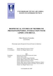
Figure 1. Schematic representation of the plant viruses andvarious aggregation states of their coat proteins. RodshapedTMV virions (I), TMV protein in helix-form (2), indisk-form (3) and as oligomers (4). Spherical CCMV, SBMV orBMV virions (5), empty capsids of CCMV, SBMV or BMV protein(6), dimers of CCMV, SBMV or BMV protein (7), empty capsidsof CCMV, SBMV or BMV protein lacking the N-terminal arm (8)and dimers of CCMV, SBMV or BMV protein lacking the N-terminal arm (9). Numbers correspond to the numbering usedfor stock solutions of virus and coat proteins. (SeeMATERIALS & METHODS).mM EDTA, pH 8.0 and then treated like CCMV and BMV. Proteins of CCMV,BMV and SBMV lacking the N-terminal arm, were obtained by trypsintreatment and checked with SDS-PAGE as reported earlier (Vriend etal., 1981), A schematic representation of the plant viruses and thevarious aggregation states of their proteins is shown in Figure 1. The numbersin this figure correspond to the following 4 mg/ml stock solutions:1) TMV virions in 50 mM sodium acetate/l50 mM NaC1, pH 5.0 and in50 mM potassium phosphate/l50 mM NaC1, pH 7.0,2) TMV protein in 50 mM sodium acetate/l50 mM NaC1, pH 5.0, mainly
helix-form (De Wit et al., 1978; 1979),3) TMV protein in 50 mM potassium phosphate/l50 mM NaC1, pH 7.0,mainly disk-form (De Wit et al., 1978; 1979),4) TMV proteln in 50 mM Tris-HC1/15O mM NaC1, pH 9.0, mainlyoligomers (De Wit et al., 1978; 1979),5) CCW, SBhN or BMV virions in 50 mM sodium acetate/l5O mM NaC1,pH 5.0 and in 50 mM potassium phosphate/l50 mM NaCl, pH 7.0,6) empty capsids of CCMV, SBMV or BMV protein in 50 mM sodiumacetate/2OO mM NaC1, pH 5.0,7) dimers of CCMV, SBMV or BMV protein in 50 mM Tris-HC1/200 mMNaCl, pH 7.5,8) empty capsids of CCMV, SBMV or BMV protein lacking the N-terminal arm in 50 mM sodium acetate/2OO mM NaC1, pH 5.0,9) dimers of CCMV, SBMV, or BMV protein lacking the N-terminal armin 50 mM Tris-HC1/200 mM NaC1, pH 7.5.Coat protein stock solutions were kept at 4OCand used within 7 daysafter preparation.(c) Samples for turbidity measurements at 550 nm and electronmicroscopy. The samples for turbidity measurements at 550 nm wereprepared by adding 250 @ virus or protein stock solution to 750 J.Uvesicle stock suspension in the same buffer. The final concentrationof lipid was 4 mg/ml buffer and of the virus or coat protein 1 mg/mlbuffer. During a 24 h period the 1-mlsamples were incubated at 18OC.The samples were measured at one-hour intervals. For each turbiditymeasurement the samples were transferred to a quartz cuvet. Afterincubation for turbidity measurements at 550 nm aliquots of the mixedsamples were taken for electron microscopy.(d) Samples for 31~ NMR. A unilamellar DLPC vesicle solution (5.33mg lipid/ml) was prepared in 10 mM Tris-HC1/15O mM NaC1/1 mM EDTA, pH9.0 by sonication in two 15-ml portions as described in section (a),Titanium particles of the sonicator and residual multilamellarvesicles were removed by centrifugation. (10,00Og, 15 min). The
supernatants were mixed resulting in a single homogeneous clearunimellar DLPC vesicle suspension of 30 ml. One 1 5 4 fraction wasincubated with 5 mlTMV protein stock solution number 4 for 24 h at1a0C and the other 15-ml fraction was incubated with 5 ml of identicalbuffer as reference. Before measurement samples were concentrated byultracentrifugation (250,00Og, 1 h), and resuspended in 10 % (v/v)2~20 containing 10 mM Tris-HCl/l5O mM NaCl/lmM EDTA buffer, pH 9.0(final volume 2 ml) and treated as indicated in Figure 5.Absorption measurements. The turbidity of the samples was measuredin a Kontron Uvikon 810 spectrophotometer at 550 nm to avoidcontributions of absorption of viral material or lipids. Turbiditiesat 550 nm of identical control vesicle suspensions not exposed toviral material (in all cases less than 0.10) and of viral materialwere subtracted if necessary.Determination of the amount of protein associated with membranes after incu-bation. After the 24-h period of incubation of the vesicle suspensionswith protein, the aggregated, fused and multilamellar bilayerstogether with associated protein were pelleted by centrifugation(8,80Og, 10 min). The fraction of residual, unassociated protein inthe supernatant wascalculated from the protein contribution at 280 nmin the UV spectrum using an extinction coefficient at 280 nm = 1.27ml.mg-l,cm-l for TMV, CCMV and BMV protein and 1.30 ml.mg-l,cm-l forSBMV protein. This fraction was subtracted from the 1 mg protein ini-tially added to obtain the complementary fraction of membrane asso-ciated protein presented in Table I.Electron microscoey. Electron micrographs were taken with a ZeissEM 109 electron microscope equipped with a transfibre photographycamera. The samples were negatively stained with a 2 %acetate in double-destilled water.Phosphorus magnetic resonance spectroscopy. The 31~ NMRsolution of uranylspectra ofthe DLPC vesicle suspension after 24 h incubation with TMV proteinwere obtained with a Bruker CXP 300 Fourier Transform spectrometer
operating at a frequency of 121.48 MHz. The spectra were taken in thepresence of broadband proton decoupling (20W/12dB) using a sweepwidthof 25,000 Hz and a 16 bs 45O pulse with a repetition rate of 1 s.RESULTSTurbidity measurements and determination of protein associated with membra-- nes. The effect of addition of plant viruses and their coat proteinsto neutral and charged SUVs under various conditions was monitored byturbidity measurements at 550 nm. In addition the percentage of proteinpresent in the pellet was determined after centrifugation of the sam-ples.(a) Effect of intact virions. Addition of TMV or CCMV to positivelycharged SUVs at pH 5 and 7 increased the turbidity, but not in thecase of neutral or negatively charged SUVs (Figure 2A,B,E,F). Theturbidity increase for CCMV-lipid mixtures was stronger at pH 7 than at5. In all cases addition of BMV or SBMV had only a small effect on theturbidity (Figure 2C,D,G,H). Pellets of positively charged SUVsobtained after incubation with TMV at pH 5 and 7 contained all theprotein initially added. Pellets of positively charged SUVs obtainedafter incubation with SBMV at pH 7 and with CCMV at pH 5 and 7contained approximately 20, 20 and 60 % of the protein, respectively.In all other pellets no protein was detected within experimental error(Table I).(b) Effect of the proteins. Addition of TMV protein at pH 5, 7(Figure 21,M) and 9 (data not shown) to positively charged SUVsincreased the turbidity rapidly. No increase of turbidity was foundwith negatively charged SUVs. Neutral SUVs caused a much slowerincrease ofthe turbidity as compared to the positively charged SUVs.The turbidity 24 h after incubation increased as a function of pHfrom
Virus Virions Coat pmfein Coat pmtein lackingpH 5.0 pH 7.0 pH 5.0 pH 20 the N- terminal armTMVpH 5.0 pH 7.0 pH5.0 pH 7.5 pH 5.0 pH 7.5BMV ,,.-------SBMVo o n a o X ) ? D 0 m a o w 2 0 0 w 2 0 1) 20t lh)Figure 2. Turbidity of suspensions (final lipid concentration4 mg/ml) of neutral vesicles (100% DLPC: solidline), positively charged vesicles (80/20 w/w DLPC/CTAB orDLPC/ PALCHOL: dotted line) and negatively charged vesicles(80/20 w/w DI,PC/DLPA or DLPC/DMPG: dashed line) after additionof virions (A-H), protein (I-P) and protein lacking theN-terminal arm (Q-V) of TMV (first row: A,E,I,M), CCMV(second row: B,F,J,N,Q,T), BMV (third row: C,G,K,O,R,U) andSBMV (fourth row: D,H,L,P,S,V) as illustrated in Figure 1.All experiments were carried out at 18OC and the same bufferas for vesicle stock suspensions was used for viral particleor protein stock solutions. A,I) pH 5.0 in 50 mM sodiumacetate/l50 mM NaCl buffer, B,C,D,J, K,L,Q,R,S) pH 5.0 in50 mM sodium acetate/200 mM NaCl buffer E,F,G,H,M) pH 7.0 in50 mM potassium phosphate/l50 mM NaCl buffer, N,O,P,T,U,V)pH 7.5 in 50 mM Tris-HC1/200 mM NaCl buffer.5 to 9. Only in pellets of positively charged SUVs obtained afterincubation with TMV protein, all the initially added coat protein wasfound (Table I).Addition of CCMV, BMV and SBMV protein at pH 5 and 7.5 topositively charged SUVs had no significant effect on the turbidity
Table I. Percentage of Coat Protein Associated with Membranesa.Virus Membrane Virions Coat ProteinChargeTMV positive **** **** **** ****NeutralNegativ.e .Virus Membrane Virions Coat Protein Coat Protein LackingChargethe N-terminal ArmpH5 pH7 pH5 pH7.5 pH5 pH7.5CCMV Positive * ***NeutralNegative . *** .-***BW Positive .NeutralNegative . ***x *SBMV Positive . *NeutralNegative . **** ***a pellets were obtained by centrifugation (8,80Og, l0min) of thesamples of Figure 2 after 24 h of incubation: . = 0 -12%,* = 12-25%, ** = 25-50%, *** = 50-75% and **** = 75-100%. ForCCMV and SBMV coat protein the values of DLPA are presented.(Figure BJ,K,L,N,O,P). For negatively charged SUVs the addition ofthese proteins increased the turbidity (Figure BJ,K,L,N,O,P) and alsoprotein was found in the pellets: 55 f 10 % for CCMV at pH 5.0 and
Figure 3. Turbidity of suspensions (final lipid concentation4 mg/ml) of neutral vesicles (100% DLPC: solid line), positivelycharged vesicles (80/20 w/w DLPC/PALCHOL: dottedline) and negatively charged vesicles (80/20 w/w DLPC/DLPA:dashed line) 30 min after addition of A) TMV protein in 50mM potassium phosphate, pH 9.0, B) CCMV protein in 50 mMTris-HC1, pH 7.5 and C) CCMV protein lacking the N-terminalarm in 50 mM Tris-HC1, pH 7.5 as function of NaCl concentration.7.5, 85 + 10 % for BMV and SBMV at pH 5, 20 + 10 % for BMV at pH 7.5and 55 + 10 % for SBMV at pH 7.5 (Table I). The addition of CCMV, BMVand SBMV protein increased the turbidity of neutral SUVs at pH 7.5(Figure 2N,O,P) and also at pH 5 for BMV protein (Figure 2K). However,no protein was found in pellets of neutral SUVs (Table I).(c) Effect of proteins lacking the N-terminal arm. After removal ofthe N-terminal arm of the CCMV, BMV and SBMV protein no significantchanges in turbidity were observed with positively and negativelycharged SUVs at pH 5 and 7.5 (Figure 2Q-V). The turbidity changesfound for neutral SUVs after incubation with intact CCMV, BMV and SBMVprotein were preserved with the cleaved proteins. Also cleaved protein
Figure 4. Electron micrographs showing typical shape of unilamellarvesicles after 24 h of incubation of the DLPC/DLPA(80/20 w/w) vesicles (A) and of multilayer structures inpellet after 24 h of incubation of the DLPC/DLPA (80/20 w/w)vesicles with empty capsids of BMV protein in 50 mM sodiumacetate/ 200 mM NaCl buffer, pH 5.0 (8).was not bound (Table I).Salt concentration dependence of turbidity induced by proteins. Theturbidity of the SUV suspensions was measured as a function of NaClconcentration 30 min after addition of TMV protein (Figure 3A), ofCCMV protein (Figure 38) and of CCMV protein lacking the N-terminalarm (Figure 3C). The turbidity reduced with increasing saltconcentration. This effect was most clear for positively charged SUVsincubated with TMVincubated with CCMV protein (Figure 38).Electron microscopy.protein (Figure 3A) and for negatively charged SUVsAfter turbidity measurements electron micrographswere taken of the samples. In Figure 4A a typical electron micrograph
Figure 5. 121.48 MHz 31~ NMR spectra of a suspension of DLPCSUVs in 10 mM Tris-HC1/15O mM NaCVl' mM EDTA buffer, pH 9.0after 24 h of incubation at 18OC without viral material (A)and (B) sample of (A) after I min of sonication; (C) as (A)with TMV protein and (D) sample of (C) after 1 min of sonication;(E) supernatant (8,80Og, 10 min) and (F) pellet(8,80Og, 10 min) of sample (D). In all cases the number ofscans was 1,000 and broadband proton decoupling (20 W/12 dB)was applied. For spectra in (A), (B) and (E) an artificialline broadening of 10 Hz and in (C), (D) and (F) of 100 Hzwas used.of a reference sample of negatively charged SUVs after 24 h of incuba-tion is shown. The diameter of the SUVs ranges from 30 to 100 nm.Identical vesicle sizes were observed for other reference vesicles(data not shown). After 24 h of incubation of negatively charged SUVswith empty capsids of BMV protein at pH 5 (Figure 4B) multilamellaraggregates were formed.Typical lnultilayer structures as in Figures 4B were observed in allelectron micrographs (data not shown) taken of samples of which theturbidity at 550 nmhad increased to values higher than 1 at the end
of the 24 h incubation period after addition of TMV protein or CCMV,BMV or SBMV protein with or without N-terminal arm (Figure 21-V).Phosphorus NMR measurements. Figure 5 shows 31~ NMRspectraobtained of DLPC SUVs after 24 h of incubation at 18OC with andwithout TMV protein, The spectrum of the reference SUVs ischaracterized by one isotropic peak with a linewidth at half height,b, of 190 Hz (Figure 5A). After 1 min of sonication A u ~ decreased to70 Hz (Figure 5B). An identical sample incubated for 24 h with TMVcoat protein , gave a typical powder spectrum characteristic forbilayers with a CSA of ca. 30 ppm (Figure 5C). No peaks were visiblethat could arise from the presence of rapidly tumbling phospholipidmolecules (Burnell et al., 1980) or HII phase lipids (Van Echteld etal., 1982). After 1 min sonication of this sample an isotropic peakwith a relative intensity of approximately 20 % was seen (Figure 5D).This peak could be separated from the underlying powder spectrum bycentrifugation (8,80Og, 10 min). This resulted in an isotropic peakwith a AVk of 370 Hz of the supernatant (Figure 5E) and a powderspectrum of the pellet (Figure 5F) with a CSA of ca. 30 ppm, similarto Figure 5C.DISCUSSIONThe turbidity measurements of neutral and charged SUVs interactingwith intact plant viruses or their coat proteins indicated:(a) Electrostatic interaction. The virus particle or protein containscharged amino acid residues, which interact with oppositely chargedmembranes. As a consequence, several vesicles are crosslinked.Also charges at the membrane are neutralized, causing aggregation,vesicle fusion and formation of multilayer structures. The precipitatedmaterial contains viral coat protein;(b) Indirect hydrophobic interaction. Dissociated protein causes neu-
tral vesicles to fuse and to form multilayer structures. The precipi-tated material does not contain protein. As will be discussed later,this effect is caused by exposed hydrophobic protein domains.For the interactions the following evidence was presented:1. Rod-shaped TMV. Positively charged vesicles interact with TMVparticles at pH 5 and 7 (Figure 2A,E). The same is observed for itscoat protein, both in helix and disk conformation (Figure 21,M) and asoligomers at pH 9 (data not shown). The pellet contains all coatprotein (Table I) whereas multilamellar vesicles are formed (Figure48). Several arguments suggest that electrostatic forces dominate inthe formation of these pellets: (a) The pI of TMV particles is 3.8(Fraenkel-Conrat & Nariba, 1958). This implies that the virus surfacehas an overall negative charge at the experimental pH values; (b)Negatively charged SUVs do not interact with TMVand its protein; (c)A reduction of protein-vesicle interaction by high salt concentrationis observed by the turbidity measurements at 550 nm as function ofsalt concentration (Figure 3A).TMVprotein with its hydrophobic areas exposed to the bulk solvent(Bloomer et al.,1978) causes the neutral SUVs to fuse after aggrega-tion (Figure 21,M). This result agrees with those of Banerjee et al.(1981a,b). In this case noprotein is detectable in the pellet. (TableI). This effect parallels the increasing exposure of hydrophobicdomains of TMV protein at higher pH values.An increase in turbidity at 550 nm can only be interpreted as anincrease in size of the aggregates. Thus on the basis of turbiditymeasurements, no discrimination between aggregation, fusion or trans-formation to multilayer structures can be made. Also, spontaneousaggregation of DLPC SUVs after preparation by sonication was reported(De Kruijff et al., 1976), which was observed as a broadening of the31~ NMR signal. Therefore, 31~ NMR spectra were recorded and EMmicrographs were taken. After 24 h of incubation of the reference DLPCSUVs the increased linewidth (190 Hz) of the 31~ NMRresonance indica-
tes aggregation of the SUVs (Figure 5A,B). The electron micrographof the reference vesicles after 24 h of incubation shows that notranformation to multilayer structures has occurred (Figure 4A).However, after 24 h of incubation with TMV protein, the 31~ NMRspectrum indicates that the majority of the vesicles (minimally ca. 80%) has become multilamellar (Figure 5F). The rest (maximally ca. 20 %)is aggregated since it appears as a relatively sharp isotropic peakafter brief sonication (Figure 5D). These results are in accordancewith the multilayer structures observed in electron micrographs ofsame samples (data not shown). Formation of multilamellar vesiclesimplies that fusion of vesicles has taken place.2. Spherical plant viruses (CCMV, BMV, SBMV). In analogy to TMV,CCMV particles interact with positively charged vesicles (Figure2B,F), which is explained by the pI value of 3.6 (Bancroft et al.,1971). As a result of the electrostatic interaction the pellets ofthese samples contain protein at pH 5 (ca. 20 %) and 7 (ca. 60 %,Table I), values at which an increasing number of negatively chargedgroups per viral particle is present. CCMV protein has an N-terminalarm of ~wenty-five amino acid residues of which nine residues arebasic. This arm, also of coat protein molecules in empty capsids, isknown to bind to negatively charged macromolecules in solution(Bancroft, 1971; Vriend et al., 1986). Therefore, binding to negativelycharged vesicles is not surprising (Figure 2J,N). Binding is lostafter removal of the N-terminal arm by trypsin (Figure 2Q,T).Dissociated CCMV protein, with and without the N-terminal arm,induces fusion of neutral SUVs and formation of multilayers (Figure2N,T). This is analogous to the effect of TMV protein on neutralvesicles (Figure 21,M).BMV and SBMV have a pI value of 6.8 (Magdolf-Fairchild, 1967) and6.0 (Rice & f or st, 1972), respectively. These viruses do not interactwith SUVs, regardless of their surface charge. SBMV is an exceptiongiving a small electrostatic effect at pH 7, if added to positiyelythe
charged SUVs (Figure 2C,D,G,H). This binding, which was alsodemonstrated by Abdel-Salam et al. (1982) in 0.1 M Tris-HC1, pH 7.5,could be due to the swollen state of SBMV. The near neutral pI valuesof BMV and SBMV are also reflected in the absence of proteinassociation to positive SUVs (Table I). BMV and SBMV protein, eitheras capsids or as dimers, interact with negatively charged SUVs(Figure 2K,L,O,P) because of the presence of their basic N-terminalarms. This interaction is lost after removal of the arm (Figure2R,S,U,V).Both BMV and SBMV protein dimers induce fusion of neutral SUVs(Figure 20,P) and this inductive capacity is completely preservedafter cleavage of the N-terminal arms (Figure 2U,V). Also BMV emptycapsids with and without the N-terminal arm react with neutral SUVsby indirect hydrophobic interaction (Figure 2K,R). This can beattributed to the strong hydrophobic character of BMV protein (Pfeiffer &Hirth, 1974). Again the induction of vesicle fusion increases withincreasing hydrophobicity of the protein,The nature of the interaction between model membranes and severalplant virusesand their coat proteins. In the discussion of theresults, electrostatic interaction was found to be quite differentfrom induction of vesicle fusion by indirect hydrophobic interaction.The electrostatic interactions are reduced to zero at high saltconcentration (Figure 3). Therefore there is no indication ofhydrophobic interaction in the formed complex, i.e, between hydrophobicprotein domains and the bilayer interior. Thus the protein islikely to be associated to the headgroups of the bilayers. This processis analogous to that found for aggregation and fusion of negativelycharged SUVs by polycations, such as polylysine (Gad et al.,1982; 1985) and polyhistidine (Wang & Huang, 1984; Uster & Deamer,1985).Induction of vesicle fusion and formation of multilayers by
indirect hydrophobicinteraction is observed only for neutral vesicles.Neutral SUVs aggregate spontaneously (De Kruijff et al., 1976). Thisis observed in the 31~ NMRspectrum (Figure 5A) as a broadening ofthe isotropic resonance. Brief sonication reverses this aggregation(Figure 5B). Vesicle fusion and further formation of multilamellarstructures is inhibited by interfacial water between the apposedbilayer surfaces of two aggregated vesicles (Le Neveu et al., 1976;Papahadjopoulos et al,, 1978). A possible mechanism . for the indirecthydrophobic interactionis that the hydrophobic sites at the proteinsin the solution remove this hydration water, enabling direct molecularcontacts of the bilayers and concomittant fusion according to thegeneral theory of vesicle fusion (Wilschut & Hoekstra, 1986). The pro-posed effect of the protein can be visualized as a pushing out of theinterfacial water resulting in a partial dehydration of the bilayersurface. This process is slow, since it requires a collision of theprotein with already apposed bilayer surfaces. The protein itself isnot inserted in the bilayer, nor does it form a complex with thelipids. Also the protein does not pass through the membrane during thefusion process, since otherwise it would have been locked in the fusedvesicles and have appeared in the final multilayer precipitate. Theaction of the protein may be compared to that of a catalyst in a che-mical reaction, enhancing only the rate of fusion between thebilayers.Since the coat protein is not found associated with the vesicles inthe case of hydrophobic interaction (Table I) this implies that thehydrophobic protein domains do not interact directly with thehydrophobic interior of the bilayers.From these observations it is concluded that, regardless of thetype of interaction (hydrophobic, electrostatic), viral coat proteinis not incorporated in the bilayer, i.e. by forming a stable complexheld together by direct hydrophobic lipid-protein interactions.
Implications of the observed interactions for earlier proposed modelsof initial interactions between virus and cell. A stable lipid-coatprotein complex in which direct hydrophobic forces dominate was notfound for any of the viruses and bilayers that were investigated. Thisstrongly disagrees with the model of virus penetration and uncoatingproposed by Durham (1978), which we therefore consider incorrect.Caspar (1963) suggested that the hydrophobic environment of thebilayer interior causes uncoating of TMV RNA in vivo. Although thismechanism of uncoating cannot be excluded rigorously, it is very unlikelyon the basis of our results. From Figure 2A-H it is clear thatviral particles can interact with the head groups of lipids in thebilayer only by electrostatic interaction, which results in coatprotein associated the outside of the bilayer. In the final complexalso no direct hydrophobic forces are involved (Figure 3). Thereforethe uncoating process is definitely not initiated and probably alsonot propagated by hydrophobic lipid-coat protein forces, replacing thehydrophobic intersubunit forces in the virus particle.Contrary to the suggested uncoating effect of the hydrophobic domainof the bilayer on the virus (Caspar, 1963), for which no informationis available from our experiments that focused on the effectof virus-membrane interactions on the membranes, a destabilizingeffect of the hydrophobic domains of the viral coat proteins on themembrane has been observed. This suggests that in vivo the virusparticle could induce membrane fusion by exposing its hydrophobicprotein domains. This could possibly be a mechanism for the virus toenter the cell.ACKNOWLEDGEMENTSWe thank B, de Kruijff, J.W. Roenhorst, T.J. Schaafsma andJ. Wilschut for valuable discussions and critical reading of the
manuscript, C.J.A.M. Wolfs and H.R. Bloksma for help with thepreparation of plant virus material and J. Groenewegen for help withelectron microscopy.REFERENCESAbdel-Salam, A,, White, J.A., & Sehgal, O.P. (1982) Phytopath. Z, 105,336-334. 'Bancroft, J.B. (1971) Adv. Virus Res. 16, 99-134.Banerjee, S., Vandenbranden, M., & Ruysschaert, J.M. (1981a) Biochim.Biophys. Acta 646, 360-364.Banerjee, S., Vandenbranden, M.,Lett. 133, 221-224. .& Ruysschaert, J.M. (1981b) FEBSBloomer, A.C., Champness, J.N., Bricogne, G., -Staden, R., & Klug, A.(1978) Nature (London) 276, 362-368.Burnell, E.E., Cullis, P.R., & De Kruijff, B. (1980) Biochim. Biophys.Acta 603, 63-69.Caspar, D.L.D. (1963) Adv. Protein Chem. 18, 87-121.De Kruijff, B., Cullis, P.R., & Radda, G.K. (1976) Biochim. Biophys.Acta 436, 729-740.De Wit, J.L., Hemminga, M.A., & Schaafsma, T.J. (1978) J. Mapn. Res.31, 97-107.De Wit, J.L., Alma, N.C.M., Hemminga, M.A., & Schaafsma, T.J. (1979)Biochemistry 18, 3973-3976.De Zoeten, G.A. (1981) in Plant Diseases and Vectors (Maramorosch, K.& Harris, K.F., Eds.) pp. 221-239, Academic Press, New York, N.Y.Durham, A.C.H., Hendry, D.A., & Von Wechmar, M.B. (1977) Virolofg 77,524-533.Durham, A.C.H. (1978) Biomedicine 3, 307-314.Fraenkel-Conrat, H., & Nariba, X. (1958) in Symposium on ProteinStructure (Neuberger, A., Ed.) pp. 249, Methuen, London.Gaard, G., & De Zoeten, G.A. (1979) Virology 96, 21-31.Gad, A.E., Sliver, B.L., Eytan, G.D. (1982) Biochim. Biophys. Acta690, 124-132.ad,^., Bental, M., Elyashir, G., & Weinberg, H. (1985)Biochemistry 24, 6277-6282.Hull, R., & Maule, A.J. (1985) in The Viruses (Francki, R.I.B., Ed.)pp. 83-115, Plenum Press, New York, N.Y.Kiho, Y., & Shimomura, T. (1976) Japan J. Microbiol. 20, 537-541.Kiho, Y., Shimomura, T., Abe, T., & Nozu, Y. (1979a) Microbiol.Immunol. 23, 735-748.Kiho, Yo, Abe, T., & Ohashi, Y. (1979b) Microbiol. Immunol. 23,
1067-1076.Kiho, Y., & Abe, T. (1980) Microbiol. Immunol. 24, 617-628.Leberman, R. (1966) Virology 30, 341-347.Le Neveu, D.M.,Rand, R.P., & Parsegian, V.A. (1976) Nature (London)259, 601-603.~a~dolf- airc child, B.S. (1967) Virology 31, 142-143.Marvin, D.A. & Wachtel, E.J. (1975) Nature (London) 253, 19-23.Papahadjopoulos, D., Portis Jr., A,, & Pangborn, W. (1978) Ann. N.Y.Acad. Sci 308, 50.Pfeiffer, P., & Hirth, L. (1974) Virolopy 58, 362-368.Rice, R.H., & Horst, J. (1972) Virolopy 49, 602-604.Shaw, J.G., Plaskitt, K.A., & Wilson, T.M.A. (1986), Virolo~y 148,326-336.Uster, P.S., & Deamer, D.W. (1985) Biochemistry 24, 1-8.Van Echteld, C.J;A., De Kruijff, B., Verkley, A.J., Leunissen-Bijvelt,J., & De Gier, J. (1982) Biochim. Biophys. Acta 692, 126-138.Van Lent, J.W.M., & Verduin, B.J.M. (1985) Neth. J. P1. Path. 91,205-13.Verduin, B.J.M. (1974) PEBS Lett. 45, 50-54.Verduin, B.J.M. (1978) J. gen. Virol. 39, 131-147.Vriend, G., Hemminga, M.A., Verduin, B.J.M., De Wit, J.L., &Schaafsma, T.J. (1981) FEBS Lett. 134, 167-171.Vriend, G., Hemminga, M.A., & Verduin, B.J.M. (1986) J, Mol. Biol.191, 453-460.~attr~.~., Dawson, J.R.O., & King. J.M. (1981) CIBA Foundationamposium 80 pp. 56-71.Watts, J.W., & King, J.M. (1984) J. gen. Virol. 65, 1709-1712.Wang, C.-Y., & Huang, L. (1984) Biochemistry 23, 4409-4416,(1986) Chem. Phys. Lipids, in press.Wilschut, J., & Hoekstra, D.Wilson, T.M.A. (1984) Virology 137, 255-265.Wilson, T.M.A. (1985) J. pen. Virol. 66, 1201-1207.
CHAPTER 3EFFECT OF THE <strong>MEMB</strong>RA<strong>NE</strong> ON BACTERIO-PHAGE MI3 COAT PROTEIN
3.1 TIME-RESOLVED TRYPTOPHAN FLUORESCENCEANISOPTROPY INVESTIGATION OF BACTERIOPHAGEMI3 COAT PROTEIN IN MICELLES AND MIXED BILAYERSKlaas P. Datema, Antonie J.W.G. Visser, Arie van HoekCor J.A.M. Wolfs, Ruud B. Spruijt and Marcus A. HemmingaABSTRACT: Coat protein of bacteriophage MI3 is examined inmicelles and vesicles by time-resolved tryptophan fluorescenceand anisotropy decay measurements and circular dichroismexperiments. Circular dichroism indicates that the coat proteinhas a-helix (60%) and @-structure (28%) in 700 mM SDSmicelles and predominantly @-structure (94%) in mixed dimyristoylphosphatidylcholine/dimyristoyl phosphatidic acid (80/20W/W) small unilamellar vesicles. The fluorescence decay at344 nm of the single tryptophan -in the coat protein afterexcitation at 295 or 300 nm is a triple exponential. In themicelles the anisotropy decay is a double exponential. Ashort, temperature ihdependent correlation time of 0.5 f 0.2ns reflects a rapid depolarization process within the coatprotein. The overall rotation of the coat protein-detergentcomplex is observed in the decay as a longer correlation timeof9.8 f 0.5 ns (at 20'~) and has a temperature dependencethat satisfies the Stokes-Einstein relation. In vesicles atall lipid to protein molar ratios in the range from 20 to 410the calculated order parameter is constant with a value of0.7 + 0.1 from 10 to ~o'c, although the lipids undergo thegel to liquid-,crystalline phase transition. The longer correlationtime decreases gradually on increasing temperature. ,,This effect probably arises from an increasing segmentalmobility within the coat protein. The results are consistentwith a model in which the coat protein has a @-structure andthe tryptophan indole rings do not experience the motion ofthe lipids in the bilayer because of protein-protein aggregation.The interaction of membrane proteins with lipids has received considerableattention in the last decade. The interest in understandingthis interaction arises from the notion that lipid composition anddynamics in membranes affect the properties of its proteins, A varietyof biophysical techiques has already been applied to membrane proteins
econstituted in model membranes (for a survey see: Watts & De Pont,1985). Model membranes can easily be prepared and are free of thecomplexity of the natural membrane. In particular, time-resolvedfluorescence anisotropy measurements (for a review see: Cundall &Dale, 1983; Beechem & Brand, 1985) and nuclear magnetic resonance (NMR)provide complementary information about motion and order of lipids inbilayer systems (for a comparison see: Wolber & Hudson, 1982; Deveax &Seigneuret, 1985).The objective of our research is to study the infection mechanismof non-enveloped viruses, like bacteriophage MI3 (Hemminga, 1987) andplant viruses (Hemminga et al., 1985), at a molecular level. Asuitable system to study the interaction of viral coat proteins withmembranes is the M13-Escherichia & system.The major (gene-8 product) MI3 coat protein is present in the longrodlike virus particle in numerous copies and functions as protectionfor its single strand DNA. The virus enters @ by leaving thecoat proteins in the cytoplasmic membrane (Marvin & Wachtel, 1975).After infection and DNA duplication the newly synthesized procoats, aprecursor form of the coat protein with a typical bacterial amino terminalleader peptide of 23 residues, also assemble in this membrane.After penetration of the N-terminus of the procoat through themembrane, the procoat is cleaved to mature coat protein by a leaderpeptidase (Geller & Wickner, 1985). Both progeny as well as parentalcoat protein are used for the membrane-bound assembly of new M13 particles(Wickner, 1976). The viral DNA is complexed with these coatproteins without lysis of the host cell.The coat protein consists of, 50 amino acid residues (M=5240): abasic C-terminus, a hydrophobic central core of 19 amino acid residuesand an acidic N-terminus (Van Asbeck et a1.,1969; Nakashima &Konigsberg, 1974). It contains a single tryptophan residue located atthe 26th position, i.e. in the hydrophobic region. The tryptophan canbe used as an intrinsic fluorescent marker.
Time-resolved fluorescence anisotropy analysis of fluorophores inmembranes (for a theoretical description see for example: Kinosita etal., 1977; 1982; Zannoni et al., 1983; Szabo, 1984; Van der Meer etal., 1984) has several features which make the technique particularlyattractive for studying the MI3 coat protein in model membranes.First, the time-dependent anisotropy provides a direct, real-timemeasurement of the, reorientational dynamics of the coat protein in the(sub)nanosecond time range. The measurable range is limited by theaverage fluorescence lifetime characterizing the tryptophan. Second,in time-resolved ' fluorescence anisotropy analysis motional rates andmolecular order manifest themselves separately, which is advantageousfor bilayer <strong>studies</strong>.We have reconstituted the coat protein of MI3 in micelles and mixedSUVs. Time-resolved .,fluorescence and fluorescence anisotropy measurementsof the single ,tryptophan residue in the coat protein werecarried out at various temperatures and protein contents. In additionCD spectra were taken of the coat protein in these micelles andbilayers to determine the secondary structure of the coat protein.Time-resolved fluorescence and fluorescence anisotropies were analysedin terms of multi-exponential functions, giving information aboutdynamics of the coat protein tryptophan and the structure of itsenvirons in these systems. In SUVs an additional residual anisotxwpyis present, which is directly related to the order of the coat proteintryptophan. The temperature dependence of the correlation times and ofthe order of the coat protein in the membrane was determined in therange in which the lipid bilayer undergoes the gel to liquid-crystallinephase transition.MATERIALS AND METHODSMaterials. DMPC (1,2-dimyristoyl-E-glycero-3-phosphocholine 99% pu-
ity) and DMPA (1,2-dimyristoyl-g-glycero-3-phosphatidic acid, 98% purity)were obtained from Sigma Chemical Co. and used without further purification.MI3 bacteriophage was purified as described by Garssen et al.(1977). The major (gene-8 product) coat protein of M13 was isolated bythe method of Knippers and Hoffmann-Berling (1966).Sample preparation. Micelles were prepared by dissolving coat proteinin 0.8, 4, 100 or 700 mM SDS, 10 mM potassium phosphate, pH 5.0.A clear, micellar solution was obtained by vortexing and heating thesample to 55'~. The samples were diluted with the same buffers toOD280nm = 0.1.Vesicles were prepared by cholate dialysis, as described by Hagenet al. (1978) with a few modifications. Stock suspensions of DMPC andDMPA vesicles (both 50 mg/ml) and coat protein (8 mg/ml) in 5.0 mMTris-HC1 buffer, pH 8.0 with 2 w% sodium cholate, 8.0 M urea, 0.1 mMEDTA and 20 mM ammonium sulphate were used. 0.16 ml of DMPC stocksuspension, 0.04 ml of DMPA stock suspension and 0.500, 0.250, 0.125,0.063, 0.031, 0.016 or 0 ml coat protein stock solution were coaddedwith 0.300, 0.550, 0.675, 0.737, 0.769, 0.784 or 0.800 ml of the samebuffer, respectively. Clear and homogeneous suspensions were obtainedby vortexing and heating the samples to 55'~. Subsequently, thesuspensions were dialysed at 4'~ against 4 times 1.5 L of 10 mMTris-HC1, pH 8.0, 0.2 mM EDTA and 10 v/v% methanol during 48 h withchanges at 12, 24 and 36 h. In the last dialysis step no methanol wasadded to the buffer. This results in opalescent dispersions of unilamellarvesicles. All the samples were sonicated under nitrogen with aBranson cell disruptor 830 (duty ,cycle: 90%, power setting 4 (max. 350W at setting 10)) to obtain SUVs. The SUV suspensions were centrifuged(8,80Og, 10 min) to precipitate unincorporated, water-insoluble coatprotein. At this point, aliquots of the samples were taken to determinethe protein and phospholipid concentration (Bartlett, 1959;Peterson, 1977), from which the L/P ratio was calculated. This proce-
dure results in samples of L/P ratios of 20, 30, 70, 140, 210, 410 oroo, respectively. The error in these ratios was less than 10%. Thesamples were homogeneous as tested by sucrose gradient centrifugation.Before measurement the samples were diluted to OD280nm = 0.1.Methods. CD spectra were recorded at room temperature on a Jobin-Yvon Auto-Dichrograph Mark V in the wavelength range 250 - 190 nm. Asample cell of I-mm path length was used. The spectra are the averageof four scans taken from the same sample. To determine the secondarystructure the CD spectra were fitted to reference spectra of Greenfieldand Fasman (1969) by a least squares fitting procedure usingspectral points in the 250 to 190 nm range with 5-nm steps. The basisfor this analysis has been described (Cantor & Schimmel, 1980).The experimental setup for the time-resolved fluorescence measurementshas been described previously (Van Hoek et al., 1983; Van Hoek &Visser, 1985; Visser et al., 1985). The excitation wavelengths Were295 and 300 nm. The emission wavelength was selected with an interferencefilter transmitting at 344 nm with a 10-nm bandpass. Pluorescencewas detected by time-correlated single-photon counting (OIConnor& Phillips, 1984).The total fluorescence decay is determined from the measured polarizedfluorescence components parallel, I and perpendicular, I*, tothe electric vector of the excitation as:in which S(t) is the convolution product of the (unconvoluted) totalfluorescence, s(t), and the excitation pulse response, P(t). The datawere analysed as a sum of three exponentials, i.e.
in which ri is the fluorescence lifetime with a pre-exponential factor,ai, indicating the relative contribution of ri to the initialintensity of the total decay. The experimental data were corrected forbackground fluorescence measured for identical vesicles without coatprotein (in all cases less than 10% of the fluorescence of the samplewith coat protein). From these data the lifetimes and relative contributionsto the total decay are obtained by a non-linear least squaresfitting procedure (Vos et al., 1987).The fluorescence anisotropy, r(t), is given by:in which i/(t) and il(t) are the (deconvoluted) parallel and perpendicularpolarized components derived from the experimental Il(t) andIl(t) components. r(t) is obtained by fitting the experimental Il(t)and Il(t) decays directly to the anisotropy-containing expressions forthese two fluorescence components with the use of one set of parameters(Gilbert, 1983; Cross & Fleming, 1984) using Fortran-'77 programs(Vos et al., 1987). The model for anisotropy decay analysis consistedof a sum of two exponentials for the coat protein in micelles and of asum of two exponentials and a constant term for the coat protein inSUVs. In the analysis it is assumed that every lifetime component iscoupled to every rotational component (Dale et al., 197'7). The qualityof the fit is determined by (1) visual inspection, (2) the weightedresiduals, (3) the number of zero passages of the auto-correlationfunction (Ameloot & Hendrickx, 1982), (4) the reduced chi-squarevalue, (5) the Durbin-Watson parameter (O1Connor & Phillips, 1984) and(6) the standard deviations. The standard deviations of the fittedparameters are estimated from the Hessian as described by Bevington(1969), which is one of the output parameters of the Fortran-'7'7programs.The orientational order parameter, S, for the coat protein in the
ilayer is calculated from the residual, r(oo), and the initial, r(0),anisotropies byas given by Heyn (1979) and Jaehnig (1979). For r(0) the average ofthe series of measurements at each excitation wavelength was used(Table IV).RESULTSCircular dichroism. The CD spectrum .of M13 coat protein in micellesis shown in Figureb. 1. Analysis of the spectrum yields 60% a-helix,28% @-structure and 12% other structure for the coat protein in 700mM SDS micelles, Below the CMC (which is at 3 mM, Helenius et al.,1979) at 0.8 mM SDS 16 % a-helix, 64% @-structure and 20% otherstructure was determined (data not shown). The CD spectrum of M13 coatprotein in mixed SUVs of DMPC/DMPA (20/80 w/w) at L/P ratio of 25 wasbest simulated by 2% a-helix, 94% @-structure and 4% other structure(Figure 1). The same CD spectra were observed for the other L/P molarratios (data not shown).Time-resolved fluorescence of M13 coat protein in micelles. Thetotal fluorescence decay at 344 nm of M13 coat protein in micellesafter excitation at 295 nm was fitted as a triple exponential function(an example of a triple exponential fit, for the decay of M13 coatprotein in mixed SUVs, is shown in Figure 4). The lifetimes thatcharacterize the total fluorescence decays of the MI3 coat protein-SDScomplexes and their relative contributions to the total decay arelisted in Table I. In 700 mM SDS the shortest lifetime, TI, and allthe amplitudes are independent of temperature. However, T2 and 73decrease at increasing temperature (Table I). Below the CMC (3 mM) all
( 8 ) degrees. cm2/dmol1Figure 1. CD spectra of M13 coat protein at 20'~ in 700 mMSDS micelles (solid line) and mixed SUVs of DMPC/DMPA (80/20W/W) at L/P ratio of 25 (dashed line). Mean residue ellipticitiesare shown.lifetimes increase, while a2, and possibly al, increase at the expenseof a3.The fluorescence anisotropy decay was fitted as a double exponentialfunction. In Figure 2 the anisotropy decay at 344 nm of M13 coatprotein in 700 mM SDS micelles at 20'~ is given. The correlation timesand their amplitudes are listed in Table I1 for various SDS . concentrationsand temperatures. The short correlation time, (0.5 1 0.2ns), is independent of both the SDS concentration and temperature. Thelonger correlation time, $2, depends on the SDS concentration as wellas temperature. The value of $2, which is significantly longer belowthe CMC (3 mM), decreases as a function of temperature (Figure 3). The
Table I.Fluorescence Decay Parameters (344 nm) of the SingleTryptophan of Bacteriophage M13 Coat Protein in SDSMicelles for Excitation at 295 nm a.SDS T a1 a2 T~ a3 r3 2.05 f.1 f.05 f.1 f,05 f.1(mM) ('c) (ns) (ns) (ns) (ns)product, kTIP2/r), in which r) is the viscosity of pure water, is givenin Table I1 and found to be constant within experimental error (10%).Time-resolved fluorescence of MI3 coat protein in vesicles. Thelifetimes and their pre-exponential factors in the range of 10 to 40°Care listed in Table 111. In Figure 4 the decay after excitation at 295nm is shown. Similar values as given in Figure 4 were also found for
TIME (NS)Figure 2. Anisotropy decay at 344 nmof the single tryptophanof bacteriophage MI3 coat protein in 700 mM SDS micelles at20% after excitation at 295 nm and the best computer fit.The parameters describing the best fit to the experimentaldecay and the estimated standard deviations are: 81 = 0.06 f0.014, b2 = 0.11 k 0.004; = 0.50 f 0.21 ns, $2 = 9.76 f0.51 ns with the Durbin-Watson parameter = 1.95 and thenumber of zero passages in the parallel channel = 97 and inthe perpendicular channel = 96 (429 channels, 42 ps/channel).The weighted residuals from experimental and fitted curve andthe auto-correlation function are given in the top of thefigure.the other L/P ratios from 20 to 410 at 20'~ (data not shown).The fluorescence anisotropy in SUVs was fitted as a double exponentialwith a constant value (r(w)). In Figure 5 the anisotropy decaysof MI3 coat protein in SUVs after excitation at 295 and 300 run areshown. Table IV summarizes the results at various temperatures. Theinitial anisotropy, r(O), is 0.16 after 295 nm and 0.23 after 300 nn
Table 11. Anisotropy Decay Parameters (344 nm) of the SingleTryptophan of Bacteriophage M13 Coat Protein inSDS Micelles for Excitation at 295 nm a.a Bi is the contribution to the initial anisotropy,r(O), of the depolarization mechanism characterizedby $i; V, the volume of the protein-micelle complexwas calculated with the Stokes-Einstein relation forspherical rotation, a2 = gV/kT in which g is theviscosity of water, T the temperature and k theBoltzmann constant.excitation and in both cases independent of temperature (Table IV).The relative contribution of fll to the total anisotropy decay, i.e.fll/r(0), is 19% at 295 nm and 26 %at 300 nm excitation. For otherL/P ratios in the range of 20 to 410 at 20°C similar results are found
Figure 3. Temperature dependence of the longer correlationtime, $12, of MI3 coat protein in 700 mM SDS obtained from thebest computer fits. For experimental details see Table I1 andthe text.(data not shown). The short correlation time, $ I was found to benearly temperature independent (0.5 f 0.2 ns). Therefore $I1 was fixedat 0.5 ns in the final computer fitting. The longer correlation time,$t2, decreases at increasing temperature (Figure 6).The order parameter, S, calculated from eq. (4), is almost constant(0.7 f 0.1) at all temperatures at 295 (Figure 6) and 300 nm excitation(Table IV).DISCUSSION
Table 111. Fluorescence Decay Parameters (344 nm) of theSingle Tryptophan of Bacteriophage MI3 CoatProtein-DMPC/DMPA (80/20 w/w) SUVs at L/P molarratio 70 a.In SDS micelles M13coat protein forms a dimer (Tanford & Reynolds,73
TIME (NSIFigure 4. Fluorescence decay at 344 nm of the single tryptophanof bacteriophage MI3 coat protein-DMPC/DMPA (80/20W/W) SUVs of L/P ratio = 70 at 20°C, the best computer fitand the excitation pulse (295 nm). The parameters describingthe best fit to the experimental decay and the estimatedstandard deviations are: a1 = 0.25 f 0.02, a2 = 0.44 f 0.02,a3 = 0.31 + 0.01; TI = 0.8 f 0.1 ns, ~2 = 2.2 f 0.1 ns, ~3 =5.5 f 0.1 ns with the Durbin-Watson parameter = 1.82 and thenumber of zero passages = 202 (959 channels, 41 ps/channel).The weighted residuals from experimental and fitted curve andthe auto-correlation function are given in the top of thefigure.1976). The secondary structure of the coat protein in the SDS micellesis partly a-helix (60%) and partly @-structure (28%, Figure 1). Thisobservation agrees with those previously reported (Nozaki et al., 1976;1978; Chamberlain et al., 1978). Below the CMC (3 mM) the amount of 8-structure increases to 64% at the expense of a-helix(16%). Under the-
Table IV. Anisotropy Decay Parameters (344 nm) of the SingleTryptophan of Bacteriophage MI3 Coat Protein-DMPC/DMPA (80/20 w/w) SUVs at L/P molar ratio 70 a.a fli is the contribution to the initial anisotropy, r(O),of the dipolarization mechanism characterized by $i.se conditions the coat protein is solubilized by SDS molecules. Thehigher amount of @-structure in the coat protein is probably due to
TIHE INS)TIME INS)Figure 5. Anisotropy decays at 344 nm of the single tryptophanof bacteriophage MI3 coat protein-DMPC/DMPA (80/20W/W) SUVs of L/P ratio = 70 at 20'~ and the best computerfits after excitation at 295 (a) and 300 nm (b). The parametersdescribing the best fit to the experimental decay andthe estimated standard deviations are, for (a): 81 = 0.03 f0.006, 82 = 0.04 f 0.006, r(w) = 0.09 f 0.007; $1 = 0.5 ns,$2 = 8 f 2 ns with the Durbin-Watson parameter = 1.87 and thenumber of zero passages in the parallel channel = 98 and inthe perpendicular channel = 118 (460 channels, 41ps/channel), for (b): = 0.06 f 0.007, f12 = 0.07 f 0.010,r(w) = 0.10 f 0.010; = 0.5 ns, $2 = 8 f 2 ns with theDurbin-Watson parameter = 1.84 and the number of zero passagesin the parallel channel = 84 and in the perpendicularchannel = 97 (432 channels, 41 ps/channel). Only the initialparts of the anisotropy decays (of totally 1024 channels) areshown. The weighted residuals from the experimental andfitted curves and the auto-correlation functions are given inthe top of the figures.the formation of larger aggregates as seen from the fluorescence anisotropydecay (to be discussed below). These aggregates could be simi- ~
Figure 6. Temperature dependence of the longer correlationtime, 42 (open circles), and order parameter, S (closedcircles), of MI3 coat protein in mixed SUVs of DMPC/DMPA(80/20 w/w) at L/P ratio of 70. For experimental details seeTable IV and the text.lar to the coat protein polymer observed in vesicles in which also flstructureis dominant (94%), consistent with the work of Nozaki etal. (1978).For all total fluorescence decays of the tryptophan in M13 coatprotein three lifetimes were required to obtain a satisfactory fitaccording to the criteria listed under Methods. This is in agreementwith previously reported multi-exponential decays for other singletryptophan proteins (Grinvald & Steinberg, 1976; Munro et al., 1979).The origin for this lies in the complex photophysics of the tryptophanresidue which is partly caused by the presence of different conformers
(Szabo & Rayner, 1980; Chang et al., 1983; Petrich et al., 1983). Wesuggest that in a similar way in the MI3 coat protein different conformersgive rise to different lifetimes. These conformers arise fromdifferent average orientations of the tryptophan indole ring withrespect to the neighbouring amide and carboxyl groups along the backboneof the coat protein.Below the CMC the fluorescence of the coat protein aggregate, solubilizedby detergent molecules, is also triply exponential, but thelifetimes are significantly longer than for the coat protein in micelles,However, a2 increases at the expense of a3. This indicatesthat in the solubilized coat protein conformation (64% /3-structure asdetermined by CD, Figure 1) the quenching of the tryptophan fluorescenceis more efficient, presumably due to changes in orientation ofneighbouring amide and carbonyl groups with respect to the indolering.The long wavelength absorption band of the indole ring consists oftwo electronic transitions, 'La and lLb. The dipole moments of thesetransitions are almost perpendicular to each other. By excitation at295 nm both energy levels become populated (Weber, 1960; Valeur & Weber,1977). Between these levels energy transfer can occur easily ona subnanosecond time scale (Cross et al., 1983). Nevertheless, it cannotbe excluded that energy transfer between these two states providesan additional mechanism for the multi-exponential decay of the fluorescence.The fluorescence anisotropy is doubly exponential. The longer correlationtime is assigned to the overall rotation of the coat proteinmicellecomplex for the following reasons. First, this correlationtime is temperature dependent (Figure 3). The constancy of kT@Z/t) (=V, see Table 11) indicates that the motion, which is characterized by42 satisfies the Stokes-Einstein relation for isotropic rotationaldiffusion in solution. Second, below the CMC, i.e. under conditions inwhich coat protein is solubilized by detergent molecules and no micel-
For the triple exponential fluorescence decay of tryptophan in M13coat protein in vesicles comparable lifetimes are found as in micelles,although the amplitude of the second lifetime, a2, has increasedsignificantly at the expense of a3. This was also observed for solubilizedcoat protein in detergent solution below the CMC. The conformationalchange towards dominiintly 8-structure (94%, Figure 1) of M13coat protein probably gives rise to a more efficient quenching process,determined by T2. The constancy of the fluorescence decay as afunction of L/P ratio indicates that the coat protein conformation invesicles that favours more efficient quenching is formed independentlyof L/P ratio. The fact that the fluorescence decays of coat protein inSUVs and detergent solution below the CMC are both dominated by r2,strongly suggests that the micro-environment of tryptophan is identicalin both aggregation states of the coat protein.The initial anisotropy, r(O), of the coat protein in vesiclesdepends on the wavelength of excitation, 0.16 (295 nm) and 0.23 (300nm), in agreement with previous measurements on other proteins (Lakowiczet al., 1983). From the initial and residual anisotropy the orderparameter of the tryptophan in the M13 coat protein is calculated tobe 0.7 f 0.1 (at 295 and 300 nm excitation) at all L/P ratios and temperaturesmeasured. The fluorescence anisotropy does not decay to zerosince the vesicle rotation is too slow on the nanosecond time scale tocontribute to the decay, in contrast to the micellar rotation (41~ inTable 11). The gel to liquid-crystalline phase transition of the lipidmixture DMPC/DMPA (80/20 w/w) was determined to be completed at 26%(unpublished results), which is 2 degrees above the phase transitionof pure DMPC. Thus, it appears from the fluorescence results that theM13 coat protein is not sensitive to the phase transition and therapid motions of lipids in the liquid-crystalline phase. This suggeststhat the majority of the indole rings do not experience the lipidmotion because of aggregation of the coat protein. The constancy ofthe order parameter is in agreement with deuterium NMR measurements of
the amide sites along the M13 coat protein backbone in similar mixedbilayers, which show that the lipid motions do not affect the order ofthe amide deuterons along the coat protein backbone (chapter 3.2). Forother single transmembrane proteins that do not aggregate, it has beenfound that the deuterium NMR order parameter reflects the gel toliquid-crystalline phase transition of the lipids (Pauls et al., 1985;Datema et al., 1986). For MI3 coat protein it is therefore concludedthat the protein in the bilayer is in an aggregated state. The conceptof such protein aggregates is not in contradiction with the rigid proteinstructure propbsed by Wolber and Hudson (1982).Steady-state fluorescence <strong>studies</strong> of MI3 coat protein in DMPCvesicles with parinaric acid have shown that the fluorescence of thetryptophan of the coat protein is quenched by radiationless energytransfer to the probe lipid (Kimelman et al., 1979). Extensive proteinproteinaggregation was excluded in that paper. This is not consistentwith our findings. However, it should be noted that, in a proteinaggregate, quenching is still possible by inter-tryptophan energytransfer and subsequent energy trapping by the lipid acceptor.The correlation times and $2 indicate an internal tryptophanflexibility within the coat protein aggregate. The larger correlationtime, 412, is most likely related to segmental mobility of the coatprotein within the aggregate. The shorter, temperature independentcorrelation time (0.5 ns), could arise from internal motion of thetryptophan indole ring, but also from energy transfer from one indolering to another in the aggregate, as demonstrated for aromatic dimersin solid solution (Visser et al., 1983).In aggregated coat protein the tryptophan rings are positionedclose to each other, i.e. halfway along the peptide in the hydrophobicregion. If is caused by energy transfer between two nearby indolerings, then the contribution of would decrease if the excitationwavelength is shifted to the red-edge of the light absorption spectrum(Weber, 1960; Weber & Shinitzky,l970). On the other hand, in the case
of rapid indole ring motion no such dependence is expected for bl,Therefore the anisotropy was measured also for 300 nm excitation.Since the relative contribution of increases if the excitationwavelength is changed from 295 to 300 nm, this observation suggeststhat arises from rapid motion of the tryptophan ring in the coatprotein,ACKNOWLEDGEMENTSWe thank dr. B.J.M. Harmsen for providing facilities to grow E,and K. Vos for an introduction to the software for fluorescence dataanalysis.REFERENCESAmeloot, M., & Hendrickx, H. (1982) J. w. m. 76, 4419-4432.Bartlett, G.R. (1959) J, Biol. Chem. 234, 466-468.Bevington, P.R. (1969) Data Reduction and Error Analysis for thePhysical Sciences, McGraw-Hill, New York, NY.Beechem, J.M., & Brand, L. (1985) &. Rev. Biochem. 54, 43-71.Cantor, C.R., & Schimmel, P.R.(1980) Biophysical Chemistry, Freeman,W.H., San FranciscoChamberlain, B.K., Nozaki, Y., Tanford, C., & Webster, R.E. (1978)Biochim. Biophys. Acta 510, 18-37.Chang, M.C., Petrich, J.W., McDonald, D.B., & Fleming, G.R. (1983) J.Am. Chem. e. 105, 3819-3824.--Cross, A.J., Waldeck, D.H., & Fleming, G.R. (1983) J. =. m. 78,6455-6467Cross, A.J. & Fleming, G.R. (1984) Biophys. J. 46, 45-56.Cundall, R.B., & Dale, R.E., Eds. (1983), Time-Resolved FluorescenceSpectroscopy Biochemistry and BioloRy, Plenum Press, New York,NY.Dale, R.E., Chen, L.A., & Brand, L. (1977) J. m. *. 252,7500-75.Datema, K.P., Pauls, K.P., & Bloom, M. (1986) Biochemistry 25,3796-3803.Devaux, P.F., & Seigneuret, M. (1985) Biochim. Biophys. Acta 822,
63-125.Garssen, G.J., Hilbers, C.W., Schoenmaker, J.G.G, & Van Boom, J.H.(1977) m. J. Biochem. 111, 453-463.Geller, B.L., & Wickner, W. (1985) J. m. m. 26q, 13281-13285.Gilbert, ' C.W. (1983) in Time-Resolved Fluorescence SpectroscopyBiochemistry and Biology (Cundall, R.B., & Dale, R.E., Eds.),pp605-606, Plenum Press, New York.Greenfield, N. & Fasman, G.D. (1969) Biochemistry g, 4108-4116.Grinvald, A,, & Steinberg, I.Z. (1976) Biochim. Biophys. Acta 427,663-678.Hagen, D.S., Weiner, J.H., & Sykes, B.D. (1978) Biochemistry l7,5860-5866.Helenius, A., McCaslin, D.R., Fries, E., & Tanford, C. (1979) s.Enzymolopy 63, 734-749.Hemminga, M.A. (1987) J. a. 3. Faraday Trans. 1, 83, 203-209.Hemminga, M.A., Datema, K.P., Ten Kortenaar, P.W.B., Kruse, J.,Vriend, G., Verduin, B.J.M., & Koole, P. (1985) in MapneticResonance in Biology and Medicine (Govil, G., Khetrapal, C.L., &Saran, A.,Eds.) pp 53-76, Tata McGraw-Hill Publishing CompanyLtd., New Delhi, India.Heyn, M. (1979) FEBS Lett. 108, 359-364.Jaehnig, P. (1979) m. m. m. @. USA 76, 6361-6365Kimelman, D., Tecoma, E.S., Wolber, P.K., Hudson, B.S., Wickner, W., &Simoni, R.D. (1979) Biochemistry l8, 5874-5880.Kinosita, K.,Jr., Kawato, S., & Ikegami, A. (19i7) Blophys. J. 20,289-305.Kinosita, K.,Jr., Ikegami, A., & Kawato, S. (1982) Biophys. J. 37,461-464.Knippers, R., & Hoffmann-Berling, H. (1966) J. s. m. a, 281-292.Lakowicz, J.R., Maliwal, B.P. Cherek, H., & Balter, A. (1983)Biochemistry 22, 1741-1752.Makino, S., Woolford, J.L.,Jr., Tanford, C., & Webster, R. (1975) J.m. m. 250, 4327.Marvin, D.A., & Wachtel, E.J. (1975) Nature 253, 19-23.Munro, I., Pecht, I., & Stryer, L. (1979) m. @&. m. g.76, 56-60.~akzhima, Y., & Konigsberg, W. (1974) J. @. m. 88, 598-600.Biophys., in press.Pauls, K.P., MacKay, A.L., Soederman, O.,USABloom, M., Tanjea, A.K., &Hodges, R.S. (1985) m. Biophys. J. 12, 1-11.Peterson, G.L. (1977) Anal. Biochem. 83, 346-356.Petrich, J.W., Chang, M.C., McDonald, D.B., & Fleming, G.R. (1983) J.@. m. 3. 105, 3824-3832.Szabo, A.G., & Rayner, D.M. (1980) J. &. m. u. 102, 554-563.Szabo, A. (1984) J. m. m, 2, 150-167.Tanford, C., Nozaki, Y., Reynolds, J.A., & Makino, S. (1974)Biochemistry l3, 2369-2376.
Tanford, C., & Reynolds, J.A. (1976) Biochim. Biophys. Acta 457,133-170.Valeur, B., & Weber, G. (1977) Photochem. Photobiol. 25, 441-444.Van Asbeck, F., Beyreuther, K., Koehler, H., Von Wettstein, G., &Braunitzer, G. (1969) Hoppe-Seyler's Z. Physiol. Chem. 350,1047-1066.Van der Meer, W., Pottel, H., Herreman, W., Ameloot, M., Hendrickx,H., & Schroeder, H. (1984) Biophys. J. 46, 515-523.Van Hoek, A,, Vervoort, J., & Visser, A.J.W.G. (1983) J. Biochem.Biophys. Methods 7, 243-254.Van Hoek, A., & Visser, A.J.W.G. (1985)&&. Instrum. (N.Y.) 3,359-378.Visser, A.J.W.G., Santema, J.S., & Van Hoek, A. (1983) Photobiochem.Photobiophys. 8, 47-55.Visser, A.J.W.G., Ykema, T., Van Hoek, A., O'Kane, D.J., & Lee, J.(1985) Biochemistry 24, 1489-1496.Vos, K., Van Hoek, A., & Visser, A.J.W.G (1987) m. J. Biochem.,in press.watts, A,, & Depont, J.J.H.H.M., Eds. (1985) Progress jg Protein-LipidInteractions, Elsevier Science Publishers, New York, NY.Weber, G. (1960) Biochem. J. 75, 335-345.USA 65,823-830.Wickner, W. (1976) m. a. m. a. USA -73, 1159-1163.Wolber, P.K., & Hudson, B.S. (1982) Biophys. J. 37, 253-262.Zannoni, C., Arcioni, A,, & Cavatorta, P. (1983)%.m. Lipids32, 179-250.Weber, G. & Shinitzky, M. (1970) m. m. m. a.-
3.2 DYNAMIC PROPERTIES OF MI3 COAT PROTEIN INMIXED BILAYERS. A DEUTERIUM NMR STUDY OFEXCHANGEABLE PROTON SITES.Klaas P. Datema, 8. Jos H. van Boxtel and Marcus A. HemmingaBacteriophage MI3 is a long rodlike nucleoprotein particle, con-sisting of a single strand of DNA protected by numerous copies of themajor(gene 8 product) coat protein. The coat protein of MI3 is small(M=5240) and contains 50 amino acid residues in its mature form: a ba-sic C-terminus, a hydrophobic central core and an acidic N-terminus(1,2). The virus enters an Escherichiw coli cell by leaving the coatproteins in the cytoplasmic membrane (3). After DNA duplication andsynthesis of new coat 'proteins both progeny as well as parental coatproteins are stored as integral membrane protein (4). During membrane-bound assembly of new virus particles the viral DNA is complexed withthese coat proteins without lysis of the host cell.This note presents a deuterium nuclear magnetic resonance ( 2 ~ NMR)study of the exchangeable proton sites at the coat protein of MI3 as asolid powder and incorporated into liposomes. It has been demonstratedthat this type of experiments characterizes the dynamical propertiesof integral membrane proteins (5-7). The objective of this study is toobtain information about the motions ofthe coat protein in phospholi-pid bilayers, which is possibly of interest for the way in which virusinfection takes place.Pig. 1 ' presents the 2~ NMR powder spectrum of MI3 coat proteindeuterated by hydrogen exchange and the corresponding DePaked spectrum(1213). The spectrum consists of a superposition of an asymmetricpowder pattern with a quadrupolar splitting Mq = 150 kHz and an asym-metry parameter r)and r)= 0.16, a symmetric powder pattern with hvq = 35 kHz= 0 and a small isotropic central peak (2% of the total spectral
I 1 1 I I I I I 1- 400 - 200 0 200 400kHzintensity). The spin-lattice relaxation (TI) decay for solid M13 coatprotein is complex. Two exponentials with time constants 50 and 830 msare measured by the inversion recovery technique (7), belonging to the35 kHz and the 150 kHz pattern, respectively, as determined from ana-
Figure 1. (a) 46.06 MHz 2~ NMR spectrum of hydrogen-exchangedeuterated MI3 coat protein as a solid powder at room temperature.The number of scans, Ns = 25,343, the relaxationdelay, Tr = 2 s and the time between the 90' pulses of 3.4 psin the quadrupolar echo, T2 = 35 ps. Spectra were recorded ona Bruker CXP300 spectrometer. The center of the spectrum wasset on resonance and a
kHzFigure 2. 46.06 MHz 2~ NMR spectra obtained from hydrogenexchangedeuterated M13 coat protein incorporated into liposomesof DMPC/DMPG (80/20 w/w, L/P = 50:l molar ratio) in thegel phase (a) and in the liquid-crystalline phase (b) preparedin 2~20. (a) 5OC, Ns = 120,000 T, = 500 ms, and thetime between the 90° pulses of 5.0 ps in the quadrupolarecho, 1-2 = 35 ps; (b) 45OC, Ns = 100,000, Tr = 333 ms, ~2 =35 ps. Samples of 150 mg of M13 coat protein incorporatedinto multilamellar llpid dispersions of DMPC/DMPG (80/20 w/w)at L/P ratio 50 were prepared by cholate dialysis (16) in2~20 buffer or at L/P ratio 15 by sonication (data notshown). In both cases the solvent was removed from theresulting unilamellar vesicles by freeze-drying and 2 ~ was 2 ~added at a ratio of 1:l (w/w). Homogeneous sanlples wereobtained by mechanically mixing of the samples and heatingthem to approximately 50°C. The final lipid to protein ratioof the sample was determined as described (17,18).
the DePaked spectrum (Fig.lb) is 7:3. This is within experimental er-ror identical to the theoretically expected ratio of 49:18. The coatprotein has 50 amino acid residues, of which five residues are lysi-nes, and a terminal amino group. Therefore, 49 exchangeable sites atthe backbone are expected to contribute to the broad powderpattern,and 18 sites at the freely rotating N ~ H groups ~ to the narrow powderpattern. In the determination of this ratio the intensity of the iso-tropic peak (2%) is neglected.2~ NMR spectra obtained with a previously described solventsubtraction technique (15) from multilamellar dispersions of dimyris-toy1 phosphatidylcholine/ dimiristoyl phosphatidylglycerol (DMPC/DMPG,80/20 (w/w)) and M13 coat protein incorporated by cholate dialysis atlipid to protein molar ratio 50 are shown in Fig. 2. Incorporation bysonication at a lipid/protein (L/P) molar ratio 15 gave identicalspectra. The spectra have a powder pattern with bq = 145 kHz and 0 =0.12. The splitting of the broad component does not change over thetemperature range from 5 to 55'C, although the lipids go through thephase transition (completed at 23OC, (19)). In contrast, the narrowaxial symmetric pattern is temperature dependent and itsdecreases from 27 kHz at 5OC to 18 kHz at 55% (Fig. 3).The 145 kHz splitting in the spectrum of M13 coat protein afterincorporation into DMPC/DMPG (80/20 (w/w)) is only slightly smallerthan the quadrupolar splitting obtained from coat protein as a powder,The 145 kHzpattern indicates little or no motion of the coat proteinbackbone on the 2~ NMR timescale s) in liquid-crystalline lipidat 45'C, in contrast to other single transmembrane polypeptidesinvestigated sofar (5-7) that show the onset of rapid axial rotationof the polypeptide at the phase transition resulting in a sharp,sharp, axially symmetric powder pattern and a strong decrease in theT2e values. In case of MI3 coat protein, however, T2e is constantthrough the whole temperature range, i.e. 90 + 10 ps from 5 to 55OC(Fig.3). These results are best explained by aggregation of the coat
Figure 3. Temperature dependence of the quadrupolar splitting,AV of the narrow axial symmetric powder pattern inthe 46.0r MHz 2H NMR spectra in Figure 2 determined from theDePaked spectra (triangles, the open and closed trianglesrepresent two different samples) and the spin-spin relaxationtime, Tz~, of the 2~ NMR quadrupolar echo arising from thedeuterons exchanged onto MI3 coat protein in DMPC/DMPG (80/20W/W) liposomes in 2~20 (solid line, open and closed circles).The phase transition of the bilayers is completed at 23OC(19). T2e values were determined by varying the delay time,'2, between the 90° pulses of the quadrupolar echo. Typicallya range of 40-500 ps was covered by twenty measurements. Eachmeasurement was the result of 1,000-4,000 free inductiondecays. The size of the quadrupolar echo signal was measuredon top of the slowly decaying 2~20 signal at each ~2 valueand plotted as a function of 2~2. All decays were monoexponential,characterized by one Tz~,
protein in the bilayer. MI3 coat protein aggregation has also beenfound with time-resolved tryptophan anisotropy measurements of MI3coat protein in similar bilayers in the liquid-crystalline state (11).Measurements on the 2~ labelled alanine side chains of bacteriophagefd coat protein (which differs from the M13 coat protein by only oneamino acid residue) in membranes have shown that the first 10 residuesat the amino terminus and three residues at the end of the carboxylterminus reorient rapidly at the 2~ NMR timescale (20). Since nomotion is observed for the coat protein backbone using a deuteriumlabel directly at, the exchangeable sites on the coat protein backbone,the alanine side chains at the amino and carboxy terminus probablyreorient to some extent independently from the coat protein backbone.This would mean that amide backbone sites are better probes for thecoat protein backbone motion than the alanine sites.The temperature dependence of the narrower axially symmetric lysinepowder pattern (Mq = 35 kHz, 1) = 0 for coat protein as a solidpowder) indicates that the lysine side chains (one in the acidicdomain at position 8 and four in the basic domain at the carboxyl terminusat the positions 40, 43, 44 and 48) are rotating more freelythan the hydrogen bonded deuterons of the rigid backbone structure ofthe coat protein. These amino groups at the end of the lysine sidechain are sensitive to motions of the surrounding lipids of the bihyer,which is seen as a decrease in the axially symetric quadrupolarsplitting in the spectra at high temperature (Fig. 3), and reflect thelysine side chain motion rather than the coat protein backbone motion.ACKNOWLEDGEMENTSThis research was supported by the Netherlands Foundation of Bio-physics, with financial aid from the Netherlands Organization for theAdvancement of Pure Research (ZWO). We thank dr. T.J. Schaafsma for
eading the manuscript, Ruud B. Spruijt and Cor J.A.M Wolfs for samplepreparation and characterization, dr. B.J.M. Harmsen for providingfacilities to grow E. and Edward Sternin for providing a copy ofhis DePakeing program.REFERENCES1 F. Van Asbeck, K. Beyreuther, H. Koehler, G. Von Wettstein andG. Braunitzer. Hoppe-Seyler's Z. Physiol. Chem. 350, 1047 (1969).2 Y. Nakashima and W. Konigsberg. J. Mol. Biol. 88, 598 (1974).3 D.A. Marvin and E.J. Wachtel. Nature 253, 19 (1975).4 W. Wickner. Proc. Natl. Acad. Sci. USA 73, 1159 (1976).5 K.P. Pauls, A.L. MacKay, 0. Soderman, M. Bloom, A.K. Tanjea andR.S. Hodges. Eur. Biophys. J. g, 1 (1985).6 M. Bloom and I.C.P. Smith. Progress in Protein-LipidInteractions (Watts, A. and Depont, J.J.H.H.M., eds.), Chapt. 2,pp.61-88, Elsevier Science Publishers, New York, NY (1985).7 K.P. Datema, K.P. Pauls and M. Bloom. Biochemistry 25, 3796 (1986).8 J.H. Davis, K.R. Jeffrey, M. Bloom, M.I. Valic and T.P. Higgs.Chem. Phys. Lett. 42, 390 (1976).9 G.J. Garssen, C.W. Hilbers, J.G.G. Schoenmaker and J.H. Van Boom.Eur. J. Biochem. 81, 45 (1977).10 R. Knippers and H. Hoffmann-Berling, J. Mol. Biol. 2l, 281 (1966).11 K.P. Datema, A.J.W.G. Visser, A. Van Hoek, C.J.A.M. Wolfs, R.B.Spruijt and M.A. Hemminga. Biochemistry, in press (1987).12 M. Bloom, J.H. Davis and A.L. MacKay. Chem. Phys. Lett. 80, 198(1981).13 E. Sternin, A.L. MacKay and M. Bloom. J. Magn. Reson. 55, 274(1983).14 M.J. Hunt and A.L. MacKay. J. Magn. Reson. 22, 295 (1976).15 P.T. Callaghan, A.L. MacKay, K.P. Pauls, 0. Soderman and M. Bloom.J. Magn. Reson. 3, 101 (1984).16 D.S. Hagen, J.H. Weiner and B.D. Sykes. Biochemistry ll, 5860(1978).17 Peterson. Anal. Biochem. 83, 346 (1,977).18 Bartlett, G.R. (J. Biol. Chem. 234, 466 (1959).19 K.P. Datema, C.J.A.M. Wolfs, D. Marsh, A. Watts and M.A. Hemminga.Biochemistry, in press (1987).20 K.G. Valentine, D.M. Schneider, G.L. Leo, L.A. Colnago and S.J.Opella. Biophys. J, 49, 36 (1986).
CHAPTER 4EFFECT OF BACTERIOPHAGE MI3 COATPROTEIN ON THE <strong>MEMB</strong>M<strong>NE</strong>
4.1 SPIN LABEL ELECTRON SPIN RESONANCE STUDY OFBACTERIOPHAGE MI3 COAT PROTEIN INCORPORATIONINTO MIXED LIPID BILAYERSKlaas P. Datema, Cor J.A.M. Wolfs, Derek Marsh, Anthony Wattsand Marcus A. HemmingaABSTRACT: The major coat protein of bacteriophage MI3 was incorporatedin mixed dimyristoyl phosphatidylcholine/dimyristoy1phosphatidyl glycerol (80/20 w/w) vesicles probed withdifferent spin labelled phospholipids, labelled on the 14-Catom of the g-2 chain. The specificity for a series of phospholipidswas determined from a motionally restricted componentseen in the ESR spectra of vesicles with the coat proteinincorporated. At 30°C and pH 8 the fraction of motionallyrestricted phosphatidic acid - spin label is 0.36, 0.52and 0.72 for li~id/protein ratios of 18, 14 and 9 mole/mole,respectively. The ESR spectra, analysed by digital subtraction,resulted in 'a phospholipid preference following thepattern cardiolipin = phosphatidic acid > stearic acid =phosphatidyl serine = phosphatidyl glycerol > phosphatidylcholine = phosphatidyl ethanolamine. The specificities foundare related to the composition of the target E. coli cytoplasmicmembrane.In vivo bacteriophage MI3 enters the Escherichia coli cell leavingits coat protein in the cytoplasmic membrane (Marvin & Wachtel, 1975).After DNA duplication and coat protein synthesis both progeny as wellas parental coat protein are stored as integral membrane proteins(Wickner, 1976). During the membrane bound assembly of new virions theviral DNA is complexed with many copies of the major coat protein,without lysis of the host cell.The major (gene 8 product) MI3 coat protein (MW=6240) consists ofa basic carboxyl terminus, a central core of 19 hydrophobic aminoacids and an acidic amino terminus (Nakashima & Koningsberg, 1974;Hagen et al., 1978). It has been shown that this hydrophobic, integralmembrane protein can only be incorporated at high levels in model
membranes containing at least 20 wX negatively charged phospholipids,which results in the formation of stable vesicles (Hagen et al., 1978;Datema et al., 1987a). Also molecular association of the MI3coat protein with negatively charged cardiolipin in the E, @ membranewas suggested from altered host lipid metabolism after insertionof the MI3 coat protein (Chamberlain & Webster, 1976).Previously, the assembly and disassembly of plant viruses, the coatprotein-nucleic acid interaction within the virus (for review see:Hemminga et al., 1985) and the interaction of coat protein with modelmembranes (Datema et al., 1987b) have been studied by magnetic resonance.An important and related problem concerns the mechanism of phageinfection of the host cell. Since, as noted above, M13 coat proteincan be incorporated at high levels into phospholipid bilayers onlyif they contain negatively-charged phospholipids (Hagen et al., 1978;Datema et al., 1987a), the interaction of the coat protein with negatively-chargedphospholipids may be of importance for the mechanismof the phage entry into the cell. Electron spin resonance (ESR) measurementswith spin-labelled phospholipids have been found to providea particularly direct means of investigating the specificity of lipidinteractions with integral proteins (Jost et al., 1973; Watts et al.,1979; Knowles et al., 1981; Marsh, 1981; Marsh & Watts, 1982; Griffithet al., 1982; Brophy et al., 1984; Marsh, 1985; Esmann et al., 1985;Pates et al., 1985; Devaux & Seigneuret, 1985). However, the methodhas not yet been applied to study the mechanism of virus infection,apart from our preliminary results with MI3 coat protein reportedrecently (Hemminga, 1987).In the present work the bacteriophage M13 coat protein has beenincorporated in mixed phospholipid membranes composed of 80 w% dimyristoylphosphatidyl choline (DMPC) and 20 w% dimyristoyl phosphatidylglycerol (DMPG), probed with a variety of different spin labelledphospholipids. The presence of the MI3 coat protein was found toinduce a second ESR spectral component, characteristic of a motionally
estricted lipid population such as has been observed previously withother integral proteins. The two-component spectra were used to investigatethe preference of the different spin labelled phospholipidsfor the coat protein. A high specificity was found for cardiolipin,a negatively-charged lipid of the target E. glJ membrane.MATERIALS AND METHODSChemicals. DMPC and DMPG were purchased from Sigma Chemical Co. andused without further purification. A series of spin labelled phospholipidslabelled on the 14-C atom of the m-2 chain was used: 14-PASL,14-PGSL, 14-PCSL, 14-CLSL, 14-PSSL, 14-PESL and 14-SASL. All phospholipidspin labels have been synthesized from lysophosphatidyl-cholineprepared from phosphatidylcholine from egg yolks, which was then acylatedon the s-2 position with the stearic acid spin label, 14-SASL.The phospholipid labels thus contain predominantly palmitic acidchains in the 9-1 position. A description of the synthesis of thespin labels can be found in Marsh & Watts (1982).Sample preparation and characterization. Bacteriophage MI3 wasgrown and isolated as described (Garssen et al., 1977). The major(gene 8 product) MI3 coat protein was isolated according to Knippers &Hoffmann-Berling (1966). MI3 coat protein was incorporated in mixed(80/20 w/w) DMPC/DMPG vesicles by cholate dialysis as described (Hagenet al., 1978) with some modifications. Typically 32 mg DMPC, 8 mg DMPGand 0.72 mg spin labelled phospholipid were suspended in 5 ml of 5 mMTris-HC1 buffer, pH 8.0 containing 8.0 M urea, 20 mM ammonium sulphate,0.2 mM EDTA and 2 w% sodium cholate. Thus the spin label amount was1.8 mol% of the total lipid. Dependent on the required lipid/proteinratio, MI3 coat protein was added and dissolved by vortexing and incubationat 550C until a clear solution was obtained. After incubation,the suspension was dialyzed for 48h at 4OC against 3 mM phosphate
uffer, pH 8.0, 0.02 mM EDTA, containing 10% (v/v) methanol, withbuffer changes at 12, 24 and 36h. In the last dialysis no methanol wasadded to the buffer. Samples were concentrated by freeze-drying andresuspended in 0.15 ml H20. To obtain homogeneous samples of multilamellarvesicles the samples were vortexed and heated through the phasetransition several times. At this point, aliquots of the samples weretaken to determine the protein and the phospholipid concentration(Bartlett, 1959; Peterson, 1977), from which the lipid/protein ratiowas calculated. Error in these ratios was less than 10%. The samplehomogeneity was checked by sucrose density gradient centrifugation.The material was loaded onto a continuous (5-40%) sucrose gradient andcentrifuged (4h, 182,000 g, 4OC). In all cases one single, sharp bandwas observed, indicating that each sample had a uniform L/P ratio. 31~NMR experiments on similar samples have indicated that all lipids arearranged in a bilayer and that the samples do not contain small vesicles,which would give rise to isotropic averaging of the chemicalshift anisotropy (Datema et al., 1987a). For ESR measurements sampleswere transferred into 0.05 ml glass capillaries.Electron spin resonance spectroscopy. ESR spectra were recorded ona Varian E-12 Century Line spectrometer with nitrogen gas flow temperatureregulation. Spectra were digitized using a Digital EquipmentCorp. LPS system and a dedicated PDP 11/10 computer with a VT 11display. ESR spectrometer settings were: 5 mW microwave power, 0.1 mTmodulation amplitude, 128 ms time constant, 240 s scan time, 10 mTscan width and 324 mT center field. Up to 13 spectra were accumulatedto improve the signalhoise ratio.RESULTSThe ESR spectra of the 14-PASL spin label in MI3 coat protein-DMPC/DMPG multilayers are shown in Figure la-d. The spectra were taken
Figure 1. ESR spectra at 30°C of the 14-PASL phosphatidicacid spin label in MI3 coat protein-DMPC/DMPG (80/20W/W) complexes of different lipid/protein ratios. a)lipid/protein = 9:l mole/mole, b) lipid/ protein = 14:lmole/mole, c) lipid/protein = 18:l mole/mole, d) lipidalone. Total scan width = 10 mT.at 30°C, which is well above the gel to liquid-crystalline phasetransition region from 19 to 23OC of the mixed DMPC/DMPG alone, asverified by the temperature dependence of the ESR spectra (data notshown). Apart from the motionally averaged sharp three-line spectrum,typical for liquid-crystalline phase lipid, the MI3 coat proteincontainingsamples display a second, broader component with stronglyrestricted motion on the ESR timescale.The two-component spectra were analysed by spectral subtraction/addition as illustrated in Figure 2 (cf. Esmann et al., 1985). Digitalsubtraction of the lipid spectrum (Figure 2b, full line) from the
Figure 2. Spectral subtraction and addition with the14-PASL spin label. Full lines are original spectra. a)MI3 coat protein in DMPC/DMPG (80/20 w/w) at alipid/protein ratio of 9:l mole/mole, recorded at 30°C,b) DMPC/DMPG (80/20 w/w) alone recorded at 26OC, c)motionally restricted component comparison spectrum (14-PCSLin sonicated DMPC vesicles at 4OC). Dashed lines aresummed and difference spectra. a) 28% lipid alonespectrum plus 72% motionally restricted spectrum, b)protein-lipid spectrum minus 72% motionally restrictedspectrum, c) protein-lipid spectrum minus 28% lipidalone spectrum. Total scan width = 10 mT.spectrum of MI3 coat protein-DMPC/DMPG multilayers (Figure 2a, fullline) yields the spectrum of the motionally restricted component(Figure 2c, dashed line). Double integration gives the percentageof this component to be 72% of the total intensity. Complementary
Table I. Fraction of Motionally Restricted 14-PASL Spin Label, f, inM13 Coat Protein-DMPC/DMPG (80/20 w/w) Complexes for VariousLipid/Protein Ratios (L/P) a.afractions were detkrmined from the ESR difference spectra of the14-PASL at 30°C. Typical error in L/P mole/mole ratio is 10%. Typicalerror in f is 5%.subtraction, ie. of a broader gel phase lipid spectrum (Figure 212,full line) yielded the spectrum of the fluid bilayer component (Figure2b, dashed line), which resembles the lipid spectrum in absence of +I13coat protein. By double integration of this spectrum the relative intensityof the fluid component was determined to be 28% of the totalspin label intensity. These complementary quantitations agree to withinan experimental error of 5%. The agreement of the addition spectrum(Figure 2a, dashed line) with the original experimental spectrum providesa further check on the consistency of the method. The fractionof motionally restricted lipid, f, obtained from the mean value of thesubtractions, is given for the different lipid/protein ratios in Table I.These values were found to vary relatively little with temperatureover the range for which satisfactory subtractions could be obtained
Figure 3. ESR spectra at 30°C of DMPC/DMPG (80/20 w/w)dispersions with incorporated MI3 coat protein at lipid/protein ratio = 9:l mole/mole and containing different6-14 position ~pin,,~,labelled lipids. a) Cardiolipin spinlabel, 14-CLSL; b) phosphatidic acid spin label, 14-PASLc) stearic acid spin label, 14-SASL; d) phosphatidylserinespin label, 14-PSSL; e) phosphatidylglycerolspin label, 14-PGSL f) phosphatidylcholine spin label,14-PCSL; g) phosphatidylethanolamine spin label, 14-PESL.The arrow? in the outer wings of the spectra indicatethe motionally restricted spin label component, which isnot present in the spectra of the lipids alone. Totalscan width = 10 mT.
Table 11. Fraction of Motionally Restricted Spin Label, f, in M13 CoatProtein-DMPC/DMPGLabels a.(80/20 w/w) Complexes for Various SpinSpin Labela fractions were determined from the ESR difference spectra of thespin label at 30°C for lipid/protein ratio 9:l mole/mole. Typicalerror in f is 5%. f/(l-f) is the ratio of motionally restricted tofluid lipid spin label component. Values are normalized to those forthe 14-PCSL spin label.(data not shown). The values bear a direct relation to the lipid/proteinratio, L/P, as seen by the approximate constancy of the product(L/P)*f in Table I.The specificity of lipid interaction with the coat protein was investigatedusing different lipid species, all with the spin label onthe 14-C atom of the s-2 chain. Figure 3 shows the ESR spectra at 30°C of the various spin labelled lipids in MI3 coat protein-DMPC/DMPGmultilayers of lipid/protein molar ratio 9:l. In all spectra a wellresolved motionally restricted component is superimposed on the fluid
component. The motionally restricted components are very similar inmaximum outer hyperfine splitting value, 2Ama, = 6.2 mT, but have differentrelative intensities. The fraction of motionally restrictedcomponent, obtained by spectral subtraction, for each of the spinlabelled lipids is given in Table 11. For comparison of the relativeselectivities the normalized ratio of motionally restricted to fluidcomponents is also given in Table 11. For a single lipid host, thesevalues would give the ratios of the relative association constants forthe different spin labelled lipids (see eg. Marsh, 1985). Because ofthe presence of the obligatory negatively-charged lipid in the mixtures,the values in Table 11, therefore represent lower limits forthe selectivity relative to the pure phosphatidyl choline.DISCUSSIONIncorporation of the MI3 coat protein at high levels in stablebilayers requires the presence of negatively-charged lipids (Hagen etal., 1978; Datema et al., 1978a). This is especially interesting inview of the lipid composition of the target E, @ membrane which is:phosphatidyl ethanolamine, 74%; phosphatidyl glycerol, 19%; and cardiolipin,3% (Burnell et al., 1980). The spin label ESR results presentedhere show that the coat protein exhibits different interactionswith the different negatively-charged spin labelled lipids. A clearsecond component is seen in the ESR spectra of vesicles containing M13coat protein. The fraction of motionally restricted component dependsdirectly on the amount of protein present in the bilayer (see Figure1). The product of the fraction of motionally restricted lipid and thelipid/protein ratio remains approximately constant (Table I), althoughfor a mixed lipid system this can only indirectly be related to thetotal number of lipids interacting with the protein. Furthermore, the
apparent temperature dependence of the fraction of motionally restrictedlipid is small (data not shown). Also the ratio of the fraction ofrestricted 14-PCSL and 14-PGSL is constant over the temperature rangeof 30 to 50°C. The influence of M13 coat protein on the phase transitionat 19 to 23OC, is small. The temperature range is only slightlybroader, 18 to 25OC in the presence of the protein (data not shown).These results clearly indicate that the motionally restricted componentrepresents lipid molecules directly influenced by the coat protein(cf. Marsh, 1985).The total percentage of motionally restricted lipid at L/P = 9mole/mole, as determined from the fractions of motionally restricted14-PCSL (f=0.38) and 14-PGSL (f=0.49) in the mixed DMPC/DMPG (80/20w/w) is approximately 40%. This means that at L/P = 9 mole/mole approximately4 moles of, lipid molecules are directly associated with theprotein per mole of MI3 coat protein, while approximately 5 moles oflipid are in a fluid lipid environment. The ratio of motionallyrestricted to fluid lipids of 0.40 is lower than one would expect onthe basis of a model in which a single M13 coat protein molecule spansthe membrane as a monomer. Therefore, the spin-label ESR results indicateprotein aggregation, in agreement with time-resolved fluorescenceanisotropy (Chapter 3.1) and 2~ NMR measurements (Chapter 3.2).The protein-associated spectrum in Figure 2c, has a hypercinesplitting of 6.2 mT, indicating a considerably reduced motion relativeto that of the bilayer lipids. Since the fluid and the restricted componentsare well resolved (see Figure 2) the rate of exchange betweenthe lipid molecules interacting with the protein and the free bilayerlipids is slow on the ESR time scale (Marsh, 1981; Devaux & Seigneuret,1985). Comparing our spectra with simulations based on two-siteexchange (Horvath, L.I., Brophy, P.J., & Marsh, D., personal communication)suggests that the exchange rate of the lipid moleculesbetween the two components is of the order of lo7 s-l.The specificity of the H13 coat protein for several phospholipids,
determined from the fractions of restricted component for the differentphospholipids can be divided into three groups as seen in Table11. 14-PCSL and 14-PESL, both zwitterionic phospholipids, show littlespecificity (f=0.37) and consequently little preferential interactionwith the coat protein. Phosphatidyl ethanolamine which accounts for74% of the lipids in the E. @ target membrane (Burnell et al.,1980), has the smallest interaction with the coat protein and, therefore,provides a good reference for comparison with the other phospholipids.The negatively charged phospholipids, all bearing one negativecharge per two fatty acid chains at pH 8, consist of two groups, onecomprising 14-PSSL, 14-PGSL and the fatty acid 14-SASL, displaying anintermediate specificity (f-0.55), and the other comprising 14-PASLand 14-CLSL, that show a larger specificity for the protein (f=0.72).Consequently, the specificity cannot simply be explained by electrostaticinteraction, and, as the hydrocarbon chains are all the samefor the phospholipid labels used, the configuration of the phospholipidheadgroup must play a role in the interaction with the protein. Apreference for negatively-charged phospholipids, as found here, isseen also for most integral membrane proteins so far investigated,with the exception of rhodopsin (Watts et al., 1979), although thedetailed specificity pattern differs between the different proteins(Marsh, 1985). The specificity of MI3 coat protein for 14-CLSL is ingood agreement with molecular association of cardiolipin with the coatprotein as suggested from altered host lipid metabolism after infectionof . @ with MI3 bacteriophage, indicating increased cardiolipinsynthesis (Chamberlain & Webster, 1976). The limited selectivityfor phosphatidyl glycerol, may also be significant, since thereis a relatively high proportion of phosphatidyl glycerol (19% of thetotal lipids) in the E, & cytoplasmic membrane (Burnell et al.,1980), which does not increase after infection (Chamberlain & Webster,1976).The lipid specificity pattern for the MI3 coat protein may there-
fore be essential for understanding the molecular mechanisms underlying(1) infection of the host cell by the bacteriophage, (2) storageof the coat protein in the host cytoplasmic membrane during reproductionof the viral DNA and (3) membrane bound assembly of the bacteriophage.ACKNOWLEDGEMENTSWe thank Dr. B.J.M. Harmsen for providing facilities to grow &.- coli and ROB. Spruijt for help with the preparation of bacteriophageM13 coat protein.REFERENCESBartlett, G.R. (1959) J. Biol. Chem. 234, 466-68.Brophy, P.J., Horvath, L.I., & Marsh, D. (1984) Biochemistry 23,860-865.Burnell, E., Van Alphen L., Verkleij, A,, & De Kruijff, B. (1980)Biochim. Biophys. Acta 597, 492-501.Chamberlain, B.K., & Webster, R.E. (1976) J. Biol. Chem. 251, 7739-45.Datema, K.P., Spruijt, R.B., Wolfs, C.J.A.M., & Hemminga, M.A. (1987a)submitted for publication.Datema, K.P., Spruijt, R.B., Verduin, B.J.M., & Hemminga, M.A. (1987b)Biochemistry, in press.Datema, K.P., Visser, A.J.W.G., Van Hoek, A,, Wolfs, C.J.A.M.,Spruijt, R.B., & Hemminga, M.A. (1987~) Biochemistry, in press.Datema, K.P., Van Boxtel, B.J.H., & Hemminga, M.A. (1987d) submittedfor publication.Devaux, P.F., & Seigneuret, M. (1985) Biochim. Bio~hys. Acta 822,63-125.Esmann, M., Watts, A., & Marsh, D. (1985) Biochemistry 24, 1386-93.Garssen, G.J., Hilbers, C.W., Schoenmaker, J.G.G, & Van Boom, J.H.(1977) Eur. J. Biochem. 81, 453-63.Griffith, O.H., Brotherus, J., & Jost, P.C. (1982) in Lipid-ProteinInteractions (Jost, P.C., & Griffith, O.H., Eds) Vol. 11, pp225-237, Wiley-Interscience, New York.Hagen, D.S., Weiner, J.H., & Sykes, B.D. (1978) Biochemistry l7,
5860-6.Hemminga, M.A., Datema, K.P., Ten Kortenaar, P.W.B., Kriise, J.,Vriend, G., Verduin, B.J.M., & Koole, P. (1985) in MagneticResonance in Biology and Medicine (Govil, G., Khetrapal, C.L., &Saran, A,, Eds.) pp. 53-76, Tata McGraw-Hill Publishing CompanyLtd, New Dehli, India.Hemminga, M.A. (1987) J. Chem. Soc., Faraday Trans. 1 83, 203-209.Jost, P.C., Griffith, O.H., Capaldi, R.A., & Vanderkooi, G.A. (1973)Proc. Natl. Acad. Sci. USA 70, 4756-4763.Knippers, R., & Hoffmann-Berling, H. (1966) J. Mol. Biol. 21, 281-292.Knowles, P.F., Watts, A,, & Marsh, D. (1981) Biochemistry 20, 5888-94.Marsh, D. (1981) in Membrane Spectroscopy (Grell, E., Ed.) pp 51-142,Springer-Verlag, Berlin-Heidelberg-New York.Marsh, D., & Watts, A. (1982) in Lipid-Protein Interactions (Jost,P.C., & Griffith, O.H, Eds.) Vol. 11, pp 53-126, Wiley-Interscience, New York.Marsh, D. (1985) in Progress in Protein-Lipid Interactions (Watts, A.,& De Pont, J.J.H.H.M., Eds.) Chapt. 4, pp 143-172, Elsevier ScienePublications, New York.Marvin, D.A., & Wachtel, E.J. (1975) Nature 253, 19-23.Nakashima, Y., & Koningsberg, W. (1974) J. Mol. Biol. 88, 598-600.Pates, R.D., Watts, A,, Uhl, R., & Marsh, D. (1985) Biochim. Biophys.Acta 814, 389-97.Peterson, G.L. (1977) Anal. Biochem. 83, 346-56.Watts, A., Volotovski, I., & Marsh, D. (1979) Biochemistry 18,5006-12.Wickner, W. (1976) Proc. Natl. Ac. Sci. USA 73, 1159-63.
that provide structural and dynamical information of the lipid moleculesin membranes [1,2]. These developments have been prompted by thenotion that the structure of membranes depends on lipid composition(for a review see: [3]) and that its functioning is influenced by thephysical properties of the lipids [4].In model membranes of well-defined mixed lipids, proteins have beenreconstituted to relate lipid-protein interactions to protein functioning,thus obtaining a better insight in the properties of integralmembrane proteins. In these <strong>studies</strong> the fatty acyl chain order andmotions have been characterized extensively in presence of a varietyof proteins. The results have been summarized and evaluated in a numberof recent reviews [5-81.The objective of our work is to study the infection mechanism ofnon-enveloped viruses, like bacteriophage M13 and plant viruses, at amolecular level. A number of stages of the virus life cycle can bestudied well by physical techniques such as NMR and spin label ESR.The assembly and disassembly of virus particles and coat proteinnucleicacid interaction within the virus itself have already beenstudied for some plant viruses in our laboratory (for a review see:[9]). A suitable system to study the interaction of viral coat proteinswith membranes is the M13-Escherichia @ system.The major (gene-8 product) MI3 coat protein is present in the longrodlike virus particle in numerous copies and functions as protectionfor its single stranded DNA.The virus enters the E. coli host cell byleaving the coat proteins in the cytoplasmic membrane [lo]. The coatprotein of MI3 (M=5240) consists of 50 amino acid residues: a basic C-terminus, a hydrophobic central cqre of 19 amino acid residues and anacidic N-terminus [11,12]. After DNA duplication and synthesis of newcoat proteins both progeny as well as parental coat proteins arestored as integral membrane protein [13]. During the membrane boundassembly of new virus particles the viral DNA is complexed with thesecoat proteins without lysis of the host cell.
Asan initial step towards a model for virus penetration into thehost cell we have reconstituted the coat protein of M13 in modelmembranes [see also: 14,151. In this study a mixed lipid system con-sisting of neutral dimyristoyl phosphatidylcholine and negativelycharged palmitic acid was chosen for the following reasons. Stablebilayers containing M13coat protein have been prepared using a mixedlipid system with , negatively charged lipids [16]. Using a free fattyacid instead of negatively charged lipids this incorporation procedureenabled us to use deuterium NMR in combination with specificallydeuterated palmitic acid as probe. Recently the effect of palmiticacid on membrane structure has been investigated by deuterium NMR,showing that palmitic acid is an accurate reporter molecule in mem-branes [IT].In the present, study perdeuterated ([u-~H~~]) andspecificallydeuterated ([2,2-2~2], [9,9-2~2] and [16,16,16-2~3]) palmitic acid inDMPCliposomes have been used with various incorporation levels of MI3coat protein. The effect of the coat protein on membrane structure andmotion is investigated with deuterium NMR of these labels and addi-tional spin label ESR of stearic acid labelled at the carbon-14 posi-tion of the acyl chain and phosphorus NMR. CD spectra were taken ofthe M13dary structure.coat protein in these bilayer systems to determine the secon-MATERIAL AND METHODSDMPC (1,2-dimyristoyl-z-glycero(3)phosphocholine, 99% purity), pal-mitic acid (99% purity) were obtained from Sigma Chemical Co. and usedwithout further purification. [u-~H~~] (99.1 atom% 2~), [2,2-2~2] (98.8atom% 2~), [9,9-2~2] (99.3 atom% 2~), and [16,16,16-2~3]palmitic acid(99 atom% 2 ~ ) were obtained from MSD Isotopes, Montreal. 14-SASL (14-doxy1 stearic acid) was prepared as decribed [la].
M13 bacteriophage was purified as described [19]. The major(gene-8 product) coat protein of M13 was isolated by the method ofKnippers and Hoffmann-Berling [20].(a) Samples for determination of the amount of coat protein instable vesicles and CD measurements. Samples were prepared by cholatedialysis, as described [16], with a few modifications. For eachDMPC/palmitic acid ratio 1 mg MI3 coat protein and a total amount of10 mg DMPC and palmitic acid were suspended in 1.0 ml of 8.0 M urea/5.0 mM Tris-HC1/ 2% (w/w) sodium cholate/ 0.1 mM EDTA/ 20 mM ammoniumsulphate buffer (pH 8.0). A clear, homogeneous suspension was obtainedby vortexing and heating the sample to 55'13.Subsequently, the suspen-sion was dialysed at 4OC against 4x1 L of 10 mM Tris-HCl/O.2 mM EDTA/10% (v/v) methanol buffer (pH 8.0) for a total of 48 h with changes at12, 24 and 36 h. In the last step no methanol was added to the buffer.This results in dispersions of unilamellar vesicles, that are opa-lescent in absence of protein. For CD measurements samples were soni-cated under nitrogen with a Branson cell disruptor B30 (duty cycle:90%, power setting 4 (max. 350 W at setting 10)) to avoid spectraldistortion by light scattering of the vesicles. The resulting disper-sion of SUVs was checked by measuring its UV absorption spectrum (300- 190 nm) and no significant light scattering was observed.(b) Samples for ESR were prepared as under (a), with small modifications.Typically, 32 mg DMPC, 8 mg palmitic acid and 0.36 mg 14-SASLwas suspended in urea/cholate buffer (pH 8.0). Thus the spin labelamounted to 1.8 mol% of the total lipid. Coat protein was added anddissolved by vortexing and incubation at 55OC until a clear solutionwas obtained. Incorporation was checked visually and no precipitate ofunincorporated, water insoluble coat protein was observed. Sampleswere concentrated by freeze-drying and redissolved in 0.15 ml H20. Toobtain homogeneous samples of multilamellar bilayers, the samples were
vortexed and heated through the phase transition several times. Beforethe ESR measurements aliquots of the samples were taken to determinethe protein [21] and phosphatidylcholine content [22] yielding the L/Pratio of the samples. The samples were homogeneous as determined bysucrose gradient centrifugation. Before measurement the samples weretransferred into 0.05 ml glass capillars.(c) Samples for 2~ and 31~ NMR were prepared as described under(a), with the following modifications. Typically, 177.2 mg DMPC, 50 mgdeuterated palmitic acid and 0, 22.7, 49.9 or 127.8 mg MI3 coat pro-tein were suspended in 10, 10, 20 or 40 ml urea/cholate buffer (pH8.0), respectively. After dialysis and removal of the solvent byfreeze-drying, 10 mM of Tris-HC1 buffer (pH 8.0) prepared from deute-riumoxide depleted water was added to the solid mixture at a ratio of2:l (w/w). Before measurement, the L/P ratio of the samples was deter-mined as described under (b) and the homogeneity of the samples waschecked by sucrose gradient centrifugation.Determination of the amount of coat protein in stable vesicles.Vesicle suspensions were centrifuged (8,80Og, 10 min) to precipitatewater insoluble aggregates of MI3 coat protein and lipid. The amountof MI3 coat protein present in stable vesicles in the supernatant wasdetermined as described [21].Determination of DMPC to palmitic acid ratio. The DMPC to palmiticacid ratio was determined by gas chromatography. After 2~ NMR measurementthe samples were freeze-dryed. Typically, 15 mg of the powder wasdissolved in 0.5 ml of 2 M sodiummethanolate/methanol to re-esterifythe myristic acid chains of DMPC. Next, 1 ml of 2 M sulfuric acid/methanol was added to the solution to also esterify the palmitic acid.The esters were extracted into hexane for gas chromatography. Afterseparation of hexane from the aqueous fraction, sodium sulphate(anhydrous) was added to remove residual water in the hexane solvent.
CD spectra were recorded at room temperature on a Jobin-Ivon Auto-Dichrograph Mark V in the wavelength range 250-190 nm. A sample cellof 1-mm path length was used. Spectra are the average of four scanstaken from the same sample. To determine the secondary structure theCD spectra were fitted to reference spectra of Greenfield and Fasman[23] by a least-square fit procedure using spectral points in the 250to 190 nm range with 5-nm steps.31~ NMR spectra of DMPC/palmitic acid systems were obtained with aBruker CXP300 Fourier Transform spectrometer at a frequency of 121.48MHz. For the spectra a multiplication equivalent to 50 Hz in the fre-quency domain was applied. All spectra were recorded in the presenceofbroadband proton decoupling (20W/12dB) using a 16 ps 45O pulse witha repetition rate of 1 s and a spectral width of 50,000 Hz. The CSAwas determined from the powder spectrum.ESR spectra were recorded as described [15].2~ NMR spectra were recorded at 46.06 MHz with a Bruker CXPBOOFourier Transform spectrometer. All spectra were recorded with thecenter of the spectrum on resonance using the quadrupolar echosequence [24,25]. For measurements of the [16,16,16-2~3]palmitic acidsamples 180° pulses were inserted at t = r2/2 to also refocus possibledephasing from dipolar interactions [26, K.P. Datema, P.A. de Jagerand M.A. Hemminga, unpublished results]. Quadrature detection was usedand both signals from the in-phase and out-of-phase channel were takenfor Fourier transformation. The temperature of the sample was regulatedby a Bruker variable temperature unit 9-VT 1000. For temperaturesbelow room temperature nitrogen gas, evaporated from a Dewar containingliquid-nitrogen, was used for temperature regulation. Measurementswere started at low temperature and after each temperature increasethe samples were allowed to equilibrate for at least half a hour.
Spin-spin relaxation times, TZe, were determined by varying thedelay time, ~ 2 , between the 90° pulses of the quadrupolar echo.Typically a range of 40-1,000 ps was covered by twenty measurements,with exception of the [16,16,16-2~3]palmitic acid samples for which arange of 40-5,000 ps was taken. Each measurement was the result of2,500-8,000 FIDs. The top of the quadrupolar echo signal was measuredat each r2 value and plotted as a function of 2r2.RESULTSMI3 coat protein incorporation in stable vesiclesThe formation of stable DMPC/palmitic acid vesicles that containMI3 coat protein at 4OC was determined as a function of the amount ofpalmitic acid. By comparison of the amount of protein found in thevesicles formed after cholate dialysis to the initial amount of proteinadded to the micellar solution (1 mg) the percentage of incorporatedprotein in the vesicles was calculated. It was found that at20% (w/w) palmitic acid or more, the protein was maximally incorporatedin vesicle structures (Figure la). It is also evident that nostable pure DMPC vesicles with coat protein are formed by cholatedialysis at 4°C (Figure la). At the ratio of 73/27 (w/w) DMPC/palmiticacid, MI3 coat protein incorporation was determined as a function ofthe amount of protein initially added (Figure lb). Up to 30% (w/w)protein incorporation is complete. At higher amounts of protein,aggregates of coat protein with lipid in non-vesicle structure wereformed, which decreased the amount of MI3 coat protein in stablevesicles.These incorporation experiments demonstrate that the initiallyadded amounts of coat protein and phosphatidylcholine do not providean accurate number for the L/P ratio in the vesicles after cholate
O/O (rnolelmole)palmi tic acidI02030 40 50 60 70I/p (molar) ratio4020 10 5 2 114111 !i1 nPb -O/' (w/w) palmitic acid20 40 60 00fraction(w1w) MI3 coat proteinFigure 1. MI3 coat protein in stable DMPC/palmitic vesiclesprepared by cholate dialysis as a function of the amount ofpalmitic acid in the DMPC bilayers at a constant coat proteincontent (10 % (w/w) of the bilayer material, (a)) and as afunction of the amount of coat protein initially added toDMPC/palmitic acid mixtures that form bilayers with a (final)ratio of 73/27 (w/w) (b). The symbols represent averagevalues of 3 different samples.dialysis. Therefore, aliquots of the 31~ NMR, 2~ NMR and spin labelESR samples were taken to determine the final L/P ratio. The phospha-tidylcholine and coat protein, that was initially added to the micel-lar solution, had a L/Pratio of 60, 30 and 15. Dialysis decreases theratios to 39, 20 and 9, respectively. These values are reproduciblewithin 10%.Bygas chromatography the myristic to palmitic acid molar ratio inthe 2~ NMR samples was found to be: 2.1 t: 0.2 for all samples in therange from 0 to 36% (w/w)coat protein. This implies that the initialweight percentage (20% (w/w)) of palmitic acid has increased to 27%(w/w).
Figure 2. Circular dichroism spectra at room temperature ofMI3 coat protein in DMPC/palmitic acid (73/27 (w/w)) SUVs atL/P ratio 56 (solid line) and 1.5 (dashed line). Spectra arethe average of four scans from the same sample. Mean residueellipticities are shown.CD spectraIn Figure 2 CD spectra are shown of MI3 coat protein incorporatedin DMPC/palmitic acid (73/27 (w/w)) SUVs at L/P ratios of 56 (solidline) and 1.5 (dashed line). Analysis of these spectra yields thefollowing secondary structure: no a-helix, 92% /3-structure, 8% otherstructure at L/P ratio 56 and no a-helix, 75% 8-structure, 25% otherstructure at L/P ratio 1.5. In between these L/P ratios intermediateamounts of secondary structure were found (data not shown).
gel phasespectra:a Aliquid-crystallinephase spectra:reference c A e r e n c eI 1 I I I , 1-80 -40 0 40 80 PPM -80 -40 0 40 80 PPMFigure 3. 121.48 MHz 31~ NMR spectra at 0°C (a,b) and 45°C(c,d) of DMPC/palmitic acid (73/27 (w/w)) systems without(a,c) and with (L/P ratio 9) M13 coat protein (b,d). Thenumber of scans was 74,676 (a), 1,500 (b), 1,539 (c) and 851(d). In all cases the spectra were recorded under protondecoupling with 20W/12dB, a relaxation delay of 1 s, a 450pulse of 16 us and a spectral width of 50,000 Hz.3 1 NMR ~ spectraFigure 3 shows 31~ NMRspectra in the gel and in the liquid-crys-talline phase of the phosphatidylcholine molecules in the DMPC/palmi-tic acid (73/27(w/w)) system with (L/P ratio 9) and without coat pro-tein. All spectra are powder spectra with a low field shoulder and ahigh field peak characteristic for DMPC organized in bilayers. At 0°Cthe spectra are broad with a CSA parameter of 66 ppm (Figure 3a,b). At4s°C the CSA parameter was 43 ppm (Figure 3c,d). No difference in CSAof the sample with and without coat protein is observed. Also in noneof the spectra intensity is seen (1) at the isotropic peak position (0ppm), that could arise from the presence of rapidly tumbling phospha-tidylcholine molecules [27], or (2) from HII phase lipids [28]. The
Figure 4. ESR spectra at 45OC of the 14-SASL stearic acidspin label in DMPC/palmitic acid (73/27 (w/w)) . systemswithout (a) and with coat protein at L/P ratio 39 (b), 20 (c)and 9 (d). Arrows indicate the motionally restricted component.phase transition, which is visible in a temperature dependent seriesas a decrease in the CSA parameter [I], ranges from 29 to 38OC in bothsamples (data not shown).ESR spectraIn Figure 4 the ESR spectra of 14-SASL spin label in DMPC/palmiticacid (73/27 (w/w)) systems with MI3 coat protein are shown at variousL/P ratios. The spectra were taken at 45OC, which is well (7°C) above
the phase transition as observed by 31~ NMR (data not shown) and 2~NMR (Figure 7, to be discussed below). Apart from the motionallyaveraged sharp three-line spectrum, typical for liquid-crystallinephase lipid, the samples with M13coat protein have a second, broadercomponent with strongly restricted motion on the ESR time scale. Theintensities of these components, determined by a previously describedanalysis [8,15], were found to be 0, 10, 19 and 36% of the total spinlabel intensity at L/P ratios of oo, 39, 20 and 9, respectively.2~ NMR spectra2~ NMR spectra of [9,9-2~2]-palmitic acid/DMPC (27/73 (w/w)) withM13 coat protein at various L/P ratios are shown in Figure 5. In thegel phase (0°C) the spectra are broadened by slow motions ('rc >s) of the labelled acyl chain [29,30]. The spectra are typical for gelphase lipid, covering a range of approximately 125 kHz (Figure 5a-d).In the liquid-crystalline phase at 45OC typical powder pattern line-shapes [5,31], are observed with quadrupolar splittings, Avq, of 28,28, 28 and 26 kHz for L/P ratios of m, 39, 20 and 9, respectively(Figure 5e-h). For samples with a low coat protein content (L/P ratio> 20) Avq is the same as for the reference sample. However, due to thecoat protein the spectra are broadened. The phase transition isvisible in a temperature dependent series as double component spectrafrom 29 to 38% for the reference sample, which is not changed by thepresence of coat protein in the range of L/P ratios tested (data notshown). The 2~ NMR spectra of all reference samples have an isotropicpeak of small intensity, which is best seen in the liquid-crystallinephase at 45OC (Figure 5e and also 6i-1, that increases at increasingcoat protein content (3, 5 and 20% intensity in Figure 5f,g,h,respectively).2~ NMR spectra of [u-~H~~], [2,2-2~2], [9,9-2~2] and [16,16,16-2~3]palmitic acid in DMPC/palmitic acid (73/27 (w/w)) systems with (L/P
gel phasespectraliquid-crys tallinephase spectraFigure 5. 46.06 MHz 2~ NMR spectra of a DMPC/[~,Q-~H~]~~~~~ticacid system (73/27 (w/w)) at O°C (a-d) and 45°C (e-h)without (a,e) and with coat protein at L/P ratio 39 (b,f), 20(c,g) and 9 (d,h). The number of scans was 40,000 (a-e),16,000 (f-h) with a relaxation delay of 333 ms, ~2 of 40 ps,a 90° pulse length of 5.8 ps, and a line broadening of 100 Hz.ratio 39) and without coat protein in the gel and liquid-crystallinephase are shown in Figure 6. In the gel phase spectra of the referencesample and the sample with MI3 coat protein a sharp powder patternwith dYq of 12 kHz of the mobile, terminal C ~ H group ~ is visible ontop of several superimposed broadened lineshapes (Figure 6a,e), thatarise from immobile sites along the acyl chain [17]. At 41°C thespectrum (Figure 61) consists of several overlapping powder patterns
gel phose spectraliquid - crystalline phose spectrareference 9 wt % M 13 coat protein reference 9 wt % M 13 coat proteinFigure 6. 46.06 MHz 2~ NMR spectra of a [u-~H~~] (a,e,i,m),[2,2-2~21 (b,f,j,n), [s,s-~H~I (c,g,k,o) and [16,16,16-2~3] (d,h,l,p)palmitic acid /DMPC (27/73 (w/w)) system without (a-d and i-1)and with (e-h and m-p) MI3 coat protein (L/P ratio 39) inthe gel phase (a-h) and in the liquid-crystalline phase (i-p).The number of scans and the temperature were 5,000 and 0°C(a), 2,000 and 10°C (b), 40,000 and 0°C (c), 20,000 and 0°C(dl, 2,000 and 0°C (el, 20,000 and 12°C (f), 40,000 and 0°C(g), 20,000 and 0°C (h), 2,500 and 41°C (i), 25,000 and 45°C(j), 40,000 and 45°C (k), 20,000 and 41 C (I), 2,000 and 21°C(m), 25,000 and 45°C (n), 16,000 and 45°C (0) and 20,000 and41°C (p). In all cases a relaxation delay of 333 ms, a r2 of40 ps, a SO0 pulse length of 5.8 ps and a line broadening of100 Hz was applied.of nine resolved quadrupolar splittings of 29, 27, 25, 24, 22, 19, 15and 11 kHz of deuterated sites along the acyl chain and 2.7 kHz ofthe terminal c2H3 site. The sample with MI3 coat protein gives iden-
116.16.16 - 'H, I palreference+ M 13 coat proteino ' . " ' " " "gel phase (%)Figure 7. Quadrupolar splittings, AVq, as a function of temperaturefrom 2~ NMR spectra of a ~~~~/[16,16,16-~~~]~almiticacid (73/27 (w/w)) system without (a, solid line) and with(a, dashed line) MI3 coat protein (L/P ratio 39). The gelphase percentage, as determined from the intensity distributionin the double component 2~ NMR spectra, is also plotted(b).tical values (Figure 6m). However, all peaks in this spectrum arebroadened due to the presence of MI3 coat protein (Figure 61,m).In the gel phase the 2~ NMR spectra of samples with palmitic acidselectively labelled at the C-2 or C-9 position resulted in broad
lineshapes covering a range of approximately 100 kHz both in thereference sample (Figure 6b,c) as well as in the sample containing MI3coat protein (Figure 6f,g). In the liquid-crystalline phase, at 45°Cboth labels give sharp single powder pattern spectra with hvq of 28kHz (Figure 63,k). Again, a distinct line broadening is observed insamples with coat protein (Figure 6n,o).2~ NMR spectra of samples with palmitic acid labelled at the C-16display a powder spectrum with a Mq of 12 kHz at O°C (Figure 6d) andof 2.7 kHz at 41°C (Figure 61), in agreement with the values found forthe terminal group in the perdeuterated sample (Figure 6a,i). InFigure 7a the quadrupolar splitting from this sample is plotted as afunction of temperature. Due to its high mobility the terminal C ~ H ~site also gives sharp powder spectra in the gel phase. This propertyis used to determine the amount of gel phase lipid as a function oftemperature (Figure 7b). The phase transition region is seen from 29to 38OC 'as double component spectra. From 29 to 35OC Avq of the fluidcomponent remains constant at 3.2 kHz and decreases above 350C withincreasing temperature for both samples, with and without coat protein(Figure 7b). In the gel phase, the .spectral width of the sample withcoat protein is slightly smaller than in the reference sample.Quadrupolar echo decay measurementsQuadrupolar echoes of samples with [2,2-2~2], [~,s-~H~] and[16,16,16-2~3]palmitic acid/DMPC (27/73 (w/w)) systems with (L/P ratio39) and without MI3 coat protein, were measured at 45°C as a functionof twice the spacing between the 90 degreepulses of the quadrupolarecho sequence, 2r2, at 45OC. The decays are mono-exponential, charac-terized by one spin-spinin this way are listed in Table I.relaxation time, TZe. The TZe values obtained
Table I. Spin-spin relaxation times along the acyl chain in mixedbilayers with M13 coat protein at 45OC.*label position T2e T2 f T2bat the (MI31 (Ref (calc)acyl chain (c(s (Ps) (c(s )* For the calculation of T2b from equation (1) the specificallydeuterated palmitic acid (27% (w/w) in DMPC bilayers) is assumed toundergo a fast two-site exchange between the sites in the bulk of thelipid bilayer and the motionally-restricted sites (10% as seen by ESRin Figure 4). The coat protein was incorporated at L/P ratio 39.Typical error in T2, and T2f is 5%.DISCUSSIONM13 coat protein incorporation in stable vesiclesFormation of stable vesicles of mixed DMPC/palmitic acid and coatprotein was maximal at 20% (w/w) palmitic acid or more. Therefore,incorporation of the coat protein at high levels in stable bilayers
equires the presence of negatively charged palmitic acid. From previousspin label ESR work it is known that the coat protein has a highpreference for the negatively charged lipids, like cardiolipin andalso for stearic acid and phosphatidylglycerol [15]. This agrees withincorporation results obtained for a lipid system containing 80% (w/w)phosphatidylcholine, 10% (w/w) phosphatidic acid and 10% (w/w) cardiolipin[16]. Maximal incorporation in vesicles is only possible atrelatively low coat protein content (L/P ratio 3 20, Figure lb). Athigh protein content not all the coat protein is incorporated in theSUVs (50% at L/P ratio 9, Figure lb). Simultaneously palmitic acidwith almost isotropic motion is seen by 2~ NMR as a broad isotropicpeak (contributing 20% of the intensity, Figure 5h). It is thereforeconcluded that the coat protein associates partly with part of thepalmitic acid molecules in a non-bilayer organization at these highprotein levels. It should be noted that the narrow isotropic peak inthe reference spectra (Figure 5e and 61-1) belongs to free palmitic insolution.MI3 coat protein secondary structureFrom the CD spectra in Figure 2 it is clear that the dominant conformationof the M13 coat protein in the lipid system is @-structure(75 - 92%) for L/P ratios from 1.5 to 56. A @-structure for anintegral membrane protein is remarkable, since many proteins are knownto span the membrane by an a-helix [7,8]. However, a @-structure conformationof fd coat protein (which differs from M13 coat protein inonly one amino acid residue) has been observed before in modelmembrane [32-351, even though the M13 and fd coat protein are known tobe entirely in an a-helix conformation in the virus [36,37]. This hasbeen explained in terms of a major conformational change during themembrane-bound assembly of the bacteriophage [32]. From time-resolved
tryptophan anisotropy measurements [35] and deuterium NMR measurementsof the exchageable sites at the coat protein backbone [38] in similarmixed bilayers, protein aggregation was concluded. In analogy, inmixed DMPC/palmitic acid bilayers the coat protein is probably alsoaggregated. Within the aggregate the positively charged C-terminus andnegatively charged N-terminus [11,12] can very well cross-link to formantiparallel 8-sheet structures. Also a U-shaped conformation of theprotein monomer, which has been considered before [34], can be presentin the protein aggregates. It is not clear whether the 8-structure isthe protein conformation & & in the E, g&i membrane, although amodel for M13 coat protein assembly has been proposed [39], whichincludes a U-shaped procoat, in which the nature coat protein domaincould well form an antiparallel @-structure conformation with its N-terminal leader sequence.Lipid structure after coat protein incorporationDMPC/palmitic acid (73/27 (w/w)) systems with M13 coat protein atL/P ratio 03, 39, 20 and 9 have been analyzed by 31~ and 2~ NMR.(1) Reference lipid system. The spectra obtained at 45'C by 31~ NMRfrom the phospholipids (Figure 3c) and by 2~ NMR from the palmiticacid labelled at the C-9 position (Figure 5e), show powder patterns,that are characteristic for lipid and palmitic acid organized in abilayer in the liquid-crystalline phase.(2) DMPC/palmitic acid mixtures with a high amount of coat protein.The 31~ NMR powder patterns (Figure 3b,d) indicate that also in pre-sence of a high amount of coat protein (L/P ratio 9) all phospholipidsare in a bilayer structure. The 2~ NMRspectrum of Figure 5h, however,displays a broad isotropic peak contributing 20% to the total spectralintensity. This shows that the major part (80%), but not all palmiticacid, is organized in a bilayer-type organization. Clearly at this
protein content and higher, part of the palmitic acid (20%) does nolonger reflect the acyl chain behaviour of the phosphatidylcholinemolecules in the bilayer.A similar 2~ NMR spectrum has been reported for a system consistingof a very high amount (67% (w/w)) of fl coat protein in DMPCat the terminal C-14labelledatom position of the 2-2 chain [40]. Since fl coatproteins has an almost identical amino acid sequence as M13 coat pro-tein it may be concluded from these results that at extremely highprotein contents the coat protein is no longer incorporated inphospholipids in a bilayer type of aggregation.(3) DMPC/palmitic acid systems with lower levels of coat protein.At L/P ratio 39 the 31~ (data not shown) and 2~ (Figure 5f) NMRspectra show that all phosphatidylcholine molecules and more than 95%of the palmitic acid are in a bilayer structure. In the liquid-crystalline phase the order parameter of the deuterons at the C-9is not changed by incorporation of the coat protein (Figure 5f).atomFree fatty acid acyl chain order and dynamics(a) Previous spin label ESR experiments have shown that incorporationof MI3 coat protein into phosphatidylcholine bilayers with negativelycharged phospholipids results in a second, motionally-restrictedcomponent [14,15]. In the 2~ NMR spectrum of Figure 5f no othercomponent is seen apart from the narrow isotropic peak, which isassigned to freely rotating palmitic acid in solution. Additional spinlabel ESR spectra were obtained from DMPC/palmitic acid (73/27 (w/w))systems probed with the 14-SASL spin label to enable direct comparisonwith the 2~ NMR spectra. The results in Figure 4 show that the coatprotein interacts preferentially with 14-SASL in the DMPC/palmiticacid bilayers. The intensity of the motionally-restricted component isproportional to the protein content. This is in agreement with the
previous results [15]. The 14-SASL spin label results can be compareddirectly with the 2~ NMR spectra of Figure 61-p. Since in the ESRspectra two distinct sites, i.e. of a motionally-restricted and of afluid component, are seen these sites are in slow exchange on the ESRtimescale (slower than s). The motionally-restricted component iswell-resolved from the fluid component, similar as previously reportedfor 14-doxy1 dimyristoyl phosphatidylglycerol spin label (14-PGSL) inmixed DMPC/dimyristoyl phosphatidylglycerol (80/20 (w/w)) bilayers[15]. Therefore the fatty acid spin label in the mixed DMPC/palmiticacid is likely tb exchange at approximately the same rate, i.e.s-I [15]. As a consequence no second component is seen in the 2~ NMRspectra of deuterated palmitic acid, i.e. The two sites, seen in thespin label ESR spectra, are in fast exchange with respect to the 2~NMR timescale s). The two fatty acid sites, seen as distinctcomponents in the ESR .spectrum, are time-averaged to reference-like 2~NMR spectra as seen in Figure 6m-p.(b) 2~ NMR. MI3 coat protein was incorporated at low protein content(L/P ratio 39) in samples with palmitic acid labelled at threedifferent positions along the acyl chain. The coat protein did notaffect the order at the C-2, C-9 and C-16 position in the liquidcrystallinephase, as reflected by the quadrupolar splitting (Figure6j and n, k and o, 1 and p). However, the presence of coat proteinincreases the linewidth of the spectra (Figure 61-p). In the liquidcrystallinephase the bilayer order decreases gradually along the acylchain [5], which is seen in the 2~ NMR spectra as a series of decreasingquadrupolar splittings. The smallest originates from the terminalc2H3 site. At all measured temperatures above the phase transition,i.e. at 35, 38, 41 (data not shown) and 45°C the order alongthe acyl chain, seen as individual splittings in the spectra from theperdeuterated sample was the same as in presence of MI3 coat protein(Figure 6i and m). These results agree with those of the samples with
C-2, C-9 and C-16 labelled acyl chains (Figure 6j-p).The phase transition region, observed by 31~ NMR(data not shown)and 2~ NMR (Figure 7b), for the mixed reference bilayers is broadenedto a range of 9°Cand shifted upward as compared to the phase tran-sition of pure DMPC (24OC). Both are caused by palmitic acid (27%(w/w)) as reported before for phospholipid bilayers containing freefatty acids [41]. Free fatty acids are assumed to fill the voids thatexist between the phospholipids. The voids arise from crowding of theheadgroups, that have a larger excluded area in the bilayer plane thanthe lipid acyl chains [42,43]. As a consequence of this, the phasetransition is shifted upward. In the phase transition region of oursamples preferential partitioning of the fatty acid in the gel phaseis observed by the constancy of the order parameter of the terminalc2H3 group from 29 to 35OC (Figure 7), consistent with previous re-sults [17]. At the onset of the phase transition the liquid-crystal-line phase is gradually enriched with palmitic acid as the tempera-ture increases at the onset ofthe phase transition. The enrichment ofthe liquid-crystalline phase with palmitic acid increases the orderparameter. This is compensated by the decrease of the bilayer order athigher temperatures, that is typical for the liquid-crystalline phase.Therefore, the order parameter is constant from 29 to 35%From Table I it is seen that in the liquid-crystalline phase thecoat protein decreases T2, at all three sites along the fatty acidacyl chain. To analyse these data we use a model in which the freefatty acid molecules are in fast two-site exchange (at a rate of lo7s-l, as concluded above) between the sites in the bulk of the lipidbilayer and the motionally-restricted sites. Then T2e is the weightedaverage of the spin-spin relaxation times of free palmitic acid in thebulk of the lipid bilayer (T2f) and at the motionally-restricted sites(T2b)t
where f is the fraction of motionally-restricted fatty acid, as seenby ESR (which is 0.10 at L/P ratio 39, Figure 4). The calculatedvalues for T2b are given in Table I. For the C-2 and C-9 position theT2b values are a factor of 2 smaller than T2f of the referencesample. At the C-16 position the difference is a factor of 6 (TableI). Assuming that the limit of fast motion applies for the relaxationbehaviour of these systems [5,44] the observed decrease of T2, valuescorresponds to a decrease in mobility. In this concept the motions ofthe palmitic acid are restricted most by the coat protein at the terminalposition.ACKNOWLEDGEMENTSThis research was supported by the Netherlands Foundation of Biophysics,with financial aid from the Netherlands Organization for theAdvancement of Pure Research (ZWO). We thank dr. T.J. Schaafsma forreading the manuscript and valuable comments, dr. D. Marsh for the giftof 14-SASL spin label and access td his ESR spectrometer, dr. B.J,M.Harmsen for providing facilities to grow E. &, and R.B.M. Koehorstand W.Ch. Melger for gas chromatography determinations,REFERENCES1 Seelig, J. and Seelig, A. (1980) Quart. Rev. Biophys. 13, 19-61.2 Jacobs, R.E. and Oldfield, E. (1981) Prog. Nucl. Magn. Reson.Spectrosc. 14, 113-136.3 De Kruijff, B., Cullis, P.R., Verkleij, A.J., Hope, M.J., VanVan Echteld, C.J.A., Taraschi, T.F., Van Hoogevest, P., Killian, J.A.,Rietveld, A,, and Van Der Steen, A.T.M. (1985) in Progress inProtein-Lipid Interactions (Watts, A. and De Pont, J.J.H.H.M.,eds.), Ch. 3, pp. 89-143, Elsevier Science Publications, NY.4 McElhaney, R.N. (1982) Curr. Top. Membr. Transp. 17, 317-380.5 Davis, J.H. (1983) Biochim. Biophys. Acta 737, 177.
6 Devaux, P.F., (1983) in Biological Magnetic Resonance (Berliner, L.J.L.J., and Reuben, J., eds.), Vol. 5, pp. 183-299, Plenum Press, NY.7 Bloom, M. and Smith, I.C.P. (1985) in Progress in Protein-LipidInteractions (Watts, A. and De Pont, J.J.H.H.M., eds.), Ch. 2, pp.61-88, Elsevier Science Publishers, NY.8 Marsh, D. (1985) in Progress in Protein-Lipid Interactions (Watts,A. and De Pont, J.J.H.H.M., eds.), Ch. 4, pp. 143-172, ElsevierScience Publications, NY.9 Hemminga, M.A., Datema, K.P., Ten Kortenaar, P.W.B., Kruse, J.,Vriend, G., Verduin, B.J.M. and Koole, P. (1985) in MagneticResonance in Biology and Medicine (Govil, G., Khetrapal, C.L.and Saran, A*, eds.), pp. 53-76, Tata McGraw-Hill PublishingCompany Ltd., New Delhy, India.10 Marvin, D.A. and Wachtel, E.J. (1975) Nature 253, 19-23.11 Van Asbeck, F., Beyreuther, K., Koehler, H., Von Wettstein, G. andBraunitzer, 0. (1969) Hoppe-Seyler's Z. Physiol. Chem. 350,1047-1066.12 Nakashima, Y. and Konigsberg, W. (1974) J. Mol. Biol. 88, 598-600.13 Wickner, W. (1976) Proc. Natl. Acad. Sci. USA 73, 1159-1163.14 Hemminga, M.A. (1987) J. Chem. Soc. Faraday Trans. 1 83, 203-21015 Datema, K.P., Wolfs, C.J.A.M., Marsh. D., Watts, A. and Hemminga,M.A. (1987), Biochemistry, in press.16 Hagen, D.S., Weiner, J.H. and Sykes, B.D. (1978) Biochemistry 17,5860-5866.17 Pauls, K.P., MacKay, A.L, and Bloom, M (1983) Biochemistry 22,6101-6109.18 Marsh, D. and Watts, A. (1982) in Lipid-Protein Interactions (Jost,P.C. and Griffith, O.H., Eds.), Vol. 2, pp 53-126,Wiley-Interscience, New York, NY.19 Garssen, G.J., Hilbers, C.W., Schoenmaker, J.G.G. and Van Boom, J.H.(1977) Eur. J. Biochem. 81, 453-463.20 Knippers, R. and Hoffmann-Berling, H. (1966) J. Mol. Biol. 21,281-292.21 Peterson, G.L. (1977) Anal. Biochem. 83, 346-56.22 Bartlett, G.R. (1959) J. Biol. Chem. 234, 466-468.23 Greenfield, N. and Fasman, G.D. (1969) Biochemistry 8, 4108-4116.24 Davis, J.H., Jeffrey, K.R., Bloom, M., Valic, M.I. and Higgs, T.P.(1976) Chem. Phys. Lett. 42, 390-394.25 Datema, K.P., Pauls, K.P. and Bloom, M. (1986) Biochemistry 25,3796-3803.26 Siminovitch, D.J., Rance, M. and Jeffrey, K.R. (1984) J. Magn.Reson. 58, 62-75.27 Burnell, E.E., Cullis, P.R. and De Kruijff, B. (1980) Biochim.Biophys. Acta 603, 63-69.28 Van Echteld, C.J.A., De Kruijff, B., Verkleij, A.J., Leunissen-Bijvelt, J. and De Gier, J. (1982) Biochim. Biophys. Acta 692,126-138.
29 Westerman, P.W., Vas, M.J., Strenk, L.M. and Doane, J.W. (1982)Proc. Natl. Acad. Sci. USA 79, 2890-2894.30 Wittebort, R.J., Blume, A., Huang, T.-H., Das Gupta, S.K., andGriffin, R.G. (1982) Biochemistry 21, 3487-3502.31 Seelig, J. and Seelig, A. (1974) Biochemistry 13, 4839-4845.32 Nozaki, Y., Chamberlain, B.K., Webster, R.E. and Tanford, C. (1976)Nature 259, 335-337.33 Nozaki, Y., Reynolds, J.A. and Tanford, C. (1978) Biochemistry 17,1239-1246.34 Chamberlain, B.K., ,Nozaki, Y., Tanford, C. and Webster, R.E. (1978)Biochim. Biophys. Acta 510, 18-37.35 Datema, K.P., Visser, A.J.W.G., Van Hoek, A, Wolfs, C.J.A.M.,Spruijt, R.B. and Hemminga, M.A. (1987) Biochemistry, in press.36 Valentine, K.G., Schneider, D.M., Leo, G.L., Colnago, L.A. andOpella, S.J. (1986) Biophys. J. 49, 36-38.37 Opella, S.J., Stewart, P.L. and Valentine, K.G. (1987) Quart.Rev. Biophys., in press.38 Datema, K.P., Van Boxtel, B.J.H. and Hemminga, M.A. (1987)submitted for publication.39 Kuhn, A,, Wickner, W. and Kreil, G. (1986) Nature 322, 335-339.40 Oldfield, E., ~ilmork, R., Glaser, M., Gutowski, H.S., Hshung,J.C., Kang, S.Y., King, T;E., Meadows, M. and Rice, D. (1978) Proc.Natl. Acad. Sci. USA 75, 4657-4660.41 Kantor, H.L. and ~restegard, J.H. (1978) Biochemistry 17, 3592-3597.42 Mabrey, S. and Sturtevant, J.M. (1977) Biochim. Biophys. Acta 486,444-450.43 McIntosh, T.J. (1980) Biophys. J. 29, 237-246.44 Brown, M.F. and Williams, G.D. (1985) J. Biochem. Biophys. Methods11, 71-81.
CHAPTER 5SUMMARIZING DISCUSSION
5 SUMMARIZING DISCUSSIONIn this thesis some new aspects of the infection process of nonenvelopedviruses are reported. The interaction of a rod-shaped (TMV)and three spherical (CCMV, BMV, SBMV) plant viruses, of the filamentousbacteriophage ?M13, and of their coat proteins with membranes havebeen investigated. A comparison is made between the infection mechanismsof these non-enveloped viruses.1 EFFECT OF PLANT <strong>VIRUS</strong>ES ON <strong>MEMB</strong>RA<strong>NE</strong>SAll plant viruse9 studied interact with membranes. This is demonstratedby turbidity measurements of small unilamellar vesicles withdifferent surface charges (chapter 2). The interaction is eitherelectrostatic or hydrophobic.Neutral vesicles always interact with viral capsids by indirecthydrophobic interaction. On the other hand, charged vesicles alwaysinteract with opposite charges at the capsids by electrostatic interaction.The location of the coat protein after interaction has beendetermined to test Durham's model for plant virus infection, in whichthe coat protein becomes an integral membrane protein, similarly asfor MI3 infection. The results indicate that as a result of the interactions,multilamellar vesicles are formed, containing all the proteinin case of electrostatic interaction. The coat protein is associatedat the bilayer surface. However, after hydrophobic interaction no proteinis found in the multilamellar vesicles. For the latter type ofinteraction a mechanism is proposed, in which the coat protein behavesas a catalyst, only enhancing the rate of fusion and multilamellarvesicle formation. Since in either case no lipid-protein complex isformed, that is stabilized by direct hydrophobic lipid-protein inter-
actions, Durham's model for plant virus infection is very likely to beincorrect.The recently proposed co-translational disassembly model for plantvirus infection, in which the viral particle dissociates during translationof its RNA by the host ribosomes, does not include a specificrole for membranes during disassembly. However, before the co-translationaldisassembly takes place, the particles need to be destabilized.At this moment no specific mechanism for this process has beenproposed. Also the fate of the coat protein after particle disassemblyremains unspecified in any model, with exception of Durham's model.The observed electrostatic interaction at the bilayer surface (e.g.with the N-terminal arm of the coat proteins, released upon assemblymay play a significant role in the infection mechanism.Prom the experiments described in this thesis no results contradictoryto co-translational disassembly have been found. Therefore, toour present opinion, the best model for plant virus infection is theco-translational disassembly model. In this view it is assumed implicitelythat the virus particles arrive intact in the cytoplasm, forexample by passage through the cell wall and the plasma membrane bylocal, transient wounding.2 EFFECT OF THE <strong>MEMB</strong>RA<strong>NE</strong> ON BACTERIOPHAGE MI3 COAT PROTEINMI3 coat protein has been incorporated as an intrinsic protein inmicelles and model membranes (chapter 3-4). The secondary structure ofcoat protein in SDS micelles is, predominantly a-helix (60%), while inmembranes (DMPC/DMPA 80/20 w/w) the structure is entirely 4-structure.In micelles at high detergent protein ratio the protein is dimericwith the central core in @-structure. The termini of the coat protein(30 residues or 60%) are in a-helix structure. The dynamics of themicellar system, investigated by time-resolved fluorescence anisotropy
measurements, is characterized by rotation of the complex on the nano-second timescale (10 ns at 20°C) and additional mobility of the Trp-26sidechain on the subnanosecond timescale (0.5 ns). The complex has atemperature dependent overall rotation, that satisfies the Stokes-Einstein relation for spherical rotation. From this dependence it hasbeen determined that the complex consists of two coat protein molecu-les and approximately 57 SDS molecules.In membranes, regardless of the lipid to protein ratio, the coatprotein is aggregated. This is concluded from three independent measu-rements. Fluorescence anisotropy decay measurements indicate that thesingle tryptophan-26 in the hydrophobic core is highly ordered on thenanosecond timescale in liquid-crystalline bilayers, whereas the sur-rounding lipids are not. 2~ NMR measurements indicate that all theexchangeable sites at the backbone are ordered on the microsecondtimescale. Both results are consistent with protein aggregation.Finally, the fraction of motionally restricted lipid, determined fromspin label ESR is too low for a monomeric state of the coat protein inthe membrane, also in agreement with protein aggregation.The state of the MI3 coat protein in model membranes used in theexperiments ofthis thesis is therefore best described as a @-polymericstate. Within the polymer the orientation of the coat protein isunknown. For comparison, & &, the coat protein in the E. &cytoplasmic membrane is known to be oriented. Its secondary structureand state of aggregation, however, are up to now unknown.3 EFFECT OF BACTERIOPHAGE MI3 COAT PROTEIN ON THE <strong>MEMB</strong>RA<strong>NE</strong>In model membranes also the effect of M13 coat protein incorporationon the lipids has been investigated (chapter 4). Upon introductionof coat protein in the membrane as an intrinsic protein a fractionof the lipid molecules becomes motionally restricted.
The spin labelled phospholipids show a difference in their selec-tivity for the coat protein: cardiolipin = phosphatidic acid >> stearicacid = phosphatidylserine = phosphatidylglycerol >> phosphatidylcho-line = phosphatidylethanolamine. The selectivities found are relatedto the composition of the target E. coli cytoplasmic membrane. Typical-ly, neutral phosphatidylethanolamine accounts for 74% of the lipid inthe membrane, constituting the bulk of the lipid, while phosphatidyl-glycerol is present for 19% and cardiolipin for 3%. The high selecti-vity of cardiolipin for the coat protein forms direct, biophysicalevidence for a previously suggested molecular association of car-diolipin with the coat protein. This wasconcluded from an increase inthe cardiolipin synthesis after infection of E. @ by M13. No in-crease in phosphatidylglycerol synthesis, the major negatively chargedlipid, is observed after infection.Using selectively deuterated palmitic acid as probe lipid, spectralbroadening has been observed in presence of M13 coat protein. Thisresult, as well as the ESR results, agrees with a two-site exchangemodel for the probe lipid between sites in the bulk of the membraneand motionally-restricted sites at the protein. The exchange rate isfast on the nanosecond timescale of the ESRtechnique, but slow on themicrosecond timescale of the 2~ NMR technique. The exchange rate of10'~ Hz, deduced from simulation of the spin label ESR spectra, is inexcellent agreement with these upper and lower limits.
SAMENVATTINGVirussen zijn onderverdeeld in membraan- en niet-membraanvirussen.Van de meeste membraanvirussen is in het algemeen het penetratiemechanismebekend: ze passeren het membraan door fusie van het eigen engastheermembraan. Van de niet-membraanvirussen, waartoe de meesteplantevirussen en sommige bacterievirussen behoren, is het penetratiemechanismeniet bekend. Zelfs voor M13, een veel bestudeerde colifaag,is dit het geval.In dit proefschrift is de interactie van enkele niet-membraanvirussen,een staafvormig (TMV) en enkele bolvormige (CCMV, BMV, SBMV)plantevirussen en de bacteriofaag M13, met membranen onderzocht metgeavanceerde spectroscopische technieken. Bovendien is een vergelijkinggemaakt tussen de infectiemechanismen van deze virussen.1 HET EFFECT VAN PLANTE<strong>VIRUS</strong>SEN OP <strong>MEMB</strong>RA<strong>NE</strong>NAlle bestudeerde plantevirussen hebben een interactie met membranen.Dit is aangetoond met turbiditeitsmetingen van kleine unilamellairevesikels met verschillende oppervlaktelading. De kleine unilamellairevesikels vormen modelmembranen voor het natuurlijke plasmamembraanvan de gastheercel.Vesikels met een neutrale oppervlaktelading hebben een interactiemet virale eiwitten door middel van een indirekte hydrofobe interactie.Vesikels met een geladen oppervlak hebben altijd een elektrostatischeinteractie met tegengestelde ladingen op de eiwitmoleculen. Alstest voor Durham's model voor penetratie van plantevirussen, waarinhet manteleiwit wordt opgeslagen als intrinsiek eiwit in het membraanop een zelfde manier als het manteleiwit bij infectie door M13, is het
lot van het manteleiwit van de plantevirussen na de interactie nagegaan.De resultaten tonen aan dat multilamellaire vesikels wordengevormd. Deze bevatten a1 het virale eiwit bij elektrostatischeinteractie: het manteleiwit is dan geassocieeerd aan het oppervlak.Echter, na hydrofobe interactie wordt geen eiwit teruggevonden in degevormde multilamellaire vesikels. Voor deze interactie wordt in ditproefschrift een mechanisme voorgesteld waarbij het manteleiwit zichgedraagt als een katalysator, die alleen de membraanfusie en multilamellairevesiltelvorming versnelt, maar waarbij het eiwit niet geincorporeerdwordt in het membraan. Aangezien in beide gevallen geen lipideeiwitcomplexgevormd is, dat gestabiliseerd wordt door direkte hydrofobeinteracties, is Durham's model voor plantevirusinfectie hoogstwaarschijnlijk onjuist.Een ander, onlangs voorgesteld "co-translational disassembly" modelvoor plantevirusinfectie, veronderstelt dat tijdens de translatie vanhet virale RNA door de gastheerribosomen het virusdeeltje dissocieert.Dit model bevat geen specifieke membraanfunctie bij de disassemblagevan het virusdeeltje, maar het virusdeeltje moet we1 vooraf gedestabiliseerdworden. Op het moment is dit mechanisme niet bekend. De bestemmingvan het manteleiwit na disassemblage van het deeltje wordt inhet midden gelaten in het "co-translational disassembly" model, terwijlDurham's model onjuist is gebleken op dit punt. De waargenomenelektrostatische interactie aan het membraan-oppervlak kan een rol vanbeteltenis spelen tijdens het infectieproces.De experimentele resultaten zijn niet tegenstrijdig met het "cotranslationaldisassembly" mechanisme. Daarom wordt dit model hier alshet beste beschouwd voor plantevirusinfectie. In dit concept is tevensimpliciet verondersteld dat de virale deeltjes intact arriveren in hetcytoplasma van de gastheercel, bijvoorbeeld door passage door decelwand en het plasmamembraan tijdens lokale, tijdelijke verwonding.
2 HET EFFECT VAN HET <strong>MEMB</strong>RAAN OP HET BACTERIOFAAG M13 MANTELEIWITMI3 manteleiwit is gelncorporeerd als intrinsiek eiwit in micellenen modelmembranen (hoofdstuk 3-4). De secundaire structuur van hetmanteleiwit in SDS micellen heeft voornamelijk a-helix karakter (69%),terwijl de secundaire structuur in membranen vrijwel geheel fl-structuuris (90%).In micellen bij hoge detergedeiwit-verhouding is het eiwit aanwezigals dimeer met het centrale deel in 6-structuur en beide uiteinden(30 residuen' ofwel 60% van het totaal) in a-helix structuur. Dedynamica van het micellaire systeem, onderzocht met tijd-opgelostefluorescentie-anisotropiemetingen, wordt gekarakteriseerd door rotatieafkomstig van het eiwit-micelcomplex ap de nanoseconde tijdschaal (10ns bij 20°C) en een extra sub-nanoseconde beweeglijkheid (0.5 ns) vande tryptofaan-26 zijketen. Het complex heeft een temperatuur-afhankelijkerotatiebeweging, die voldoet aan de Stokes-Einstein relatie vooreen bol. Met behulp van deze afhankelijkheid is bepaald dat hetcomplex bestaat uit twee eiwit sub-eenheden en 57 detergens moleculen.In membranen is het manteleiwit geaggregeerd. Dit is waargenomenmet drie onafhanltelijke technieken. Tijd-opgeloste fluorescentieanisotropiemetingenlaten zien dat het tryptofaan-26 in het hydrofobe gedeeltevan het manteleiwit in hoge mate geordend is op de nanosecondetijdschaal, ook als dit niet zo is voor de omringende lipiden. Deuterium-NMRmetingen, gevoelig voor beweeglijkheid op de microsecondetijdschaal, laten zien dat alle uitwisselbare plaatsen van de[-CO-NH-CR-1-keten van het eiwit eveneens geordend zijn. Beide resultatenzijn in overeenstemming met eiwitaggregatie. Tenslotte blijkt defractie van de lipiden, die in hun beweeglijkheid worden beperkt doorde aanwezigheid van het manteleiwit, gekwantificeerd met spinlabel-ESRmetingen, te laag te zijn voor een monomere toestand van het eiwit inhet membraan. Ook dit is in overeenstemming met eiwitaggregatie.De toestand van het MIS-manteleiwit in de gebruikte modelmembraan
systemen, wordt het best beschreven als een 8-polymere toestand. Ineen dergelijk polymeer is de orientatie van het eiwit echter onbekend.Ter vergelijking, is van het M13-manteleiwit bekend dat hetgeorienteerd voorkomt in het cytoplasmamembraan van &. @. Integenstelling tot de orientatie zijn de secundaire structuur en aggregatietoestandvan het manteleiwit tot nu toe onbekend.3 HET EFFECT VAN HET BACTERIOFAAG MI3 MANTELEIWIT OP HET <strong>MEMB</strong>RAANHet effekt van M13-manteleiwit op de lipiden in modelmembranen isonderzocht in hoofdstuk 4. Zodra M13-manteleiwit gelncorporeerd is alsintrinsiek eiwit in het membraan wordt een fractie van de lipiden beperktin hun beweeglijkheid. Fosfolipiden, voorzien van een spinlabel,laten een verschil zien in hun selectiviteit voor het M13-manteleiwit:cardiolipine = fosfatidezuur >> stearinezuur = fosfatidylserine = fosfatidylglycerol>> fosfatidylcholine = fosfatidylethanolamine. Het waargenomenselectiviteitspatroon is duidelijk gerelateerd aan de samenstellingvan het E. @ cytoplasms membraan. Dit bestaat voor 74%uit fosfatidylethanolamine, 19% fosfatidylglycerol en 3% cardiolipine.De zeer hoge specificiteit voor cardiolipine vormt een direkt, biofysischbewijs voor een a1 eerder veronderstelde moleculaire associatievan cardiolipine met MI3 manteleiwit. Dit werd destijds door andereonderzoekers geconcludeerd uit een toename van de synthese van cardiolipinena infectie van 5. @ met M13.Met gebruikmaking van selectief deuterium-gelabeld palmitinezuurals probe is een verbreding van ,het deuterium-NMR spectrum waargenomenin aanwezigheid vam M13-manteleiwit. Dit resultaat, evenals de spinlabel-ESRresultaten, stemmen overeen met een uitwisselingsmodel voorde gelabelde lipiden in de bulk van het membraan en naast he9 eiwit,waar de beweeglijkheid van de lipiden beperkt is. De uitwisselingssnelheidis snel op de nanoseconde tijdschaal (ESR), maar langzaam op
de microseconde tijdschaal (deuterium NMR). De uitwisselingssnelheidvan 10'~ Hz, bepaald door simulatie van de spin-label ESR spectra zijnzeer goed in overeenstemming met deze onder- en bovengrens.
ABBREVIATIONSA550nmBMV13cCCKVCD14-CLSLCMCCSACTABDLP ADLPCDMPADMPCDMPGDNAAuD20EDTAEMESR17fFID+H;11H2~2~20L/P ratioLpc/P ratioMM13NMR0~280nmPALCHOL3 1 ~14-PASLturbidity at 550 nmbrome mosaic viruscarbon-13cowpea chlorotic mottle viruscircular dichroism1-(3-~-phosphatidyl)-3-[l-acyl-2-[14-(4,4-dimethyloxazol~dine-~-oxyl)stearoyl]glycero-3-phospho]-~-glycerolcritical micelle concentrationchemical shift anisotropycetyl trimethyl ammonium bromide1,2-dflauroyl-2-glycero-3-phosphatidic acid1,2-dilauroyl-SJ-glycero-3-phosphocholine1,2-dimyristoyl-SJ-glycero-3-phosphatidic acid1,2-dimyristoyl-g-glycero-3-phosphocholine1,2-dimyristoyl-g-glycero-3-phosphoglyceroldesoxyribonucleic acidresidual .@Aquadrupolar ,splittingdeuterium oxideethylene diamine tetraacetic acidelectron microscopyelectron spin resonanceasymmetry parameterfractionfree induction decaycorrelation timespin hamiltonianhexagonal phase of type I1protondeuteriumdeuterium oxidephospholipid to coat protein molar ratiophosphatidylcholine to coat protein molar ratiomolecular weightbacteriophage MI3nuclear magnetic resonancenumber of scansoptical density at 280 nmpalmitoyl cholinephosphorus-311-acyl-2-[14-(4,4-dimethyloxazolidine--1--sn-glycero-3-phosphoric acid1-acyl-2-[14-(4,4-dimethyloxazoli~ne-N_-oxyl)stearoyl]-- sn-glycero-3-phosphocholine
RN Arr(0)r(m)SSDSSDS-PAGE14-SASLSBMVsuvsTMVTris1-acyl-2-[14-(4,4-dimethyloxazolidine-~)stearoyl]-- sn-glycero-3-phosphoethanolamine1-acyl-2-[14-(4,4-dimethyloxazolidine-~-oxyl)stearoyl]-- sn-glycero-3-phosphoglyceroliso-electric pointparts per million1-acyl-2-[14-(4,4-dimethyloxazolidine-~-oxyl)stearoyl]-sn-glycero-3-phosphoserine~bonucleic acidradiusinitial anisotropyresidual anisotropyorder parametersodium dodecyl sulphatesodium dodecyl sulphate poly acryl amide gel electrophoresis14-(4,4-dimethyloxazolidine-tj-oxy1)stearic acidsouthern bean mosaic virussmall unilamellar vesiclesprincipal axis chemical shift anisotropy tensor valuestriangulation number or Teslatobacco mosaic virustris(hydroxymethy1)aminomethanespin-lattice relaxation timespin-spin relaxation time of bound palmitic acid, motionallyrestricted by coat proteinspin-spin relaxation time determined from the decay of thequadrupolar echospin-spin relaxation time of free palmitic acid in thebulk of the lipid bilayermean residue ellipticitiesrotational-correlation timefluorescence lifetimerelaxation delaydelay between 90' pulses of quadrupolar echo sequenceprincipal axis quadrupolar tensor values
CURRICULUM VITAEKlaas Pieter Datema werd geboren op 8 februari 1959 in Kesteren. In1977 legde hij het eindexamen gymnasium-8 af, waarna hij begon met destudie biologie aan de Landbouwuniversiteit in Wageningen.In 1981 behaalde hij het kandidaatsexamen biologie (oridMatie cel)en moleculaire wetenschappen (fysisch-chemische orientatie). De doktoraalvakkenvoor moleculaire wetenschappen waren: moleculaire fysica(hoofdvak), biochemische genetica (hoofdvak) en wiskunde (bijvak). Depraktijktijd bracht hij door in het NMR Laboratory, Department ofPhysics, University of British Columbia in Vancouver, Canada. In 1983legde hij het ingenieursexamen af.Van september 1983 tot september 1987 verrichtte hij, in dienst vande Stichting van Biofysica met financiele hulp van de NederlandseOrganisatie voor Zuiver Wetenschappelijk Onderzoek (ZWO), het onderzoekdat leidde tot dit proefschrift. Vanaf september 1987 werkt hijals onderzoeker in de NMR groep van het Koninklijke/Shell-laboratoriumin Amsterdam.