

The evolution of the cytoskeleton - University of Oxford
The evolution of the cytoskeleton - University of Oxford
The evolution of the cytoskeleton - University of Oxford

Create successful ePaper yourself
Turn your PDF publications into a flip-book with our unique Google optimized e-Paper software.
Published August 22, 2011JCB: ReviewEvolution<strong>The</strong> <strong>evolution</strong> <strong>of</strong> <strong>the</strong> <strong>cytoskeleton</strong>Bill Wickstead 1,2 and Keith Gull 11 Sir William Dunn School <strong>of</strong> Pathology, <strong>University</strong> <strong>of</strong> <strong>Oxford</strong>, <strong>Oxford</strong> OX1 3RE, England, UK2 Centre for Genetics and Genomics, <strong>University</strong> <strong>of</strong> Nottingham, Nottingham NG7 2UH, England, UKTHE JOURNAL OF CELL BIOLOGY<strong>The</strong> <strong>cytoskeleton</strong> is a system <strong>of</strong> intracellular filaments crucialfor cell shape, division, and function in all three domains<strong>of</strong> life. <strong>The</strong> simple <strong>cytoskeleton</strong>s <strong>of</strong> prokaryotesshow surprising plasticity in composition, with none <strong>of</strong> <strong>the</strong>core filament-forming proteins conserved in all lineages.In contrast, eukaryotic cytoskeletal function has beenhugely elaborated by <strong>the</strong> addition <strong>of</strong> accessory proteinsand extensive gene duplication and specialization. Much<strong>of</strong> this complexity evolved before <strong>the</strong> last common ancestor<strong>of</strong> eukaryotes. <strong>The</strong> distribution <strong>of</strong> cytoskeletal filamentsputs constraints on <strong>the</strong> likely prokaryotic line that madethis leap <strong>of</strong> eukaryogenesis.Introduction<strong>The</strong> view that <strong>the</strong> <strong>cytoskeleton</strong> was a feature unique to eukaryoteswas dramatically overturned about 20 years ago by <strong>the</strong>discovery that bacteria possess homologues <strong>of</strong> both tubulin(de Boer et al., 1992; RayChaudhuri and Park, 1992; Mukherjeeet al., 1993) and actin (Bork et al., 1992). Since that time, a combination<strong>of</strong> bioinformatics, structural data, and advanced cellimaging has cemented <strong>the</strong> idea that both bacteria and archaeahave active and dynamic <strong>cytoskeleton</strong>s. However, as more informationhas emerged regarding <strong>the</strong> function <strong>of</strong> prokaryotic filamentsand <strong>the</strong> distribution <strong>of</strong> cytoskeletal components, it hasbecome clear that <strong>the</strong>re is no simple relationship between <strong>the</strong><strong>cytoskeleton</strong>s <strong>of</strong> prokaryotes and eukaryotes. Moreover, <strong>the</strong>re isconsiderable diversity in both composition and function between<strong>cytoskeleton</strong>s in different lines <strong>of</strong> prokaryotes and eukaryotes.Like eukaryotic actin-based micr<strong>of</strong>ilaments and tubulinbasedmicrotubules, several <strong>of</strong> <strong>the</strong> filaments <strong>of</strong> <strong>the</strong> bacterial<strong>cytoskeleton</strong> are intrinsically “cytomotive” (Löwe and Amos,2009); i.e., <strong>the</strong> filaments <strong>the</strong>mselves can act as linear motorsdriven by <strong>the</strong> kinetics <strong>of</strong> polymerization/depolymerization.In eukaryotes, this activity has been hugely augmented by <strong>the</strong><strong>evolution</strong> <strong>of</strong> multiple classes <strong>of</strong> motors, as well as a menagerie<strong>of</strong> nucleators, severing agents, tip-binding factors, and(de)polymerases. O<strong>the</strong>r cytoskeletal filaments appear to be morestructural in function, providing resistance to external force or actingas a scaffold. Such filaments may still be dynamic in cellswithout being intrinsically cytomotive. <strong>The</strong> most closely studied<strong>of</strong> <strong>the</strong>se are <strong>the</strong> intermediate filaments <strong>of</strong> animal cells, but aprotein <strong>of</strong> bacteria, crescentin, also builds intermediate filamentlikestructures that function in cell shape determination.In this review, we discuss <strong>the</strong> relationships between <strong>the</strong>major components <strong>of</strong> <strong>the</strong> bacterial, archaeal, and eukaryotic<strong>cytoskeleton</strong>s. We compare <strong>the</strong> function <strong>of</strong> filaments in <strong>the</strong>sethree groups and also interrogate <strong>the</strong> distribution <strong>of</strong> key componentsacross <strong>the</strong> tree <strong>of</strong> life. Finally, we examine what can beinferred with respect to <strong>the</strong> origins <strong>of</strong> cytoskeletal componentsand discuss <strong>the</strong> means by which <strong>the</strong> simple prokaryotic <strong>cytoskeleton</strong>might have evolved into <strong>the</strong> elaborate system <strong>of</strong> filaments,motors, and accessory proteins that is characteristic <strong>of</strong><strong>the</strong> eukaryotic cell.Filaments I: tubulin-related proteinsEukaryotic microtubules are constructed from prot<strong>of</strong>ilamentsresulting from <strong>the</strong> polymerization <strong>of</strong> heterodimers <strong>of</strong> - and-tubulin. Most microtubules consist <strong>of</strong> 13 prot<strong>of</strong>ilaments thatinteract laterally to form a hollow tube. Heterodimers added to<strong>the</strong> plus end <strong>of</strong> microtubules contain GTP in both subunits. Subsequenthydrolysis <strong>of</strong> GTP bound to <strong>the</strong> subunit encourages aconformational change in <strong>the</strong> heterodimer that is resisted by <strong>the</strong>geometry <strong>of</strong> <strong>the</strong> microtubule, thus trapping energy in <strong>the</strong> lattice(Howard and Hyman, 2003). This difference in <strong>the</strong> free energy<strong>of</strong> GTP- and GDP-bound polymers is <strong>the</strong> cause <strong>of</strong> microtubuledynamic instability—whereby <strong>the</strong> presence <strong>of</strong> unhydrolyzed GTPat <strong>the</strong> plus end <strong>of</strong> microtubules promotes fur<strong>the</strong>r polymerization,but cap loss induces rapid depolymerization (Erickson andO’Brien, 1992; Howard and Hyman, 2009).<strong>The</strong> first evidence for a bacterial homologue <strong>of</strong> tubulincame with <strong>the</strong> finding that FtsZ, an essential cell division protein<strong>of</strong> Escherichia coli, bound and hydrolyzed GTP and possessesa conserved seven-residue sequence nearly identical toDownloaded from jcb.rupress.org on November 18, 2011Correspondence to Bill Wickstead: bill.wickstead@path.ox.ac.ukAbbreviations used in this paper: ARP, actin-related protein; DHC, dynein heavychain; ESCRT-III, endosomal sorting complex required for transport, type III; IF,intermediate filament; LECA, last eukaryotic common ancestor; WACA, Walker Acytoskeletal ATPase.© 2011 Wickstead and Gull This article is distributed under <strong>the</strong> terms <strong>of</strong> an Attribution–Noncommercial–Share Alike–No Mirror Sites license for <strong>the</strong> first six months after <strong>the</strong> publicationdate (see http://www.rupress.org/terms). After six months it is available under aCreative Commons License (Attribution–Noncommercial–Share Alike 3.0 Unported license,as described at http://creativecommons.org/licenses/by-nc-sa/3.0/).<strong>The</strong> Rockefeller <strong>University</strong> PressJ. Cell Biol. Vol. 194 No. 4 513–525www.jcb.org/cgi/doi/10.1083/jcb.201102065 JCB 513
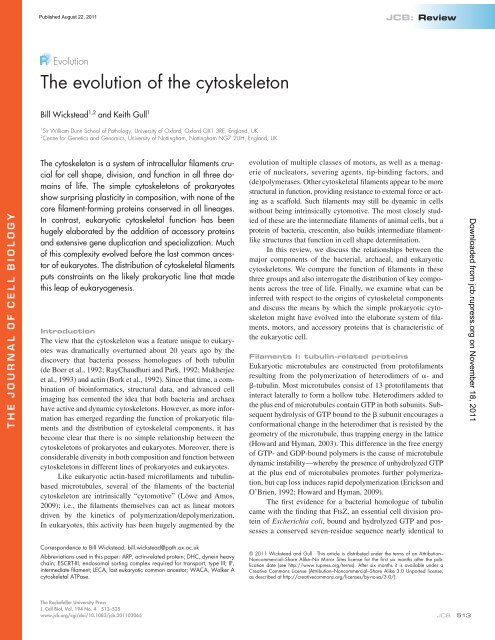
Published August 22, 2011Figure 1. Homology between prokaryotic and eukaryotic cytoskeletal filaments. Despite low levels <strong>of</strong> sequence similarity, <strong>the</strong> homologous cytoskeletalproteins FtsZ/TubZ/tubulin (top) and MreB/ParM/actin (bottom) have considerable conservation <strong>of</strong> folding and also longitudinal interaction. ParM andactin form similar helical filaments, but with opposite chirality (Orlova et al., 2007). Structures <strong>of</strong> filament subunits are derived from <strong>the</strong> following ProteinData Bank accession numbers: 1W5A (FtsZ; Oliva et al., 2004), 1JFF (/-tubulin; Löwe et al., 2001), 1JCG (MreB; van den Ent et al., 2001), and 1YAG(actin; Vorobiev et al., 2003).an N-terminal tubulin signature motif (de Boer et al., 1992;RayChaudhuri and Park, 1992; Mukherjee et al., 1993). Subsequentalignments showed that <strong>the</strong> similarities between FtsZ andtubulin sequences extended beyond <strong>the</strong> tubulin signature motif(Mukherjee and Lutkenhaus, 1994). Never<strong>the</strong>less, tubulin andFtsZ are very divergent in primary sequence, sharing only10% identity compared with >40% between most FtsZ sequences(Vaughan et al., 2004; Erickson, 2007). Despite lowsequence similarity, <strong>the</strong> determination <strong>of</strong> <strong>the</strong> crystal structures<strong>of</strong> tubulin and FtsZ revealed proteins with near identical folds(Fig. 1; Löwe and Amos, 1998; Nogales et al., 1998a,b).Unlike tubulin, FtsZ does not assemble into microtubules,but it does form a range <strong>of</strong> o<strong>the</strong>r structures in vitro using lateralinteractions between prot<strong>of</strong>ilaments (Bramhill and Thompson,1994; Mukherjee and Lutkenhaus, 1994; Erickson et al., 1996;Löwe and Amos, 1999, 2000). In vivo, FtsZ forms a dynamic“Z ring” between prospective daughter cells during cytokinesis(Fig. 2 A). This ring acts as a scaffold for <strong>the</strong> recruitment <strong>of</strong><strong>the</strong> “divisome”, which gradually constricts to divide <strong>the</strong> cell(Adams and Errington, 2009). This system <strong>of</strong> cytokinesis wasclearly an <strong>evolution</strong>ary success because FtsZ is both widely distributedand highly conserved. Its presence in most lineages <strong>of</strong>bacteria is indicative <strong>of</strong> it being an ancient protein. It has evenbeen suggested that an over-representation <strong>of</strong> amino acids withsimpler biosyn<strong>the</strong>tic pathways in conserved positions <strong>of</strong> FtsZmight be evidence that <strong>the</strong> protein predates <strong>the</strong> completion <strong>of</strong><strong>the</strong> 20–amino acid standard code itself (Davis, 2002).FtsZ is also encoded by many archaeal genomes, but it isnot ubiquitous in prokaryotes. Most notably, FtsZ (and o<strong>the</strong>rtubulin homologues) are entirely absent from one <strong>of</strong> <strong>the</strong> majorclades <strong>of</strong> archaea, <strong>the</strong> Crenarchaeota (Margolin, 2000; Vaughanet al., 2004)—with <strong>the</strong> possible exception <strong>of</strong> a highly divergentFtsZ-like gene in Sulfolobus solfataricus probably acquired byhorizontal gene transfer (Makarova et al., 2010). FtsZ is alsoabsent from at least one sequenced euryarchaeon (Picrophilustorridus; Fig. 3), as well as <strong>the</strong> bacterial groups Chlamydiae,Planctomycetes, and some Mycoplasmataceae (Vaughan et al.,2004; Adams and Errington, 2009). In <strong>the</strong> Crenarchaeota, FtsZindependentdivision is possible because <strong>of</strong> an alternative cytokinesismachinery provided by ESCRT-III (endosomal sortingcomplex required for transport, type III; Lindås et al., 2008;Samson et al., 2008). ESCRT-III proteins are conservedbetween archaea and eukaryotes, and eukaryotic proteins <strong>of</strong>this complex form dynamic polymers that have a role in membranescission events, including cell abscission during cytokinesis(Hurley and Hanson, 2010). In contrast to <strong>the</strong> lack <strong>of</strong>FtsZ in Crenarchaeota, <strong>the</strong> repertoire in Euryarchaeota has beenduplicated to form three monophyletic families (FtsZ1, 2, and 3),with FtsZ3 being <strong>the</strong> most divergent and least widely distributed(Vaughan et al., 2004).Downloaded from jcb.rupress.org on November 18, 2011 514JCB • VOLUME 194 • NUMBER 4 • 2011
Published August 22, 2011Figure 2. Bacterial, archaeal, and eukaryotic <strong>cytoskeleton</strong>s. Schematicrepresentations are shown for a small number <strong>of</strong> model organisms fromeach <strong>of</strong> <strong>the</strong> three domains <strong>of</strong> life (A–C), showing <strong>the</strong> organization <strong>of</strong> <strong>the</strong><strong>cytoskeleton</strong> in dividing and nondividing cells (right and left <strong>of</strong> each pair,respectively). Homologous filaments are colored similarly. Also shown is<strong>the</strong> possible organization <strong>of</strong> <strong>the</strong> <strong>cytoskeleton</strong> in <strong>the</strong> LECA (D), highlighting<strong>the</strong> ancestral families <strong>of</strong> microtubule motors.FtsZ is <strong>the</strong> most common tubulin homologue in prokaryotes(Fig. 3). However, <strong>the</strong>re are at least four o<strong>the</strong>r tubulin-likeprotein families in bacteria, most <strong>of</strong> <strong>the</strong>m with a restricted distribution.Several Bacillus plasmids encode tubulin-like proteins,which are very divergent in sequence from one ano<strong>the</strong>r and als<strong>of</strong>rom FtsZ and tubulins (Larsen et al., 2007). <strong>The</strong>se proteins includeTubZ and RepX, which play important roles in <strong>the</strong> stability<strong>of</strong> <strong>the</strong> plasmids that encode <strong>the</strong>m (Tinsley and Khan, 2006;Larsen et al., 2007). Monomeric TubZ has a structure very similarto /-tubulin (Ni et al., 2010), but forms right-handed helixfilaments consisting <strong>of</strong> two prot<strong>of</strong>ilaments (Aylett et al., 2010).An exception to <strong>the</strong> low level <strong>of</strong> sequence conservationbetween prokaryotic and eukaryotic cytoskeletal proteins areBtubA and B, found in <strong>the</strong> Verrucomicrobia Pros<strong>the</strong>cobacter(Jenkins et al., 2002). <strong>The</strong>se proteins are much more similar insequence to eukaryotic tubulins than are FtsZ, TubZ, or RepX.This high similarity in sequence, <strong>the</strong>ir restriction to Pros<strong>the</strong>cobacter,and phylogenetic analyses <strong>of</strong> FtsZ/tubulin all stronglysuggest that BtubA/B were acquired by horizontal gene transferfrom a eukaryote and are not descendents <strong>of</strong> bacterial FtsZ/TubZ (Jenkins et al., 2002; Vaughan et al., 2004; Schlieperet al., 2005; Pilh<strong>of</strong>er et al., 2007a). It is likely that BtubA is ahomologue <strong>of</strong> -tubulin and BtubB <strong>of</strong> -tubulin (Sontag et al.,2009), but <strong>the</strong> eukaryotic lineage that acted as <strong>the</strong> donor for<strong>the</strong>se proteins is not known. <strong>The</strong>y retain <strong>the</strong> ability to formprot<strong>of</strong>ilaments (not microtubules), both in vitro and in vivo(Sontag et al., 2005; Sontag et al., 2009), and <strong>the</strong>ir continuedexpression and functional conservation suggests that <strong>the</strong>y are<strong>of</strong> some benefit to Pros<strong>the</strong>cobacter cells. However, <strong>the</strong>y havenot displaced <strong>the</strong> native tubulin-like <strong>cytoskeleton</strong> in <strong>the</strong>seorganisms, but ra<strong>the</strong>r coexist alongside conventional FtsZ(Pilh<strong>of</strong>er et al., 2007b).Many eukaryotes possess FtsZ genes <strong>of</strong> prokaryotic origin(Fig. 3) inherited from <strong>the</strong> endosymbiotic ancestors <strong>of</strong> mitochondriaand chloroplasts. <strong>The</strong>se genes have subsequentlymoved from <strong>the</strong> organellar genomes to <strong>the</strong> nucleus, but <strong>the</strong>irorigins are still apparent in FtsZ phylogenies, where <strong>the</strong>y groupwith sequences from ei<strong>the</strong>r -proteobacteria or cyanobacteria(Kiefel et al., 2004; Vaughan et al., 2004; <strong>the</strong> bacterial groupsthat gave rise to mitochondria and chloroplasts, respectively).<strong>The</strong>se eukaryotic FtsZs function in <strong>the</strong> division <strong>of</strong> mitochondriaand chloroplasts by forming a division ring with striking similarityto that found in bacteria (Margolin, 2005). Redundancy <strong>of</strong>this FtsZ-based division machinery with <strong>the</strong> eukaryotic dynaminfission apparatus may provide a mechanism for multipleindependent losses <strong>of</strong> mitochondrial FtsZ since <strong>the</strong> eukaryoticroot (Osteryoung and Nunnari, 2003; Praefcke and McMahon,2004). In contrast, chloroplast FtsZ genes are more widely conserved(Fig. 3).Very recently, <strong>the</strong> FtsZ-tubulin family has been expandedfur<strong>the</strong>r by <strong>the</strong> identification <strong>of</strong> two new FtsZ-like protein familiesin prokaryotes—named FtsZl1 and FtsZl2 (Makarova andKoonin, 2010). Both <strong>of</strong> <strong>the</strong>se families are divergent from tubulin,FtsZ, or o<strong>the</strong>r previously identified families. One <strong>of</strong> <strong>the</strong> new families,FtsZl2 is found only in proteobacteria, but <strong>the</strong> FtsZl1 familyhas a much wider distribution that includes several bacterialgroups and Euryarchaeota. <strong>The</strong> function <strong>of</strong> FtsZl1 and FtsZl2 isunknown. It is possible that <strong>the</strong>y do not form FtsZ/tubulin-likefilaments, given <strong>the</strong>ir predicted structural divergence from o<strong>the</strong>rmembers <strong>of</strong> <strong>the</strong> superfamily (Makarova and Koonin, 2010).Filaments II: <strong>the</strong> actin superfamilyActin is a ubiquitous eukaryotic filament-forming protein.Actin filaments (also called micr<strong>of</strong>ilaments or F-actin) consist<strong>of</strong> two prot<strong>of</strong>ilament polymers wound toge<strong>the</strong>r in a righthandedhelix (Fig. 1). ATP hydrolysis by actin causes a muchless dramatic change in polymer stability than is seen for GTPhydrolysis in microtubules. As a result, pure actin shows littledynamic instability in vitro (Mitchison, 1992), but ra<strong>the</strong>rundergoes “treadmilling” through <strong>the</strong> polarized addition <strong>of</strong> ATPboundsubunits. In vivo, actin is much more dynamic than in vitroDownloaded from jcb.rupress.org on November 18, 2011<strong>The</strong> <strong>evolution</strong> <strong>of</strong> <strong>the</strong> <strong>cytoskeleton</strong> • Wickstead and Gull515
Published August 22, 2011Downloaded from jcb.rupress.org on November 18, 2011Figure 3. <strong>The</strong> distribution <strong>of</strong> key components <strong>of</strong> <strong>the</strong> <strong>cytoskeleton</strong> across <strong>the</strong> tree <strong>of</strong> life. Filled circle indicates presence <strong>of</strong> an identifiable member <strong>of</strong> aprotein family in an organism; open circle indicates absence/not found. Organisms are identified by genera only and are grouped into higher taxonomicgroups. Emiliania huxleyi has not been placed into one <strong>of</strong> <strong>the</strong> eukaryotic taxonomic groups in reflection <strong>of</strong> uncertainty as to <strong>the</strong> placement <strong>of</strong> Haptophyta.“MreB” includes MreB-like (Mbl/MreBH) sequences, which colocalize with MreB and are very similar in sequence (Carballido-López and Errington, 2003;Carballido-López et al., 2006). <strong>The</strong> archaeal sequences identified as MreB using <strong>the</strong> arCOG technique (arCOG04656; Makarova et al., 2007, 2010)have a closer affinity to Hsp70 sequences and are not included. Archaeal crenactin (*) is orthologous to <strong>the</strong> single common ancestor <strong>of</strong> eukaryotic actinand ARPs (Yutin et al., 2009; Ettema et al., 2011), but has been entered as actin for clarity. <strong>The</strong> distributions <strong>of</strong> <strong>the</strong> large number <strong>of</strong> prokaryotic actin-likeproteins o<strong>the</strong>r than MreB and FtsA (such as AlfA, Alp6/7/8) are not included here because <strong>of</strong> current difficulties in resolution <strong>of</strong> individual families (Dermanet al., 2009; Yutin et al., 2009). <strong>The</strong>re are possible orthologues <strong>of</strong> MinD in Euryarchaeota (Leipe et al., 2002), but <strong>the</strong>ir true membership is still unclear.due to <strong>the</strong> presence <strong>of</strong> monomer-binding factors, filamentseveringagents, and capping/stabilizing agents.Eukaryotic actin is a member <strong>of</strong> a large and diversesuperfamily <strong>of</strong> ATPases that includes Hsp70 chaperones andseveral classes <strong>of</strong> sugar/sugar alcohol kinases (Flaherty et al.,1991; Bork et al., 1992), as well as eukaryotic actin-related proteins(ARPs; Frankel and Mooseker, 1996; Schafer and Schroer,1999). Also identified as members <strong>of</strong> this superfamily were 516JCB • VOLUME 194 • NUMBER 4 • 2011
Published August 22, 2011several o<strong>the</strong>r bacterial proteins, including MreB, FtsA, andParM (Bork et al., 1992). Since initial identification, <strong>the</strong>superfamily <strong>of</strong> actin-like proteins in bacteria has proved tobe very complex, encompassing more than 20 classes <strong>of</strong> protein(Derman et al., 2009).<strong>The</strong> most common prokaryotic homologue <strong>of</strong> actin isMreB. As for tubulin/FtsZ, actin and MreB are very divergentin primary sequence but have similar structures (Fig. 1), basedon <strong>the</strong> “actin-fold” that unites <strong>the</strong> superfamily (Kabsch andHolmes, 1995). In vitro MreB forms assemblies <strong>of</strong> two prot<strong>of</strong>ilamentsthat are similar in structure to F-actin, but lack <strong>the</strong>helical twist (van den Ent et al., 2001; Esue et al., 2005). <strong>The</strong>conserved function <strong>of</strong> bacterial MreB (and closely related proteinssuch as Mbl and MreBH) appears to be in maintenance <strong>of</strong>cell shape (Jones et al., 2001; Graumann, 2007; Margolin,2009). MreB filaments form a helix below <strong>the</strong> cell membraneand influence <strong>the</strong> position <strong>of</strong> cell wall syn<strong>the</strong>sis (Fig. 2 A; Joneset al., 2001; Daniel and Errington, 2003). Consistent with thisfunction, MreB is generally conserved only among rod-shapedbacteria, but absent from spherical cocci. Because extant rodshapedbacterial lineages are probably more ancient, it has beensuggested that <strong>the</strong> coccal forms have been derived from <strong>the</strong>mmultiple times by <strong>the</strong> loss <strong>of</strong> MreB and associated genes(Margolin, 2009). However, <strong>the</strong> correlation between prokaryoteshape and MreB is not strict: some cocci still possess MreB and<strong>the</strong>re are rod-shaped bacteria that lack MreB (Margolin, 2009).ParM (previously StbA) is a plasmid-borne bacterial actinhomologue with a filament-forming role. ParM is only 20%identical to MreB, which is comparable to <strong>the</strong> degree <strong>of</strong> conservationbetween actin and ei<strong>the</strong>r ParM or MreB. None<strong>the</strong>less,ParM assembles twisted polymers in vitro that are very reminiscent<strong>of</strong> F-actin (van den Ent et al., 2002), but with a left-handedra<strong>the</strong>r than right-handed twist (Orlova et al., 2007). <strong>The</strong> energy<strong>of</strong> polymerization <strong>of</strong> ParM is used in <strong>the</strong> segregation <strong>of</strong> <strong>the</strong> R1plasmid (and o<strong>the</strong>rs containing <strong>the</strong> parMRC operon) by pushingnewly syn<strong>the</strong>sized plasmid to <strong>the</strong> cell poles (Garner et al., 2007;Salje and Löwe, 2008).To date, unambiguous ParM homologues are restricted toa close group <strong>of</strong> -proteobacteria, <strong>the</strong> Enterobacteriaceae. It islikely that <strong>the</strong> annotated “ParM” genes in <strong>the</strong> genomes <strong>of</strong>Firmicutes, -proteobacteria, and cyanobacteria represent o<strong>the</strong>rclasses <strong>of</strong> bacterial actin-like proteins, ra<strong>the</strong>r than true ParMorthologues. In support <strong>of</strong> this interpretation, “ParM” encodedon <strong>the</strong> Staphylococcus aureus pSK41 plasmid shares only 19%identity with -proteobacterial ParM sequences and appears tobe more structurally related to archaeal actin-like proteins (Poppet al., 2010).Both MreB and FtsA are almost exclusively restricted tobacteria (Fig. 3). Clear examples <strong>of</strong> MreB are also found ineuryarchea <strong>of</strong> <strong>the</strong> genera Methanopyrus, Methanobrevibacter, andMethano<strong>the</strong>rmobacter (Yutin et al., 2009). Given <strong>the</strong>ir limiteddistribution and close similarity to bacterial sequences, <strong>the</strong>seare most likely <strong>the</strong> result <strong>of</strong> horizontal gene transfer from bacteria.However, it cannot be entirely ruled out that <strong>the</strong>y are rarebut highly conserved genes <strong>of</strong> linear descent. Several o<strong>the</strong>r archaealsequences have been identified as possible MreB orthologuesusing <strong>the</strong> “archaeal cluster <strong>of</strong> orthologous groups” technique(Makarova et al., 2007, 2010). <strong>The</strong>se sequences have a closeraffinity to Hsp70 sequences than to MreB from bacteria orMethanopyrus and <strong>the</strong>ir grouping in arCOG04656 may be anartifact <strong>of</strong> <strong>the</strong> technique. <strong>The</strong>se sequences are not considered astrue MreB members herein (Fig. 3).Several o<strong>the</strong>r actin superfamily members exist in bacteria—notably: MamK, which is required for magnetosome organizationin Magnetospirillium (Komeili et al., 2006; Pradel et al.,2006); AlfA, which is involved in plasmid segregation in Bacillussubtilis in a manner similar to ParM (Becker et al., 2006);and Ta0583 from <strong>the</strong> euryarchaeon <strong>The</strong>rmoplasma acidophilum(Roeben et al., 2006; Hara et al., 2007). <strong>The</strong>se proteins haveei<strong>the</strong>r a limited phylogenetic distribution or <strong>the</strong>ir families haveyet to be well delimited (Derman et al., 2009; Yutin et al., 2009).<strong>The</strong> possibility that Ta0583 might be <strong>the</strong> closest prokaryotichomologue to eukaryotic actin (Hara et al., 2007) has not beensupported by subsequent analyses (Yutin et al., 2009; Ettemaet al., 2011). However, recently an archaeal actin-like familyhas been described that is monophyletic with eukaryotic actinand actin-related proteins (Yutin et al., 2009; Ettema et al.,2011). This protein family, dubbed “crenactin,” has a localizationin Pyrobaculum cells very similar to MreB in bacteria (Fig. 2 B;Ettema et al., 2011), but is more closely related to eukaryoticactin than to MreB or ParM. <strong>The</strong> possible implications <strong>of</strong> thisfamily are discussed fur<strong>the</strong>r below.Filaments III: intermediate filamentsEukaryotic intermediate filaments (IFs) are unlike microtubulesand micr<strong>of</strong>ilaments in structure, biochemistry, and phylogeneticdistribution. Unlike actin and tubulin, which are globular proteinsthat form polarized prot<strong>of</strong>ilaments, IF proteins are extendeddimers that overlap to form unpolarized cables. <strong>The</strong>re are manytypes <strong>of</strong> IF protein in vertebrates, most <strong>of</strong> which can be groupedinto five classes: (1) type I (acidic) keratins; (2) type II (basic)keratins; (3) vimentin and desmin; (4) -internexin and neur<strong>of</strong>ilamentproteins; and (5) lamins (Fuchs and Weber, 1994). Laminsare also present in protosomes, suggesting that all IF proteinfamilies are derived from a single lamin-like sequence in <strong>the</strong>common metazoan ancestor (Weber et al., 1989; Dodemontet al., 1994; Bovenschulte et al., 1995). Significantly, however,eukaryotic IF proteins have only been found unambiguously inanimals and <strong>the</strong>ir relatives (Erber et al., 1998), suggesting that<strong>the</strong>y are an innovation specific to this lineage and not present in<strong>the</strong> last eukaryotic common ancestor (LECA; see Fig. 3).<strong>The</strong> bacterium Caulobacter crescentus encodes a proteinwith a predicted arrangement <strong>of</strong> coiled-coils similar to that inanimal lamin A (Ausmees et al., 2003). This protein, CreS orcrescentin, forms helical filaments that are necessary for <strong>the</strong>vibrioid or helical cell shapes adopted by Caulobacter. Althoughcrescentin was originally described only as “IF-like”, <strong>the</strong>function and predicted secondary structure <strong>of</strong> <strong>the</strong> protein have<strong>of</strong>ten been interpreted as evidence that bacteria possess ancienthomologues <strong>of</strong> eukaryotic IFs. However, <strong>the</strong>re are several argumentsagainst such an interpretation. First, CreS has a veryrestricted distribution (Fig. 3); to date, it has only been found inCaulobacter. Given this restricted range, if lamin A andcrescentin are truly homologous, <strong>the</strong>n CreS would most likelyDownloaded from jcb.rupress.org on November 18, 2011<strong>The</strong> <strong>evolution</strong> <strong>of</strong> <strong>the</strong> <strong>cytoskeleton</strong> • Wickstead and Gull517
Published August 22, 2011represent a lateral transfer <strong>of</strong> a eukaryotic gene to Caulobacterra<strong>the</strong>r than a true bacterial homologue. Second, identifiablehomologues <strong>of</strong> eukaryotic IF proteins are restricted to metazoa(or possibly holozoa; Fig. 3) and may not have been present in<strong>the</strong> LECA—precluding vertical inheritance from prokaryotes.Third, similarities in predicted protein architecture (in this instance,coiled-coil positions) are not equivalent to fold homology.At present, it is not possible to compare eukaryotic IFproteins and crescentin for evidence <strong>of</strong> fold homology, as <strong>the</strong>reare no representatives <strong>of</strong> ei<strong>the</strong>r family for which full structureshave been determined. Moreover, although orthologues <strong>of</strong> crescentinhave not been identified outside <strong>of</strong> Caulobacter, <strong>the</strong>re isevidence that CreS is a member <strong>of</strong> a larger family <strong>of</strong> bacterialproteins (Bagchi et al., 2008). All <strong>of</strong> <strong>the</strong>se proteins are predictedto contain long stretches <strong>of</strong> coiled-coils, but <strong>the</strong> vast majorityhave no striking architectural similarity to lamin A (or o<strong>the</strong>rvertebrate IF proteins). Hence, <strong>the</strong> distribution <strong>of</strong> coiled-coilregions in crescentin and lamin A is more likely an example <strong>of</strong>convergence than a reflection <strong>of</strong> shared ancestry.Filaments IV: WACA proteinsProkaryotes have a fourth class <strong>of</strong> filament-forming proteinsknown as Walker A cytoskeletal ATPases (WACAs; Michie andLöwe, 2006). WACA proteins are a diverse family <strong>of</strong> ATPases(Koonin, 1993), which are <strong>the</strong>mselves part <strong>of</strong> <strong>the</strong> extremelylarge superclass <strong>of</strong> P-loop proteins including signal recognitionparticle proteins, Rho/Ras GTPases and cytoskeletal motors(Leipe et al., 2002). <strong>The</strong> WACA MinD is an active ATPase(de Boer et al., 1991) that forms dynamic filaments around <strong>the</strong>cell periphery in E. coli and inhibits Z ring formation (Pich<strong>of</strong>fand Lutkenhaus, 2001; Shih et al., 2003). In Bacillus subtilis,MinD is statically associated with <strong>the</strong> cell poles (Marston et al.,1998; Marston and Errington, 1999), making it unclear if dynamicpolymerization is an <strong>evolution</strong>arily conserved feature <strong>of</strong>MinD biology. MinD sequences are found in many groups <strong>of</strong>bacteria (Fig. 3) and <strong>the</strong>re are putative orthologues identified inEuryarchaeota (Leipe et al., 2002). However, <strong>the</strong> distinctions between<strong>the</strong> prokaryotic WACA families are yet to be fully resolved,so it is presently unclear if <strong>the</strong>se archaeal sequences are monophyleticwith bacterial MinD. Even if not strict MinD orthologues,examples <strong>of</strong> WACA proteins do appear to be present inboth Euryarcheaota and Crenarchaeota (Makarova et al., 2010).Several o<strong>the</strong>r bacterial WACA proteins have been described.ParA, ParF, and Soj all play roles in DNA segregationby different mechanisms (Pogliano, 2008; Löwe and Amos,2009), demonstrating an apparent versatility <strong>of</strong> this system forsegregation. It is perhaps surprising, <strong>the</strong>refore, that <strong>the</strong>re are noeukaryotic WACA filaments. <strong>The</strong> fold <strong>of</strong> MinD and Soj is distantlyrelated to that <strong>of</strong> eukaryotic septins (Cordell and Löwe,2001; Leonard et al., 2005; Löwe and Amos, 2009), but thismost likely reflects <strong>the</strong> distant relationship shared by all P-loopGTPases (Leipe et al., 2002). <strong>The</strong>re are, however, MinD genes<strong>of</strong> bacterial ancestry in Viridiplantae (green algae and land plants)and some stramenopiles (Fig. 3), which play a role in plastiddivision (Marrison et al., 1999; Colletti et al., 2000; Kanamaruet al., 2000). It is noteworthy that all eukaryotes that possessMinD genes also encode endosymbiont-derived FtsZ.Prokaryotic division: a common problemwith many solutionsIf <strong>the</strong>re is an overarching <strong>the</strong>me running through <strong>the</strong> <strong>evolution</strong><strong>of</strong> <strong>the</strong> prokaryotic <strong>cytoskeleton</strong>, it appears to be this: plasticity.Each <strong>of</strong> <strong>the</strong> major families <strong>of</strong> cytomotive filament-formingproteins consists <strong>of</strong> several paralogues, which are <strong>of</strong>ten as divergentfrom each o<strong>the</strong>r as <strong>the</strong>y are from eukaryotic homologues.Some <strong>of</strong> <strong>the</strong> classes—such as FtsA (from <strong>the</strong> MreB/actinsuperfamily) or <strong>the</strong> newly identified FtsZ-like families—maynot form filaments in vivo at all. However, among thosefamilies that do polymerize, lateral interactions in <strong>the</strong> corefilament appear to be quite malleable over <strong>evolution</strong>ary timescaleswithout disrupting polymerization (for example, straightMreB filaments, against left-handed twisting ParM and righthandedactin).<strong>The</strong>re is also plasticity in <strong>the</strong> biological function forwhich filaments are used—FtsZ and MreB are involved in celldivision and morphology, while <strong>the</strong>ir homologues TubZ andParM play roles in plasmid segregation. <strong>The</strong> result is that prokaryotesuse different systems to solve common problems. Inbacteria, active DNA segregation can be achieved by at leastthree types <strong>of</strong> segregation machinery (Ebersbach and Gerdes,2005; Hayes and Barillà, 2006). Type I systems use WACAproteins, such as ParA and Soj. Type II systems are based on<strong>the</strong> actin homologue ParM. Type III systems are those fromBacillus that use tubulin-like homologues, such as TubZ andRepX. In a striking example <strong>of</strong> apparent convergence, bothParM (type II) and TubZ (type III) form similar helical filamentsthat probably act to push apart plasmids (van den Entet al., 2002; Aylett et al., 2010). However, it is type I systemsthat are by far <strong>the</strong> most wide spread in bacteria. It is interesting,<strong>the</strong>n, that filaments based on WACA proteins are notconserved in eukaryotes, and may not be widely used in chromosomesegregation in archaea (Bernander, 2000; Makarovaet al., 2010). Significantly, none <strong>of</strong> <strong>the</strong> cytoskeletal proteinfamilies is ubiquitous to all prokaryotes, or even one <strong>of</strong> <strong>the</strong>two prokaryotic domains <strong>of</strong> life. <strong>The</strong> role for FtsZ in cytokinesisappears to be <strong>the</strong> function under most selective pressurefor retention (Erickson, 2007), but even this protein has beenlost from several lineages <strong>of</strong> bacteria and <strong>the</strong> entire crenarchaealclade.Eukaryogenesis: families and specializationDespite being based on homologous filaments, <strong>the</strong> eukaryotic<strong>cytoskeleton</strong> is not simply a more extensive version <strong>of</strong> <strong>the</strong> prokaryoticone (Fig. 2 C). <strong>The</strong> complex eukaryotic <strong>cytoskeleton</strong> isactually based on a smaller set <strong>of</strong> ancestral cytomotive filamentsthan that <strong>of</strong> prokaryotes. With <strong>the</strong> notable exception <strong>of</strong> <strong>the</strong> prokaryote-likedivision machineries associated with some plastids,only one paralogue <strong>of</strong> an MreB/crenactin family proteinand one FtsZ/TubZ protein seems to have founded <strong>the</strong> eukaryotic<strong>cytoskeleton</strong>. However, this small selection has undergoneseveral rounds <strong>of</strong> gene duplication and specialization from thisancestral set.Heterodimers <strong>of</strong> - and -tubulin make up <strong>the</strong> vast majority<strong>of</strong> tubulin in eukaryotic cells. <strong>The</strong>y are sufficient for <strong>the</strong> production<strong>of</strong> microtubules in vitro, and it is very likely that <strong>the</strong>yDownloaded from jcb.rupress.org on November 18, 2011 518JCB • VOLUME 194 • NUMBER 4 • 2011
Published August 22, 2011were <strong>the</strong> first types to evolve from <strong>the</strong> single proto-tubulinancestor. However, <strong>the</strong>y are not <strong>the</strong> only ancestral tubulins.Analyses suggest that <strong>the</strong> tubulin family encompasses at least sixclasses, named , , , , , and , with a fur<strong>the</strong>r two divergenttypes ( and ) being found in some organisms (Vaughan et al.,2000; Dutcher, 2003). -Tubulin plays an essential role in microtubulenucleation (through <strong>the</strong> action <strong>of</strong> <strong>the</strong> conserved -tubulinring complex) and is, like - and -, ubiquitous to all eukaryotes.In contrast, - and -tubulin have centriolar roles (Dutcherand Trabuco, 1998; Chang and Stearns, 2000; Ruiz et al., 2000)and are conserved in nearly all organisms that build centrioles/basal bodies and absent from organisms that have lost cilia/flagella (Hodges et al., 2010). At least five <strong>of</strong> <strong>the</strong> classes <strong>of</strong> tubulin(, , , , and ) can be traced back to <strong>the</strong> last commoneukaryotic common ancestor (Fig. 3). All <strong>of</strong> <strong>the</strong> tubulin typesare more closely related to one ano<strong>the</strong>r than to FtsZ/TubZ(Vaughan et al., 2000; Dutcher, 2003), implying that <strong>the</strong>y alldescended from a common proto-tubulin by gene duplication.Both duplication and specialization into functionally distinctclasses must have occurred before <strong>the</strong> LECA.<strong>The</strong>re are striking parallels between <strong>the</strong> proto-eukaryotic<strong>evolution</strong> <strong>of</strong> tubulins and that <strong>of</strong> actin. <strong>The</strong> proteins most similarin sequence to conventional actin are <strong>the</strong> ARPs. <strong>The</strong> ARPs coverat least eight major families (Sehring et al., 2007) that are foundonly in eukaryotes. Four ARP families (ARP4, 5, 6/7, and 8/9)are not associated with cytoplasmic actin, but are nuclear proteinsinvolved in chromatin remodeling (Chen and Shen, 2007;Dion et al., 2010). <strong>The</strong> remaining families—ARP1, 2, 3, and10/11—have important roles in modification or extension <strong>of</strong> cytoplasmicactin function. ARP1 is an integral part <strong>of</strong> <strong>the</strong> dynactincomplex, which links <strong>the</strong> actin- and tubulin-based <strong>cytoskeleton</strong>(Schroer, 2004). Yeast Arp10p and metazoan Arp11 (ARP10/11family members) are also part <strong>of</strong> <strong>the</strong> dynactin complex (Eckleyand Schroer, 2003; Clark and Rose, 2006). In contrast, a complex<strong>of</strong> seven subunits formed around a heterodimer <strong>of</strong> ARP2 andARP3 is a major F-actin nucleator in most eukaryotes (Pollardand Beltzner, 2002; Goley and Welch, 2006). <strong>The</strong> only ARP thatappears to form filaments is Arp1p (Schafer et al., 1994; Binghamand Schroer, 1999). <strong>The</strong>se filaments are much shorter than thoseseen for actin, but have similar morphology.As for tubulin, <strong>the</strong>re was a single actin-like protein in <strong>the</strong>proto-eukaryote which evolved into multiple paralogous formsbefore <strong>the</strong> LECA. In doing so, both <strong>the</strong> actin- and tubulin-based<strong>cytoskeleton</strong> independently invented complexes containing divergentforms <strong>of</strong> filament subunits that were specialized fornucleation (ARP2/3 and -tubulin ring complexes). However,in <strong>the</strong> case <strong>of</strong> <strong>the</strong> actin-based <strong>cytoskeleton</strong>, this nucleatingcomplex appears to be expendable in some circumstances becauseorthologues <strong>of</strong> ARP2 and ARP3 have been lost multipletimes across eukaryotes (Fig. 3; note that organisms alwayslose both, as would be expected for a complex). Cross talk betweenmicrotubules and micr<strong>of</strong>ilaments via <strong>the</strong> dynactin complexhas also been lost multiple times, as can be seen from <strong>the</strong>distribution <strong>of</strong> ARP1.<strong>The</strong> history <strong>of</strong> <strong>the</strong>se core eukaryotic cytoskeletal familiesalso shows some degree <strong>of</strong> plasticity, but it is ra<strong>the</strong>r differentfrom that seen in prokaryotes. <strong>The</strong> eukaryotic paralogues were,for <strong>the</strong> most part, present in <strong>the</strong> LECA. Although rare divergenttypes do exist (for example - and -tubulin), <strong>the</strong>y do not have<strong>the</strong> same level <strong>of</strong> divergence from core types as is seen for divergentprokaryotic proteins. Moreover, <strong>the</strong>ir biological roleseems more static. Divergence between <strong>the</strong> distribution <strong>of</strong> familiesin extant eukaryotes is largely a product <strong>of</strong> loss <strong>of</strong> function(e.g., loss <strong>of</strong> dynactin complex and ARP1; loss <strong>of</strong> centrioles and/-tubulin).Cytoskeletal motorsProkaryotes use <strong>the</strong> intrinsic cytomotive capacity <strong>of</strong> cytoskeletalfilaments to do work. Eukaryotic cells also use <strong>the</strong> energy <strong>of</strong>polymerization/depolymerization (McIntosh et al., 2010). However,in eukaryotes <strong>the</strong> intrinsic dynamics <strong>of</strong> <strong>the</strong> filaments hasbeen augmented by <strong>the</strong> addition <strong>of</strong> <strong>the</strong> cytoskeletal motors—dyneins, kinesins, and myosins—which derive energy fromATP hydrolysis to perform various cellular tasks. Each <strong>of</strong> <strong>the</strong>semotors is a superfamily containing multiple classes <strong>of</strong> specializedproteins.Kinesins and myosins share a similar fold structure, suggestingthat <strong>the</strong>y have common ancestry (Kull et al., 1996,1998). This is ra<strong>the</strong>r surprising given that <strong>the</strong>y now act exclusivelyon microtubules and F-actin, respectively, and presentssomething <strong>of</strong> a conundrum in terms <strong>of</strong> <strong>evolution</strong>ary cell biology.It is only plausible for motors to evolve after <strong>the</strong> filamentson which <strong>the</strong>y act have been established. As has been discussedabove, filaments made from tubulin/FtsZ and actin/MreBhomologues were already well established in prokaryotes before<strong>the</strong> emergence <strong>of</strong> eukaryotes, yet both motor classes evolvedonly later in <strong>the</strong> proto-eukaryote. This requires that ei<strong>the</strong>r: (1) asingle “ur-kinesin-myosin” originally walked on both tubulinandactin-based filaments and only later specialized into separatefamilies; (2) a motor evolved for one type <strong>of</strong> filament andsubsequently developed an ability to move on <strong>the</strong> o<strong>the</strong>r; or (3)both motor families evolved independently from <strong>the</strong> same family<strong>of</strong> NTPases. <strong>The</strong> large differences in structure and sequencebetween tubulin/FtsZ and actin/MreB filaments suggest that <strong>the</strong>last scenario is most plausible, even if less parsimonious. <strong>The</strong>reis also a precedent for this because this family <strong>of</strong> P-loopNTPases also gave rise to many eukaryote-specific GTPases(including Arf, Ras/Rab, Rho/Ran, and G protein families;Leipe et al., 2002).Dyneins have a very different structure to that <strong>of</strong> myosinsand kinesins. <strong>The</strong> core <strong>of</strong> <strong>the</strong> dynein complex is <strong>the</strong> dyneinheavy chain (DHC), which is a member <strong>of</strong> ano<strong>the</strong>r large and diversesuperclass <strong>of</strong> NTPases, <strong>the</strong> AAA+ proteins (a family includingATPases associated with various cellular activities).DHCs contain six AAA+ domains that form an intramolecularhexameric ring (Samsó et al., 1998; King, 2000; Burgess et al.,2003), implying that DHCs originally evolved by ei<strong>the</strong>r intragenedomain duplication or fusion <strong>of</strong> genes encoding AAA+proteins. Because all classes <strong>of</strong> DHC have <strong>the</strong> same structure,this duplication occurred before <strong>the</strong> creation <strong>of</strong> functional paralogues.<strong>The</strong> dynein superfamily is also different from kinesinsand myosins in that all but one <strong>of</strong> <strong>the</strong> nine classes are associatedwith one specific organelle—<strong>the</strong> cilium. Only cytoplasmicdynein 1 has a conserved role outside <strong>of</strong> <strong>the</strong> cilium; <strong>the</strong> o<strong>the</strong>rDownloaded from jcb.rupress.org on November 18, 2011<strong>The</strong> <strong>evolution</strong> <strong>of</strong> <strong>the</strong> <strong>cytoskeleton</strong> • Wickstead and Gull519
Published August 22, 2011families being <strong>the</strong> retrograde motor <strong>of</strong> intraflagellar transport(for historic reasons known as cytoplasmic dynein 2) and sevenfamilies <strong>of</strong> dyneins built into motile axonemes.Given <strong>the</strong>ir central role in <strong>the</strong> eukaryotic cell, it was surprisingto find from post-genomic analyses <strong>of</strong> motor repertoiresthat none <strong>of</strong> <strong>the</strong> families is ubiquitous to all eukaryotes. Indeed,myosin or dynein superfamilies have been lost in <strong>the</strong>ir entiretyfrom some lineages (see Fig. 3; Lawrence et al., 2001; Matsuzakiet al., 2004; Richards and Cavalier-Smith, 2005; Wickstead andGull, 2007). To date, kinesins are encoded by all sequencedgenomes, but <strong>the</strong> repertoire in each lineage can be very different(Wickstead and Gull, 2006; Wickstead et al., 2010)—for example,Entamoeba histolytica encodes only members <strong>of</strong> kinesin-5,-14, and -15 families (and no dynein at all), whereas <strong>the</strong> apicomplexan<strong>The</strong>ileria parva encodes kinesin-8 and -13.<strong>The</strong> axonemeOne <strong>of</strong> <strong>the</strong> iconic structures <strong>of</strong> <strong>the</strong> eukaryotic <strong>cytoskeleton</strong> is<strong>the</strong> axoneme—canonically, nine microtubule doublets radiallyplaced around a central apparatus containing two singlet microtubules(Haimo and Rosenbaum, 1981). <strong>The</strong> axoneme is <strong>the</strong>structure around which all eukaryotic cilia and flagella areformed. <strong>The</strong> <strong>evolution</strong> <strong>of</strong> flagella/cilia forms part <strong>of</strong> a review inthis series by Carvalho-Santos et al. (2011) and will not bediscussed at length here. However, <strong>the</strong> axoneme represents amajor cytoskeletal innovation and aspects <strong>of</strong> its <strong>evolution</strong> arehighly pertinent to <strong>the</strong> emergence <strong>of</strong> <strong>the</strong> <strong>cytoskeleton</strong> in <strong>the</strong>proto-eukaryote.From its distribution in extant organisms, it is clear that<strong>the</strong> axoneme evolved before <strong>the</strong> last common ancestor <strong>of</strong> eukaryotes.Since that time, it has been independently lost frommultiple eukaryotic lineages (notably, from many lineages <strong>of</strong>plants, fungi, and amoebae). <strong>The</strong>se losses are closely correlatedto losses <strong>of</strong> ancestral kinesin and dynein families. <strong>The</strong> axonemeevolved from <strong>the</strong> microtubules <strong>of</strong> <strong>the</strong> cytoplasm (Cavalier-Smith,1978, 1982, 2006). It is usually inferred that <strong>the</strong> earliest axonemelikestructures were microtubule-based protrusions from <strong>the</strong>cell body with a solely sensory function, similar to <strong>the</strong> immotilecilia found on many types <strong>of</strong> differentiated animal cells(Rosenbaum and Witman, 2002; Jékely and Arendt, 2006; Satiret al., 2007). Axonemal motility, <strong>the</strong>refore, only arises laterwith <strong>the</strong> <strong>evolution</strong> <strong>of</strong> <strong>the</strong> specialized axonemal dynein classes.If it is assumed that cytoplasmic dynein 1 evolved before <strong>the</strong>o<strong>the</strong>r classes, <strong>the</strong>n dynein phylogenies support this hypo<strong>the</strong>sis,suggesting <strong>the</strong> <strong>evolution</strong> <strong>of</strong> intraflagellar transport (IFT) beforeaxonemal dynein (Wilkes et al., 2008; Hartman and Smith,2009). This rooting is also consistent with <strong>evolution</strong> <strong>of</strong> <strong>the</strong>proto-cilium as a motile organelle, but where movement wasoriginally based on gliding driven by <strong>the</strong> IFT machinery (Mitchell,2004, 2007). However, rooting <strong>of</strong> DHC phylogenies with <strong>the</strong>closest eukaryotic relative <strong>of</strong> dynein, midasin (Garbarino andGibbons, 2002; Iyer et al., 2004), suggests that microtubulesliding was much closer to <strong>the</strong> origin <strong>of</strong> ciliary <strong>evolution</strong>,emerging before <strong>the</strong> specialized machinery for IFT (Wicksteadand Gull, 2011). This implies that <strong>the</strong> proto-cilium evolved notfrom an immotile protrusion, but from a motile cytoplasmicmicrotubule bundle analogous to <strong>the</strong> axostyles <strong>of</strong> oxymonads(Grimstone and Cleveland, 1965; McIntosh, 1973), presumablybeing assembled in an IFT-independent manner as are someaxonemes today (Witman, 2003; Briggs et al., 2004).<strong>The</strong> proto-eukaryotic r<strong>evolution</strong>One <strong>of</strong> <strong>the</strong> most surprising results <strong>of</strong> our increasing ability toprobe <strong>the</strong> characteristics <strong>of</strong> <strong>the</strong> LECA has been how much <strong>of</strong><strong>the</strong> biological complexity in extant cells can be traced back tothis ancestral cell. <strong>The</strong> LECA possessed much <strong>of</strong> <strong>the</strong> complexitynow seen in <strong>the</strong> replisome (Liu et al., 2009), <strong>the</strong> spliceosome(Collins and Penny, 2005), and <strong>the</strong> endocytic system (Dackset al., 2009), as well as <strong>the</strong> machineries necessary for meiosis(Ramesh et al., 2005) and phagotrophy (Cavalier-Smith, 2002b;Yutin et al., 2009). Moreover, comparative analysis <strong>of</strong> <strong>the</strong>genome <strong>of</strong> <strong>the</strong> free-living excavate Naegleria gruberi identified4,000 protein groups that probably were present in <strong>the</strong> LECA(Fritz-Laylin et al., 2010).This “complexity early” model <strong>of</strong> eukaryotic <strong>evolution</strong> ismirrored in <strong>the</strong> <strong>cytoskeleton</strong> (Fig. 2 D). Somewhere in <strong>the</strong> <strong>evolution</strong>aryspace between prokaryotes and <strong>the</strong> LECA, singleproto-tubulin and proto-actin molecules diversified into multiplespecialized forms. Three classes <strong>of</strong> motors arose independently,and evolved to include at least nine classes <strong>of</strong> dynein,eleven classes <strong>of</strong> kinesin, and three classes <strong>of</strong> myosin (Richardsand Cavalier-Smith, 2005; Wickstead and Gull, 2007; Wicksteadet al., 2010). As well as <strong>the</strong>se, <strong>the</strong> axoneme formed, with 100–200 associated proteins (Avidor-Reiss et al., 2004; Pazour et al.,2005; Broadhead et al., 2006), many <strong>of</strong> which have no prokaryoticorthologues. Between <strong>the</strong> prokaryotes and <strong>the</strong> LECA, ar<strong>evolution</strong> occurred in cytoskeletal biology.Such complexity cannot have appeared fully formed, butarose by stepwise elaborations <strong>of</strong> cell structure (and geneticrepertoire). However, <strong>the</strong> large number <strong>of</strong> simpler intermediateforms that must have existed appear to have left no descendants.This is perhaps because a great many <strong>of</strong> <strong>the</strong>se changes occurredin a relatively short time, with one innovation creating a favorablelandscape for <strong>the</strong> <strong>evolution</strong> <strong>of</strong> <strong>the</strong> next (Cavalier-Smith,2006). Alternatively, all descendants <strong>of</strong> <strong>the</strong>se intermediateforms have been simply out-competed by <strong>the</strong> arrival <strong>of</strong> <strong>the</strong>LECA, with its mitochondrial endosymbiont, endomembranesystem, and sophisticated <strong>cytoskeleton</strong>. What is clear is that sincethis complex LECA, <strong>the</strong> diversification into many eukaryoticlineages has <strong>of</strong>ten been accompanied not by <strong>the</strong> addition <strong>of</strong> fur<strong>the</strong>rclasses, but by loss <strong>of</strong> ancestral ones. Some <strong>of</strong> <strong>the</strong>se lossesare associated with loss <strong>of</strong> specific structures or functions (suchas axonemal motility), but <strong>the</strong>re appears to be a remarkable flexibilityin <strong>the</strong> precise repertoire <strong>of</strong> many <strong>of</strong> <strong>the</strong>se ancient familiesthat is required for eukaryotic cell function.Although <strong>the</strong>y are constructed from homologous proteins,<strong>the</strong> functions <strong>of</strong> prokaryotic and eukaryotic filaments are notbroadly homologous. In eukaryotes, DNA segregation is ubiquitouslyperformed by <strong>the</strong> tubulin-based <strong>cytoskeleton</strong>, whereascytokinesis involves actin–myosin. In contrast, most prokaryoticcytokinesis is based on <strong>the</strong> tubulin homologue FtsZ, whereasactin-like, tubulin-like, or WACA proteins may be used forDNA segregation. This suggests that a switch must have occurredin <strong>cytoskeleton</strong> function in <strong>the</strong> proto-eukaryote (Löwe andDownloaded from jcb.rupress.org on November 18, 2011 520JCB • VOLUME 194 • NUMBER 4 • 2011
Published August 22, 2011Amos, 2009). However, with several divergent forms occurringin both filament families, and increasing evidence for plasticityin prokaryotic cytoskeletal function, this was perhaps not sucha dramatic transition as it might at first appear.FtsZ and tubulin are highly conserved proteins that areconstrained in sequence, yet are very divergent from one ano<strong>the</strong>r(Erickson, 2007). Because this is <strong>the</strong> case, how did FtsZ (or aprokaryotic homologue <strong>the</strong>re<strong>of</strong>) evolve into eukaryotic tubulin?It has been suggested that eukaryotic tubulins evolved from aTubZ-like sequence (Cavalier-Smith, 2010). Such a scenario hasmuch to recommend it. Because TubZ functions in DNA segregation,<strong>the</strong>re is a plausible <strong>evolution</strong>ary transition to <strong>the</strong> development<strong>of</strong> <strong>the</strong> tubulin-based mitosis <strong>of</strong> eukaryotes. Moreover,TubZ and FtsZ coexist in bacterial cells, meaning that a tubulinlikefunction might evolve for a descendant <strong>of</strong> TubZ without <strong>the</strong>loss <strong>of</strong> FtsZ-based cell scission function. Alternatively, a redundantFtsZ, resulting from ei<strong>the</strong>r gene duplication or <strong>the</strong> <strong>evolution</strong><strong>of</strong> actin-based cytokinesis, could have provided <strong>the</strong> necessaryrelaxation <strong>of</strong> sequence constraints such that FtsZ might evolveinto eukaryotic tubulin (Doolittle, 1995; Erickson, 2007).Interestingly, <strong>the</strong> increase in cell size that accompaniedeukaryogenesis may have been one <strong>of</strong> <strong>the</strong> factors favoring<strong>the</strong> replacement <strong>of</strong> FtsZ-based cytokinesis with an actin-basedsystem. To date, no Z ring has been described that is muchlarger than 1 µm in diameter. Even very large bacteria requireonly <strong>the</strong>se small Z rings for division because <strong>the</strong>y form newcells by budding <strong>of</strong>f small endospores (Angert and Losick,1998; Robinow and Angert, 1998). This has led to <strong>the</strong> tentativesuggestion that <strong>the</strong>re may be a maximum diameter beyondwhich <strong>the</strong> Z ring cannot function efficiently (Erickson, 2007).Eukaryotic originsAlongside <strong>the</strong> many genes that seem to have specifically arisenduring <strong>the</strong> proto-eukaryotic r<strong>evolution</strong>, eukaryotic genomes containa mixture <strong>of</strong> genes with apparent archaeal ancestry and genes<strong>of</strong> bacterial origin (Koonin, 2010). Genes <strong>of</strong> apparent bacterialorigin are more numerous (Esser et al., 2004; Rivera and Lake,2004; Makarova et al., 2005). Because all extant eukaryotes aredescended from a single ancestor that already contained <strong>the</strong> mitochondrialendosymbiont, this provides a good candidate source formany <strong>of</strong> <strong>the</strong> genes <strong>of</strong> bacterial origin (although <strong>the</strong> phylogeneticaffinities <strong>of</strong> <strong>the</strong>se genes are actually ra<strong>the</strong>r mixed and not onlyfrom <strong>the</strong> -proteobacteria; Koonin, 2010). In contrast, much <strong>of</strong> <strong>the</strong>machinery for replication, transcription, and translation appears tobe fundamentally archaeal, or ra<strong>the</strong>r archaeal-like (Ribeiro andGolding, 1998; Rivera et al., 1998; Yutin et al., 2008), suggestingthat <strong>the</strong> eukaryotic nuclear genome originated ei<strong>the</strong>r from withinArchaea or from a sister group to it.<strong>The</strong> eukaryotic <strong>cytoskeleton</strong> more likely evolved from <strong>the</strong>archaeal-like host than a bacterial endosymbiont. If we assumethat <strong>the</strong> mitochondrion was acquired by a phagocytosis-like processin a proto-eukaryote, <strong>the</strong>n <strong>the</strong> close association betweenactin and endomembrane systems in eukaryotes would stronglysuggest that a primitive actin <strong>cytoskeleton</strong> was already inexistence (and <strong>the</strong>refore <strong>the</strong> actin <strong>cytoskeleton</strong> is host- and notendosymbiont-derived). Such a phagocytic origin provides abiologically plausible mechanism for acquisition <strong>of</strong> mitochondriaduring eukaryogenesis (Cavalier-Smith, 2002b; Yutin et al.,2009), although <strong>the</strong> presence <strong>of</strong> prokaryotes that live in o<strong>the</strong>r prokaryotesdemonstrates that phagocytosis is not necessarily a prerequisitefor acquisition (von Dohlen et al., 2001). <strong>The</strong> origin fortubulin is less clear. However, at least initially FtsZ/TubZ filamentsfrom <strong>the</strong> incoming -proteobacterium would have beentrapped on <strong>the</strong> “wrong side” <strong>of</strong> both <strong>the</strong> phagosome and bacterialmembranes. This would make <strong>the</strong>m unavailable for functions inDNA or organelle movements in <strong>the</strong> host until gene transfer to<strong>the</strong> host genome and subsequent translation in <strong>the</strong> host cytoplasm.<strong>The</strong> fact that several eukaryotic lines also still possess identifiablemitochondrial FtsZ with functions in organelle division suggeststhat FtsZ from <strong>the</strong> endosymbiont was constrained by a conservedfunction and did not evolve directly into cytoplasmic tubulin.Phylogenomic studies variously support <strong>the</strong> origin <strong>of</strong> <strong>the</strong>archaea-like part <strong>of</strong> <strong>the</strong> eukaryotic genome from within Crenarchaeota(Cox et al., 2008; Foster et al., 2009), Euryarchaeota(Pisani et al., 2007), or basal to ei<strong>the</strong>r clade (Yutin et al., 2008;Kelly et al., 2010). Alternatively, both Archaea and eukaryotesmight be derived from an extinct “neomuran” line (Brown andDoolittle, 1997; Cavalier-Smith, 2002a). If both eukaryotic actinand tubulin are derived from an archaea-like host ra<strong>the</strong>r than <strong>the</strong>endosymbiont, <strong>the</strong>n what does this tell us about <strong>the</strong> phylogeneticposition <strong>of</strong> <strong>the</strong> proto-eukaryote? Because FtsZ (and allo<strong>the</strong>r identified tubulin homologues) are missing from Crenarchaeota(Fig. 3), this would exclude an origin for eukaryotesfrom within this group (<strong>the</strong> “eocytes”; Lake, 1988). Conversely,<strong>the</strong> recently identified archaeal protein crenactin, which is monophyleticwith eukaryotic actin/ARPs and is <strong>the</strong> closest extanthomologue <strong>of</strong> eukaryotic actin, is absent from Euryarchaeota(Yutin et al., 2009). Given <strong>the</strong>se data, <strong>the</strong> presence <strong>of</strong> both crenactinand FtsZ in Korarcheota, deep-branched archaea thatappear to be nei<strong>the</strong>r Euryarchaeota nor Crenarchaeota (Barnset al., 1996; Elkins et al., 2008), is particularly interesting (seeFig. 3). Significantly, <strong>the</strong> sequenced korarchaeon also containskey components <strong>of</strong> eukaryotic RNA polymerases (Koonin et al.,2007) and histones (Bell and White, 2010). Such inferencesbased on distribution are by no means definitive (it is noteworthythat <strong>the</strong> sequenced korarchaeon lacks ESCRT-III[Makarova et al., 2010], which is found in eukaryotes). However,<strong>the</strong>y do lend support to a root for <strong>the</strong> eukaryotes ei<strong>the</strong>rembedded in a basal archaeal line, or as an earlier neomuran cell.Over <strong>the</strong> last 20 years our model <strong>of</strong> <strong>the</strong> <strong>evolution</strong> <strong>of</strong> <strong>the</strong><strong>cytoskeleton</strong> has changed greatly. <strong>The</strong> prokaryotic <strong>cytoskeleton</strong>has been shown not only to exist, but to be dynamic and diverse.Surprisingly, it has also turned out to be expendable—at least inits canonical forms. During eukaryogenesis a huge amount <strong>of</strong>complexity was built up before <strong>the</strong> LECA, but <strong>the</strong> compositionin individual extant organisms shows a remarkable flexibility.As more biological data are ga<strong>the</strong>red and it becomes possible toanalyze sequences from a greater range <strong>of</strong> genomes, it seemslikely that more surprises will emerge. For prokaryotes, it maybe that some <strong>of</strong> <strong>the</strong> players have yet to join <strong>the</strong> stage.<strong>The</strong> authors wish to thank Flavia Moreira-Leite (<strong>University</strong> <strong>of</strong> <strong>Oxford</strong>, <strong>Oxford</strong>,UK) for assistance in development <strong>of</strong> <strong>the</strong> manuscript and Stephen D. Bell (<strong>University</strong><strong>of</strong> <strong>Oxford</strong>) for helpful critical review.This work was supported by <strong>the</strong> Wellcome Trust.Downloaded from jcb.rupress.org on November 18, 2011<strong>The</strong> <strong>evolution</strong> <strong>of</strong> <strong>the</strong> <strong>cytoskeleton</strong> • Wickstead and Gull521
Published August 22, 2011Submitted: 11 February 2011Accepted: 20 June 2011ReferencesAdams, D.W., and J. Errington. 2009. Bacterial cell division: assembly, maintenanceand disassembly <strong>of</strong> <strong>the</strong> Z ring. Nat. Rev. Microbiol. 7:642–653.doi:10.1038/nrmicro2198Angert, E.R., and R.M. Losick. 1998. Propagation by sporulation in <strong>the</strong> guineapig symbiont Metabacterium polyspora. Proc. Natl. Acad. Sci. USA.95:10218–10223. doi:10.1073/pnas.95.17.10218Ausmees, N., J.R. Kuhn, and C. Jacobs-Wagner. 2003. <strong>The</strong> bacterial <strong>cytoskeleton</strong>:an intermediate filament-like function in cell shape. Cell. 115:705–713. doi:10.1016/S0092-8674(03)00935-8Avidor-Reiss, T., A.M. Maer, E. Koundakjian, A. Polyanovsky, T. Keil, S.Subramaniam, and C.S. Zuker. 2004. Decoding cilia function: definingspecialized genes required for compartmentalized cilia biogenesis. Cell.117:527–539. doi:10.1016/S0092-8674(04)00412-XAylett, C.H.S., Q. Wang, K.A. Michie, L.A. Amos, and J. Löwe. 2010. Filamentstructure <strong>of</strong> bacterial tubulin homologue TubZ. Proc. Natl. Acad. Sci.USA. 107:19766–19771. doi:10.1073/pnas.1010176107Bagchi, S., H. Tomenius, L.M. Belova, and N. Ausmees. 2008. Intermediate filament-likeproteins in bacteria and a cytoskeletal function in Streptomyces.Mol. Microbiol. 70:1037–1050.Barns, S.M., C.F. Delwiche, J.D. Palmer, and N.R. Pace. 1996. Perspectiveson archaeal diversity, <strong>the</strong>rmophily and monophyly from environmentalrRNA sequences. Proc. Natl. Acad. Sci. USA. 93:9188–9193. doi:10.1073/pnas.93.17.9188Becker, E., N.C. Herrera, F.Q. Gunderson, A.I. Derman, A.L. Dance, J. Sims,R.A. Larsen, and J. Pogliano. 2006. DNA segregation by <strong>the</strong> bacterialactin AlfA during Bacillus subtilis growth and development. EMBO J.25:5919–5931. doi:10.1038/sj.emboj.7601443Bell, S.D., and M.F. White. 2010. Archaeal chromatin organization. In BacterialChromatin. Dame, R.T., and C.J. Dorman, editors. Springer. 205–217.Bernander, R. 2000. Chromosome replication, nucleoid segregation and celldivision in archaea. Trends Microbiol. 8:278–283. doi:10.1016/S0966-842X(00)01760-1Bingham, J.B., and T.A. Schroer. 1999. Self-regulated polymerization <strong>of</strong> <strong>the</strong>actin-related protein Arp1. Curr. Biol. 9:223–226. doi:10.1016/S0960-9822(99)80095-5Bork, P., C. Sander, and A. Valencia. 1992. An ATPase domain common toprokaryotic cell cycle proteins, sugar kinases, actin, and hsp70 heatshock proteins. Proc. Natl. Acad. Sci. USA. 89:7290–7294. doi:10.1073/pnas.89.16.7290Bovenschulte, M., D. Riemer, and K. Weber. 1995. <strong>The</strong> sequence <strong>of</strong> a cytoplasmicintermediate filament (IF) protein from <strong>the</strong> annelid Lumbricus terrestrisemphasizes a distinctive feature <strong>of</strong> protostomic IF proteins. FEBSLett. 360:223–226. doi:10.1016/0014-5793(95)00108-LBramhill, D., and C.M. Thompson. 1994. GTP-dependent polymerization <strong>of</strong>Escherichia coli FtsZ protein to form tubules. Proc. Natl. Acad. Sci. USA.91:5813–5817. doi:10.1073/pnas.91.13.5813Briggs, L.J., J.A. Davidge, B. Wickstead, M.L. Ginger, and K. Gull. 2004. Morethan one way to build a flagellum: comparative genomics <strong>of</strong> parasiticprotozoa. Curr. Biol. 14:R611–R612. doi:10.1016/j.cub.2004.07.041Broadhead, R., H.R. Dawe, H. Farr, S. Griffiths, S.R. Hart, N. Portman, M.K.Shaw, M.L. Ginger, S.J. Gaskell, P.G. McKean, and K. Gull. 2006.Flagellar motility is required for <strong>the</strong> viability <strong>of</strong> <strong>the</strong> bloodstream trypanosome.Nature. 440:224–227. doi:10.1038/nature04541Brown, J.R., and W.F. Doolittle. 1997. Archaea and <strong>the</strong> prokaryote-to-eukaryotetransition. Microbiol. Mol. Biol. Rev. 61:456–502.Burgess, S.A., M.L. Walker, H. Sakakibara, P.J. Knight, and K. Oiwa. 2003.Dynein structure and power stroke. Nature. 421:715–718. doi:10.1038/nature01377Carballido-López, R., and J. Errington. 2003. <strong>The</strong> bacterial <strong>cytoskeleton</strong>: in vivodynamics <strong>of</strong> <strong>the</strong> actin-like protein Mbl <strong>of</strong> Bacillus subtilis. Dev. Cell.4:19–28. doi:10.1016/S1534-5807(02)00403-3Carballido-López, R., A. Formstone, Y. Li, S.D. Ehrlich, P. Noirot, and J.Errington. 2006. Actin homolog MreBH governs cell morphogenesisby localization <strong>of</strong> <strong>the</strong> cell wall hydrolase LytE. Dev. Cell. 11:399–409.doi:10.1016/j.devcel.2006.07.017Carvalho-Santos, Z., J. Azimzadeh, J.B. Pereira-Leal, and M. Bettencourt-Dias.2011. Origin and <strong>evolution</strong> <strong>of</strong> <strong>the</strong> centriole, cilia, and flagella. J. CellBiol. 194:165–175. doi:10.1083/jcb.201011152Cavalier-Smith, T. 1978. <strong>The</strong> <strong>evolution</strong>ary origin and phylogeny <strong>of</strong> microtubules,mitotic spindles and eukaryote flagella. Biosystems. 10:93–114.doi:10.1016/0303-2647(78)90033-3Cavalier-Smith, T. 1982. <strong>The</strong> <strong>evolution</strong>ary origin and phylogeny <strong>of</strong> eukaryoteflagella. Symp. Soc. Exp. Biol. 35:465–493.Cavalier-Smith, T. 2002a. <strong>The</strong> neomuran origin <strong>of</strong> archaebacteria, <strong>the</strong> negibacterialroot <strong>of</strong> <strong>the</strong> universal tree and bacterial megaclassification. Int. J. Syst.Evol. Microbiol. 52:7–76.Cavalier-Smith, T. 2002b. <strong>The</strong> phagotrophic origin <strong>of</strong> eukaryotes and phylogeneticclassification <strong>of</strong> Protozoa. Int. J. Syst. Evol. Microbiol. 52:297–354.Cavalier-Smith, T. 2006. Cell <strong>evolution</strong> and Earth history: stasis and r<strong>evolution</strong>.Philos. Trans. R. Soc. Lond. B Biol. Sci. 361:969–1006. doi:10.1098/rstb.2006.1842Cavalier-Smith, T. 2010. Origin <strong>of</strong> <strong>the</strong> cell nucleus, mitosis and sex: roles <strong>of</strong>intracellular co<strong>evolution</strong>. Biol. Direct. 5:7. doi:10.1186/1745-6150-5-7Chang, P., and T. Stearns. 2000. Delta-tubulin and epsilon-tubulin: two newhuman centrosomal tubulins reveal new aspects <strong>of</strong> centrosome structureand function. Nat. Cell Biol. 2:30–35. doi:10.1038/71350Chen, M., and X. Shen. 2007. Nuclear actin and actin-related proteins in chromatindynamics. Curr. Opin. Cell Biol. 19:326–330. doi:10.1016/j.ceb.2007.04.009Clark, S.W., and M.D. Rose. 2006. Arp10p is a pointed-end-associated component<strong>of</strong> yeast dynactin. Mol. Biol. Cell. 17:738–748. doi:10.1091/mbc.E05-05-0449Colletti, K.S., E.A. Tattersall, K.A. Pyke, J.E. Froelich, K.D. Stokes, and K.W.Osteryoung. 2000. A homologue <strong>of</strong> <strong>the</strong> bacterial cell division sitedeterminingfactor MinD mediates placement <strong>of</strong> <strong>the</strong> chloroplast divisionapparatus. Curr. Biol. 10:507–516. doi:10.1016/S0960-9822(00)00466-8Collins, L., and D. Penny. 2005. Complex spliceosomal organization ancestralto extant eukaryotes. Mol. Biol. Evol. 22:1053–1066. doi:10.1093/molbev/msi091Cordell, S.C., and J. Löwe. 2001. Crystal structure <strong>of</strong> <strong>the</strong> bacterial cell division regulatorMinD. FEBS Lett. 492:160–165. doi:10.1016/S0014-5793(01)02216-5Cox, C.J., P.G. Foster, R.P. Hirt, S.R. Harris, and T.M. Embley. 2008. <strong>The</strong>archaebacterial origin <strong>of</strong> eukaryotes. Proc. Natl. Acad. Sci. USA. 105:20356–20361. doi:10.1073/pnas.0810647105Dacks, J.B., A.A. Peden, and M.C. Field. 2009. Evolution <strong>of</strong> specificity in <strong>the</strong>eukaryotic endomembrane system. Int. J. Biochem. Cell Biol. 41:330–340. doi:10.1016/j.biocel.2008.08.041Daniel, R.A., and J. Errington. 2003. Control <strong>of</strong> cell morphogenesis in bacteria:two distinct ways to make a rod-shaped cell. Cell. 113:767–776.doi:10.1016/S0092-8674(03)00421-5Davis, B.K. 2002. Molecular <strong>evolution</strong> before <strong>the</strong> origin <strong>of</strong> species. Prog.Biophys. Mol. Biol. 79:77–133. doi:10.1016/S0079-6107(02)00012-3de Boer, P.A., R.E. Crossley, A.R. Hand, and L.I. Rothfield. 1991. <strong>The</strong> MinDprotein is a membrane ATPase required for <strong>the</strong> correct placement <strong>of</strong> <strong>the</strong>Escherichia coli division site. EMBO J. 10:4371–4380.de Boer, P., R. Crossley, and L. Rothfield. 1992. <strong>The</strong> essential bacterial celldivisionprotein FtsZ is a GTPase. Nature. 359:254–256. doi:10.1038/359254a0Derman, A.I., E.C. Becker, B.D. Truong, A. Fujioka, T.M. Tucey, M.L. Erb,P.C. Patterson, and J. Pogliano. 2009. Phylogenetic analysis identifiesmany uncharacterized actin-like proteins (Alps) in bacteria: regulatedpolymerization, dynamic instability and treadmilling in Alp7A. Mol.Microbiol. 73:534–552. doi:10.1111/j.1365-2958.2009.06771.xDion, V., K. Shimada, and S.M. Gasser. 2010. Actin-related proteins in <strong>the</strong> nucleus:life beyond chromatin remodelers. Curr. Opin. Cell Biol. 22:383–391. doi:10.1016/j.ceb.2010.02.006Dodemont, H., D. Riemer, N. Ledger, and K. Weber. 1994. Eight genes andalternative RNA processing pathways generate an unexpectedly largediversity <strong>of</strong> cytoplasmic intermediate filament proteins in <strong>the</strong> nematodeCaenorhabditis elegans. EMBO J. 13:2625–2638.Doolittle, R.F. 1995. <strong>The</strong> origins and <strong>evolution</strong> <strong>of</strong> eukaryotic proteins. Philos.Trans. R. Soc. Lond. B Biol. Sci. 349:235–240. doi:10.1098/rstb.1995.0107Dutcher, S.K. 2003. Long-lost relatives reappear: identification <strong>of</strong> new members<strong>of</strong> <strong>the</strong> tubulin superfamily. Curr. Opin. Microbiol. 6:634–640. doi:10.1016/j.mib.2003.10.016Dutcher, S.K., and E.C. Trabuco. 1998. <strong>The</strong> UNI3 gene is required for assembly<strong>of</strong> basal bodies <strong>of</strong> Chlamydomonas and encodes delta-tubulin, a newmember <strong>of</strong> <strong>the</strong> tubulin superfamily. Mol. Biol. Cell. 9:1293–1308.Ebersbach, G., and K. Gerdes. 2005. Plasmid segregation mechanisms. Annu.Rev. Genet. 39:453–479. doi:10.1146/annurev.genet.38.072902.091252Eckley, D.M., and T.A. Schroer. 2003. Interactions between <strong>the</strong> <strong>evolution</strong>arilyconserved, actin-related protein, Arp11, actin, and Arp1. Mol. Biol. Cell.14:2645–2654. doi:10.1091/mbc.E03-01-0049Elkins, J.G., M. Podar, D.E. Graham, K.S. Makarova, Y. Wolf, L. Randau, B.P.Hedlund, C. Brochier-Armanet, V. Kunin, I. Anderson, et al. 2008. A korarchaealgenome reveals insights into <strong>the</strong> <strong>evolution</strong> <strong>of</strong> <strong>the</strong> Archaea. Proc.Natl. Acad. Sci. USA. 105:8102–8107. doi:10.1073/pnas.0801980105Downloaded from jcb.rupress.org on November 18, 2011 522JCB • VOLUME 194 • NUMBER 4 • 2011
Published August 22, 2011Erber, A., D. Riemer, M. Bovenschulte, and K. Weber. 1998. Molecular phylogeny<strong>of</strong> metazoan intermediate filament proteins. J. Mol. Evol. 47:751–762. doi:10.1007/PL00006434Erickson, H.P. 2007. Evolution <strong>of</strong> <strong>the</strong> <strong>cytoskeleton</strong>. Bioessays. 29:668–677.doi:10.1002/bies.20601Erickson, H.P., and E.T. O’Brien. 1992. Microtubule dynamic instability and GTPhydrolysis. Annu. Rev. Biophys. Biomol. Struct. 21:145–166. doi:10.1146/annurev.bb.21.060192.001045Erickson, H.P., D.W. Taylor, K.A. Taylor, and D. Bramhill. 1996. Bacterial celldivision protein FtsZ assembles into prot<strong>of</strong>ilament sheets and minirings,structural homologs <strong>of</strong> tubulin polymers. Proc. Natl. Acad. Sci. USA.93:519–523. doi:10.1073/pnas.93.1.519Esser, C., N. Ahmadinejad, C. Wiegand, C. Rotte, F. Sebastiani, G. Gelius-Dietrich, K. Henze, E. Kretschmann, E. Richly, D. Leister, et al. 2004.A genome phylogeny for mitochondria among alpha-proteobacteria anda predominantly eubacterial ancestry <strong>of</strong> yeast nuclear genes. Mol. Biol.Evol. 21:1643–1660. doi:10.1093/molbev/msh160Esue, O., M. Cordero, D. Wirtz, and Y. Tseng. 2005. <strong>The</strong> assembly <strong>of</strong> MreB, aprokaryotic homolog <strong>of</strong> actin. J. Biol. Chem. 280:2628–2635. doi:10.1074/jbc.M410298200Ettema, T.J.G., A.C. Lindås, and R. Bernander. 2011. An actin-based <strong>cytoskeleton</strong>in archaea. Mol. Microbiol. 80:1052–1061. doi:10.1111/j.1365-2958.2011.07635.xFlaherty, K.M., D.B. McKay, W. Kabsch, and K.C. Holmes. 1991. Similarity <strong>of</strong><strong>the</strong> three-dimensional structures <strong>of</strong> actin and <strong>the</strong> ATPase fragment <strong>of</strong> a70-kDa heat shock cognate protein. Proc. Natl. Acad. Sci. USA. 88:5041–5045. doi:10.1073/pnas.88.11.5041Foster, P.G., C.J. Cox, and T.M. Embley. 2009. <strong>The</strong> primary divisions <strong>of</strong> life: aphylogenomic approach employing composition-heterogeneous methods.Philos. Trans. R. Soc. Lond. B Biol. Sci. 364:2197–2207. doi:10.1098/rstb.2009.0034Frankel, S., and M.S. Mooseker. 1996. <strong>The</strong> actin-related proteins. Curr. Opin.Cell Biol. 8:30–37. doi:10.1016/S0955-0674(96)80045-7Fritz-Laylin, L.K., S.E. Prochnik, M.L. Ginger, J.B. Dacks, M.L. Carpenter,M.C. Field, A. Kuo, A. Paredez, J. Chapman, J. Pham, et al. 2010. <strong>The</strong>genome <strong>of</strong> Naegleria gruberi illuminates early eukaryotic versatility.Cell. 140:631–642. doi:10.1016/j.cell.2010.01.032Fuchs, E., and K. Weber. 1994. Intermediate filaments: structure, dynamics,function, and disease. Annu. Rev. Biochem. 63:345–382. doi:10.1146/annurev.bi.63.070194.002021Garbarino, J.E., and I.R. Gibbons. 2002. Expression and genomic analysis <strong>of</strong>midasin, a novel and highly conserved AAA protein distantly related todynein. BMC Genomics. 3:18. doi:10.1186/1471-2164-3-18Garner, E.C., C.S. Campbell, D.B. Weibel, and R.D. Mullins. 2007. Reconstitution<strong>of</strong> DNA segregation driven by assembly <strong>of</strong> a prokaryotic actin homolog.Science. 315:1270–1274. doi:10.1126/science.1138527Goley, E.D., and M.D. Welch. 2006. <strong>The</strong> ARP2/3 complex: an actin nucleatorcomes <strong>of</strong> age. Nat. Rev. Mol. Cell Biol. 7:713–726. doi:10.1038/nrm2026Graumann, P.L. 2007. Cytoskeletal elements in bacteria. Annu. Rev. Microbiol.61:589–618. doi:10.1146/annurev.micro.61.080706.093236Grimstone, A.V., and L.R. Cleveland. 1965. <strong>The</strong> fine structure and function <strong>of</strong><strong>the</strong> contractile axostyles <strong>of</strong> certain flagellates. J. Cell Biol. 24:387–400.doi:10.1083/jcb.24.3.387Haimo, L.T., and J.L. Rosenbaum. 1981. Cilia, flagella, and microtubules. J. CellBiol. 91:125s–130s. doi:10.1083/jcb.91.3.125sHara, F., K. Yamashiro, N. Nemoto, Y. Ohta, S. Yokobori, T. Yasunaga, S.Hisanaga, and A. Yamagishi. 2007. An actin homolog <strong>of</strong> <strong>the</strong> archaeon<strong>The</strong>rmoplasma acidophilum that retains <strong>the</strong> ancient characteristics <strong>of</strong> eukaryoticactin. J. Bacteriol. 189:2039–2045. doi:10.1128/JB.01454-06Hartman, H., and T.F. Smith. 2009. <strong>The</strong> <strong>evolution</strong> <strong>of</strong> <strong>the</strong> cilium and <strong>the</strong> eukaryoticcell. Cell Motil. Cytoskeleton. 66:215–219. doi:10.1002/cm.20344Hayes, F., and D. Barillà. 2006. <strong>The</strong> bacterial segrosome: a dynamic nucleoproteinmachine for DNA trafficking and segregation. Nat. Rev. Microbiol.4:133–143. doi:10.1038/nrmicro1342Hodges, M.E., N. Scheumann, B. Wickstead, J.A. Langdale, and K. Gull. 2010.Reconstructing <strong>the</strong> <strong>evolution</strong>ary history <strong>of</strong> <strong>the</strong> centriole from proteincomponents. J. Cell Sci. 123:1407–1413. doi:10.1242/jcs.064873Howard, J., and A.A. Hyman. 2003. Dynamics and mechanics <strong>of</strong> <strong>the</strong> microtubuleplus end. Nature. 422:753–758. doi:10.1038/nature01600Howard, J., and A.A. Hyman. 2009. Growth, fluctuation and switching at microtubuleplus ends. Nat. Rev. Mol. Cell Biol. 10:569–574. doi:10.1038/nrm2713Hurley, J.H., and P.I. Hanson. 2010. Membrane budding and scission by <strong>the</strong>ESCRT machinery: it’s all in <strong>the</strong> neck. Nat. Rev. Mol. Cell Biol. 11:556–566. doi:10.1038/nrm2937Iyer, L.M., D.D. Leipe, E.V. Koonin, and L. Aravind. 2004. Evolutionary historyand higher order classification <strong>of</strong> AAA+ ATPases. J. Struct. Biol.146:11–31. doi:10.1016/j.jsb.2003.10.010Jékely, G., and D. Arendt. 2006. Evolution <strong>of</strong> intraflagellar transport from coatedvesicles and autogenous origin <strong>of</strong> <strong>the</strong> eukaryotic cilium. Bioessays.28:191–198. doi:10.1002/bies.20369Jenkins, C., R. Samudrala, I. Anderson, B.P. Hedlund, G. Petroni, N. Michailova,N. Pinel, R. Overbeek, G. Rosati, and J.T. Staley. 2002. Genes for <strong>the</strong>cytoskeletal protein tubulin in <strong>the</strong> bacterial genus Pros<strong>the</strong>cobacter. Proc.Natl. Acad. Sci. USA. 99:17049–17054. doi:10.1073/pnas.012516899Jones, L.J., R. Carballido-López, and J. Errington. 2001. Control <strong>of</strong> cell shape inbacteria: helical, actin-like filaments in Bacillus subtilis. Cell. 104:913–922. doi:10.1016/S0092-8674(01)00287-2Kabsch, W., and K.C. Holmes. 1995. <strong>The</strong> actin fold. FASEB J. 9:167–174.Kanamaru, K., M. Fujiwara, M. Kim, A. Nagashima, E. Nakazato, K. Tanaka, andH. Takahashi. 2000. Chloroplast targeting, distribution and transcriptionalfluctuation <strong>of</strong> AtMinD1, a Eubacteria-type factor critical for chloroplastdivision. Plant Cell Physiol. 41:1119–1128. doi:10.1093/pcp/pcd037Kelly, S., B. Wickstead, and K. Gull. 2010. Archaeal phylogenomics providesevidence in support <strong>of</strong> a methanogenic origin <strong>of</strong> <strong>the</strong> Archaea anda thaumarchaeal origin for <strong>the</strong> eukaryotes. Proc. Biol. Sci. 10.1098/rspb.2010.1427.Kiefel, B.R., P.R. Gilson, and P.L. Beech. 2004. Diverse eukaryotes have retainedmitochondrial homologues <strong>of</strong> <strong>the</strong> bacterial division protein FtsZ.Protist. 155:105–115. doi:10.1078/1434461000168King, S.M. 2000. AAA domains and organization <strong>of</strong> <strong>the</strong> dynein motor unit.J. Cell Sci. 113:2521–2526.Komeili, A., Z. Li, D.K. Newman, and G.J. Jensen. 2006. Magnetosomes arecell membrane invaginations organized by <strong>the</strong> actin-like protein MamK.Science. 311:242–245. doi:10.1126/science.1123231Koonin, E.V. 1993. A superfamily <strong>of</strong> ATPases with diverse functions containingei<strong>the</strong>r classical or deviant ATP-binding motif. J. Mol. Biol. 229:1165–1174. doi:10.1006/jmbi.1993.1115Koonin, E.V. 2010. <strong>The</strong> origin and early <strong>evolution</strong> <strong>of</strong> eukaryotes in <strong>the</strong>light <strong>of</strong> phylogenomics. Genome Biol. 11:209. doi:10.1186/gb-2010-11-5-209Koonin, E.V., K.S. Makarova, and J.G. Elkins. 2007. Orthologs <strong>of</strong> <strong>the</strong> smallRPB8 subunit <strong>of</strong> <strong>the</strong> eukaryotic RNA polymerases are conservedin hyper<strong>the</strong>rmophilic Crenarchaeota and “Korarchaeota”. Biol. Direct.2:38. doi:10.1186/1745-6150-2-38Kull, F.J., E.P. Sablin, R. Lau, R.J. Fletterick, and R.D. Vale. 1996. Crystalstructure <strong>of</strong> <strong>the</strong> kinesin motor domain reveals a structural similarity tomyosin. Nature. 380:550–555. doi:10.1038/380550a0Kull, F.J., R.D. Vale, and R.J. Fletterick. 1998. <strong>The</strong> case for a common ancestor:kinesin and myosin motor proteins and G proteins. J. Muscle Res. CellMotil. 19:877–886. doi:10.1023/A:1005489907021Lake, J.A. 1988. Origin <strong>of</strong> <strong>the</strong> eukaryotic nucleus determined by rate-invariant analysis<strong>of</strong> rRNA sequences. Nature. 331:184–186. doi:10.1038/331184a0Larsen, R.A., C. Cusumano, A. Fujioka, G. Lim-Fong, P. Patterson, and J.Pogliano. 2007. Treadmilling <strong>of</strong> a prokaryotic tubulin-like protein, TubZ,required for plasmid stability in Bacillus thuringiensis. Genes Dev.21:1340–1352. doi:10.1101/gad.1546107Lawrence, C.J., N.R. Morris, R.B. Meagher, and R.K. Dawe. 2001. Dyneinshave run <strong>the</strong>ir course in plant lineage. Traffic. 2:362–363. doi:10.1034/j.1600-0854.2001.25020508.xLeipe, D.D., Y.I. Wolf, E.V. Koonin, and L. Aravind. 2002. Classification and<strong>evolution</strong> <strong>of</strong> P-loop GTPases and related ATPases. J. Mol. Biol. 317:41–72. doi:10.1006/jmbi.2001.5378Leonard, T.A., P.J. Butler, and J. Löwe. 2005. Bacterial chromosome segregation:structure and DNA binding <strong>of</strong> <strong>the</strong> Soj dimer—a conserved biologicalswitch. EMBO J. 24:270–282. doi:10.1038/sj.emboj.7600530Lindås, A.C., E.A. Karlsson, M.T. Lindgren, T.J.G. Ettema, and R. Bernander.2008. A unique cell division machinery in <strong>the</strong> Archaea. Proc. Natl. Acad.Sci. USA. 105:18942–18946. doi:10.1073/pnas.0809467105Liu, Y., T.A. Richards, and S.J. Aves. 2009. Ancient diversification <strong>of</strong> eukaryoticMCM DNA replication proteins. BMC Evol. Biol. 9:60.doi:10.1186/1471-2148-9-60Löwe, J., and L.A. Amos. 1998. Crystal structure <strong>of</strong> <strong>the</strong> bacterial cell-divisionprotein FtsZ. Nature. 391:203–206. doi:10.1038/34472Löwe, J., and L.A. Amos. 1999. Tubulin-like prot<strong>of</strong>ilaments in Ca 2+ -inducedFtsZ sheets. EMBO J. 18:2364–2371. doi:10.1093/emboj/18.9.2364Löwe, J., and L.A. Amos. 2000. Helical tubes <strong>of</strong> FtsZ from Methanococcus jannaschii.Biol. Chem. 381:993–999. doi:10.1515/BC.2000.122Löwe, J., and L.A. Amos. 2009. Evolution <strong>of</strong> cytomotive filaments: <strong>the</strong> <strong>cytoskeleton</strong>from prokaryotes to eukaryotes. Int. J. Biochem. Cell Biol. 41:323–329. doi:10.1016/j.biocel.2008.08.010Downloaded from jcb.rupress.org on November 18, 2011<strong>The</strong> <strong>evolution</strong> <strong>of</strong> <strong>the</strong> <strong>cytoskeleton</strong> • Wickstead and Gull523
Published August 22, 2011Löwe, J., H. Li, K.H. Downing, and E. Nogales. 2001. Refined structure <strong>of</strong> alphabeta-tubulin at 3.5 A resolution. J. Mol. Biol. 313:1045–1057. doi:10.1006/jmbi.2001.5077Makarova, K.S., and E.V. Koonin. 2010. Two new families <strong>of</strong> <strong>the</strong> FtsZ-tubulinprotein superfamily implicated in membrane remodeling in diverse bacteriaand archaea. Biol. Direct. 5:33. doi:10.1186/1745-6150-5-33Makarova, K.S., Y.I. Wolf, S.L. Mekhedov, B.G. Mirkin, and E.V. Koonin. 2005.Ancestral paralogs and pseudoparalogs and <strong>the</strong>ir role in <strong>the</strong> emergence<strong>of</strong> <strong>the</strong> eukaryotic cell. Nucleic Acids Res. 33:4626–4638. doi:10.1093/nar/gki775Makarova, K.S., A.V. Sorokin, P.S. Novichkov, Y.I. Wolf, and E.V. Koonin. 2007.Clusters <strong>of</strong> orthologous genes for 41 archaeal genomes and implicationsfor <strong>evolution</strong>ary genomics <strong>of</strong> archaea. Biol. Direct. 2:33. doi:10.1186/1745-6150-2-33Makarova, K.S., N. Yutin, S.D. Bell, and E.V. Koonin. 2010. Evolution <strong>of</strong> diversecell division and vesicle formation systems in Archaea. Nat. Rev.Microbiol. 8:731–741. doi:10.1038/nrmicro2406Margolin, W. 2000. <strong>The</strong>mes and variations in prokaryotic cell division. FEMSMicrobiol. Rev. 24:531–548. doi:10.1111/j.1574-6976.2000.tb00554.xMargolin, W. 2005. FtsZ and <strong>the</strong> division <strong>of</strong> prokaryotic cells and organelles.Nat. Rev. Mol. Cell Biol. 6:862–871. doi:10.1038/nrm1745Margolin, W. 2009. Sculpting <strong>the</strong> bacterial cell. Curr. Biol. 19:R812–R822.doi:10.1016/j.cub.2009.06.033Marrison, J.L., S.M. Ru<strong>the</strong>rford, E.J. Robertson, C. Lister, C. Dean, and R.M.Leech. 1999. <strong>The</strong> distinctive roles <strong>of</strong> five different ARC genes in <strong>the</strong> chloroplastdivision process in Arabidopsis. Plant J. 18:651–662. doi:10.1046/j.1365-313x.1999.00500.xMarston, A.L., and J. Errington. 1999. Selection <strong>of</strong> <strong>the</strong> midcell division site inBacillus subtilis through MinD-dependent polar localization and activation<strong>of</strong> MinC. Mol. Microbiol. 33:84–96. doi:10.1046/j.1365-2958.1999.01450.xMarston, A.L., H.B. Thomaides, D.H. Edwards, M.E. Sharpe, and J. Errington.1998. Polar localization <strong>of</strong> <strong>the</strong> MinD protein <strong>of</strong> Bacillus subtilis and itsrole in selection <strong>of</strong> <strong>the</strong> mid-cell division site. Genes Dev. 12:3419–3430.doi:10.1101/gad.12.21.3419Matsuzaki, M., O. Misumi, T. Shin-I, S. Maruyama, M. Takahara, S.Y.Miyagishima, T. Mori, K. Nishida, F. Yagisawa, K. Nishida, et al. 2004.Genome sequence <strong>of</strong> <strong>the</strong> ultrasmall unicellular red alga Cyanidioschyzonmerolae 10D. Nature. 428:653–657. doi:10.1038/nature02398McIntosh, J.R. 1973. <strong>The</strong> axostyle <strong>of</strong> Saccinobaculus. II. Motion <strong>of</strong> <strong>the</strong> microtubulebundle and a structural comparison <strong>of</strong> straight and bent axostyles.J. Cell Biol. 56:324–339. doi:10.1083/jcb.56.2.324McIntosh, J.R., V. Volkov, F.I. Ataullakhanov, and E.L. Grishchuk. 2010.Tubulin depolymerization may be an ancient biological motor. J. CellSci. 123:3425–3434. doi:10.1242/jcs.067611Michie, K.A., and J. Löwe. 2006. Dynamic filaments <strong>of</strong> <strong>the</strong> bacterial <strong>cytoskeleton</strong>.Annu. Rev. Biochem. 75:467–492. doi:10.1146/annurev.biochem.75.103004.142452Mitchell, D.R. 2004. Speculations on <strong>the</strong> <strong>evolution</strong> <strong>of</strong> 9+2 organelles and <strong>the</strong>role <strong>of</strong> central pair microtubules. Biol. Cell. 96:691–696. doi:10.1016/j.biolcel.2004.07.004Mitchell, D.R. 2007. <strong>The</strong> <strong>evolution</strong> <strong>of</strong> eukaryotic cilia and flagella as motileand sensory organelles. Adv. Exp. Med. Biol. 607:130–140. doi:10.1007/978-0-387-74021-8_11Mitchison, T.J. 1992. Compare and contrast actin filaments and microtubules.Mol. Biol. Cell. 3:1309–1315.Mukherjee, A., and J. Lutkenhaus. 1994. Guanine nucleotide-dependent assembly<strong>of</strong> FtsZ into filaments. J. Bacteriol. 176:2754–2758.Mukherjee, A., K. Dai, and J. Lutkenhaus. 1993. Escherichia coli cell divisionprotein FtsZ is a guanine nucleotide binding protein. Proc. Natl. Acad.Sci. USA. 90:1053–1057. doi:10.1073/pnas.90.3.1053Ni, L., W. Xu, M. Kumaraswami, and M.A. Schumacher. 2010. Plasmid proteinTubR uses a distinct mode <strong>of</strong> HTH-DNA binding and recruits <strong>the</strong>prokaryotic tubulin homolog TubZ to effect DNA partition. Proc. Natl.Acad. Sci. USA. 107:11763–11768. doi:10.1073/pnas.1003817107Nogales, E., K.H. Downing, L.A. Amos, and J. Löwe. 1998a. Tubulin andFtsZ form a distinct family <strong>of</strong> GTPases. Nat. Struct. Biol. 5:451–458.doi:10.1038/nsb0698-451Nogales, E., S.G. Wolf, and K.H. Downing. 1998b. Structure <strong>of</strong> <strong>the</strong> alphabeta tubulin dimer by electron crystallography. Nature. 391:199–203.doi:10.1038/34465Oliva, M.A., S.C. Cordell, and J. Löwe. 2004. Structural insights into FtsZ prot<strong>of</strong>ilamentformation. Nat. Struct. Mol. Biol. 11:1243–1250. doi:10.1038/nsmb855Orlova, A., E.C. Garner, V.E. Galkin, J. Heuser, R.D. Mullins, and E.H. Egelman.2007. <strong>The</strong> structure <strong>of</strong> bacterial ParM filaments. Nat. Struct. Mol. Biol.14:921–926. doi:10.1038/nsmb1300Osteryoung, K.W., and J. Nunnari. 2003. <strong>The</strong> division <strong>of</strong> endosymbiotic organelles.Science. 302:1698–1704. doi:10.1126/science.1082192Pazour, G.J., N. Agrin, J. Leszyk, and G.B. Witman. 2005. Proteomic analysis<strong>of</strong> a eukaryotic cilium. J. Cell Biol. 170:103–113. doi:10.1083/jcb.200504008Pich<strong>of</strong>f, S., and J. Lutkenhaus. 2001. Escherichia coli division inhibitor MinCDblocks septation by preventing Z-ring formation. J. Bacteriol. 183:6630–6635. doi:10.1128/JB.183.22.6630-6635.2001Pilh<strong>of</strong>er, M., A.P. Bauer, M. Schrallhammer, L. Richter, W. Ludwig, K.H.Schleifer, and G. Petroni. 2007a. Characterization <strong>of</strong> bacterial operons consisting<strong>of</strong> two tubulins and a kinesin-like gene by <strong>the</strong> novel two-step genewalking method. Nucleic Acids Res. 35:e135. doi:10.1093/nar/gkm836Pilh<strong>of</strong>er, M., G. Rosati, W. Ludwig, K.H. Schleifer, and G. Petroni. 2007b.Coexistence <strong>of</strong> tubulins and ftsZ in different Pros<strong>the</strong>cobacter species.Mol. Biol. Evol. 24:1439–1442. doi:10.1093/molbev/msm069Pisani, D., J.A. Cotton, and J.O. McInerney. 2007. Supertrees disentangle <strong>the</strong>chimerical origin <strong>of</strong> eukaryotic genomes. Mol. Biol. Evol. 24:1752–1760.doi:10.1093/molbev/msm095Pogliano, J. 2008. <strong>The</strong> bacterial <strong>cytoskeleton</strong>. Curr. Opin. Cell Biol. 20:19–27.doi:10.1016/j.ceb.2007.12.006Pollard, T.D., and C.C. Beltzner. 2002. Structure and function <strong>of</strong> <strong>the</strong> Arp2/3complex. Curr. Opin. Struct. Biol. 12:768–774. doi:10.1016/S0959-440X(02)00396-2Popp, D., W. Xu, A. Narita, A.J. Brzoska, R.A. Skurray, N. Firth, U.Ghoshdastider, Y. Maéda, R.C. Robinson, and M.A. Schumacher. 2010.Structure and filament dynamics <strong>of</strong> <strong>the</strong> pSK41 actin-like ParM protein:implications for plasmid DNA segregation. J. Biol. Chem. 285:10130–10140. doi:10.1074/jbc.M109.071613Pradel, N., C.L. Santini, A. Bernadac, Y. Fukumori, and L.F. Wu. 2006.Biogenesis <strong>of</strong> actin-like bacterial cytoskeletal filaments destined for positioningprokaryotic magnetic organelles. Proc. Natl. Acad. Sci. USA.103:17485–17489. doi:10.1073/pnas.0603760103Praefcke, G.J.K., and H.T. McMahon. 2004. <strong>The</strong> dynamin superfamily: universalmembrane tubulation and fission molecules? Nat. Rev. Mol. Cell Biol.5:133–147. doi:10.1038/nrm1313Ramesh, M.A., S.B. Malik, and J.M.J. Logsdon Jr. 2005. A phylogenomic inventory<strong>of</strong> meiotic genes; evidence for sex in Giardia and an early eukaryoticorigin <strong>of</strong> meiosis. Curr. Biol. 15:185–191.RayChaudhuri, D., and J.T. Park. 1992. Escherichia coli cell-division gene ftsZencodes a novel GTP-binding protein. Nature. 359:251–254. doi:10.1038/359251a0Ribeiro, S., and G.B. Golding. 1998. <strong>The</strong> mosaic nature <strong>of</strong> <strong>the</strong> eukaryotic nucleus.Mol. Biol. Evol. 15:779–788.Richards, T.A., and T. Cavalier-Smith. 2005. Myosin domain <strong>evolution</strong> and <strong>the</strong>primary divergence <strong>of</strong> eukaryotes. Nature. 436:1113–1118. doi:10.1038/nature03949Rivera, M.C., and J.A. Lake. 2004. <strong>The</strong> ring <strong>of</strong> life provides evidence for a genomefusion origin <strong>of</strong> eukaryotes. Nature. 431:152–155. doi:10.1038/nature02848Rivera, M.C., R. Jain, J.E. Moore, and J.A. Lake. 1998. Genomic evidencefor two functionally distinct gene classes. Proc. Natl. Acad. Sci. USA.95:6239–6244. doi:10.1073/pnas.95.11.6239Robinow, C., and E.R. Angert. 1998. Nucleoids and coated vesicles <strong>of</strong> “Epulopiscium”spp. Arch. Microbiol. 170:227–235. doi:10.1007/s002030050637Roeben, A., C. K<strong>of</strong>ler, I. Nagy, S. Nickell, F.U. Hartl, and A. Bracher. 2006.Crystal structure <strong>of</strong> an archaeal actin homolog. J. Mol. Biol. 358:145–156. doi:10.1016/j.jmb.2006.01.096Rosenbaum, J.L., and G.B. Witman. 2002. Intraflagellar transport. Nat. Rev.Mol. Cell Biol. 3:813–825. doi:10.1038/nrm952Ruiz, F., A. Krzywicka, C. Klotz, A. Keller, J. Cohen, F. Koll, G. Balavoine,and J. Beisson. 2000. <strong>The</strong> SM19 gene, required for duplication <strong>of</strong> basalbodies in Paramecium, encodes a novel tubulin, eta-tubulin. Curr. Biol.10:1451–1454. doi:10.1016/S0960-9822(00)00804-6Salje, J., and J. Löwe. 2008. Bacterial actin: architecture <strong>of</strong> <strong>the</strong> ParMRC plasmidDNA partitioning complex. EMBO J. 27:2230–2238. doi:10.1038/emboj.2008.152Samsó, M., M. Radermacher, J. Frank, and M.P. Koonce. 1998. Structuralcharacterization <strong>of</strong> a dynein motor domain. J. Mol. Biol. 276:927–937.doi:10.1006/jmbi.1997.1584Samson, R.Y., T. Obita, S.M. Freund, R.L. Williams, and S.D. Bell. 2008. A rolefor <strong>the</strong> ESCRT system in cell division in archaea. Science. 322:1710–1713. doi:10.1126/science.1165322Satir, P., C. Guerra, and A.J. Bell. 2007. Evolution and persistence <strong>of</strong> <strong>the</strong> cilium.Cell Motil. Cytoskeleton. 64:906–913. doi:10.1002/cm.20238Schafer, D.A., and T.A. Schroer. 1999. Actin-related proteins. Annu. Rev. CellDev. Biol. 15:341–363. doi:10.1146/annurev.cellbio.15.1.341Downloaded from jcb.rupress.org on November 18, 2011 524JCB • VOLUME 194 • NUMBER 4 • 2011
Published August 22, 2011Schafer, D.A., S.R. Gill, J.A. Cooper, J.E. Heuser, and T.A. Schroer. 1994.Ultrastructural analysis <strong>of</strong> <strong>the</strong> dynactin complex: an actin-related proteinis a component <strong>of</strong> a filament that resembles F-actin. J. Cell Biol.126:403–412. doi:10.1083/jcb.126.2.403Schlieper, D., M.A. Oliva, J.M. Andreu, and J. Löwe. 2005. Structure <strong>of</strong> bacterialtubulin BtubA/B: evidence for horizontal gene transfer. Proc. Natl.Acad. Sci. USA. 102:9170–9175. doi:10.1073/pnas.0502859102Schroer, T.A. 2004. Dynactin. Annu. Rev. Cell Dev. Biol. 20:759–779. doi:10.1146/annurev.cellbio.20.012103.094623Sehring, I.M., J. Mansfeld, C. Reiner, E. Wagner, H. Plattner, and R. Kissmehl.2007. <strong>The</strong> actin multigene family <strong>of</strong> Paramecium tetraurelia. BMCGenomics. 8:82. doi:10.1186/1471-2164-8-82Shih, Y.L., T. Le, and L. Rothfield. 2003. Division site selection in Escherichiacoli involves dynamic redistribution <strong>of</strong> Min proteins within coiled structuresthat extend between <strong>the</strong> two cell poles. Proc. Natl. Acad. Sci. USA.100:7865–7870. doi:10.1073/pnas.1232225100Sontag, C.A., J.T. Staley, and H.P. Erickson. 2005. In vitro assembly and GTPhydrolysis by bacterial tubulins BtubA and BtubB. J. Cell Biol. 169:233–238. doi:10.1083/jcb.200410027Sontag, C.A., H. Sage, and H.P. Erickson. 2009. BtubA-BtubB heterodimer isan essential intermediate in prot<strong>of</strong>ilament assembly. PLoS ONE. 4:e7253.doi:10.1371/journal.pone.0007253Tinsley, E., and S.A. Khan. 2006. A novel FtsZ-like protein is involved in replication<strong>of</strong> <strong>the</strong> anthrax toxin-encoding pXO1 plasmid in Bacillus anthracis.J. Bacteriol. 188:2829–2835. doi:10.1128/JB.188.8.2829-2835.2006van den Ent, F., L.A. Amos, and J. Löwe. 2001. Prokaryotic origin <strong>of</strong> <strong>the</strong> actin<strong>cytoskeleton</strong>. Nature. 413:39–44. doi:10.1038/35092500van den Ent, F., J. Møller-Jensen, L.A. Amos, K. Gerdes, and J. Löwe. 2002.F-actin-like filaments formed by plasmid segregation protein ParM.EMBO J. 21:6935–6943. doi:10.1093/emboj/cdf672Vaughan, S., T. Attwood, M. Navarro, V. Scott, P. McKean, and K. Gull.2000. New tubulins in protozoal parasites. Curr. Biol. 10:R258–R259.doi:10.1016/S0960-9822(00)00414-0Vaughan, S., B. Wickstead, K. Gull, and S.G. Addinall. 2004. Molecular <strong>evolution</strong><strong>of</strong> FtsZ protein sequences encoded within <strong>the</strong> genomes <strong>of</strong> archaea,bacteria, and eukaryota. J. Mol. Evol. 58:19–29. doi:10.1007/s00239-003-2523-5von Dohlen, C.D., S. Kohler, S.T. Alsop, and W.R. McManus. 2001. Mealybug-proteobacterial endosymbionts contain -proteobacterial symbionts.Nature. 412:433–436. doi:10.1038/35086563Vorobiev, S., B. Strokopytov, D.G. Drubin, C. Frieden, S. Ono, J. Condeelis,P.A. Rubenstein, and S.C. Almo. 2003. <strong>The</strong> structure <strong>of</strong> nonvertebrateactin: implications for <strong>the</strong> ATP hydrolytic mechanism. Proc. Natl. Acad.Sci. USA. 100:5760–5765. doi:10.1073/pnas.0832273100Weber, K., U. Plessmann, and W. Ulrich. 1989. Cytoplasmic intermediate filamentproteins <strong>of</strong> invertebrates are closer to nuclear lamins than are vertebrateintermediate filament proteins; sequence characterization <strong>of</strong> twomuscle proteins <strong>of</strong> a nematode. EMBO J. 8:3221–3227.Wickstead, B., and K. Gull. 2006. A “holistic” kinesin phylogeny reveals newkinesin families and predicts protein functions. Mol. Biol. Cell. 17:1734–1743. doi:10.1091/mbc.E05-11-1090Wickstead, B., and K. Gull. 2007. Dyneins across eukaryotes: a comparativegenomic analysis. Traffic. 8:1708–1721. doi:10.1111/j.1600-0854.2007.00646.xWickstead, B., and K. Gull. 2011. <strong>The</strong> <strong>evolution</strong>ary biology <strong>of</strong> dyneins. In Dyneins:Structure, biology and disease. King, S., editor. Elsevier. 89–121.Wickstead, B., K. Gull, and T.A. Richards. 2010. Patterns <strong>of</strong> kinesin <strong>evolution</strong>reveal a complex ancestral eukaryote with a multifunctional <strong>cytoskeleton</strong>.BMC Evol. Biol. 10:110. doi:10.1186/1471-2148-10-110Wilkes, D.E., H.E. Watson, D.R. Mitchell, and D.J. Asai. 2008. Twenty-fivedyneins in Tetrahymena: A re-examination <strong>of</strong> <strong>the</strong> multidynein hypo<strong>the</strong>sis.Cell Motil. Cytoskeleton. 65:342–351. doi:10.1002/cm.20264Witman, G.B. 2003. Cell motility: deaf Drosophila keep <strong>the</strong> beat. Curr. Biol.13:R796–R798. doi:10.1016/j.cub.2003.09.047Yutin, N., K.S. Makarova, S.L. Mekhedov, Y.I. Wolf, and E.V. Koonin. 2008.<strong>The</strong> deep archaeal roots <strong>of</strong> eukaryotes. Mol. Biol. Evol. 25:1619–1630.doi:10.1093/molbev/msn108Yutin, N., M.Y. Wolf, Y.I. Wolf, and E.V. Koonin. 2009. <strong>The</strong> origins <strong>of</strong>phagocytosis and eukaryogenesis. Biol. Direct. 4:9. doi:10.1186/1745-6150-4-9Downloaded from jcb.rupress.org on November 18, 2011<strong>The</strong> <strong>evolution</strong> <strong>of</strong> <strong>the</strong> <strong>cytoskeleton</strong> • Wickstead and Gull525

