PDF(1.5mb) - Malaysian Dental Association
PDF(1.5mb) - Malaysian Dental Association
PDF(1.5mb) - Malaysian Dental Association
- No tags were found...

Create successful ePaper yourself
Turn your PDF publications into a flip-book with our unique Google optimized e-Paper software.
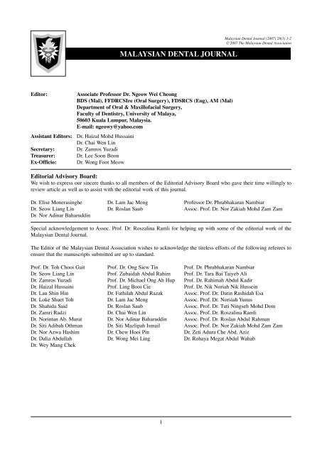
MALAYSIAN DENTAL JOURNAL<strong>Malaysian</strong> <strong>Dental</strong> Journal (2007) 28(1) 1-2© 2007 The <strong>Malaysian</strong> <strong>Dental</strong> <strong>Association</strong>Editor:Associate Professor Dr. Ngeow Wei CheongBDS (Mal), FFDRCSIre (Oral Surgery), FDSRCS (Eng), AM (Mal)Department of Oral & Maxillofacial Surgery,Faculty of Dentistry, University of Malaya,50603 Kuala Lumpur, Malaysia.E-mail: ngeowy@yahoo.comAssistant Editors: Dr. Haizal Mohd HussainiDr. Chai Wen LinSecretary: Dr. Zamros YuzadiTreasurer: Dr. Lee Soon BoonEx-Officio: Dr. Wong Foot MeowEditorial Advisory Board:We wish to express our sincere thanks to all members of the Editorial Advisory Board who gave their time willingly toreview article as well as to assist with the editorial work of this journal.Dr. Elise Monerasinghe Dr. Lam Jac Meng Professor Dr. Phrabhakaran NambiarDr. Seow Liang Lin Dr. Roslan Saub Assoc. Prof. Dr. Nor Zakiah Mohd Zam ZamDr. Nor Adinar BaharuddinSpecial acknowledgement to Assoc. Prof. Dr. Roszalina Ramli for helping up with some of the editorial work of the<strong>Malaysian</strong> <strong>Dental</strong> Journal.The Editor of the <strong>Malaysian</strong> <strong>Dental</strong> <strong>Association</strong> wishes to acknowledge the tireless efforts of the following referees toensure that the manuscripts submitted are up to standard.Prof. Dr. Toh Chooi Gait Prof. Dr. Ong Siew Tin Prof. Dr. Phrabhakaran NambiarDr. Seow Liang Lin Prof. Zubaidah Abdul Rahim Prof. Dr. Tara Bai Taiyeb AliDr. Zamros Yuzadi Prof. Dr. Michael Ong Ah Hup Prof. Dr. Rahimah Abdul KadirDr. Haizal Hussaini Prof. Ling Booi Cie Prof. Dr. Nik Noriah Nik HusseinDr. Lau Shin Hin Dr. Fathilah Abdul Razak Assoc. Prof. Dr. Datin Rashidah EsaDr. Loke Shuet Toh Dr. Lam Jac Meng Assoc. Prof. Dr. Norsiah YunusDr. Shahida Said Dr. Roslan Saub Assoc. Prof. Dr. Tuti Ningseh Mohd DomDr. Zamri Radzi Dr. Chai Wen Lin Assoc. Prof. Dr. Roszalina RamliDr. Norintan Ab. Murat Dr. Nor Adinar Baharuddin Assoc. Prof. Dr. Roslan Abdul RahmanDr. Siti Adibah Othman Dr. Siti Mazlipah Ismail Assoc. Prof. Dr. Nor Zakiah Mohd Zam ZamDr. Nor Azwa Hashim Dr. Chew Hooi Pin Dr. Zeti Adura Che Abd. AzizDr. Dalia Abdullah Dr. Wong Mei Ling Dr. Rohaya Megat Abdul WahabDr. Wey Mang Chek
<strong>Malaysian</strong> <strong>Dental</strong> <strong>Association</strong> Council 2006-2007President:Dr. Wong Foot MeowPresident-elect:Dato’ Dr. Low TeongImmediate Past President: Dr. Shubon Sinha RoyHon. General Secretary: Dr. How Kim ChuanAsst. Hon. Gen. Secretary: Dr. Abu Razali bin SainiHon. Financial Secretary: Dr. Lee Soon BoonAsst. Hon. Finan Secretary: Dr. Sivanesan SivalingamHon. Publication Secretary: Assoc. Prof. Dr. Ngeow Wei CheongChairman, Northern Zone: Dr. Koh Chou HuatSecretary, Northern Zone: Dr. Neoh Gim BokChairman, Southern Zone: Dr. Steven Phun Tzy ChiehSecretary, Southern Zone: Dr. Leong Chee SanElected Council Member: Dr. Sorayah SidekElected Council Member: Dr. Chia Ah ChikNominated Council Member: Dr. V. NedunchelianNominated Council Member: Dr. Xavier JayakumarNominated Council Member: Dr. Hj. Marusah JamaludinNominated Council Member: Dr. Lim Chiew WooiThe PublisherThe <strong>Malaysian</strong> <strong>Dental</strong> <strong>Association</strong> is the official Publication of the <strong>Malaysian</strong> <strong>Dental</strong> <strong>Association</strong>. Please address allcorrespondence to:Editor,<strong>Malaysian</strong> <strong>Dental</strong> Journal<strong>Malaysian</strong> <strong>Dental</strong> <strong>Association</strong>54-2, (2nd Floor), Medan Setia 2,Plaza Damansara, Bukit Damansara,50490 Kuala LumpurTel: 603-20951532, 20947606, Fax: 603-20944670Website address: http://mda.org.myE-mail: mda@po.jaring.mymda@streamyx.com
MALAYSIAN DENTAL JOURNAL<strong>Malaysian</strong> <strong>Dental</strong> Journal (2007) 28(1) 3© 2007 The <strong>Malaysian</strong> <strong>Dental</strong> <strong>Association</strong>Aim And ScopeThe <strong>Malaysian</strong> <strong>Dental</strong> Journal covers all aspects of work in Dentistry and supporting aspects of Medicine. Interactionwith other disciplines is encouraged. The contents of the journal will include invited editorials, original scientificarticles, case reports, technical innovations. A section on back to the basics which will contain articles covering basicsciences, book reviews, product review from time to time, letter to the editors and calendar of events. The mission isto promote and elevate the quality of patient care and to promote the advancement of practice, education and scientificresearch in Malaysia.PublicationThe <strong>Malaysian</strong> <strong>Dental</strong> Journal is an official publication of the <strong>Malaysian</strong> <strong>Dental</strong> <strong>Association</strong> and is published halfyearly (KDN PP4069/12/98)SubscriptionMembers are reminded that if their subscription are out of date, then unfortunately the journal cannot be supplied. Sendnotice of change of address to the publishers and allow at least 6 - 8 weeks for the new address to take effect. Kindly usethe change of address form provided and include both old and new address. Subscription rate: Ringgit Malaysia 60/- foreach issue, postage included. Payment in the form of Crossed Cheques, Bank drafts / Postal orders, payable to <strong>Malaysian</strong><strong>Dental</strong> <strong>Association</strong>. For further information please contact :The Publication Secretary<strong>Malaysian</strong> <strong>Dental</strong> <strong>Association</strong>54-2, (2nd Floor), Medan Setia 2, Plaza Damansara, Bukit Damansara,50490 Kuala LumpurBack issuesBack issues of the journal can be obtained by putting in a written request and by paying the appropriate fee. Kindly sendRinggit Malaysia 50/- for each issue, postage included. Payment in the form of Crossed Cheques, Bank drafts / Postalorders, payable to <strong>Malaysian</strong> <strong>Dental</strong> <strong>Association</strong>. For further information please contact:The Publication Secretary<strong>Malaysian</strong> <strong>Dental</strong> <strong>Association</strong>54-2, (2nd Floor), Medan Setia 2, Plaza Damansara, Bukit Damansara,50490 Kuala LumpurCopyright© 2007 The <strong>Malaysian</strong> <strong>Dental</strong> <strong>Association</strong>. All rights reserved. No part of this publication may be reproduced, storedin a retrieval system or transmitted in any form or by means of electronic, mechanical photocopying, recording orotherwise without the prior written permission of the editor.Membership and change of addressAll matters relating to the membership of the <strong>Malaysian</strong> <strong>Dental</strong> <strong>Association</strong> including application for new membershipand notification for change of address to and queries regarding subscription to the <strong>Association</strong> should be sent toHon General Secretary, <strong>Malaysian</strong> <strong>Dental</strong> <strong>Association</strong>, 54-2 (2nd Floor) Medan Setia 2, Plaza Damansara,Bukit Damansara, 50490 Kuala Lumpur. Tel: 603-20951532, 20951495, 20947606, Fax: 603-20944670,Website Address: http://www.mda.org.my. Email: mda@po.jaring.my or mda@streamyx.comDisclaimerStatements and opinions expressed in the articles and communications herein are those of the author(s) and notnecessarily those of the editor(s), publishers or the <strong>Malaysian</strong> <strong>Dental</strong> <strong>Association</strong>. The editor(s), publisher and the<strong>Malaysian</strong> <strong>Dental</strong> <strong>Association</strong> disclaim any responsibility or liability for such material and do not guarantee, warrantor endorse any product or service advertised in this publication nor do they guarantee any claim made by themanufacturer of such product or service.
MALAYSIAN DENTAL JOURNAL<strong>Malaysian</strong> <strong>Dental</strong> Journal (2007) 28(1) 4© 2007 The <strong>Malaysian</strong> <strong>Dental</strong> <strong>Association</strong>CONTENTEditorial : Changes Over Time for Journals - Are We Ready for e-MDJ? 5Ngeow WCSurvey on Readership of Professional Journals among <strong>Malaysian</strong> Dentists 7Part I: Readership of Professional <strong>Dental</strong> JournalsNgeow WC, Mohd Noor NS, Mohd Tahir NNSurvey on Readership of Professional Journals among <strong>Malaysian</strong> Dentists6Part II: Readership of the <strong>Malaysian</strong> <strong>Dental</strong> JournalNgeow WC, Mohd Noor NS, Mohd Tahir NNEfficacy of Training <strong>Dental</strong> Officers in the Index of Orthodontic Treatment Need (IOTN)4Loke STThe Expert Says………IOTN as a Tool in Prioritizing Orthodontic Treatment 32Rohaya MAWDiagnostic Overlay Removable Partial Denture in the Management of Tooth Wear: A Clinical Report 34Yahya AN, Radzi Z, Yusof ZYMPeriodontal ReferralShashikiran ND, Subba VV, Patil RA Retrospective Clinico-Statistical Analysis of Lip Mucoceles Occurrence in Children Below 3816 Years Seen at a Government Hospital Based Paediatric <strong>Dental</strong> Clinic.Sockalingam GThe Prevalence of Actinobacillus Actinomycetemcomitans in a Healthy Malay Population In Malaysia 45Zahari NM, Ismail R, Bunyarit SS, Shafiei Z, Al Rawenduzy KCRadiographic Considerations in Endodontics 51Yusof ZYM, Nambiar P.The Occurrence of Patients Presenting with Broken/lost Removable Appliances 59Tan L, Awang CFContinuing Professional Development Quiz 63Instruction to contributors 65MDJ Cover page : Clinical picture showing a person with tooth wear, before and after treatment. Picture courtesy ofNor Azlin Yahya, Zamri Radzi and Zamros Yuzadi (of article: Diagnostic overlay removable partial denture in themanagement of tooth wear: A clinical report)
MALAYSIAN DENTAL JOURNAL<strong>Malaysian</strong> <strong>Dental</strong> Journal (2007) 28(1) 5-6© 2007 The <strong>Malaysian</strong> <strong>Dental</strong> <strong>Association</strong>Editorial : changes over time for Journals - are we ready for e-mdj?For more than three centuries, printed journals are theprimary medium of research communication and a meansfor Continuing Professional Development/Education(CPD/CPE). It has remained unchanged in form andfunction since the first scholarly journal, the Journal desScavans was published in 1665. 1 Despite its benefits tothe academic and research community, printed journalshave been subjected to criticism from many angles such asthe peer review process, delays in publication, escalatingcosts, lack of selectivity, stoppage of subscriptions bylibraries and commercial publishers holding copyrights. 2And needless to say, the MDJ is currently facing some ofthese problems.Nowadays, the scholarly scientific, technical, and medical/dental journal systems are undergoing tremendouschange. With steady increases in the price of printsubscriptions, the number of subscriptions has declinedcorrespondingly. 3 A clear cut alternative currently availableis the publication of electronic journals (e-journals). Ashas been highlighted by Dr. Lee Soon Boon, the HonoraryFinancial Secretary of the <strong>Malaysian</strong> <strong>Dental</strong> <strong>Association</strong>in his march 2007 message to the members, the inflatingcost of printing and postage to around RM 125,000 inFY 2006 against RM 92,188.00 in FY 2005 should bea point for all of us to ponder. He thinks that electronicpublishing should be the way forward as embraced bymany leading <strong>Association</strong>s, citing the Commonwealth<strong>Dental</strong> <strong>Association</strong> as being the latest one to do so. Froma financial point of view, electronic or digital publicationcan be produced and circulated relatively inexpensively,and can reach a readership far wider than small-scale printpublication. Moreover, it is not subjected to the risk of lateor undelivery that often plagued the conventional mailingsystem.The electronic journal is a version of the traditional print orpaper-based journal which is disseminated electronicallyin some form or other directly to the end-user. 1 Althoughe-journals have been in existence since 1976, full fledgede-journals only came into the limelight in the 1990s. 4Nasir defined e-journal as a regularly issued publication that isavailable in electronic form, with or without its print equivalentand is accessed online. 5 Any journal produced, published,distributed and received via an electronic medium is alsoconsidered an e-journal. 6 In comparison to print journals,e-journals have the following advantages: 1a) easily accessible;b) easy to publish,c) no physical barriers,d) time saving,e) low cost (sometimes free),f) and authors and readers get closer easilyWith the emergence of the Internet, publishing has becomevery easy, quick and cheap in a medium that can beaccessed easily by everyone from anywhere. With thesteady growth of e-journals on the Internet, it was foundthat creativity and productivity has improved due tonetwork technologies. 7 Scholars have understood the powerof electronic journals and seem to have accepted the newmedium for communicating research ideas and resultsamong fellow professionals. 1There are lots of dental professional journals that are nowavailable online, such as the Brazilian <strong>Dental</strong> Journal,the British <strong>Dental</strong> Journal, the European Journal of<strong>Dental</strong> Education, the New York State <strong>Dental</strong> Journal, theInternational Journal of Pediatric etc. Some of the onlinejournals can be browse freely e.g. the Australian <strong>Dental</strong>Journal, the Journal of The Canadian <strong>Dental</strong> <strong>Association</strong>,
and the Journal of Oral Sciences, but some of them needto be subscribed.So, are we ready for e-journal, in particular if the <strong>Malaysian</strong><strong>Dental</strong> Journal (MDJ) is only available electronic form? Inthe finding of a survey that is going to be published in thisissue of the MDJ, the answer is a sad, “not yet”. With that,I leave all the readers of the MDJ to ponder upon our nextcourse of direction. Thank you.Acknowledgement:I would like to acknowledge the research work done byNorhidayah @ Norzahidah Mohd Tahir and Nora SakinaMohd Nor that enables the write-up of this editorial.Associate Professor Dr. Ngeow Wei Cheong,Editor,<strong>Malaysian</strong> <strong>Dental</strong> Journal.References:1. Rao MK. Scholarly communication and electronic journals: issues and prospects for academic and research libraries. LibraryReview. 2001;50:169-75.2. Harter, Stephen P, Kim HJ. Electronic journal and scholarly communication: a citation and reference study. Paper presented atthe Mid Year Meeting of the American Society for Information Science, San Diego, May 20-22. 1996: 299-315 (also available at:http://php.indiana.edu/~harter/harter-asis96midyear.html).3. Tenopir C, King DW, Bush A. Medical faculty’s use of print and electronic journals: changes over time and in comparison withscientists. J Med Libr Assoc. 2004;92:2.4. Turoff M, Hiltz SR. The electronic journal: a progress report. Journal of the American Society for Information Sciences. 1982;33:195-202.5. Nasir NE. The electronic journal. Network information. 1996;1:15-28.6. Ngah ZA, Che Nasir NE, Ling TC, Phang KK. Making <strong>Malaysian</strong> scholarly journals more visible: the case of MJCS and MJLISonline. Paper presented at Persidangan Majlis Penerbit Ilmiah Malaysia (MAPIM), 17 August 2000, Institut Pengajian Siswazahdan Penyelidikan, Universiti Malaya, Kuala Lumpur.7. Schauder D. Electronic publishing of professional articles: attitudes of academics and implications for the scholarly communicationindustry. Journal of the American Society for Information Sciences. 1994;45:73-100.
MALAYSIAN DENTAL JOURNAL<strong>Malaysian</strong> <strong>Dental</strong> Journal (2007) 28(1) 7-15© 2007 The <strong>Malaysian</strong> <strong>Dental</strong> <strong>Association</strong>Survey on Readership of Professional Journals among <strong>Malaysian</strong> DentistsPart I: Readership of Professional <strong>Dental</strong> JournalsNgeow WC. BDS, FFDRCSI, FDSRCS, Lecturer, Department of Oral & Maxillofacial Surgery, Faculty of Dentistry,University of Malaya, 50603 Kuala Lumpur, Malaysia.Mohd Noor NS. Final Year <strong>Dental</strong> Students 2007. Faculty of Dentistry, University of Malaya, 50603 Kuala Lumpur,Malaysia.Mohd Tahir NN. Final Year <strong>Dental</strong> Students 2007. Faculty of Dentistry, University of Malaya, 50603 Kuala Lumpur,Malaysia.ABSTRACTThe objective of this survey was to understand the current trend of readership of professional dental journals among<strong>Malaysian</strong> dentists. A total of 225 questionnaires were sent out to <strong>Malaysian</strong> dentists who attended various dentalrelated conferences throughout Peninsular Malaysia from February 2006 to July 2006. Questionnaires comprisedof questions relating to dentists’ socio-demographic status and a list of journal(s) read by them. <strong>Malaysian</strong> dentists’view on the content and quality of a particular dental journal, i.e. the <strong>Malaysian</strong> <strong>Dental</strong> Journal (MDJ) was alsoenquired. The details of this finding are highlighted in Part II of this study. A total of 156 questionnaires werereturned; the respondents were made up of 61 male and 91 female dentists. Almost 80% of the respondents agedbetween 20-49 year-old and most respondents (n= 132; 84.62%) only had a basic Bachelor of <strong>Dental</strong> Surgery orequivalent degree while another 19 (12.18%) had in addition, a post-graduate degree. Almost equal numbers ofrespondents were working in the Ministry of Health (MOH) or Armed Force (n=73; 46.8%) and private practice(n=74; 47.4%). Also, equal number of respondents (n=67; 42.95%) were found to be working as single-handedpractitioner and in a partnership/assistant/working-with-other specialties type of practice Almost two-thirds(n=103; 66%) of the respondents read more than one professional journal, and a majority of them worked in theprivate sector. The percentage of readers reading more than one journal from the private practice (n=67, 60.0%)was close to twice of that from the MOH (n=36, 35.0%). No specific age-group pattern was present but the leastnumber of subscribers were from those 60 year-old and above (n=3), whereby none of them subscribed to anyprofessional dental journal/magazine. The highest percentage of subscribers were from those in the age groupof 40-49 year-old, whereby 86.49% (n=32) of dentists in this age-group subscribed to at least one professionaldental journal/magazine. Out of the list of journals/magazines provided, it was found that the MDJ has the mostnumber of readers. The MDJ was most read by dentists in the private practice while the Annals of Dentistry ofthe University of Malaya was most read by dentists in the MOH. In conclusion, it was found that almost two-thirdof the respondents read more than one professional journal, with the MDJ receiving the most number of readers.More dentists in the private practice read professional dental journals than dentists in the MOH.Key words:dental journal, readership, survey, continuing professional development
Readership of Professional <strong>Dental</strong> JournalsIntroductionThe pursuit of ongoing professional educationor updating is referred to as continuing professionaldevelopment (CPD) or continuous professional education(CPE).1 In the United Kingdom, CPD is defined as aprocess of “lifelong learning” for all individuals and teamswhich meets the needs of patients and delivers the healthoutcomes and healthcare priorities of the United KingdomNational Health Services (NHS). 1 CPD is becomingincreasingly recognised by professional institutions asessential in ensuring that their members remain up to dateand maintain their professional competence. 2,3Madden and Mitchell 4 had identified two models ofCPD, namely the sanctions model and the benefits model.The former represents some mandatory requirement whichmust be met if the individual is to be able to demonstrateconsistent professional competence. The latter modelis founded on actions where the individual can take aconscious decision whether or not to pursue some particularform of CPD. The sanction model is usually found in theolder, well-established professional institutions. 5,6 Thismodel may be adopted by the Ministry of Health ofMalaysia (MOH) which is currently in the midst of makingCPD compulsory before dental practitioners renew theirAnnual Practising Certificate. 7The benefits model, which is usually found in newor developing professional institutions, emphasises thebenefits of CPD to the individual, and CPD is thereforeencouraged as a voluntary activity. The <strong>Malaysian</strong> <strong>Dental</strong><strong>Association</strong> (MDA) has over the years informally adoptedthis model whereby it organizes CPD for membersthroughout their career. Attendance is on voluntary basis.Another means of achieving the benefit model is throughjournal readership. The MDA has been contributing tothis by regularly publishing the <strong>Malaysian</strong> <strong>Dental</strong> Journal(MDJ) and the MDA Newsletter.Around the world, dental registration bodies arebeginning to demand that dentists keep up-to-date and beable to show proof of doing so. It is quite clear that in thefuture, dentists will not be able to coast along once theyhave their basic qualification but will be expected to stayabreast of current knowledge. 8Leggate and Russel 9 in a survey reported thatmost of their respondents indicated an interest in usingseveral different learning methods. As might be expected,a preference for ‘hands-on’ was strongly expressed forclinical procedures, although even for practical topicsmany respondents favoured using a wide variety oflearning methods. They also reported that lectures werestill favoured as one of several useful formats, with smallgroup tutorials, books and journals also popular with themajority. Videos were also valued by over half of theirrespondent as a useful tool for many different learningscenarios. 9In the United Kingdom, there are several types ofCPD activity that the dentists usually participated in, suchas journal reading, course attendance, participation in localand/or international professional associations and societies,formal discussion of professional matters with colleagues,purchase of books, watching professional videos, accessingto the internet for information or online journals, attendingconferences with self assessment, joining journal clubsand/or study groups, and organising peer review or clinicalaudit, either via formal collaboration or individually. 1Razali et al. 7 stated that the <strong>Malaysian</strong> dentist showedhighest preference to CPD activity that entailed learningfrom experience and from peers as compared to readingprofessional journals. They claimed that about 42 percentof their respondents did not subscribe to any professionaljournal while the rest subscribed to at least one professionaljournal. In terms of total number of journals read/commented/written in the past year, about 20 percent oftheir respondents did not engage in any of these activities.They also stated that, on the average, the respondents read/commented/wrote about only 3 journals per year. 7Hence it is the aims of this study to understand thecurrent trend of readership of professional dental journal(s)and magazine(s) among <strong>Malaysian</strong> dentists.MATERIALS AND METHODSA total of 225 pre-tested questionnaires were sentout to <strong>Malaysian</strong> dentists who attended dental relatedconferences/continuing professional development (CPD)throughout the Peninsular Malaysia (Kuala Lumpur,Kuantan, Johor, Melaka, Seremban and Alor Star) betweenFebruary 2006 and July 2006. This form had been pretestedamong dentists attending CPD in Penang andmodified according to their feedback before actual use.The questionnaires comprised of questions relatingto dentists’ socio-demographic status and a list of journal(s)read by them. In details, the variables collected were asfollows:i) genderii) ageiii) marital statusiv) ethnicv) professional qualificationsvi) number of years of practicevii) types of practice (*single-handed orpartnership)viii) place of practice (**MOH/Armed forces orprivate practice)ix) Subscription to professional dental journals/magazines.x) various types of professional journals/magazines read* The participants were categorized as either working in asingle-handed practice or in partnership (with colleague[s]),irrespective of their place of work; be it in the MOH orprivate practice. Hence, a dental officer working alonein the MOH but has no contact with any colleague wasdeemed to work in a single-handed practice.** The word MOH will be used in the text to denoterespondents who worked in either the Ministry of Healthof Malaysia or the Royal Armed Force of Malaysia.
Ngeow / Mohd Noor / Mohd TahirThe data received were then compiled, entered and analyzed using SPSS version 12.0. Descriptive statistic was employedwhen necessary.RESULTSSocio-demographic background of respondentsOut of the 156 survey forms that were returned, 39.1% (n=61) were from male respondents, 58.33% (n=91) fromfemale respondents and 2.56% (n=4) from respondents who did not state their gender. Their ethnic distributions areshown in Figure 1.Figure 1 : Ethnic distributions of the respondents70n=7044.87%6050n=5937.82%Number403020n=2113.46%100n=21.28%n=42.56%Malay India Chinese Other not availableEthnicAge-groupsAlmost 80% of the respondents aged between 20-49 year-old, with the following breakdown: 32.69% (n=51) were 20-29year-old; 22.44% (n=35) were 30-39 year-old; and 23.72% (n=37) 40-49 year-old (Figure 2).Figure 2 : Age groups of respondents605040Number30n=5132.69%20n=3522.44%n=3723.72%100n=2214.1%n=31.92%20-29 year old 30-39 year old 40-49 year old 50-59 year old 60 year oldand aboveAge-groupsn=85.13%not available
Readership of Professional <strong>Dental</strong> JournalsProfessional qualificationsMost respondents (n= 132; 84.62%) only had a basic Bachelor of <strong>Dental</strong> Surgery or equivalent degree whileanother 19 (12.18%) had in addition, a post-graduate degree. One respondent (0.64%) was still undergoing his/herpostgraduate study, while the professional qualification(s) of 4 respondents were not available.Working experience and location & type of practiceIn terms of working experience, the results are as follows: 19.23% (n=30) were respondents with less than 1 yearof working experience, 17.95% (n=28) had between 1 to 5 years of working experience, and 8.97% (n=14) had been inpractice for between 6 to 10 years. Another 26 (16.67%) had been in practice for between 11 to 15 years, 21 (13.45%)had been in practice for between 16 to 20 years, and 33 (21.15%) had been working for more than 20 years (Figure 3).Figure 3: Working experience of respondents40Number3020n=3019.23%n=2817.95%n=2616.67%n=2113.46%n=3321.15%10n=148.97%0< 1 year1-5 6-10 11-15 years 16-20 years >20 years not availableNumber of years in practicen=42.56%Almost equal numbers of respondents were working in the Ministry of Health or Armed Force [denoted as MOH] (n=73;46.8%) and private practice (n=74; 47.4%). Only three respondents (1.94%) work as part-time private practitioners whilethe working information for the remaining 6 respondents (3.8%) was unavailable. The breakdown of gender and ethnicityof respondents in relation to work place are as follows: in the MOH, 55 respondents were Malays (7 males, 48 females),7 were Indians (3 males, 4 females), and 11 were Chinese (4 males, 7 females). The gender and ethnicity breakdownof 74 respondents from full time private practice are as follows: 14 respondents were Malay (4 males, 10 females), 13were Indians (7 males, 6 females), 45 were Chinese (32 males, 13 females) and for the two “other” respondents, bothwere male. Equal number of respondents (n=67; 42.95%) were found to be working as single-handed practitioner and inpartnership/assistant/working-with-other-specialties type of practice (Figure 4).Figure 4 : Type of practice706042.95%n=6742.95%n=6750Number40302014.1%n=22100Single handed practicePartnership/assistant/togetherwith other specialtiesType of Practicenot available10
Ngeow / Mohd Noor / Mohd TahirIn the MOH, only 23.9% (n=16) were single-handed practitioners while more than two-thirds (n=47; 70.1%) werein partnership/assistant/working-together-with-other-specialties type of practice.In comparison, almost three quarters (n=50; 74.6%) full time private dental practitioners were single-handed practitioner,while another 28.4% (n=19) were in partnership/assistant/working-with-other-specialties type of practice. Where as in part timeprivate dental practice, equal number of respondents work as single-handed practitioner and in partnership/assistant/workingwith-other-specialtiestype of practice (n=1;1.5% each) (Table 1).Table 1 : Breakdown of practitioners working in single-handed practice or partnership/assistant/together –withother-specialtiesin relation to place of practicePlace of practice MOH Private practice Private practice(full time) (part time)not availableNo. % No. % No. % No. %Type of practiceSingle-handed practice 16 23.9% 50 74.6% 1 1.5%Partnership/assistant/together with otherspecialties47 70.1% 19 28.4% 1 1.5%Not available 10 45.5% 5 22.7% 1 4.5% 6 27.3%Subscription to professional dental journals/dental magazinesWhen asked about current subscription(s) to any professional dental journal(s)/magazines(s), almost two-thirds(n=103; 66.03%) claimed doing so, while another 30.77% (n=48) did not (Figure 5). Those who subscribed weremainly from full time private practice (n=61; 59.22%), while readers from the MOH make up another 38.83% (n=40) ofsubscribers. No specific age-group pattern was present but the least number of subscribers were from those 60 year-oldand above (n=3), whereby none of them subscribed to any professional dental journal/magazine. The highest percentageof subscribers were from those in the age group of 40-49 year-old, whereby 86.49% (n=32) of dentists in this age-groupsubscribed to at least one professional dental journal/magazine.Figure 5 : Subscription to professional dental journal(s)/magazines12010066.03%n=10380Number604030.77%n=482003.21%n=5Subscribe Do not subscribe not available<strong>Dental</strong> journal/magazine Subscription11
Readership of Professional <strong>Dental</strong> JournalsReadership of professional dental journals/dental magazinesA list of journals and magazines were given and it was found that 64.74% (n=101) respondents read the <strong>Malaysian</strong><strong>Dental</strong> Journal (MDJ), 61.5% (n=96) read the MDA Newsletter, 10.3% (n=16) read the Annals of Dentistry of theUniversity Malaya, 32.1% (n=50) read Asian Dentist Magazine, 31.4% (n=49) read <strong>Dental</strong> Asia Magazine while 16.7%(n=26) read other journals like the British <strong>Dental</strong> Journal (BDJ)(Figure 6).Figure 6: Readership of various professional dental journals/magazinesPercentage ofRespondents100.00%80.00%60.00%40.00%20.00%0.00%35.3%5564.7%101ReadMDJ20.5%3217.9%2861.5%96Read MDANewsletter20.5%3269.2%10810.3%16Read Annalsof Dentistry ofUniversity ofMalaya20.5%3247.4%7432.1%50Read AsiaDentistMagazine20.5%3248.1%7531.4%49Read<strong>Dental</strong>AsiaMagazine20.5%3262.8%9816.7%26Read OthersNot AvailableNoName of JournalsYesOf all (n=101; 64.74%) of the respondents who claimed that they read the MDJ, 30 (29.70%) were from the MOH,two-third (n=64; 63.37%) were full time private practitioner and 3 (2.97%) was in part time private practice. The profileof another 4 respondents was unavailable.Of the 96 respondent who claimed that they read the MDA News, 30 (31.3%) were from the MOH, 63 (65.6%)were from full time private practice, while 3 (3.1%) were from part time private practice.For the Annals of Dentistry of University of Malaya, only 10.3% (n=16) respondents gave positive response. Morethan two-third (n=11; 68.8%) readers were from the MOH, another quarter (25.0%) were from full time private practiceand only 1 person (6.3%) was from part time private practice.Almost one third of the respondents read Asia Dentist Magazine (n=50, 32.1%), of which, 30% (n=15) of themwere from the MOH, 68% (n=34) were from full time private practice and 2% (n=1) were from part time privatepractice.<strong>Dental</strong> Asia Magazine received 31.4% (n=49) positive responses. Out of 49 respondents, 40.8% (n=20) werefrom the MOH while 55.1% (n=27) were from full time private practice and 4.1% (n=2) were from part time privatepractice.It was found that 66.0% (n=103) of the respondents read more than one journal. The percentage of readers readingmore than one journal from the private practice, (n=67, 60.0%) was close to twice of that from the MOH (n=36, 35.0%).There were 3 respondents who stated that they read all the professional journals/magazines listed and they were from theyounger age group of 20-29 year-old. They consisted of 1 Malay male, 1 Malay female and 1 Chinese female dentists.They all had only 1 basic degree and were working in the MOH.12
Ngeow / Mohd Noor / Mohd TahirDISCUSSIONAs at the end of 2003, a total of 2418 dental practitioners were serving in both the government sector and privatepractice, giving dentists to population ratio as 1:10,612.10 Of that total, 992 (41%) were government dentists. Themajority of government dentists were female (n=736; 74%), with only 256 male dentists (26%). Almost 60% of thedentists in Malaysia were private practitioners (n=1,426). As shown in Figure 7, male dentists accounted for 845 (59%)of the private practitioners while the remaining 581 were female. As can be noted, the demographic distribution ofrespondents in this survey is not directly in proportion to the distribution of dental practitioners in Malaysia.Figure 7: Distribution of dental practitioners in Malaysia by genderDistribution of dental practitioners in Malaysia bygender900Number of people800700600500400300200256845736581governmentprivate1000malefemaleGenderSource: MDC (as at 31/12/2003 )Alreck and Settle 11 stated that it was seldomnecessary to sample more than 10 percent of the populationfor a study. They disputed the logic that sample size wasnecessarily dependant upon population size. However,choice of sample size is often as much a budgetaryconsideration as a statistical one. Hence it is necessaryto think of all resources needed namely time, space,energy and money. Razali et al. 7 stated that generallychoice of sample size is as much a function of budgetaryconsiderations as it is statistical considerations. When theycan be afforded, large samples are usually preferred oversmaller ones. Based on these reasons, we sent out about225 questionnaires but only 156 dentists responded.We would like to highlight that for this survey,the samples were not selected randomly from the dentistslisted on the <strong>Malaysian</strong> <strong>Dental</strong> Council due to difficulty inaccessing them as well as basing on past experiences ofthe MDA, whereby survey questionnaires sent out weremainly unanswered. Instead, we resorted to making directapproach by asking participants at various continuingprofessional development (CPD) sessions organized bythe MDA to volunteer for this survey. Hence, it mustbe emphasized that there is a certain degree of businessin these samples in that the respondents may be moremotivated to participate in CPD as compared to those whonever or attended very few CPD sessions. Nevertheless, wewould like to treat this study as a pilot project and woulddefinitely like to extend it to cover randomized sampleswhen the problem of time and financial constrain areaddressed to.The findings of this survey are indeed veryencouraging. A list of journals and magazines were givenand it was found that 64.74% (n=101) of respondents readthe MDJ, 61.5% (n=96) read the MDA Newsletter, 10.3%(n=16) read the Annals of Dentistry of the UniversityMalaya, 32.1% (n=50) read Asian Dentist Magazine,31.4% (n=49) read <strong>Dental</strong> Asia Magazine while 16.7%(n=26) read other journals like the British <strong>Dental</strong> Journal(BDJ). This makes the MDJ the most read dental journalsamong the respondents, and if extrapolated, may indicatethe same for the whole country.13
Readership of Professional <strong>Dental</strong> JournalsIn fact, almost two-third (n=103; 66%) of therespondents claimed they read more than one professionaldental journal/magazine. Only a slightly lower number(n=101; 64.74%) of respondents were MDJ readers.Free dental magazines like the Asian Dentist and <strong>Dental</strong>Asia seemed to be able to attract more readers, perhapsbecause of the nature of their easiness to gain access to,as compared to other journals that needed subscriptionlike the Annals of Dentistry of the University of Malayaand other foreign dental journals, which of course couldbe costly. This is in contrast to the finding of a survey byRazali et al. 7 whereby about 42 percent of their respondentsdid not subscribe to any professional journal. However,direct comparison cannot be made on this matter asRazali et al. 7 specifically looked into subscription of dentaljournals whereas in this survey, we concentrated more intoreadership, with subscription being just 1 of the questionsasked. And one has to bear in mind that readership doesnot equal subscription as a dentist may still be able to readany professional dental journals without subscribing to it.Even so, the percentage of dentists claiming to subscribeto at least 1 dental journal is higher in this survey ascompared to that observed by Razali et al. 7Of the respondents who claimed to read morethan one professional journal, a majority of them workedin the private sector. The percentage of readers readingmore than one journal from the private practice (n=67,60.0%) was close to twice of that from the MOH (n=36,35.0%). This may re-emphasize the usefulness of having aprofessional dental journal as a source of CPD, especiallyfor private practitioners, in our local context. Leggateand Russel 9 reported that journal is a popular means ofCPD. It has been accepted as one of the official meansof CPD in the United Kingdom. 1 Unlike their colleaguesin the government services where ward rounds, in-houseseminars and workshops and internal courses are moreeasily available, our general dental practitioners have torely on professional dental journals and CPD talks for theircontinuing professional development.The finding that the highest percentage of subscriberswere from those in the age group of 40-49 year-old, whereby86.49% (n=32) of dentists in this age-group subscribed toat least one professional dental journal/magazine is also apositive finding. This again indicates the need for CPD, asthis group of dental practitioners would have left dentalschools 15-20 years ago and certainly would need to keepthemselves abreast with development in dental technology,skill and current concept of management and treatment.As the dental practitioners reaches near the age ofretirement, their need for CPD reduces. This is suggestedby the finding that respondents 60 year-old and above, didnot subscribe to any professional dental journal/magazine.Lastly, it has to be noted that the list of journals/magazines provided in this survey may not reflects thecomplete list of professional dental journals/magazinesavailable in Malaysia. Moreover, even though these dentaljournals and magazines are listed together as “professionaldental journals/magazines”, they are not of equal standard.Two of them were professional journals that were peerreviewed(the MDJ and the Annals of Dentistry of theUniversity of Malaya) where as two others were nonrefereeddental magazines and one was a newsletter. Theywere group together as “professional dental journals/magazines” out of convenience that all of them providethe same objective, i.e. to provide CPD updates for thedental practitioners and their content solely concentratedon dentistry.Nevertheless, it is interesting to note that the MDJwas most read by dentists in the private practice whilethe Annals of Dentistry of the University of Malaya wasmost read by dentists in the MOH. This may be due to theeasiness to access to these journals in the two differentworking environments.CONCLUSIONIn conclusion, it was found that almost two-third ofthe respondents read more than one professional journal,with the MDJ having the most number of readers. Moredentists in the private practice read professional dentaljournals than dentists in the MOH. The MDJ was mostread by dentists in the private practice while the Annals ofDentistry of the University of Malaya was most read bydentists in the MOH.ACKNOWLEDGEMENTData of this article has been presented at the8th <strong>Dental</strong> Student’s Scientific Conference held in theFaculty of Dentistry of the University of Malaya between15th and 16th December 2006. This survey has beenundertaken as part of the elective project assignment of thelatter two authors. We would like to extend our appreciationto Dr. Shubon Sinha Roy, the Immediate Past President andthe current President, Dr. Wong Foot Meow for supportingthe effort taken to understand the current readership ofprofessional dental journals in Malaysia. Also gratefulthanks to all participants who were willing to spend a littlebit of their time giving us these feedbacks; without them,we won’t be able to have any data for analysis. Lastly, wewould like to thank the <strong>Malaysian</strong> <strong>Dental</strong> <strong>Association</strong> forsupporting this project financially.14
Ngeow / Mohd Noor / Mohd TahirREFERENCES1. Bullock A, Firmstone V, Fielding A, Frame J, Thomas D,Belfield C. Participation of UK Dentists in ContinuingProfessional Development. Br Dent J. 2003;194:47-51.2. Welsh L, Woodward P. Continuing Professional Development:Towards a National Strategy, Glasgow Planning Exchange,Glasgow. 1989.3. Jones N, Fear N. Continuing Professional Development:Perspectives from Human Resource Professionals. PersonnelReview. 1994;23:49-60.4. Madden CA, Mitchell VA. Professions, Standards andCompetence: A Survey of Continuing Education for theProfessions, University of Bristol, Bristol. 1993.5. The Law Society. Continuing Professional Development:Information Pack, The Law Society, Redditch. 1994.6. Institute of Chartered Accountants in England and Wales.Guidelines on Continuing Professional Education, Instituteof Chartered Accountants in England and Wales, MiltonKeynes. 1994.7. Razali AS, Rozima Y, Shamsuddin A. <strong>Malaysian</strong> privatedental practitioners in continuing professional educationcontext. Malays Dent J. 2006;27:6-13.8. Lawrence A. There is more to continuing professionaldevelopment than just scoring hours. Evidence BasedDentistry. 2003;4:40-1.9. Leggate M, Russell E. Attitudes and trends of primary caredentists to continuing professional development: A reportfrom the Scottish dental practitioners survey 2000. Br <strong>Dental</strong>J. 2002;193:65-469.10. Information and Documentation System Unit, Planning &Development Division. Ministry of Health Malaysia. (2003).Health Facts. Available from URL: www.gov.moh.my.11. Alreck PL, Settle RB. The Survey Research handbook, 2ndEd. Chicago: Irwin Press, 1995.Address for correspondence:Associate Professor Dr. Wei Cheong NgeowHonorary Publication Secretary MDA & Editor MDJ,54-2 (2nd Floor), Medan Setia 2,Plaza Damansara, Bukit Damansara,50490 Kuala Lumpur,Malaysia.E-mail: ngeowy@yahoo.comTel : 603-2095 1532Fax : 603-2094 467015
MALAYSIAN DENTAL JOURNAL<strong>Malaysian</strong> <strong>Dental</strong> Journal (2007) 28(1) 16-23© 2007 The <strong>Malaysian</strong> <strong>Dental</strong> <strong>Association</strong>Survey on Readership of Professional Journals among <strong>Malaysian</strong> DentistsPart II. Readership of the <strong>Malaysian</strong> <strong>Dental</strong> JournalNgeow WC. BDS, FFDRCSI, FDSRCS, Lecturer, Department of Oral & Maxillofacial Surgery, Faculty of Dentistry,University of Malaya, 50603 Kuala Lumpur, Malaysia.Mohd Noor NS. Final Year <strong>Dental</strong> Students 2007. Faculty of Dentistry, University of Malaya, 50603 Kuala Lumpur,Malaysia.Mohd Tahir NN. Final Year <strong>Dental</strong> Students 2007. Faculty of Dentistry, University of Malaya, 50603 Kuala Lumpur,Malaysia.ABSTRACTThe objective of this part of the study was to understand the current trend on readership of the <strong>Malaysian</strong> <strong>Dental</strong>Journal (MDJ) among <strong>Malaysian</strong> dentists. Their views on the contents and quality of the <strong>Malaysian</strong> <strong>Dental</strong> Journalwere enquired. We also enquired the reasons they chose-to/chose-not-to read the MDJ. Of the 225 dentists surveyed,the number of MDJ readers was 101; with only 24.75% reading all issues published. The editorial sectionwas rated as “useful” by 70.3% of readers, while 79.2%, 87.1%, 87.1% and 80.2% of readers rated the researcharticle section, the review article section, the case reports section and book recommendation section similarlyrespectively. Feedback from readers indicated that they wanted more case reports, more review articles on “howto do it” and on medical problems in dentistry. More than half (55.45%) of the MDJ readers preferred to receivethe journal in both hard and soft copies. For the non-readers, the most common reasons cited for not reading theMDJ was not being able to access to the journal, followed by not having time to read. Our finding suggested thatthe respondents preferred to learn from colleagues’ experience and to read article that can improve their clinicalknowledge and skill.Key words:continuing professional education, journal, readership, AsiaIntroductionThe <strong>Malaysian</strong> <strong>Dental</strong> Journal (MDJ) is a publicationof the <strong>Malaysian</strong> <strong>Dental</strong> <strong>Association</strong> (MDA) and ispublished twice yearly. It provides a means for continuingprofessional development/education (CPD/CPE) due to thenature of its content. Other means of CPD available in thiscountry include seminars, workshops, conferences, journalclub discussions and ward rounds.This report details the finding of a readershipsurvey of the MDJ, undertaken between February and July2006. The reason this survey was undertaken was to bridgethe gap between the publisher (MDA) and the readers, sothat the readers may voice their opinions on the qualityand what they expect from the journal. Unless a journalarouses criticism, it hardly achieves the objects for whichit is published. 1 With statements like this more and morejournal are doing readership surveys to get more positivefeedback and criticism from their readers.In the United Kingdom, the British <strong>Dental</strong> Journal(BDJ) is the most popular journal when compared to itscounterparts. Even so, the BDJ has been undertakingreadership survey continuously since 1992 to get feedbacksfrom its readers in order to improve itself and for makingsure the readers’ needs are met. 2-6In Malaysia, even though the MDJ has beenpublished for more than three decades, no readershipsurvey has been undertaken. The authors are of the opinionthat such an exercise is inevitable as dental technologyand needs of dentists have changed tremendously since itsinaugural issue as the former <strong>Dental</strong> Journal of Malaysiain 1974. Hence, this exercise of getting feedback fromreaders was done to obtain their views on the content andquality of the MDJ and what they want in future issues.16
Ngeow / Mohd Noor / Mohd TahirIn essence, the objectives of this study were:• To seek <strong>Malaysian</strong> dentists’ views on the<strong>Malaysian</strong> <strong>Dental</strong> Journal (MDJ).• To find out reasons why <strong>Malaysian</strong> dentistschoose-to/choose-not-to read a particularprofessional journal (MDJ).• To find out most favourable ways to attract newreaders and reach existing readers.MATERIALS AND METHODSThis is a continuation to the materials and methodsdescribed in Part I of these series. Similarly, a total of225 pre-tested questionnaires were sent out to <strong>Malaysian</strong>dentists who attended dental related conferences/continuing professional development (CPD) throughoutthe Peninsular Malaysia (Kuala Lumpur, Kuantan, Johor,Melaka, Seremban and Alor Setar) between February 2006and July 2006. This questionnaire has been pre-testedamong dentists attending CPD in Penang and modifiedaccording to their feedback before actual use.The part relating to dentists’ socio-demographicstatus and a list of journal(s) read by them has been dealtwith in Part I of this study. In this second part, theirreadership, perceived quality, preference mode of deliveryand the reasons why they choose-to/choose-not-to read theMDJ were obtained. In details, the variables collected wereas follows:A) Readers sectioni) Frequency reading the MDJii) Sections ratingsiii) Frequency of sections readiv) Perceived quality of the MDJv) Preference mode of delivery (soft/hard copy)vi) Suggestion for future issues of the MDJB) Non-readers Sectioni) Reasons not reading the MDJii) How to attract non-readers’ to read the MDJiii) Interest in purchasing the MDJiv) Interest in browsing through the MDJ webpage.The data received were then compiled, entered andanalyzed using SPSS version 12.0. Descriptive statisticwas employed where necessary.RESULTSI. Feedback on the readership of the MDJAlmost two-third (n=101; 64.7%) of the 156 respondentswere readers of the MDJ. A quarter of the readers (n=25;24.75%) claimed to read all issues of the MDJ whilealmost half (n=49; 48.51%) read only selective article(s).Another 22.77% (n=23) said they only browsed throughthe MDJ while no information was obtainable from theremaining 4 respondents.i) Ratings on various sections of the MDJReaders of the MDJ were asked to rate theusefulness of various sections of the MDJ, namely, theeditorial, research article, review article, case reports andbook recommendation. The editorial section was rated asbetween “somewhat useful” to “most useful” by 70.3% ofreaders, while 79.2%, 87.1%, 87.1% and 80.2% of readersrated the research article section, the review article section,the case reports section and book recommendation sectionsimilarly respectively. The breakdown on the ratings for allthese sections is shown in Table 1.Table 1. Ratings on various sections of the MDJ.Section Not useful Some whatusefulUseful Most useful InformationNot availableNumber (n)Editorial 14 44 26 1 16Research article 9 26 41 13 12Review article 5 26 48 14 8Case report 4 15 51 22 9Book recommendation 5 45 34 2 15The editorial section was rated as “useful” by morethan two-third of respondents from both the Ministry ofHealth, Malaysia (MOH) (73.33%) and full-time privatepractices (71.88%). In contrast, more respondents whorated the research, review article and case report sectionsas “useful” were from the MOH than full-time privatepractices. [* “useful” denotes between “somewhat useful”to “most useful”]A higher percentage of respondents from the MOH(20%) rated the editorial section as “not useful”, so ascompared to full-time private practitioners (10.94%). Lesserrespondents rated research articles (8.9%), review articles(5%), case reports (4%) and book recommendations (5%)as “not useful”. However, a higher percentage of full-timeprivate practitioners (12.5%) gave this rating as comparedrespondents from the MOH (3.33 %) for the research17
Readership of the <strong>Malaysian</strong> <strong>Dental</strong> Journalarticle section. As a matter of facts, the respondents whorated the case reports and book recommendation sectionsas “not useful” were all from full-time private practice.When analyzing negative feedbacks from the respondents,it was noted that they were not made consistently by thesame person. However, 6 respondents gave two or morenegative answers. Four of them were overseas graduatesand were all in the 50-59 year-old age group while theother two local graduates are younger dentist, (one from30-39 year-old, and one from 40-49 year-old groups). Interms of gender, 4 of the respondents were male while twoother were female.ii) Readership of various sections of the MDJReaders were asked to indicate which section(s) of the MDJ they read most. The result is shown in Table 2.Table 2. Readership of various sections of the MDJ.Section Not useful SomewhatusefulUseful Most useful InformationNot availableNumber (n)Editorial 16 59 12 12 14Research article 45 42 3 6 11Review article 48 42 2 2 9Case report 63 30 1 1 7Book recommendation 18 60 7 7 16All the sections surveyed were rather well read(includes “always read” and “sometimes read”) by therespondents with the percentage of readership that variesbetween 74.2% for the editorials to 92.1% for the casereports. However, compared to others, the editorial sectionrecorded the highest percentage of respondents (11.9%;n=12) who claimed that they never read this section.A higher percentage of respondents from the MOH(93.33%) read research article section as compared torespondents from full time private practice (84.38%).In contrast, only a very slightly higher percentage ofrespondents from the MOH (93.33%) read the reviewarticles section as compared to respondents from full timeprivate practice (90.63%).The case reports section seemed to be the mostpopular section with 92.1% respondents reading it. It alsorecorded the most respondents (n=63; 62.4%) who claimedthat they “always read” this section. The percentage ofreaders who were full-time private practice (92.19%) wascomparable to those working in the MOH (93.33%).The distribution for readership for the bookrecommendation section is the same from that of theeditorial section (Table 2). Similar to other sections, ahigher percentage of positive respondents were from theMOH (86.67%) as compared to respondents from full-timeprivate practice (76.56%).iii) Perceived Quality of the MDJRespondents were also requested to give their opinionon their perceived quality of the MDJ. The majority(n=90; 89.11%) rated it between “ok” (n=63; 62.38%)and “good” (n=27; 26.73%). Only two respondents (2%)thought it was “bad” (Figure 1). Further analysis showedthat, these 2 respondents rated negatively on most part ofthe questionnaires. They were both private practitioners,were overseas graduates and were in the 50-59 year-oldage group.Figure 1 : Respondents’ perceived quality of MDJ.18
Ngeow / Mohd Noor / Mohd Tahiriv) Preference mode of delivery of the MDJThis questionnaire survey made an attempt to look into respondents’ preferred means of publication i.e. hard copyor e-journal. It is interesting to note that more than half of the respondents (n=56; 55.45%) preferred to receive the MDJin both the hard and soft copies (Figure 2). Only 8 respondents were ready to go fully electronics, while the remaining31 respondents (30.69%) preferred to receive printed copies only (Figure 2).Figure 2 : Respondents’ preferred means of publication.v) Respondents’ needs for future issues of the MDJReaders were finally asked on what would they want in future issues of the MDJ (Figure 3). Most of therespondents answered more than one option. Only 19 (18.81%) respondents chose a single option. Generally, this groupof readers chose to have more case reports followed by asking for more review articles on ‘how to do it’.Figure 3 : What readers want from future issues of the MDJ.19
Readership of the <strong>Malaysian</strong> <strong>Dental</strong> JournalWhilst 80 (79.21%) respondents suggested morethan one option, 2 (1.98%) respondents did not answerthe questions at all. The majority of these respondentspreferred to have more review articles on ‘how to do it’and more case reports, followed by more review articleson ‘medical problems in dentistry’.II. Feedback on non-readership of the MDJWhile the MDJ readers were asked specificquestions reading to the contents and perceived qualityof the MDJ, non-readers had to answer a different set ofquestions enquiring why they did not read the MDJ.i) Reasons for non-readershipThe most common reasons cited for not reading theMDJ was the inability to access to the journal (81.8%) aswell as not having time to read it (36.4%). Another 1.8%dentists stated that it was “too scientific”, while 5.5%stated that it was not relevant to their practice, or that theywere not interested in it respectively.Among the 80% of dentists who gave a singlereason on why they did not read the MDJ, majority ofthem work in the MOH (n=34; 77.27%) while 8 (18.18%)were from full-time private practice. The profile of tworespondents was not available for analysis.Only 11 (20%) respondents gave multiple reasonson why they decided not to read the journal. Theircombined reasons included not being able to access to theMDJ and not having time to read it. These respondentswere mainly from the MOH (n=9; 81.81%) with theremaining 2 (18.18%) were full-time private practitioners.ii) Potential means to attract non-readersNon-readers were then asked on what can be doneto arouse their interest to read the MDJ and results are asshown in Table 3. In essence, the most common requestswere for the MDJ to provide useful internet web pages(49.1%), to publish review articles in “medical problems indentistry” (45.5%) and to publish “Questions & Answers”section on clinical cases (38.2%). More respondents (n=34;61.81%) gave more than one way to attract future readersas compared to single suggestions (n=21; 38.18%).Table 3 : Articles that may attract new readers of the MDJ.AttractionsFrequencyNumber (n) Percentage (%)Publish More Frequently 4 7.3Publish on Time 6 10.9Publish Review Articles on ‘How to Do It’ 16 29.1Publish Review Articles on ‘Medical Problems in Dentistry’ 25 45.4Publish Research Articles of Special Interests 4 7.3Publish Review Articles of Special Interests 2 3.6Publish Q & A 21 38.2Publish More Case Reports 14 25.5Publish More Book Recommendation 9 16.4Publish Abstract Review 8 14.5Provide Useful Internet Webpage 27 49.1Others 7 13.220
Ngeow / Mohd Noor / Mohd Tahiriii) Accessibility of the MDJWhen non-readers were asked whether they wouldbe interested to purchase the MDJ if it was available forsale, almost three quarters 74.5% (n=41) claimed theywould do so while 12.7% said they would not. Another12.7% did not respond to this question.They were also asked whether they would beinterested to browse through the journal if the MDJ wasfreely available on the MDA webpage. A big majority(n=50; 90.9%) replied “yes”, while 5.5% said “no” and3.6% did not respond to this question.Only one full-time private practitioner in the 60years and above age group declined to read the MDJ evenif he were able to buy it or download free from the web. Hehowever, reads MDA Newsletter, Asia Dentist magazineand <strong>Dental</strong> Asia Magazine. The other respondents mostlysaid they were not interested to buy the MDJ but if it werefreely available on the webpage, they would read it.Lastly, another 2 respondents stated that they werenot interested to read the journal even if it was freelyavailable on the MDA webpage. They were from fulltimeprivate practice and they did not answer questionsin previous sections. They only answered this questionmaking the previous question data unavailable for analysis.Both were female dentists in the 40-49 and 30-39 year-oldage groups.DISCUSSIONThe questions used in this survey were very similarto the one used by the BDJ in 2002 in order to allowcomparison. 2 However, some modifications had to bedone to ours as the MDJ has limited sections to analyzeas compared to the BDJ. In essence, readers in our surveywere asked how often they read different sections of theMDJ and the answers provided were, “always read”,“sometimes read” and “never read”, similar to that usedby the BDJ. Readers were also asked to rate the usefulnessof each section using a scale of 1 (not useful) to 4 (mostuseful), again similar to that used in the BDJ surveyquestions. However, unlike the BDJ survey that onlytargeted their readers, our study also looked into gettingfeedback from non-MDJ readers, as we believe that theiropinion and needs would also have to be taken care of.We hope, with their feedback, we can reach out to moredentists in Malaysia.In 2002, the BDJ sent out 1000 questionnaires andreceived 587 responds, 2 while in comparison, we sent out225 questionnaires and received 156 responds which makeit a 69.33% response. However, their questionnaires weresent out via mail, employing a similar survey methodologyused by Razali et al. 7 We managed to get a higherpercentage of response than the BDJ as we approachedpotential volunteers personally.Some of our findings were comparable to thatof the BDJ survey. 2 For example, the majority of ourrespondents worked in general dental practice (75.68% offull time private practitioners & 32.88% of dental officersin the MOH). However, more of our respondents workedin solo dental practice (46.5%) as compared to the 16%found reported in the BDJ survey. Results from the BDJsurvey showed that the typical BDJ reader was between30 and 50 year-old (58%), male (60%) and married (71%).In comparison, the MDJ readers were mostly 40-49 yearsold (30.69%), male (54.46%) and married (77.23%). Thisfinding suggests that our profile of target audience is notdissimilar from that of the BDJ, and therefore, the editorialteam could consider adopting changes that the BDJ hasmade, if any to the future issues of the MDJ.Similar to the BDJ survey, readers of the MDJ wereasked to indicate how many issues they read. Sadly, thepercentage of the MDJ readers who read all or most issuesof the MDJ was lower than that reported in the BDJ survey(MDJ, 24.8% versus BDJ, 91%). This implies to us that<strong>Malaysian</strong> dentists only read selectively, perhaps due totheir time constrain working in a solo practice.Among the 101 MDJ readers, 55 (54.4%) weremale and 43 (42.6%) were female; the gender for theremaining 3 respondents was unknown. When comparedto all respondents whereby there were more female (n=91)than male (n=61), our finding seemed to indicate that therewere more male respondents (in term of percentage) whowere readers of the MDJ as compared to female dentists.This echoed a similar finding by the BDJ. 2 A total of 48female respondents and 6 male respondents were noted inthe non-MDJ readers category to read at least one otherprofessional dental journal, if not the MDJ.Several reasons were speculated as to why there wasa lack of female dentists interested in reading professionaljournals in general. Among them could be due to timeconstrain dividing themselves between their career andhomely duties as most of these non-readers were found tobe between 20-29 year-old (n= 32, 66.67%), working in theMOH and the majority of them were Malays.Out of the 101 MDJ readers, 82.17% (n=83),had basic degree who also made up the highest numberof readers, followed by dentist who had postgraduatequalifications (n=15; 14.9%). This is almost in proportionto the number of dentists with and without post-graduatequalification surveyed. Hence, we did not find any corelationshipbetween the number of degrees obtained andreadership of the MDJ.In terms of quality, 93.3% of respondents from theMOH and 89.1% of respondents from full-time privatepractice were happy with its quality indicating that theMDJ had been providing enough information and latestupdates to dentists. Even though the positive respond wasoverwhelming, there were two senior dentists workingin full-time private practice who commented the MDJas “bad”. Both were male dentist in the age group of 50-59 year-old. As the questionnaires incorporated reasonsand suggestions for improvement in the event a negativefeedback was received, we were able to obtain a positivesuggestion from one of them, whereby he suggested of21
Readership of the <strong>Malaysian</strong> <strong>Dental</strong> Journal‘making the journal more interesting and having moreclinical dentistry in it’. The second dentists seemed to bemore focused on local situation when he remarked thatthere was a ‘lack of local clinical articles’ in it. The latterperson commented that he was not interested to read thejournal at all unless the quality improves. Unfortunately,he did not elaborate on what needed to be done to improvethe quality of the MDJ. As these two senior dentistswere overseas graduate, they might be using a moresophisticated and established professional journals forcomparison. Nevertheless, the editorial team will look intothese negative comments as a challenge to improve thequality of the future MDJs.Dentists in Malaysia seemed to prefer articleswith clinical application rather than editorial and bookrecommendations. These were case reports (62.4%always read, 29.7% sometimes read), research articles(44.6% always read, 41.6% sometime read) and reviewarticles (47.5% always read, 41.6% sometimes read),more preferred probably because they were relevant tothe dentists’ daily practice. Moreover, by reading thesesections, the dentists might encounter new technology andget updates on new research being done in their area ofinterests. Case reports seemed to receive the most positivereading preference, especially among those in the 40-49year-old age group (n=30, 96.77% of them) because it wasnot a heavy reading as compared to the other two sections.On the same note, Razali et al. 7 in a questionnaire surveyamong private dental practitioners in Malaysia found that<strong>Malaysian</strong> dentists showed highest preference to learningfrom experience and from peers. Case reports, in a way,are documentation of experiences in managing cases in aperson’s clinical setting. This is akin to learning indirectlyfrom peers. It was also found that dentist in the privatepractice made up the highest number of readers (n=61;64.89%) for case reports. It was also more popular amongmale readers as compared to female readers.Interestingly, we found a higher percentage ofreaders in Malaysia who always read research articles, casereports and book reviews as compared that always read bythe BDJ readers. For example, 44.6% of the MDJ readersstated that they always read the ‘research articles’ sectionas compared to only 24% of the BDJ readers. Similarly,the BDJ reported that 42% of their readers always readthe case reviews section while our survey indicated that62.4% of our readers always read this section. As for bookreview sections, the percentage of readers who always readthe book review section in the MDJ was twice of that forthe BDJ. No comparison can be done for “review article”section, as the BDJ does not have one in their survey.When analyzing the “sometime read” option, theBDJ readers opted to do so more often for research articles(69% versus 41.6% of the MDJ readers), case reports (57%versus 29.7% of the MDJ readers) and book reviews (70%versus 59.4% of the MDJ readers). Both these findingsare, nevertheless, very positive considering that <strong>Malaysian</strong>dentists generally read the MDJ selectively, indicating thatthey may still be able to get the best information out of the‘little’ that they read.However, when analyzing the usefulness of each sections,i.e. the amalgamation of scores 3 & 4 (useful and mostuseful), the percentage obtained did not differ muchbetween the MDJ readers and the BDJ readers for everysection analyzed, although in general our percentage waslower. For example, the BDJ found that 77% of theirreaders rated the case reports useful where as in our case,72.3% of the MDJ stated the same. Two-third (67%) ofBDJ readers reported that they found the research articlesection useful while 53.5% of the MDJ readers stated thesame. Lastly, 59% of the BDJ readers stated that the bookrecommendation section was useful while only slightlymore than one-third (35.7%) of the MDJ readers stated thesame. This implies that even though <strong>Malaysian</strong> dentistsread more than their British counterparts did, they foundthe quality and content read was not as useful as the BDJ.Hence, the editorial team needs to do more improvement inorder to increase the ratings of each section of the MDJ.The BDJ survey asked its readers if more or lesscoverage was needed of their 17 topics/subjects. 2 Similarly,we put up a list of topics/subjects, which we feel ourreaders may want more/less and asked for their feedback.The top three requests were for the MDJ to publish morecase reports (n=62; 61.4%), more review articles on “howto do it” (n=67; 66.3%), and more review articles on“medical problems in dentistry” (n=51; 50.5%). This iscomparable to the request by the BDJ readers wherebythey thought there should be more coverage of practicalclinical techniques and ‘How to do it' articles and fewernon dental topics. 2 This finding also reinforced the findingby Razali et al. 7 that <strong>Malaysian</strong> dentists preferred to learnfrom the peers’ experiences.Lastly, this questionnaire survey also looked intorespondents’ preferred means of publication i.e. hard copyor e-journal. It is interesting to note that more than halfof the respondents (n=56; 55.45%) preferred to receivethe MDJ in both the hard and soft copies with only 8respondents were ready to go fully electronics, while theremaining 31 respondents (30.69%) preferred to receiveprinted copies only. The reluctance to access professionaldental journals electronically was also manifested amongthe readers of the BDJ. Most of their respondents (57%)accessed the BDJ website occasionally and 39% neveraccessed it at all. 2 We believe the arises by the lack ofneed to use the computer and the internet in dentistry ascompared to other professions. Moreover, some of thedentists concerned might be computer illiterate. Morestudies need to be done to understand the use of computeramong dentists.Slightly more than one-third (n=55; 35.3%) ofthe respondents were non-readers of the MDJ. However,we found that all of the non-readers read at least oneprofessional journal/magazine such as the MDA Newsletter(21.82%), the Annals of Dentistry of the University ofMalaya (12.73%), the Asia Dentist Magazine (12.73%),the <strong>Dental</strong> Asia Magazine (20.0%) and others. (14.55%)[see Part I for details]. An attempt was made to analyze theprofiles of these non-readers and to understand the reasonfor their reluctance to read the MDJ. It is interesting to22
Ngeow / Mohd Noor / Mohd Tahirnote that the majority of non-readers of the MDJ wereMalay respondents (n=40; 72.7%) and were those whoworked in the MOH (n=43; 78.2%). More specifically, wefound 83.72% of non-readers who were Malays and wereworking in the MOH as well.The most common reason (n=38; 88.4%) cited bynon-readers from the MOH for not reading the MDJ wasinability to gain access to the journal. This could be due tothe fact that the MDJ is provided as part of the membershipbenefit to the member MDA, and as these dentists werenot members of the MDA, they were unable to gain accessto it. Moreover, government hospitals general do not haveresources to subscribe to the MDJ. In addition, readingjournal is not the only means of CPD for dentists in theMOH. They may learn from ward rounds, journal clubdiscussion and attend seminars as at the one where theywere recruited into this study. 8,9The second most common reason (n=17; 39.5%)cited by the non-readers from the MOH was that theyhad no time to read the journals. This could be due to thepacked working hours and endless number of patientsneeding attention, especially if these respondents wererequired to work in the rural areas. These respondents weremostly working in partnership/assistant/together with otherspecialist (n=13, 76.47%) as compared to solo practice(n=3, 17.65%). However, our survey forms did not lookinto the location of their practice; hence we are unable toascertain if the majority of these respondents work in busydental clinics. In contrast, only 1 respondent from fulltimeprivate practice stated that she had no time to read.Surprisingly, a higher number of single dentists(n=31; 56.4%) was found not reading the MDJ as comparedto married dentists (n=22, 40%). Three married dentists(13.6%) and 15 single dentists (48.4%) claimed that theydid not have time to do so. All the married dentists werefemale of no particular age group. For single dentist,most of them were 20-29 years old female dentists (n=13,86.67%). However, the good news was that only 1 marriedrespondent and 2 single respondents stated that they werenot interested in reading the MDJ at all. This indicates thatthere is still potential hope to promote the MDJ and otherprofessional dental journals as a means of CPD to thesedental professionals.CONCLUSIONSlightly below two-third (n=101; 64.74%) ofrespondents were readers of the MDJ readers. Only aquarter of MDJ readers read every/most issues of theMDJ. The most frequently read section of the MDJwas case report followed by the review article section.Overall, readers wanted more coverage of practical clinicaltechniques and “how to do it” articles. Finally, thepossibility of publishing the MDJ on the web could belooked into as some respondents were interested in readingthe MDJ hosted on the internet. As for non-readership ofthe MDJ, the reason cited was inability to gain accessto it as well as practitioners not having time to read thejournal.ACKNOWLEDGEMENTData of this article has been presented at the 8th<strong>Dental</strong> Student’s Scientific Conference held in the Facultyof Dentistry of the University of Malaya between 19th and20th December 2006. This survey has been undertaken aspart of the elective project assignment of the latter twoauthors. We would like to extend our appreciation to Dr.Shubon Sinha Roy, the Immediate-Past President and thecurrent President, Dr. Wong Foot Meow for supportingthe effort taken to understand the current readership ofprofessional dental journals in Malaysia. Also gratefulthanks to all participants who were willing to spend a littlebit of their time giving us these feedbacks; without them,we won’t be able to have any data for analysis. Lastly, wewould like to thank the <strong>Malaysian</strong> <strong>Dental</strong> <strong>Association</strong> forsupporting this project financially.REFERENCES1. Anonymous. Why a College Journal? [Editorial] J R CollGen Pract. 1969;17:1-2.2. Stott JC. What do readers want from their journal? BDJreadership survey 2002. Br Dent J. 2003;194:311-3.3. Morris S. Readership survey results. Br Dent J. 1993;174:144-5.4. Desmond J. Results of the readers' survey. Br Dent J. 1995;180:193-4.5. Desmond J. BDJ readership survey. Br Dent J. 1998;184:563-4.6. Desmond J. BDJ readership survey. Br Dent J. 2000;188:233-4.7. Razali AS, Rozima Y, Shamsuddin A. <strong>Malaysian</strong> privatedental practitioners in continuing professional educationcontext. Malays Dent J. 2006;27:6-13.8. Leggate M, Russell E. Attitudes and trends of primary caredentists to continuing professional development: A reportfrom the Scottish dental practitioners survey. Br Dent J.2002;193:465-9.9. Bullock A, Firmstone V, Fielding A, Frame J, ThomasD, Belfield C. Participation of UK dentists in continuingprofessional development. Br Dent J. 2003;194:47-51.Address for correspondence:Associate Professor Dr. Wei Cheong NgeowHonorary Publication Secretary MDA & Editor MDJ,54-2 (2nd Floor), Medan Setia 2,Plaza Damansara, Bukit Damansara,50490 Kuala Lumpur,Malaysia.E-mail: ngeowy@yahoo.comTel : 603-2095 1532Fax : 603-2094 467023
MALAYSIAN DENTAL JOURNAL<strong>Malaysian</strong> <strong>Dental</strong> Journal (2007) 28(1) 24-31© 2007 The <strong>Malaysian</strong> <strong>Dental</strong> <strong>Association</strong>Efficacy of Training <strong>Dental</strong> Officers in the Index of Orthodontic Treatment Need(IOTN)Loke ST. BDS(Malaya), MScOrtho(London), M.OrthRCS(Edin), M.OrthRCS(Eng).Head of Orthodontic Specialist Unit, Telok Wanjah <strong>Dental</strong> Clinic, 05100 Alor Star, Kedah, Malaysia.ABSTRACTIntroduction: There is generally inconsistent appropriate orthodontic referral among local dentists. Orthodonticindices are not routinely used to assess the need for treatment. The aim of this pilot study is to evaluate theefficacy of the Index of Orthodontic Treatment Need (IOTN) as an educational tool to improve theirability to assess orthodontic treatment need. Methodology: Local dental officers assessed 30 study models on twooccasions (before and after IOTN training) and their findings compared with an expert group for agreementin IOTN scores and referral decisions. Training comprised oral/visual presentation, instruction manuals,‘hands-on’ and self-study. Kappa statistic (κ) was used to assess agreement. Results: As a group there was nosignificant improvement in referral decisions after training with only ‘moderate’ agreement (κ=0.47), althoughhalf of the subjects improved. Agreement was better with aesthetic (κ=0.51) than functional (κ=0.41) assessment.‘Sensitivity’ was 82.4% and ‘Specificity’ was 58.2% but both were not statistically significant before and aftertraining. Conclusion: The IOTN has potential as an educational tool for improving the diagnostic skills of dentalofficers. More accurate assessment of the <strong>Dental</strong> Health Component with the IOTN ruler and familiarity with theAesthetic Component has to be emphasized in future training.Key words:IOTN, dental officer, efficacyIntroductionThe provision of orthodontic treatment is justifiedon the grounds of potentially improved dental and facialaesthetics, occlusal function, long-term dental healthand psychosocial benefits 1,2 . However, the efficacy oforthodontic treatment at improving dental health andfunction is still somewhat controversial 3 . Most researchsuggests that patients seek treatment mainly for aestheticimprovement and the principle benefits perceived bypatients are related to aesthetics 4,5 . The spectrum ofmalocclusion ranges from mild to markedly deviant and sothe justification for treatment for an individual will vary.The point at which the potential risks of treatment outweighthe potential benefits is a matter of contention and mustbe judged on an individual basis. Often, clinicians maybe influenced by non-clinical factors such as individualpatient demands and pressure when assessing borderlinecases 6 .Shaw et al. 7 found that about 70% of orthodonticpatients were advised to seek orthodontic treatment bytheir general dentists. In our local government orthodonticclinics, all patients are referred from the general dentistsand doctors. Thus it is important that the referrals wereappropriate. In 1989, Brook and Shaw 8 developed theIndex of Orthodontic Treatment Need (IOTN) for a moreconsistent and standardized orthodontic assessment. ThisIndex attempts to quantify malocclusions objectively bymeasuring and scoring certain important occlusal featureswhich may be detrimental to dental health and subsequentlygrading the results according to severity 9,10,11 . The IOTN alsoconsider the psychological aspects of aesthetic impairmentby incorporating an aesthetic component (AC) in the formof photographs of malocclusions in descending order ofattractiveness 1,12 . However, this Index reflects currentBritish opinion for orthodontic treatment need, although ithas been proven valid and reliable in other countries 2,13,14 .In Great Britain, 75% of orthodontic consultants useorthodontic indices to determine which patients willreceive treatment from a long waiting list 15 . In Norway, theNorwegian orthodontic treatment index is used to establishthe severity of a malocclusion and this determines the levelof governmental payment for orthodontic treatment 16 .24
LokeThere is high demand for orthodontic treatmentin local government dental clinics 17 . It is important todetermine objectively how public funds are used andwhy only certain patients should receive orthodontictreatment in public health facilities since treatment isheavily subsidized. Prioritization and selective eligibilityfor treatment is not only necessary for a more equitabledelivery of orthodontic service to the public, but is crucialto the survival of the specialist health service in the future.Providing public-funded orthodontic care to everyone issimply not pragmatic or feasible due to limited financialand human resources. Treatment benefit has to be weighedwith cost in each case.In recent years, undergraduate dental students froma local university were given an introduction to IOTN,but graduates from other universities and very seniorofficers in-service have no or limited exposure to IOTN.The frequency of patient referral for orthodontic treatmentdepends very much on the diagnostic skills of the clinicianbesides patient demands. An inconsistent standard ofreferral may result in unnecessary referral while othersmay be missing out on timely intervention. The IOTN wasthe index of choice in the present study as the majority oflocal orthodontists use this index and it has been shown tobe effective as an educational tool to improve orthodonticdiagnostic skills of dental undergraduates 14 .This pilot study evaluates the IOTN as an educationaltool for improving our dental officers’ ability to assessappropriate orthodontic treatment need. It is hoped thatunnecessary orthodontic consultations and waiting lists canbe cut down with reduced diagnostic shortcomings.MATERIAL AND METHODSSelection of study modelsThe original IOTN by Brooke and Shaw 8 , modifiedby Richmond1 and later re-categorized into three gradesby Lunn et al. 18 reflects current British dental opinion.The IOTN has two parts, the Aesthetic (AC) and the <strong>Dental</strong>Health (DHC) components. The AC (Fig.1) consists of10 photographs ranked in level of dental attractiveness, 1being most attractive and 10 least attractive. Photographs1-4 represent ‘no/slight need’, 5-7 a ‘borderline need’, and8-10 a ‘definite need’ for treatment on aesthetic grounds.The DHC grades the necessity for orthodontic treatmentbased on 5 occlusal traits, which is believed to be relatedto the deleterious dental health effects of malocclusion(Table 1). The 5 occlusal traits assessed are missing teeth,overjet, overbite, displacement of contact points (crowding)and crossbite. A grade is allocated according to the severityof the worst single trait present in the malocclusion. Thereare 5 grades; grades 1 & 2 indicate ‘no need’, grade 3,‘borderline need’ and grades 4 & 5, ‘definite need’ fororthodontic treatment on dental health grounds. The gradealso describes the priority for treatment.Ideally, the expert group has to undergo the IOTNcalibration course in the United Kingdom before theycan carry out the exercise 14 . Since this was not possiblein the present study, a ‘local standard’ was establishedby agreement between two senior orthodontists (expertgroup) using local study models. Thirty study models weresorted according to severity of malocclusion to representthe different grades in the AC and DHC. Two additionalFig 1 : Aesthetic Component of the Index of Orthodontic Treatment Need25
Efficacy of Training <strong>Dental</strong> Officers in IOTNTable 1 : The <strong>Dental</strong> Health Component of the Index of Orthodontic Treatment Need (IOTN)Grade 5 (Need treatment)5.i Impeded eruption of teeth (except for third molars) due to crowding, displacement, the presence of supernumerary teeth, retained deciduous teeth and any pathological cause.5.h Extensive hypodontia with restorative implications (more than 1 tooth missing in any quadrant) requiringpre-restorative orthodontics.5.a Increased overjet greater than 9 mm.5.m Reverse overjet greater than 3.5 mm with reported masticatory and speech difficulties.5.p Defects of cleft lip and other craniofacial anomalies.5.s Submerged deciduous teeth.Grade 4 (Need treatment)4.h Less extensive hypodontia requiring pre-restorative orthodontics or orthodontic space closure to obviate the needfor a prosthesis4.a Increased overjet greater than 6 mm, but less than or equal to 9 mm.4.b Reverse overjet greater than 3.5 mm with no masticatory and speech difficulties4.m Reverse overjet greater than 1 mm, but less than 3.5 mm with recorded masticatory and speech difficulties.4.c Anterior or posterior crossbites with greater than 2 mm discrepancy between retruded contact position andintercuspal position.4.l Posterior lingual crossbite with no functional occlusal contact in one or both buccal segments.4.d Severe contact point displacements greater than 4 mm.4.e Extreme lateral or anterior open bites greater than 4 mm.4.f Increased and complete overbite with gingival or palatal trauma.4.t Partially erupted teeth, tipped and impacted against adjacent teeth.4.x Presence of supernumerary teeth.Grade 3 (Need treatment)3.a Increased overjet greater than 3.5 mm, but less than or equal to 6 mm with incompetent lips.3.b Reverse overjet greater than 1 mm, but less than or equal to 3.5 mm.3.c Anterior or posterior crossbites with greater than 1 mm, but less than or equal to 2 mm discrepancy betweenretruded contact position and intercuspal position.3.d Contact point displacements greater than 2 mm, but less than or equal to 4 mm.3.e Lateral or anterior open bite greater than 2 mm, but less than or equal to 4 mm.3.f Deep overbite complete on gingival or palatal tissues, but no trauma.Grade 2 (Little Need)2.a Increased overjet greater than 3.5 mm, but less than or equal to 6 mm with competent lips2.b Reverse overjet greater than 0 mm, but less than or equal to 1 mm.2.c Anterior or posterior crossbite with less than or equal to 1 mm discrepancy between retruded contact position andintercuspal position.2.d Contact point displacements greater than 1 mm, but less than or equal to 2 mm.2.e Anterior or posterior openbite greater than 1 mm, but less than or equal to 2 mm.2.f Increased overbite greater than or equal to 3.5 mm without gingival contact.2.g Prenormal or postnormal occlusions with no other anomalies (includes up to half a unit discrepancy).Grade 1 (No need)1. Extremely minor malocclusions including contact point displacements less than 1 mm.26
Lokemalocclusions that are not reflected in the British AC, thebialveolar dental protrusion and Class III malocclusionswere included to reflect the cases commonly seen in thelocal population 19,20 .Grading of selected study modelsThe selected study models were rated using the ACand DHC by the author and another senior orthodontist(expert group). Both have more than 8 years of orthodonticworking experience. Rating of the DHC is facilitated bythe IOTN ruler. The raters then made decisions of ‘yes’or ‘no’ for orthodontic referral for each case. The AC andDHC are two discrete components and the scores cannotbe combined. The decision to refer is based on either theseverity of the AC or DHC. This rating was repeated atanother time with an interval of more than a month. Anymajor discrepancies were examined and re-graded foragreement as the ‘local standard’.Subject selectionThe target group was all dental officers in Kota Stardistrict, Kedah, and who have not previously undergoneany training or calibration in the IOTN. Senior officers-inchargeand specialists were excluded. To facilitate teachingthe subjects were divided into two groups.Assessment before (T1) and after (T2) IOTN trainingAt T1 the selected 30 study models were arrangedon tables in random numbered order that had no relationto treatment need. The subjects were asked to determinethe classification of the malocclusion and orthodontictreatment need. The instructions given were as follows:“You are to assess these study models from theaesthetic and occlusal aspects. If you think that orthodontictreatment, whether in the form of removable or fixedappliances are required, you should refer this case. Ifyou think other forms of treatment is required insteadof orthodontic treatment, for example, extractions only,conservative means, prosthodontics or the malocclusionis deemed too mild for orthodontic treatment, do not referthis case.”All subjects were asked to make ‘yes’ or ‘no’decisions for orthodontic referral. The participants wereinstructed not to discuss the cases with each other or withthe other group who has not been assessed. At T2 thesubjects reevaluated the models for orthodontic referral.This time, they were to record both the AC and DHC gradesusing the AC photographs and DHC with the IOTN rulerrespectively. An interval of about two weeks between T1and T2 was deemed adequate to lessen the probability ofsubjects remembering the study models whilst minimizingthe deterioration of their knowledge and training.Training in IOTNAfter T1 the subjects were given a generalintroduction to orthodontic indices followed by a detailedexplanation on grading with the IOTN. Instruction manualssimilar to the visual presentation were also provided. Timefor question & answer was allotted after the presentationand subjects encouraged to ask questions and to clarifydoubts. Seven sets of selected study models were used for‘hands-on’ training in the grading of AC and DHC anddecision to refer. The acronym MOCDO (missing, overjet,crossbite, displacement of contact points, and overbite)was used to enhance learning in assessing DHC. Thetraining exercise was continued until all the participantsdemonstrated correct grading of each case. The lecture andhands-on session was limited to about 2 hours. Self-studywas encouraged with the instruction manuals.Statistical analysisThe κ statistic (kappa coefficient) was used todetermine the level of agreement between differentexaminers 14,21,22 . In this study it is used to assess subject/orthodontist agreement in referral decisions and the IOTNgrades. This study used a binary yes or no decision withno rank ordering, so a simple K was used 14 . It is designedspecifically to look at agreement beyond chance. Inter- andintra-orthodontist reliability tests were not done. HoweverK statistic was calculated in the expert group for agreementbetween the two evaluation periods. One-way analysis ofvariance (ANOVA) was used to compare before and aftertraining group differences. Statistical significance was atp
Efficacy of Training <strong>Dental</strong> Officers in IOTNRESULTSThe sample size was reduced to 13 subjects (3males, 10 females) due to failure in attendance of one ofthe participants at T1. Some subjects failed to score on thedecision to refer or not in some of the cases. It is not knownwhether this was a deliberate omission due to indecisionor carelessness in scoring. Eight subjects were first-yeardental officers with ≤ 1 year in-service and five subjectswith 2-10 years in-service. Although there is no universalagreement on interpreting kappa scores, the scale of Landisand Koch 22 is widely accepted as an interpretation of thestrength of agreement for kappa scores (Table 2). Therewas ‘substantial’ agreement (κ=0.79) within the expertgroup at repeat evaluations.Table 2 Evaluation of kappa (κ) statistics 22κ valuesStrength of agreement
LokeTable 3 κ values in scoring Aesthetic component (AC) and <strong>Dental</strong> Health component (DHC), referralbefore (T1) and after (T2) IOTN training.Subjectκ valuesReferral at T1 referral at T2 AC DHC1 0.51 0.54 0.58 0.272 0.46 0.56 0.61 0.643 0.52 0.46 0.54 0.564 0.52 0.50 0.56 0.475 0.31 0.51 0.51 0.476 0.38 0.49 0.53 0.317 0.37 0.40 0.48 0.278 0.63 0.16 0.47 0.229 0.58 0.48 0.44 0.5410 0.63 0.46 0.50 0.5411 0.57 0.43 0.67 0.4012 0.72 0.49 0.57 0.3513 0.31 0.58 0.17 0.25Group Mean 0.50 Mean 0.47 Mean 0.51 Mean 0.41N=13 S.D. 0.13 S.D. 0.10 S.D. 0.12 S.D. 0.14Table 4 ‘Accuracy’ of referral before (T1) and after (T2) IOTN trainingSubject Accuracy at T1 (%) Accuracy at T2 (%)1 76.7 73.32 73.3 76.73 76.7 73.34 76.7 63.35 70.0 73.36 70.0 76.77 70.0 70.08 80.0 56.79 80.0 76.710 76.7 76.711 76.7 73.312 86.7 76.713 66.7 80.0Group Mean 75.4 Mean 72.8N=13 S.D. 5.37 S.D. 5.6729
Efficacy of Training <strong>Dental</strong> Officers in IOTNTable 5 Sensitivity and Specificity before (T1) and after (T2) IOTN trainingSubject Sensitivity Sensitivity Specificity SpecificityAt T1 (%) At T2 (%) At T1 (%) At T2 (%)1 83 83 67 582 72 61 75 1003 78 89 75 504 78 67 75 585 72 72 67 756 72 89 67 587 78 83 58 508 89 78 67 259 83 94 75 5010 78 100 75 4211 78 83 75 5812 89 89 83 5813 72 83 58 75Group Mean 78.6 Mean 82.4 Mean 70.7 Mean 58.2N=13 S.E. 1.66 S.E. 2.9 S.E. 2.01 S.E. 4.00In the present study, about half of the group didimprove with training. The fact that the other half of thegroup performed worse after training suggested that theymight be confused in their decisions when they had toevaluate two different components in which the scorescannot be combined 12 . Perhaps the 2-hour training sessionwas adequate for basic understanding of the IOTN butmore skill is required for accuracy in measurement withthe IOTN ruler and familiarity with the AC.CONCLUSIONThe baseline performance level of the dentalofficers as a group was ‘moderate’ with no statisticallysignificant difference before and after IOTN training,although variation in K scores within the group rangedfrom ‘slight’ to ‘substantial’ agreement. Teaching dentalofficers the IOTN was beneficial even though only abouthalf of them improved. It was feasible to teach dentalofficers with a combination of lectures and ‘hands-on’training in a limited time frame within the constraintsof public health service. Training in future may need toemphasize on diagnostic and measurement skills in theDHC in order to have better objectivity and agreement.ACKNOWLEDGEMENTThe author would like to thank Dr. Khoo ChooiSee, Head of Orthodontic specialist unit in Penang, for hercontribution in the standardization and calibration of thestudy models. The assistance of dental nurse, Tan Bee Beeand dental technologist, Chua Phaik Lean in the smoothrunning of the study is greatly appreciated.REFERENCES1. Richmond S. A critical evaluation of orthodontic treatmentin the General <strong>Dental</strong> Services of England and Wales PhDThesis, University of Manchester 1990.2. Richmond S, Roberts CT, Andrews N. Use of the Index ofOrthodontic Treatment Need (IOTN) in assessing the needfor orthodontic treatment pre- and post-appliance therapy.Br J Orthod. 1994;21:175-84.3. Katz RV. An epidemiological study of the relationshipbetween various states of occlusion and the pathologicalconditions of dental caries and periodontal disease J DentRes. 1978;57:433-9.4. Shaw WC. Factors influencing desire for orthodontictreatment Euro J Orthod. 1981;3:151-62.30
Loke5. Tulloch JFC, Shaw WC, Underhill C, Smith A, Jones G,Jones M. A comparison of attitudes toward orthodontictreatment in British and American communities Am J OrthodDentofacial Orthop. 1984;85:253-9.6. Richmond S, Daniels C. International comparisons ofprofessional perceptions in orthodontics Part 1: treatmentneed. Am J Orthod Dentofacial Orthop. 1998;113:180-5.7. Shaw WC, Gabe MJ, Jones BM. The expectations oforthodontic patients in South Wales and St.Louis, MO. Br JOrthod. 1980;7:75-80.8. Brook PH, Shaw WC. The development of an index oforthodontic treatment priority. Eur J Orthod. 1989;11:309-20.9. Summers CJ. The occlusal index- a system for identifyingand scoring occlusal disorders. Am J Orthod. 1971;59:552-67.10. Shaw WC, Richmonnd S, O’Brien K, Brook P, StephensCD. Quality control in orthodontics: indices of orthodontictreatment need and treatment standards Br Dent J.1991;170:107-12.11. O’Brien KD, Shaw WC, Roberts CT. The use of occlusalindices in assessing the provision of orthodontic treatment bythe hospital orthodontic service of England and Wales Br JOrthod. 1993;20:22-35.12. Jenny J, Cons NC. Comparing and contrasting two orthodonticindices, the Index of Orthodontic Treatment Need and the<strong>Dental</strong> Aesthetic Index Am J Orthod Dentofac Orthop.1996;110:410-6.13. Younis JW, Vig KW, Rinchuse DJ, Weyant RJ. A validationstudy of three indexes of orthodontic treatment need in theUnited States. Community Dent Oral Epidemiol. 1997;25:358-60.14. Bentele MJ, Vig KWL, Shanker S, Beck FM. Efficacy oftraining dental students in the index of orthodontic treatmentneed Am J Orthod Dentofacial Orthop. 2002;122:456-62.15. Holmes A, Willmot D. The Consultant Orthodontists Group1994 survey of the use of the index of orthodontic treatmentneed (IOTN) Br J Orthod. 1996;23:57-9.16. Espeland LV, Ivarsson K, Stenvik A. A new Norwegian indexof orthodontic treatment need related to orthodontic concernamong 11-year-olds and their parents Community Dent OralEpidemiol. 1992;20:274-9.17. Health Management Information System (<strong>Dental</strong> sub-system)Annual Report 2001, 2002. Appendix 20-21.18. Lunn H, Richmond S, Mitropolous C. The use of the Index ofOrthodontic Treatment Need (IOTN) as a public health tool:a pilot study. Comm <strong>Dental</strong> Health. 1993;10:111-21.19. Woon KC, Thong YL, Abdul Kadir R. Permanent dentitionocclusion in Chinese, Indian and Malay groups in Malaysia.Aust Orthod J. 1989;11:45-8.20. Esa R, Abdul Razak I, Allister JH. Epidemiology ofmalocclusion and orthodontic treatment need of 12-13-year-old <strong>Malaysian</strong> schoolchildren. Comm Dent Health.2001;18:31-6.21. Cohen AJ. A coefficient of agreement for nominal scales.Educational and Psychological measurements. 1960;20:37-46.22. Landis JR, Koch GG. The measurement of observer agreementfor categorical data. Biometrics. 1977;33:159-74.23. Firestone AR, Beck FM, Beglin FM, Vig KW. Evaluationof the peer assessment rating (PAR) index as an index oforthodontic treatment need Am J Orthod Dentofacial Orthop.2002;122:463-9.24. Norzakiah MZ, Norini B, Mazlina H, Zamri R. Assessmentof Orthodontic Treatment Need using aesthetic component ofIOTN. <strong>Malaysian</strong> Dent J. 2004;25:47-51.Address for correspondence:Dr. Loke Shuet Toh.BDS(Malaya), MScOrtho(London),M.OrthRCS(Edin), M.OrthRCS(Eng),Head of Orthodontic Specialist Unit,Telok Wanjah <strong>Dental</strong> Clinic,05100 Alor Star, KedahTel: 04-731 7233Fax: 04-731 9219E-mail: shuetl@yahoo.com31
MALAYSIAN DENTAL JOURNAL<strong>Malaysian</strong> <strong>Dental</strong> Journal (2007) 28(1) 32-33© 2007 The <strong>Malaysian</strong> <strong>Dental</strong> <strong>Association</strong>The Expert Says............. IOTN as a Tool in Prioritizing Orthodontic TreatmentDr Rohaya bt Megat Abdul Wahab. BDS (Mal), MDentSci (Leeds), MOrthRCSEd,Head of department/lecturer Department of Orthodontic, Faculty of Dentistry, Universiti Kebangsaan Malaysia, 50300Kuala Lumpur, Malaysia.IntroductionDeviations from normal occlusion are known asmalocclusion. Orthodontics treatment usually is the choiceof management of irregularities and abnormalities of theirrelation to the surrounding structures i.e malocclusions.Patient or parent commonly seeks orthodontic treatment foraesthetic reasons rather than functional problems such astemporomandibular joint dysfunction. With greater demandfor orthodontic treatment due to greater awareness of thepeople towards dental health, good orthodontic treatmentoutcome would be expected. Good orthodontic treatmentoutcome usually related to good clinical management ofthe patients.The categorization of malocclusions is importantfor describing and documenting a patient’s occlusionwhich help assessment of need, difficulty and successof orthodontic treatment. Indices are developed to easethe recording and standardization of the prevalence ofmalocclusion. The indices should fulfill the criteria: 1)clinically valid and reliable, 2) objective and reproducibility,3) quick to apply and 4) acceptable to both profession andthe patient.One of the commonly used Indices to categorizemalocclusion is Index of Orthodontic Treatment Need(IOTN). The purpose of the index was to determine thelikely impact of a malocclusion on an individual’s dentalhealth and psychosocial well-being. It comprises of twocomponents; 1) <strong>Dental</strong> health and 2) Aesthetic¹. It ranksthe malocclusions in terms of the significance of variousocclusal traits of an individual’s dental health and perceivedaesthetic impairment. This is to identify individuals whowould most likely benefit out of orthodontic treatment.The aesthetic component (AC) of the IOTN consistsof a visual 10-point scale that represents a wide rangeof dental attractiveness. It is illustrated by a series of 10front view of oral photographs arranged from number 1to 10 with number 1 for most attractive and number 10for least attractive. The photographs were selected on thebasis of the attractiveness ratings of six non-dental judgesof a sample of 1000 photographs of 12-year-old subjects¹.During orthodontic consultation, the child would be asked:“Here is a scale of 10 photographs of teeth showingdifferent levels of attractiveness. Number 1 is consideredmost attractive and number 10 the least attractive. Wherewould you put your teeth on this scale?”². The scores arecategorized according to need of treatment as follows: score1 or 2 – none, score 3 or 4 – slight, 5, 6 or 7 – moderate /borderline and score 8, 9 or 10 – definite.The <strong>Dental</strong> Health component (DHC) is amodification of the index used by the Swedish <strong>Dental</strong>Board. The DHC records the various occlusal traits ofmalocclusion that would increase the morbidity of thedentition and surrounding structures. It has five gradesranging from grade one, “no need” for treatment, tograde five, “very great need”. Cleft palate, severe overjetgreater than 9 mm would fall into grade 5 whereasdisplacements between contact points less than 1 mmwould fall into grade 1. A hierarchical scale of occlusalanomalies was developed to help identify the occlusalfeature. The hierarchical scale is as follows: 1) missingteeth (including congenital, ectopic and impacted teeth),2) overjet (including reverse overjet), 3) crossbite, 4)displacement of contact points, 5) overbite (including openbite). The acronym “MOCDO” was used to rememberthe hierarchical scale, constructed from the first letter ofFigure 1 : The IOTN Ruler30 i 4 522 c3 44 - ms - 55 Defect of CLP5 Non-eruption of teeth5 Extensive hypodontia4 Less extensive hypodontia4 Crossbite > 2mm discrepancy4 Scissors bite4 O.B with G + P trauma3 O.B with NO G + P trauma3 Crossbite 1.2mm discrepancy2 O.B.>------------2 Dev. From full interdig2 Crossbite < 1mm discrepancyIOTN Manchester (clinical)Displacementopen biteV4 3 2 132
Rohaya MAWeach category. The purposes of using hierarchical scaleare firstly, to provide guide which enables the examiner tosurvey the dentition in a systematic manner and ensuresall relevant occlusal anomalies are identified. Secondly,to record DHC grade of higher up in the order of occlusalanomaly if more than one occlusal anomalies are found.A ruler called the <strong>Dental</strong> Health Component ruler(Figure 1) was designed for clinical setting to identifycompetence of lips, displacement on closure and masticatory/ speech problems. It also contained all the informationnecessary to record the DHC. For overjet assessment, theruler is held parallel to the occlusal plane where the mostprominent incisor is recorded. In the absence of patient,dental cast could be used to record the DHC, with theassumption of worst scenario, for example if overjet is 3.5– 9 mm, assume the lips are incompetence and recorded as3a. If there is crossbite, assume that there is a discrepancybetween retruded contact position and intercuspal positionof greater than 2 mm and recorded as 4c and if there isreverse overjet, assume presence of masticatory or speechproblems.The reliability of IOTN, according to Brook andShaw¹ is that in general, the reproducibility of the DHCwas good in ideal clinic settings but less ideal in schoolsetting. They also reported that the AC showed good interexaminerand intra-examiner reproducibility when dentistrated a child for aesthetic impairment. However, Holmes³found that the between the examiner and the childrenon the aesthetic score rating in only 32% of cases. Anaverage score can be taken from the two components, butthe DHC alone is widely used. Furthermore, the aestheticcomponent has been criticized for being subjective, anddifficult to assess Class III malocclusion or anterior openbite due to all photographs are only Class I and Class IIcases.In conclusion, IOTN could be a useful tool forprioritizing treatment due to lack of manpower. This willalso reduce the long waiting lists for orthodontic treatment,especially in government clinics.REFERENCES1. Brook PH, Shaw WC. The development of an index oforthodontic treatment priority. Eur J Orthod. 1989;11:309-96.2. Lunn H, Richmond S, Mitropoulos C. The use of the Index oforthodontic Treatment Need (IOTN) as a public health tool: apilot study. Community Dent Health. 1993;10:111-21.3. Holmes A. The prevalence of orthodontic treatment need.Br J Orthod. 1992;19:177-82.Address for correspondence:Dr Rohaya bt Megat Abdul Wahab.BDS (Mal), MDentSci (Leeds), MOrthRCSEd,Head of department/lecturer Department of Orthodontic,Faculty of Dentistry, Universiti Kebangsaan Malaysia,50300 Kuala Lumpur.Tel: 03-4040 5756/4040 572733
MALAYSIAN DENTAL JOURNAL<strong>Malaysian</strong> <strong>Dental</strong> Journal (2007) 28(1) 34-37© 2007 The <strong>Malaysian</strong> <strong>Dental</strong> <strong>Association</strong>Diagnostic Overlay Removable Partial Denture in the Management of ToothWear: A Clinical ReportYahya AN. Department of Conservative Dentistry, Faculty of Dentistry, University of Malaya 50603, Kuala Lumpur.Radzi Z. Department of Children’s Dentistry & Orthodontics, Faculty of Dentistry, University of Malaya 50603, KualaLumpur.Yusof ZYM. Department of General <strong>Dental</strong> Practice and Oral & Maxillofacial Imaging, Faculty of Dentistry, Universityof Malaya 50603, Kuala Lumpur.ABSTRACTThis clinical case report describes an aspect of restorative management of worn teeth. It illustrates the use ofdiagnostic overlay removable partial denture or DORPD, which has the same function as occlusal splint but withadvantages of providing immediate aesthetic improvement and function.Key words:tooth wear, vertical dimension of occlusion (VDO), diagnostic overlay removable partial denture (DORPD)IntroductionTooth wear is a clinical problem that is becomingincreasingly important in aging populations. 1 Thisphenomenon can occur as a natural physiological processwhere the average wear rates on occlusal contact areaswere estimated to be 29 μm per year for molars and 15 μmper year for premolars. 2 Tooth wear is considered excessiveor pathologic when compared with the amount of weartypical for the patient’s age and when an intervention isnecessary for cosmetic and functional purposes.Tooth wear has been classified into the following types: 3(1) erosion, the result of chemical damage (acids)excluding chemicals produced by bacteria, (2) attrition,the physical wear of one tooth against another, (3) abrasion,the physical wear of the tooth surface by something otherthan another tooth.However, a differential diagnosis is not alwayspossible because there may be a combination of theseprocesses occurring at the same time. 4,5Loss of vertical dimension of occlusion (VDO)caused by physiologic tooth wear is usually compensatedby continuous tooth eruption and alveolar bone growth. 6In situations where tooth wear exceeds compensatorymechanisms, loss of VDO occurs. If the VDO is stillacceptable, treatment may include crown lengthening,orthodontic movement with limited intrusion, andplacement of crowns and bridges. 7To determine whether VDO has been altered,the following aspects should be observed in a patient: 7(1) loss of posterior support, (2) history of tooth wear,(3) phonetic evaluation, (4) interocclusal distance, and(5) facial appearance.A carefully monitored trial period with removableocclusal splints, followed by final restorations shouldbe performed when clinical evaluation demonstrates thenecessity to restore VDO. 8-10 Often a simple acrylicdiagnostic appliance is sufficient to assess the patient’sability to cope with the necessary increase. 11 For mostpatients, moderate alterations to the VDO may well betolerated and final restorations can then be executed at thismodified condition. 7,10This clinical report describes the treatment ofa patient who was clinically monitored to evaluate theadaptation to the diagnostic overlay removable partialdenture (DORPD) that have the same function as removableocclusal splint during a 2-month trial period.CLINICAL REPORTThe patient, a 44-year-old man, was referred by hisgeneral dental practitioner with a view to reconstruction.The chief complaints included a desire to improve aestheticsand function, and eliminate tooth sensitivity. The medicaland dental histories were recorded, and a complete series34
Yahya / Radzi / Yusofof radiographs were taken. History of high consumption offruit juices and carbonated drinks and history of bruxingwere reported.Extraoral examination showed overclosurefacial appearance with a 5-mm loss of VDO. Clinicaldetermination of the VDO was achieved with the followingmethods: phonetics, interocclusal distance, swallowing,patient preferences, and facial appearance. The loss ofVDO could be caused by a combination of attrition anderosion. In addition, there was also a discrepancy betweencentric relation (CR) and centric occlusion (CO).The dentition showed signs of severe tooth wearespecially on upper front teeth (Fig. 1). The worn insicaledges of the upper and lower teeth were in close fit withsome evidence of “cupped” appearance. Active carioussites were found and a number of teeth were missing (14to 18, 24 to 26, 28, 36, 37, 46 and 47). Recurrent carieswas detected underneath large composite restoration on11, whereas arrested caries were found at cervical marginsof several lower teeth. Abrasion cavities can be seen at 41,42, 31 and 32. There was inadequate coronal seal to theexisting endodontic filling at 23. Existing metal ceramiccrown on 22 had lost half of its porcelain facing. Inaddition, the crown was short and worn down.Fig. 1: Intraoral anterior view of patient presentingworn maxillary and mandibular dentitiontransfer. The treatment plan was to restore the worn teethby increasing the actual VDO by 2 mm and to provide3 mm of interocclusal space. Thus, the new VDO wasset by a 2 mm increase in the incisal guidance pin of thearticulator.To evaluate the patient’s tolerance to the newposition, the DORPD was fabricated using heatpolymerizingacrylic resin. The finished and polishedDORPD was then fitted (Fig. 2 & 3) in the patient. Thepatient was advised to wear it for a 2-month trial period.He was instructed to wear the DORPD 24 hours a day,removing it only for oral hygiene. During the trial period,the condition of the tooth wear was monitored and minorrestorative works such as composite restorations weredone where indicated. The occlusion of the DORPD wasassessed and adjusted accordingly at each appointment toharmonize with the natural occlusion. The improvementin mastication, speech, and facial esthetics at eachappointment indicated the patient’s tolerance to the newmandibular position at the restored VDO. In addition,the DORPD also helped in managing the patient’sparafunctional habit. Based on these observations,it was then decided to undertake the definitive oralrehabilitation.Fig. 2: Intraoral anterior view of DORPD on maxillaryteeth.In this case, the first consideration was to identifyand eliminate or reduce any factor(s) that might contributeto the excessive wear. The patient was informed of thenature of the problem. A two-week dietary analysis wascarried out to identify possible dietary causes of tooth wearand the result indicated high consumption of carbonateddrinks and fruit juices. Therefore, a preventive regimeconsisting of dietary advice and instruction were given toreduce consumption of acidic drinks and to improve diet.At subsequent appointment, impressions of botharches were taken using stock trays and irreversiblehydrocolloid impression material. The diagnostic castsobtained were then articulated in a semi adjustablearticulator, using a centric relation record and a face-bowFig. 3: Occlusal view of DORPD35
Diagnostic Overlay Removable Partial Denture in the Management of Tooth WearSurgical crown lengthening of 13 to 23 followedby crowning of these teeth at the increased VDO wascarried out. The aim was to increase the height of theclinical crowns in order to improve retention and aestheticof the permanent restorations. Metal ceramic crowns werefabricated for the maxillary anterior teeth (Fig. 4, 5, 6).The missing maxillary posterior teeth were replaced with aremovable cobalt chrome partial denture. No attempt wasmade to replace the missing teeth (36, 37, 46 and 47) atthe lower arch. After 2 review appointments (postinsertion)that included minor adjustments, the patient was thenplaced on a 6-month recall.Fig. 4: Intraoral anterior view of restored maxillarydentition.Fig. 5: Occlusal view of maxillary arch afterrehabilitation.Fig. 6: Close up view of patient smiling after treatment.DISCUSSIONThis case has illustrated the use of DORPD as aninterim management for the treatment of worn dentition. Inthis case, a moderately complex approach was adopted, inwhich it was deemed necessary to reorganize the patient’socclusion to improve function, speech and aesthetics.Despite warnings against increasing the VDO, thereis evidence from long-term observations which supportsthe view that, as a general rule, the patient adapts to suchan increase and that the new VDO is stable. 11,12A monitoring period is important in order to assessthe patient’s tolerance to the new VDO. Although a simpleacrylic diagnostic appliance is sufficient, it will not solvethe problem of aesthetic or missing teeth during the trialperiod. In this case, the patient was clinically monitored toevaluate the adaptation to the DORPD during a 2-monthtrial period. Once the compatibility to the new VDO hasbeen achieved, permanent reconstruction could then beinitiated. In this clinical report, a satisfactory clinicalresult was obtained by restoring the VDO, which has giventhe patient an improvement in aesthetics, function andcomfort.DORPD provides immediate aesthetic results,re-establishes the occlusion by replacing missing teeth, andallows the patient to become familiar with the new VDObefore delivery of the definitive prostheses. 13,14,15 The maindisadvantage of DORPD is the inadequate appearancecreated by the teeth themselves and the butt joint madeby the DORPD on the incisor and canine teeth. Therefore,DORPD should be seen as a transitional procedure.36
Yahya / Radzi / YusofCONCLUSIONA case report has been used to describe aspects ofrestorative management of worn teeth. It illustrates the useof DORPD which has the same function as occlusal splintbut with an additional benefit of providing immediateaesthetic improvement and function.REFERENCES1. Donachie MA, Walls AWG. Assessment of tooth wear in anageing population. J Dent. 1995;23:157-64.2. Lambrechts P, Braem M, Vuylsteke-Wauters M, VanherleG. Quantitative in vivo wear of human enamel. J Dent Res.1989;68:1752-54.3. Smith BG, Bartlett DW, Robb ND. The prevalence, etiologyand management of tooth wear in the United Kingdom. JProsthet Dent. 1997;78:367-72.4. Smith BG, Knight JK. A comparison of patterns of tooth wearwith aetiological factors. Br Dent J. 1984;157:16-9.5. Lewis KJ, Smith BG. The relationship of erosion and attritionin extensive tooth tissue loss. Case reports. Br Dent J.1973;135:400-4.6. Murphy T. Compensatory mechanisms in facial heightadjustment to functional tooth attrition. Aust Dent J.1959;4:312-23.7. Turner KA, Missirlian DM. Restoration of the extremelyworn dentition. J Prosthet Dent. 1984;52:467-74.8. Hotta TH, Bataglion A, Bataglion C, Bezzon OL. Involvementof dental occlusion and trigeminal neuralgia: a clinical report,J Prosthet Dent. 1997;77:343-45.9. Windchy AM, Morris JC. An alternative treatment with theoverlay removable partial denture: a clinical report. J ProsthetDent. 1998;79:249-53.10. Brown KE. Reconstruction considerations for severe dentalattrition, J Prosthet Dent. 1980;44:384-88.11. Watson IB, Tulloch EN. Clinical assessment of cases of toothsurface loss. Br Dent J. 1985;159:144-8.12. Ibbetson R. Tooth surface loss: Treatment planning. Br DentJ. 1999;186:552-8.13. Windchy AM, Morris JC. An alternative treatment withoverlay removable partial denture: A clinical report. J ProsthetDent. 1998;79:249-53.14. Sato S, Hotta T H, Pedrazzi V. Removable occlusal overlaysplint in the management of tooth wear: A clinical report. JProsthet Dent. 2000;83:392-5.15. Ganddini MR, Al-Mardini M, Graser GN, Almog D. Maxillaryand mandibular overlay removable partial dentures for therestoration of worn teeth. J Prosthet Dent. 2004;91:210-4.Address for correspondence:Dr. Nor Azlin YahyaLecturerDepartment of Conservative Dentistry,Faculty of Dentistry, University of Malaya,50603, Kuala Lumpur.Tel : 603-7967 4814 / 4806Fax : 603-7967 4533E-mail : nazlin@um.edu.my37
MALAYSIAN DENTAL JOURNAL<strong>Malaysian</strong> <strong>Dental</strong> Journal (2007) 28(1) 38-40© 2007 The <strong>Malaysian</strong> <strong>Dental</strong> <strong>Association</strong>Periodontal ReferralShashikiran ND. MDS, Professor Department of Pedodontics and Preventive Dentistry,College of <strong>Dental</strong> Sciences, Davangere - 577 004, Karnataka, India.Subba Reddy VV. MDS, Principal, Professor and Head, Dept. of Pedodontics and Preventive Dentistry,College of <strong>Dental</strong> Sciences, Davangere - 577 004, Karnataka, India.Patil R. MDS, Asst. Professor, Dept. of Pediatric Dentistry, SDM College of <strong>Dental</strong> Sciences, Dharwad - 580009,Karnataka, India.ABSTRACTA dentist in his busy dental practice is responsible, not only for diagnosis of periodontal diseases, but also decisionsabout treatment and their implementation. This article highlights the issue when to refer a patient to a periodontist,factors affecting decision to refer, selection of a periodontist and necessities of the referral process, which are veryessential for a healthy professional relationship between a periodontist and a general dentistKey words:Periodontal referral, general dentist, periodontist, team approachIntroductionPeriodontal diseases are some of the most commonailments to affect mankind. An overwhelming numberof patients who need treatment to avoid the loss ofappearance and function that is associated with missingteeth is due to periodontal disease. 1,2 Ideally patient welfareand dental health should be of paramount importance.Comprehensive management of the patient by a team ofvaries dental specialists can provide optimum health careof the dentition and supporting tissues of the mouth. 3General dentist is not only responsible for thediagnosis of periodontal diseases but also must makedecisions about treatment and who is to implement it.When a patient presents with complex dental problems, adentist must articulate his concepts of the problem, goalsin seeking the care and must have a realistic concept ofhis own abilities and limitations. If the treatment planhas a significant periodontal component, the dentist mustdecide whether he has the required skills to treat theperiodontal problem or if better care could be provided bya specialist.It is not right to treat a disease without an adequateunderstanding of the disease process and its effect on theindividual. When to treat and when to refer should bebased on honest appraisal of skill levels and preferences oftreatment. There are no rules for referring a patient andeach dentist must be accountable with the decision to treator refer. Also patient will be benefited since he is receivingthe correct treatmentWhen to refer a patient to a periodontistThe question, ‘when to refer?’ is a very individualisticfor which there are no specific formulae that have beenproven by the test of time. 4 Since many patients areaffected by periodontal problems, the general dentist mustfirst examine and evaluate the need for referral based onthe severity and extent of the disease. 3 General dentistshould be trained to perform basic periodontal therapylike scaling, root planning and curettage. 5 The reverseis certainly true in the treatment and management ofmore advanced disease states should be managed by aperiodontist 3A general practitioner who performs the necessarytreatment with the informed consent is bound to disclose tothe patient if the treatment is not successful. He must referthe patient to a specialist as soon as he becomes aware thatthe therapy he has initiated is not proving to be effective ascould be expected in the hands of a specialist.If the dentist feels that he has the skills to managethe periodontal care but better care could be provided bya periodontist, he must determine whether the patient islikely to accept the referral and can afford treatment by aspecialist. If a significant periodontal problem exists, hemust first determine whether any aspects of the problemare acute /symptomatic in nature. Acute problems requireimmediate attention that might preclude further treatmentplanning. An acute periodontal abscess must be managedat the time of occurrence, while a diagnosis of refractory38
Shashikiran / Subba Reddy / Patilperiodontal disease may be reached only after treatment attempts have been completed. If the patient refuses the referral,the dentist must decide whether to treat a case he feels unequipped to handle well (which is unwise but sometimesnecessary) or to refuse to accept the person as his patient. 6 The factors affecting decision to refer are given in Table 1. 7Factors affecting decision to refer 7A) GENERAL DENTIST’S B) PATIENT’S C) PERIODONTIST’SSelection of Periodontist)• Knowledge and skills• Lack of success in closedinstrumentation’s and confidencein advanced periodontal therapy.• Personal ethical standards and adesire to share responsibility.• Business and economic pressures.• Desire to transfer a difficultpatient.• Location of periodontist.• Discussion with colleagues• Prior experience withperiodontists.• Preference and finances.• Preconceptions / prejudices.• Confidence in the general dentist.• Discussion with other patients.• Location of periodontist.• Knowledge, skill andqualification.• Communication abilities andrapport with general dentists.• Attitude and willingness to beflexible.• Tolerance, gentleness, compassionand concern for patient welfare.• Personality and staff.• Reputation and commitment• Fees• Office location and facilities• Availability in emergencyTWO SYSTEMS OF REFERRING:1. CONSULTATION :Consultation occurs when two or more doctorsconfer about the diagnosis or treatment of a patientcondition. Consultants are responsible for carrying outthe necessary diagnostic and treatment planning requests,returning the patient and information to the referringdentist.2. TREATMENT:Referral for treatment is process where diagnosisand management is done by a specialist. This process isvery important to preserve doctor-patient relationships anddoctor-doctor relationships. 8REFERRAL FROM A SPECIALIST TO ASPECIALIST:The practice of modern dentistry is basically amulti-disciplinary domain. To make this practice reallyfruitful, patient should be managed comprehensively andsystematically by different specialists. Clinical modalitiessuch as advanced endo-perio lesions, crown lengtheningprocedures, bone grafting and implantology etc, requirereferral to a Periodontist. We can take a leaf out of the modernday dentistry in the west, which treats periodontologyas an integral link of aesthetic dentistry. Periodontalconsultation is necessary before orthodontic correction ora fixed prosthodontic rehabilitation. 8The systematic process of referral:There should be a systematic way of referringa patient. General dentist cannot simply tell “go see aperiodontist” A concise, relevant summary of oral healthand dental history, physical and radiographic findingsmust be sent to a consultant before the actual visit by thepatient. Nothing is more frustrating to any dentist thanhaving patient say, ‘I don’t know why I am here’. It is notreasonable for the referring dentist to expect the consultantto build a patient relation from such an inauspiciousbeginning. The patient will be better managed if theperiodontist receives the case early before any treatment isinitiated by the dentist, because the dentist could institutetreatment that conflicts with the periodontists choice. 3SUITABLE TIMING:As soon as the dentist’s diagnosis is completedand the extent of disease is determined, a judgment mustbe made regarding treatment. Delay in a referral couldchange a treatment situation into a hopeless one. Timelyinclusion of a specialist in the therapy may preserve apatient’s confidence as well as patient’s dentition. 7SELECTION OF A PERIODONTIST:A patient cannot rely only on his own resources tochoose a specialist. Once having informed the patient ofhis need for special care, the dentist is obliged to assistthe patient in making a prudent choice. Knowledge ofthe staff, facilities, location, doctor personality and his/39
Periodontal Referralher philosophy of practice and patient management arenecessary. It is wise to avoid giving too many details abouta specialist skills and techniques to the patient, since theperiodontist will discuss all pertinent facts with the patient.Any discussion of the fees involved in the periodontaltreatment should be avoided.Refer each patient to only one periodontist, asmultiple referrals confuse patients and may dilute theirtrust. 2 The patient should be informed of the specializedadditional training of the periodontist. Patient trust startsin the referring dentist office and building up trust alwaysmakes the profession look better.TEAM APPROACH IN THE PROCESS:The referral system will be successful only ifcommunication is good. Referring dentist requesting thespecialist to limit his/her scope of treatment will createuneasiness for the periodontist. When a periodontist isasked to treat selected teeth or region in oral cavity, it isknown as ‘Prescription periodontics’ 2 Such dictations onwhat is to be done should be avoided. Referring dentist hasthe responsibility of allowing the periodontist to diagnoseall periodontal problems of referred patient. Treatment canthen be determined in consultation with the patient and thereferring dentist.The periodontist also has many responsibilitiesin the referral relationship. However, treatment does notalways go exactly as planned. There is no room for faultand blame in these relationships. Periodontists shouldnot be blamed and criticized by the general dentist whenthe referred patient does not return to the general officefor continued restorative care after completion of theperiodontal treatment. A clinician must remember thathe/she cannot force patients to receive dental treatment,patients can make their own decisions and many patientschoose to go elsewhere. 3LEGAL ASPECTS OF REFERRAL:Periodontal referral is an ethical responsibility ofthe dentist, based on honest appraisal of skill levels andpreferences of treatment. Each year, when malpracticestatistics are compiled, failure to inform the patientof periodontal disease is high on the list of causesfor lawsuits. 9 The absence of specialists in a particulargeographic locale does not relieve a dentist of the legalduty to refer. 1 If the referral is refused, the reason shouldbe recorded. The patient is a co-therapist in preventingand managing periodontal disease. Therefore a patient islegally obligated to follow a dentists responsible advice,instructions and specialty referrals. 1 A good dentist whodeliver dental care with reasonable competence and whosubstantiate their treatment with well documented recordshave little to fear from litigation.A successful referral system is based on goodcommunication. Comprehensive periodontal therapy ina co-operative team of periodontist, dentist and patientshares the reward of addressing a health problem that hasbeen grossly overlooked and ignored throughout the ages.Perhaps the application of the principle that “one should doto others as they would have others do unto them”, is thekey to unlocking the door to our common goal.ACKNOWLEDGEMENTThe authors thank Dr. Vandana KL, Professor,Department of Periodontics, College of <strong>Dental</strong> Sciences,Davangere, for reviewing the manuscript.REFERENCES1. Zimman E. <strong>Dental</strong> & Legal considerations in periodontaltherapy. Periodontology 2000. 2001;25:114-30.2. Glicksman MA. Referral of the periodontal patients to theperiodontist. Periodontology 2000. 2001;25:110-3.3. Fisher ET. General dentist and periodontist working together.Compendium. Contin. Edu. Dent. 1990;11:454-6.4. Starr CB. Collins JF. Determining when to refer periodontalpatients–clinical guidelines. General Dentistry. 1999;47:381-4.5. Brown IS, Salkin LM, Vanderveer R. The current status ofprofessional relationships between periodontists and generaldentists. JADA. 1981;102:854-8.6. Hall WB. Decision making in periodontology. B.C. DeckerInc. Toronto. Philadelphia. 1988;67-8.7. Wilson RD. Referrals to specialists. In Wilson TG. KornamKS. Fundamentals of periodontics. Chicago. QuintessencePublishing Co. Inc. 1996;457-9.8. Wathen WF. Patient referral. JADA. 1989;119:336.9. Comer L. Referral to periodontists : The need to documentCompendium Contin Edu Dent. 1990;11;182-4.Address for correspondence:Professor Dr. Shashikiran ND. MDS.Professor Department of Pedodontics andPreventive Dentistry,College of <strong>Dental</strong> Sciences, Davangere - 577 004,Karnataka, India.40
MALAYSIAN DENTAL JOURNAL<strong>Malaysian</strong> <strong>Dental</strong> Journal (2007) 28(1) 41-44© 2007 The <strong>Malaysian</strong> <strong>Dental</strong> <strong>Association</strong>A Retrospective Clinico-Statistical Analysis of Lip Mucoceles Occurrence inChildren Below 16 Years Seen at a Government Hospital Based Paediatric <strong>Dental</strong>Clinic.Sockalingam G. Paediatric <strong>Dental</strong> Surgeon, <strong>Dental</strong> Specialist Clinic, Hospital Sultanah Aminah 80100 Johor Bahru,Johor, Malaysia.ABSTRACTThe objective of this study is to report the occurrence, demographic and clinical findings of lip mucoceles inchildren. A restrospective study was conducted at the paediatric dental clinic situated at Hospital Sultanah Aminah,Johor Bahru. The period of study was 3 years beginning 2003 to 2005. Out of 1407 new cases seen over the periodof study 17 (1.2%) patients presented with lip mucoceles. All lesions occurred in the lower lip. There was no sexor racial predilection. The average duration of the lesion prior to seeking treatment was 2.8 ± 2.8 months. Mostpatients (76.5%) first saw a medical practitioner for the problem. No spontaneous resolution of the lesions in any ofthe patients was noted. The preferred method of treatment of lower lip mucoceles in children is surgical excision ofthe involved minor salivary glands. Salivary gland mucoceles in children predominantly involve the lower lip andcan be treated successfully by complete removal of the involved and associated minor salivary glandsKey words:Mucoceles, Salivary gland, childrenIntroductionMucoceles are benign soft tissue lesions. Clinicallythey appear as raised, well demarcated, non tender,translucent swellings in the oral mucosa. Most lesions arebluish in color due to the mucinous content. Long standinglesions may present with a whitish keratinized surface.Typically the swelling can vary in size depending on theamount of saliva pooling beneath the mucosa. Mucocelesarise from the minor salivary glands. It is thought thattrauma due to lip biting or other minor injury results inextravasation of mucous into the surrounding connectivetissue. Two types of mucoceles have been described i.e.mucous extravasation type where mucous has extruded intothe connective tissue and is surrounded by a granulationtissue envelope and mucous retention type where mucousis retained within the ductal epithelium. This classificationis only of histological interest and does not influence theclinical course or management of the lesion. 1-4It has been reported that mucoceles have no sex orracial predilection. The most common site reported in theliterature is the lower lip although other sites may be thebuccal mucosa, floor of the mouth, ventral aspect of thetongue, palate and retromolar area. Its size at presentationcan range from a few millimeters to over one centimeter indiameter. 2, 4Mucoceles are seldom self resolving. Treatmentconsists of removal in toto of the involved and associatedminor salivary glands. It must be remembered that inadequateremoval can result in recurrence of the lesion. 5-7A study was conducted at the paediatric dentalclinic Hospital Sultanah Aminah Johor Bahru, to determinethe occurrence, referral patterns and clinical features of lipmucoceles in children. This clinic is a referral centre andprovides specialist oral care for children from 0-16 yearsof age.MATERIAL AND METHODTreatment cards (L.P 8-2 Pin.7/97 and L.P 8- Pin.6/89) of all patients seen at the clinic in the years 2003,2004 and 2005 were retrieved. All cards of patients inwhom a clinical diagnosis of lip mucoceles was madewere then selected. Required data was recorded andsubsequently analyzed.41
A Retrospective Clinico-Statistical Analysis of Lip Mucoceles Occurrence in ChildrenRESULTSThe total number of new patients seen at the paediatric dental clinic from 2003 to 2005 was 1407.Of these 17 (1.2%) patients presented with lip mucoceles. Table 1 illustrates the demographical data of the patients.Table 1: Demographic data of the patients presenting with salivary mucocelesPatientNoAge Sex Race ReferredFrom1 11 F M Medicalofficer2 9 F M Medicalofficer3 11 F M Medicalofficer4 12 M M Medicalofficer5 10 M M Medicalofficer6 5 M M <strong>Dental</strong>officer7 10 M M Medicalofficer8 9 F M Medicalofficer9 11 F M Medicalofficer10 7 M I Medicalofficer11 4 F C <strong>Dental</strong>officer12 11 F M Medicalofficer13 10 M I Medicalofficer14 4 M I <strong>Dental</strong>officer15 7 F M Medicalofficer16 5 F M Medicalofficer17 11 F M Medicalofficer18 3 M M Medicalofficer19 8 M C Medicalofficer20 13 F M <strong>Dental</strong>officer21 10 F M MedicalofficerSiteSize(cm)Duration(Months)TreatmentLower lip 1.0 2 Excision LA NoLower lip 0.5 2 Excision LA NoLower lip 0.6 3 Excision LA NoLower lip 0.5 2 Excision LA NoLower lip 0.5 1.5 Excision LA NoLower lip 0.5 2 Excision LA NoLower lip 1.0 1 Excision LA NoLower lip 0.5 1 Excision LA NoLower lip 0.5 3 Excision LA NoLower lip 1.5 1 Excision LA NoLower lip 0.3 1 CO2 laser/LA/SedationReccurrence3 MonthsRecallNoLower lip 0.5 4 FTA NoLower lip 1.0 1 Excision LA NoLower lip 1.0 3 Excision LA/sedationLower lip 0.5 1 FTALower lip 0.3 ? CO2 laser/LA/SedationNoNo*Lower lip 0.5 6 Excision LA NoAnt tongue 0.5 3 None ResolvedspontaneouslyAnt tongue 0.7 1 None ResolvedspontaneouslyAnt tongue 0.5 2 Laserablation/LAAnt tongue 0.5 1 Laserablation/LANoNo* Recurrence at 3 months FTA-failed to attend M-Malay C-Chinese I- Indian42
SockalingamDemographic findingsThe age of patients ranged from 4 to 12 years. The meanage was 8.6 ± 2.7 years. There were 7 males and 10females with a male: female ratio of 1: 1.4. The patientsconsisted of 13 (76.5%) Malays, 3 (17.6%) Indians and 1(5.9%) Chinese.Clinical findings14 (82.4%) patients were referred by medical colleaguesand 3 (17.6%) patients were referred by dental colleagues.13 (76.5%) patients gave a positive history of trauma to thelower lip, eight of whom had a lip biting habit. 4 (23.5%)patients did not give a known cause. The time from onsetof lesion to consultation ranged from 1 month to 1 year.The mean duration was 2.8 ± 2.8 months. The size of thelesion at presentation ranged from 0.3 cm in diameter to1.5 cm in diameter with a mean size of 0.67 ± 0.3 cmManagement13 (76.4%) patients had excision of the lesion underlocal anesthetic with or without oral sedation. 2 (11.8%)patients had the lesion ablated using CO2 laser under localanesthetic and oral sedation. One patient was 4 years oldand the other 5 years old. Both patients presented withlesions of about 0.3 cm in diameter. 2 (11.8%) patientsfailed to attend for treatment. Only 1 patient (5.9%) hada recurrence. This was the 5 year old female patient whohad the lesion initially ablated with CO2 laser. A newlesion was noted two months later and was subsequentlysurgically excised under local anesthetic.DISCUSSIONMucoceles are not a common occurrence in childrenas commonly believed. In this study out of 1407 newpatients seen over the period of study only 17 (1.2%)patients presented with lower lip mucoceles.Mucoceles however make up a major componentof soft tissue pathology seen in the oral cavity of children.In this study out of 59 oral pathological biopsies sent forhistopathological examination during the same period, 17(28.8%) were mucoceles. In the study by Jones AV (2006)whereby 4406 biopsy specimens in children from 0-16years were analyzed over a period of 30 years, mucocelescomprised the largest single diagnostic group accountingfor over 16% of the specimens. Other studies have reportedthis figure to range from 5%-21%. 1 As with other studiesthe most common site of mucoceles seen is the lowerlip. 3,5,6 Out of 21 mucoceles seen over the study period, 17(80.9%) were in the lower lip and 4 (19.1%) on the ventralaspect of the anterior tongue. No upper lip mucoceles werenoted during the study period. According to FinkelsteinMW et al (1984), 80% of patients seek treatment withinsix months of the onset of the lesion. 6 In this study therange was one month to one year with the average timebeing 2.8 ± 2.8 months. As with other reports this study didnot find any sex or racial predilection for lip mucoceles.The male: female ratio being 1:1.4. 76.5% patients inthis study comprised Malays, 17.6% Indians and 5.9%Chinese. This breakdown however does not reflect theracial population breakdown in the state of Johor. The lownumber of chinese patients seen in this study may be dueto the management of some of these patients by the privatesector. It is interesting to note from this study that 82.4%patients first seeked consultation with a medical doctor.It may be that the public may consider the lips as not beingwithin the scope of care given by dental practitioners.The average size of the lesion at presentation was 0.67 ±0.09 cm. As the lower lip is relatively exposed, it may bethat lesions in the lower lip are often visible and seen earlyby either the patient or parents.Excision of the involved minor salivary glandwith associated glands in the vicinity of the lesion isthe treatment of choice. 7 13 (76.5%) of the patients inthis study underwent excision of the lesion under localanesthetic. No recurrence was noted in all patients at 3months recall. Surgical excision requires a certain degreeof cooperation from the patient. It is important that thepatient is able to remain relatively still for the procedureto be carried out successfully. Other techniques that havebeen described in the literature for the treatment of lipmucoceles are laser ablation 7 and cryosurgery. 8 It is theauthors opinion that in the very young patient who isunable to give adequate cooperation for surgical excisionlaser ablation of the lesion may be considered. Thedisadvantage with laser ablation is that associated glandsin the vicinity of the lesion may not be visualized andmay inadvertently be traumatized. This may then resultin a recurrence. It is therefore imperative that parents arealways warned of the possibility of recurrences. In childrenhowever laser ablation is particularly advantageous ashaemostasis is achieved without the need for suturing andpostoperative pain is said to be minimal as compared withsurgical excision. 7In this study two (11.8%) patients failed to attendfor surgical excision of the lesion. It can only be inferredthat the lesions either resolved spontaneously or that theyhad seeked treatment elsewhere. In conclusion lowerlip mucoceles are the most frequent orofacial soft tissuepathology seen in the child population. In most instancesdiagnosis can be made with a great degree of accuracybased on clinical findings alone. Mucoceles can be treatedwith adequate surgical removal of the involved minorsalivary glands. However the possibility of occurrence ofnew mucoceles or recurrent lesions after treatment shouldalways be considered.43
A Retrospective Clinico-Statistical Analysis of Lip Mucoceles Occurrence in ChildrenACKNOWLEDGEMENTThe author wishes to thank the Director General ofHealth and Director of Oral Health, Malaysia for grantingpermission to publish this study.REFERENCES1. Jones AV, Franklin CD. An analysis of oral and maxillofacialpathology found in children over a 30 year period. Int J PaedDent. 2006;16:19-30.2. Shafer WG, Hine MK, Levy BM. A textbook of oralpathology. fourth edition. Philadelphia. WB Saunders co.1983:557-9.3. Finkelstein MW, Hammond HL, Ebetino F. Congenitalmucous retention phenomenon. Journal of Dentistry forChildren. Sept-Oct. 1984:379-81.4. Prabhu SR, Wilson DF, Daftary DK, Johnson NW.oral diseases in the tropics. Oxford University Press. 1993:669-70.5. Harrison JD. Salivary mucoceles. Oral Surg Oral med Oralpathol. 1975;39:268-78.6. Standish SM, Shafer WG. The mucous retention phenomenon.J Oral Surg. 1959;17:15-22.7. Kaban LB, Troulis MJ. Pediatric oral and maxillofacialsurgery. Saunders 2004:153-7.8. Toida M, Ishimaru JI, Hobo.N. A simple cryosurgical methodfor treatment of oral mucous cysts. Int J Oral MaxillofacSurg. 1993;22:353-5.Address for correspondence:Dr Ganasalingam Sockalingam,Paediatric <strong>Dental</strong> Surgeon,<strong>Dental</strong> Specialist Clinic,Hospital Sultanah Aminah80100 Johor BahruJohor, Malaysia.E-mail : ganasalingam@hotmail.com44
MALAYSIAN DENTAL JOURNAL<strong>Malaysian</strong> <strong>Dental</strong> Journal (2007) 28(1) 45-50© 2007 The <strong>Malaysian</strong> <strong>Dental</strong> <strong>Association</strong>The Prevalence of Actinobacillus Actinomycetemcomitans in a Healthy MalayPopulation in MalaysiaZahari NM. Faculty of Dentistry, UKMIsmail R. Faculty of Dentistry, UKMBunyarit SS. Lecturer, Department of Clinical Oral Biology, Faculty of Dentistry, UKMShafiei Z. Lecturer, Department of Clinical Oral Biology, Faculty of Dentistry, UKMAl Rawenduzy KCMA. Head of Department Periodontal Surgery, Faculty of Dentistry, UKMABSTRACTActinobacillus actinomycetemcomitans is considered a major pathogen in periodontal disease. The aim of this studywas to determine the prevalence of A. actinomycetemcomitans from 46 subjects aged 20-24 years old of who wereall periodontally healthy Malays.Key words:Actinobacillus actinomycetemcomitans, periodontology, Malay, subgingival plaque, prevalence.IntroductionThe prevalence of subgingival A.endocarditis. 6 due to differences in techniques used. 12,13,14actinomycetemcomitans has been studied extensively.Oral flora comprises a diverse group of organisms One study in Singapore in an ethnic adult Chinese populationwhich includes bacteria, fungi, mycoplasmas, protozoa andshowed a high prevalence of A. actinomycetemcomitanspossibly a viral flora. 1 Bacteria are by far the predominant(78%) in periodontally healthy patients. 7 Another study ingroup of organisms and there are probably 350 differentSouthern Thailand also demonstrated a high prevalencecultivable species and a further proportion of uncultivableof A. actinomycetemcomitans (88%) in healthy patientsand indicated this species as part of the oral flora for thisspecies, which are currently being identified usingmolecular techniques. 1 population.Oral bacterial flora can be classifiedHowever in Taiwan, results showed a 5.5%primarily as gram-negative or -positive and secondarily asprevalence of A. actinomycetemcomitans, which increasedeither anaerobic or facultatively anaerobic according toafter the eruption of first molars and peaked near puberty. 9their oxygen (O 2 ) requirements. 1The same study reported no significant differences in theActinobacillus actinomycetemcomitans falls into thedetection of A. actinomycetemcomitans in terms of gender,gram-negative rod, facultatively anaerobic group and growsPlaque Index (PI) and Gingival Index (GI). 9best in an anaerobic environment enriched with 5 - 10%Elsewhere, a cross-sectional study in an Australiancarbon dioxide (CO 2 ) with optimal temperature of 37°Cpopulation demonstrated recognized periodontal pathogenover a pH range of 7.0 - 8.5. A. actinomycetemcomitans (A. actinomycetemcomitans – 22.8%) as part of the florais 1.0 - 1.5 x 0.4 - 0.5 µm in size and occurs singly, of subgingival plaque. 10 While in Indonesia, a studyin pairs, or in small clumps2. The primary habitat of revealed that the prevalence of A. actinomycetemcomitansA. actinomycetemcomitans is most probably dental plaque was 40%, male and female distributions being 52%in the gingival crevice. 2and 33%, respectively. 11 The prevalence ofA. actinomycetemcomitans is considered a major A. actinomycetemcomitans may be associated with anpathogen in localized aggressive periodontitis previously increased chance of disease progression. 11known as localized juvenile periodontitis. 3,4 It was shown Prevalence of subgingival A.that some strains of A. actinomycetemcomitans associatedwith localized aggressive periodontitis produced aleucotoxin, which can lyse polymorphonuclear cells. 5This organism has also been known to cause infectiveactinomycetemcomitans in both periodontally healthyand diseased individuals in populations using variousmicrobiological techniques differed from study to study.Much of the discrepancies between various data may be45
The Prevalence of Actinobacillus Actinomycetemcomitans in MalaysFor this study, the conventional method of culturetechniques combined with biochemical testing has beenused. This investigation was conducted to determine theprevalence of A. actinomycetemcomitans as a human oralmicroflora in a Malay population that consisted of onlyperiodontally healthy subjects.MATERIALS AND METHODS1. Patient selectionA total of 46 young adults of Malay origin aged20-24 years old were drawn principally from thedental student population of <strong>Dental</strong> Faculty, UniversitiKebangsaan Malaysia. Subjects who had used systemicantibiotics in the previous six months or required antibioticcover prior to sampling or smoked or were daily users ofmouth rinse were excluded from the study.Before collecting the dental plaque samples, theperiodontal status of sample sites including Gingival Index(GI), 15 Plaque Index (PI), 16 Pocket Depth (PD) and criteriaof the gingival appearance as stated below were recorded.The healthy gingiva is pink, firm in texture, andextends from the free gingival margin to the mucogingivalline. It has a knife-edged margin. The interdental papillaeare pyramidal in shape and occupy the interdental spacebeneath the tooth contact points. Gingiva is keratinised andstippling is frequently present. The gingiva comprises freeand attached gingiva. Free gingiva is the most coronal bandof unattached tissue sometimes demarcated by the gingivalgroove. Attached gingiva is firmly bound to underlyingcementum and alveolar bone, and extends apically fromthe free gingival groove to the mucogingival junction.The width of the attached gingiva varies considerablythroughout the mouth. 17Subjects with PD ≤ 3 mm, mean PI ≤ 0.5, meanGI < 0.15 and no evidence of attachment loss were selected.Subjects that were selected in the study were in goodhealth and all of them with the presence of first and secondmolars. Subjects’ selection, history taking and clinicalassessment were performed by the same examiner.2. Collection of subgingival plaque samplesPrior to sampling, supragingival plaque was firstremoved from the site by means of prophylaxis or scalingand polishing or tooth-brushing. The site was then cleanedwith cotton pellets and dried before sampling. In order toreduce bias and errors, only one trained examiner did thesampling. Here, the single blind method was used whichmeans that the subject did not know the purpose of thisresearch.A subgingival plaque sample was obtained fromeither mesial or distal aspect of all first molars using asterile Gracey curette (Hu-Friedy, Chicago, IL. USA). 10In the periodontally healthy group, subgingival plaque wasobtained from a site that did not show any sign of bleedingon probing.3. Processing of subgingival plaque samplesImmediately, the sample was transferred into a vialtube containing Stuart’s transport medium (Oxoid Ltd.,Basingstoke, UK.). On the same day (not more than twohours after sampling), the sample was then spread ontothe surface of a selective medium Tripticase Soy Brothwith 75mg/l Bacitracin and 5mg/l Vancomycin (TSBV). 8The TSBV (Scharlau Chemie, S.A. Barcelona) plate wasthen placed in an anaerobic jar (Oxoid Ltd., Basingstoke,UK) and stored in an incubator at 37°C for 3 - 5 days.A. actinomycetemcomitans grows best in an anaerobicenvironment enriched with 5 – 10% CO 2 compared to O 2 .On the TSBV agar, A. actinomycetemcomitans wouldappear as white, translucent colonies with a star-shapedinner structure. They adhere to agar surface and aredifficult to break up.4. Biochemical testingBiochemical tests were done to confirm theidentification of A. actinomycetemcomitans. 18,19 Theseidentifications were supported based on Bergey’s Manual, 19and included sugar fermentation and assimilation reactions,acid-end products of carbohydrate metabolism as well asenzyme profiling.The catalase reaction test was performed after thetested organism was exposed to the air for 30 minutes. Itwas performed by mixing a part of the colony with onedrop of 15% hydrogen peroxide (H 2 O 2 ). The chemicalreaction that occurred would be:2H 2 O 2 → 2H 2 O + O 2 ↑ (the production of oxygen gasbubble)The isolates that showed positive catalase reactionswere gram-stained and inspected under the light microscopeat 100X. In light microscopy, bacterial stains were used tovisualize bacteria clearly and to categorize them accordingto staining properties. A. actinomycetemcomitans is agram-negative bacterium. Gram-negative bacteria losethe violet stain during decolorization and are thereforecounterstained with pink, the color of safranin stain, asobserved under the microscope. At the same time, thiscatalase-positive and gram-negative isolate was subjectedto further biochemical testing (oxidase test).Oxidase test was done to confirm the presence ofcytochrome enzyme from the bacteria. The oxidase reagent(tetramethyl-p-phenylene-diamine-dihydrochloride) isoxidized by this enzyme in which the reaction solutionwould change from colourless to purple in a few seconds.The cytochrome enzyme system is usually present only inaerobic organisms, producing a positive oxidase test result.Obligate anaerobes and a few certain facultative anaerobicorganisms lack this oxidase activity.The motility test was done to determine whether anorganism is motile or nonmotile. Bacteria are motile bymeans of flagella. In this study, the isolate was incubatedin agar media at room temperature (22°C) for 24 hours.Motile organisms would migrate from the inoculation line46
Zahari / Ismail / Bunyarit / Shafiei / Al Rawenduzyand diffuse into the medium, causing turbidity. On theother hand, nonmotile organisms exhibit bacterial growthalong the inoculation line whereas surrounding mediumremains clear.The isolate was also incubated on a common mediaMacConkey agar (Oxoid, Basingstoke, Hampshire). Thismedium contains crystal violet dye to inhibit the growth ofgram-positive bacteria and fungi, and allows many typesof gram-negative bacilli to grow. The pH indicator, neutralred, provides this medium with a differential capacity.Bacterial fermentation of lactose results in acid production,which decreases medium pH and causes the neutral redindicator to give bacterial colonies a pink to red color.Non-lactose fermenters remain colorless and translucent.Carbohydrate fermentation tests were performedto determine the ability of the organism to ferment aspecific carbohydrate incorporated in a basal mediumproducing acid or acid with visible gas. In this study, theDurham tube was included to test for gas production.Bacteria that ferment carbohydrate were usually facultativeanaerobes. A variety of carbohydrates may be utilizedsuch as lactose, sucrose, maltose and mannitol. Using apH indicator (Andrade) with a specific carbohydrate, itcan be determined whether the bacteria had degraded thecarbohydrate to various end products by observing for avisible colour change in the medium.Positive result of acid end products with variablegas production was pinkish red. Negative results rangedfrom yellow to colourless while delayed result was lightpink in colour.The urease test was done to determine the abilityof an organism to split urea, forming two molecules ofammonia by the action of the enzyme urease resultingin alkalinity. The organism was inoculated into the ureabroth and was incubated for 24 - 48 hours. Negative resultshowed no colour change (yellow-orange) while positiveresult showed intense pink-red colour throughout thebroth.The triple sugar iron (TSI) test principally is todetermine the ability of organism to attack a specificcarbohydrate incorporated in a basal growth media, with orwithout the production of gas, along with the determinationof possible hydrogen sulfide (H2S) production. TSIcontains three carbohydrates, which are glucose, sucroseand lactose (1:10:10) including the red phenol indicator.Fermentation occurs both aerobically (on the slant) andanaerobically (in the butt). In this study, we only focusedon the hydrogen sulfide (H2S) presence, which wasproduced by A. actinomycetemcomitans in this media.Presence of H2S can be seen as a visible, black colourreaction along the inoculation line.discs diffuse into the media in a circle surrounding thediscs. If the antibiotic is capable of killing the organism,the growth of the organism will be inhibited provided thatthe concentration of the antibiotic on the disc is sufficient.Large zones of inhibition indicate a higher level ofantimicrobial activity or greater diffusibility of the drug.This organism is sensitive to a certain drug. The actionsof specific antimicrobial drugs namely clindamycin,metronidazole, tetracycline, penicillin and erythromycinagainst A. actinomycetemcomitans can be measuredquantitatively. This species is known to be sensitiveto tetracycline6 (MIC < 1.5μg/ml) and amoxycillin(MIC < 0.5μg/ml), but it is resistant to metronidazole(MIC > 64μg/ml).6. Data analysisThe clinical parameters at the baseline of theperiodontally healthy subjects were recorded for thisstudy. These were based on the characteristics of gingivalappearance, pocket depth and all the inclusion criteria asstated above.The microbiological data were analyzed forprevalence of A. actinomycetemcomitans investigated inthe present study by establishing the proportion of subjectspositive for this microorganism based on the biochemicaltests after Bergey’s manual. The proportion of positivecultivable A. actinomycetemcomitans was determined interms of percentage.RESULTSA total of 46 subjects were recruited comprising 13(28%) males and 33 (72%) females aged 20-24 years old.The subjects did not have medical and periodontal problems,had not used systemic antibiotics in the previous six months,and did not smoke. The resultant 46 subgingival plaquesamples were cultured in order to determine the presenceof A. actinomycetemcomitans. Two of the plaque samples(4.35%) were positive for A. actinomycetemcomitans andsensitive to tetracycline. Figure I shows the prevalenceof A. actinomycetemcomitans as a normal constituent ofhuman oral microflora percentage.Negative 95.65% Positive 4.35%5. Antibiotic sensitivity reactionsPositive finding of A. actinomycetemcomitans wasfurther tested by antibiotic sensitivity reaction. In this test,the isolate of positive A. actinomycetemcomitans colonywas seeded over the entire surface of an agar plate and adrug-impregnated filter disc was applied. The antimicrobialFigure 1: Prevalence of A. actinomycetemcomitans in aperiodontally healthy selected Malay population.47
The Prevalence of Actinobacillus Actinomycetemcomitans in MalaysThe identification was based on the specific colonymorphology on TSBV agar. Theoretically, TSBV yieldedwhite, translucent colonies which have star-shaped internalstructures. However, in the present study, the star-shapedinternal structure was not readily found and was lost onsubculture. In addition, A. actinomycetemcomitans adheredto the agar surface and were difficult to break up. Growthof A. actinomycetemcomitans was improved by CO 2 .It did not grow on the common media, MacConkey agar.Table 1 shows that A. actinomycetemcomitans wascatalase-positive, oxidase-negative and nonmotile at thetemperature of 22°C. This species fermented maltose andmannitol sugars without gas production, but it did notferment lactose and sucrose. A. actinomycetemcomitansproduced H 2 S in the triple sugar iron media, but it wasunable to split urea giving a negative result for urease test.These characterizations of A. actinomycetemcomitans weresupported by Bergey’s manual. 20Table 1: Result of biochemical tests for the identificationof A. actinomycetemcomitansNo. Biochemical testsA. actinomycetemcomitans1. Motility at 22°C -2. Catalase +3. Oxidase -4. Growth on Mac Conkey -5. Growth improved by CO 2 +6. Lactose -7. Maltose +8. Mannitol D9. Sucrose -10. Urease -11. H 2 S +Colonies of gram-negative rods with negativecatalase reaction were also observed and classified asbelonging to the Haemophilus spp. 8 They are more likeeach other than the genera names they bear. In morphology,both organisms are small gram-negative coccobacilli,which grow adherent to each other and stick to the medium.Furthermore, growth of both organisms is improved byCO 2 . Other species such as Capnocytophaga spp. whichare gram-negative filaments and Neisseria spp. which aregram-negative cocci were also identified from the samples. 8A. actinomycetemcomitans was sensitive to tetracycline.Tetracycline is a broad-spectrum antibiotic that is generallybacteriostatic (inhibits bacterial growth).DISCUSSIONUsing the cultural and selective medium(TSBV), our study revealed a low prevalence ofA. actinomycetemcomitans (4.35%) in a periodontallyhealthy Malay population. Although the prevalence ofA. actinomycetemcomitans in general has been extensivelystudied around the globe, there is still a lack of standardsregarding the prevalence of this species as part of thehuman oral microflora. This species is believed to beassociated with a distinct racial bias as well as manypotentially virulent factors and virulent mechanisms.The present study stood in contrast to a study in anethnic adult Chinese population using polymerase chainreaction (PCR) technique. The study revealed a 78%prevalence of A. actinomycetemcomitans in periodontallyhealthy subjects in this population. 7 A study in Taiwanusing the PCR technique, however, showed that theprevalence of A. actinomycetemcomitans on the permanentfirst premolar was only 5.5% in the school children. 9This observation may suggest that the occurrence ofA. actinomycetemcomitans may vary among races andcountries. In addition, the subjects consisted of a differentcomposition in terms of age and gender, where childrenwere the subjects in the Taiwan study while adults were thesubjects in the Chinese study. Nevertheless, the differencein sampling and methods of analysis may have causedvariations in the first place, as the levels of sensitivityused were different. A different composition of subjects interms of age and gender 12 could also have contributed tothe variation.In yet another study, 13 it was reported thatin a Chinese population a prevalence of 13% forA. actinomycetemcomitans was obtained usingconventional culture method. However, using checkerboardDNA-DNA hybridization 14 method, a prevalence ofA. actinomycetemcomitans of 83.17% for the same Chinesepopulation was reported. These two contrasting resultsclearly demonstrate the effect that different techniques canhave on the outcome of a research.In general, among the different methods for analysisincluding culture, DNA probing, PCR and ELISA, PCRhas been proven to be highly sensitive, specific andefficient compared to the others. 21 For a large-scale study,PCR is the best method to be used. Furthermore, it candetect specific microorganisms. However, PCR may notbe ideal for clinical monitoring of treatment outcomesand disease activity as it has no ability in differentiatingserotypes and antibiotic sensitivity.The enzyme-linked immunosorbent assay (ELISA) utilizingpathogen-specific monoclonal antibodies was also highlyspecific for all the concerned organisms tested as in a studythat showed a comparatively high prevalence of 22.8%of A. actinomycetemcomitans. 10 The ELISA technique is48
Zahari / Ismail / Bunyarit / Shafiei / Al Rawenduzya modification of the immunofluorescent test in whichthe fluorescent dye tagged to the antibody is replaced byan enzyme. The organism binds to the antibody and thetagged enzyme, and the amount of bound enzyme canbe demonstrated by reaction with the enzyme substrate.This technique however needs a large amount of viablemicroorganisms to be tested.Cultural methods that have been used in the presentstudy were conventionally popular but proved to betime-consuming and demanding. Culture data on largerpopulations are rare. Two studies using culture on selectivemedium, revealed 13% and 28% prevalence rates ofA. actinomycetemcomitans in Chinese populations andadult Kenyan populations from small subject samples,respectively. 13,23Meanwhile, a recent report on the same Chinesepopulation as previously mentioned, which had usedcheckerboard DNA-DNA hybridization in a larger samplesize, revealed the prevalence rates of 88% and 92.7% forA. actinomycetemcomitans. 8,12 However, a methodologicalpaper reporting a direct comparison between culture andcheckerboard techniques revealed an underestimation inA. actinomycetemcomitans detection using DNAhybridization. The detection level in the checkerboardmethod is estimated to be 10 3 -10 4 cells per sample,while selective media may detect even fewer than 100cells per sample. 20 Ironically, TSBV agar also favorsextensive growth of other bacterial species such asNeisseria spp. 8,24 that may render the identification ofA. actinomycetemcomitans impossible. A comparison ofall the studies performed by various researchers has beensummarized in Table 2.Table 2: Summary of the prevalence of A. actinomycetemcomitans of various studies and methodsStudy Method Sample Prevalence (%)Tan et al., (2001) 6 PCR Periodontally unhealthy 69.0Tan et al., (2001) 6 PCR Periodontally healthy 78.0Yuan et al., (2001) 8 PCR Taiwanese school 5.5Hamlet et al., (2001) 9 ELISA Australian 22.8Timmermen et al., (2001) 10 Immunofluorescence assay Indonesian 40.0Papapanou et al., (1997) 13Checkerboard DNA-DNAhybridizationChinese, Thailand83.17Dahlén et al., (2002) 7DNA hybridizationCheckerboard DNA-Chinese, Thailand88.0Papapanou et al., (2002) 11DNA hybridizationCheckerboard DNA-Chinese, Thailand92.7Dahlén et al., (1995) 12 Conventional culture Chinese 13.0Dahlén et al., (1989) 16 Conventional culture Kenyan adult 28.0CONCLUSIONThe low prevalence of A. actinomycetemcomitans(4.35%) in this study suggested that it may not be part ofthe normal constituent of human oral microflora amonghealthy Malays. Future studies could be performed on thesame subjects employing more sensitive methods. Ideallyhowever, a further prospective longitudinal study shouldbe carried out on the positive subjects to ascertain thepresence of periodontal disease progression.REFERENCES1. Samaranayake LP, Essential Microbiology for Dentistry 2ndedn. Churchill Livingstone. 1996:207.2. Slots J, Reynolds HS, Genco RJ. Actinobacillusactinomycetemcomitans in human periodontal disease: across sectional microbiological investigation. Infect Immun1980;29:1013-20.3. Slots J, Ting M. Actinobacillus actinomycetemcomitans andPorphyromonas gingivalis in human periodontal disease:occurance and treatment. Periodontology 2000. 1999;20:82-121.4. Leung, WK, Ngai VKS, Yau JYY, Cheung BPK, TsangPWK, Corbet EI. Characterization of Actinobacillusactinomycetemcomitans isolated from young Chineseaggressive periodontitis patients. J Periodont Res. 2005;40:258-68.5. Taichman NS, Dean RT, Sanderson CJ. Biochemical andmorphological characterization of the killing of humanmonocytes by a leukotoxin derived from Actinobacillusactinomycetemcomitans. Infec & Immun. 1981;28:258-78.49
The Prevalence of Actinobacillus Actinomycetemcomitans in Malays6. Van Winkelhoff AJ, Slots J. Actinobacillusactinomycetemcomitans and Porphyromonas gingivalis innonoral infections. Periodontology 2000. 1999;20:122-35.7. Tan KS, Woo CH, Ong G, Song KP. Prevalence ofActinobacillus actinomycetemcomitans in an ethnic adultChinese population. J Clin Periodontol. 2001;28:886-90.8. Dahlén G, Widar F, Teanpaisan R, Papapanou PN, BaelumV, Fejerskov O. Actinobacillus actinomycetemcomitans in arural adult population in Southern Thailand. Oral MicrobiolImmunol. 2002;17:137-42.9. Yuan K, Hsu P-C, Tseng C-C, Kiang D, Wang J-R. Detectionrate of Actinobacillus actinomycetemcomitans on thepermanent 1st molars of primary school children in Taiwanby polymerase chain reaction. J Clin Periodontol. 2001;28:348-352.10. Hamlet SM, Cullinan MP, Westerman B, Lindeman M, BirdPS, Palmer J, Seymour GJ. Distribution of Actinobacillusactinomycetemcomitans, Porphyromonas gingivalis andPrevotella intermedia in an Australian population. J ClinPeriodontol. 2001;28:1163-71.11. Timmerman MF, Van der Weijden GA, Arief EM, ArmandS, Abbas F, Winkel EG, Van Winkelhoff AJ, Van der VeldenU. Untreated periodontal disease in Indonesian adolescents:subgingival microbiota in relation to experienced progressionof periodontitis. J Clin Peridontol. 2001;28:617-27.12. Papapanou PN, Teanpaisan R, Obiechina NS,Pithpornchaiyakul W, Pongpaisal S, Pisuithanakan S, BaelumV, Fejerskov O, Dahlén G. Periodontal microbiota and clinicalperiodontal status in a rural sample in southern Thailand.Eur J Oral Sci, 2002;110:345-52.13. Dahlén G, Luan WM, Baelum V, Fejerskov O, Chen X.Periodontopathogens in elderly Chinese with differentperiodontal disease experience. J Clin Periodontal. 1995;22:188-200.14. Papapanou PN, Baelum V, Luan WM, Madianos PN, ChenX, Fejerskov O. Subgingival microbiota in adult Chinese:prevalence and relation to periodontal disease progression.J Periodontal 1997;68:651-66.15. Jacquelyn M, Microbiology: The Clinical Laboratory ManualSeries 1st edn. Delmar Publishers Inc.1995.16. Jean F. Mac F, Biochemical Tests for Identification ofMedical Bacteria 2nd edn. Williams & Wilkins 1980.17. Heasman P, Preshaw P and Smith D, Colour GuidePeriodontology. Churchill Livingstone. 2003:1.18. Holt JG, Krieg NR, Sneath PHA, Staley, JT, Williams ST.Gram negative aerobic/microaerophilic rods and cocci. InBergey.s Manual of Determinative Bacteriology 9th edn.Williams & Wilkins. 1994:73.19. Riggio MP, Macfarlane TW, Mackenzie D, Lennon A,Smith AJ, Kinane DF. Comparison of polymerase chainreaction and culture methods for detection of Actinobacillusactinomycetemcomitans and Porphyromonas gingivalis insubgingival plaque samples. J of Periodontal Research 1996;31:496-501.20. Papapanou PN, Madianos PN, Dahlén G, Sandros J.‘Checkerboard’ versus ‘culture’: a comparison between twomethods for identification of subgingival microbiota. Eur JOral Sci. 1996;105:389-96.21. Dahlén G, Manji F, Baelum V, Fejerskov O. Blackpigmented Bacteroides species and Actinobacillusactinomycetemcomitans in subgingival plaque of adultKenyans. J Clin Periodontol. 1989;16:305-10.22. Slots J. Selective medium for isolation of Actinobacillusactinomycetemcomitans. J Clin Microbiol. 1982;606-11.23. Loe, H. & Silness, J. Periodontal disease in pregnancy I.Prevalence and severity. Acta Odontol Scan. 1963;21:533-51.24. Silness, J. & Loe, H. Periodontal disease in pregnancy II.Correlation between oral hygiene and periodontal condition.Acta Odontol Scan. 1964;22:533-51.Address for correspondence:Dr Zaleha ShafieiLecturerDepartment of Clinical Oral Biology,Faculty of Dentistry, UKMJalan Raja Muda Abd Aziz50300 Kuala Lumpur50
MALAYSIAN DENTAL JOURNAL<strong>Malaysian</strong> <strong>Dental</strong> Journal (2007) 28(1) 51-58© 2007 The <strong>Malaysian</strong> <strong>Dental</strong> <strong>Association</strong>Radiographic Considerations in EndodonticsYusof ZYM. MSc (London). Lecturer, Department of General <strong>Dental</strong> Practice and Oral & Maxillofacial Imaging,Faculty of Dentistry, University of Malaya, Kuala Lumpur, Malaysia.Nambiar P. MSc Dent (Radiology) (Western Cape). Professor and Head of Department, Department of General <strong>Dental</strong>Practice and Oral & Maxillofacial Imaging, Faculty of Dentistry, University of Malaya, Kuala Lumpur, Malaysia.ABSTRACTQuality radiographs of diagnostic value are important in root canal treatment. The dentist who has knowledgeand skills in the use of radiographs for diagnostic purposes has a professional responsibility to ensure that theradiographs are obtained with minimum risk of radiation dose to and for the benefit of the patient. This articlereviews the effectiveness of radiography techniques required for successful root canal treatment with the patient’sinterest in mind. Awareness of effectual radiographic techniques, their constraints and applicable techniques forimprovements are discussed. It seeks to reduce potentially harmful ionising radiation dose to patients and optimisethe use of X-rays to produce diagnostic radiographs during root canal treatment.Key words:radiography, endodonticsIntroductionAs people are getting aware and conscious of theirappearance, the importance of teeth could not be moreemphasised. Changes in diet and sugars intake togetherwith improved education, knowledge and attitudes towardsoral health have encouraged individuals to keep theirteeth for as long as possible. Consequently, teeth withdeep caries and non-vital pulp are more often than notbeing saved. This has become possible with the advent ofendodontic treatment.Successful root canal treatment (RCT) dependson several factors. Apart from knowledge and skills, theability to obtain accurate radiographs is crucial. Goodradiographs serve as the “eye” of the dentist duringdiagnosis, treatment and recall. As the number of RCTsbeing carried out by dentists increased considerably overpast decades, the implication of ionising x-ray exposure topatients and dentists could not be ignored. From 1975 to1990, the number of root canal treatment performed eachyear by NHS practitioners in England and Wales has risenfrom 5.4 ×10 5 to 1.2 ×10 6 , an increase of over 100%. 1There is no such record involving <strong>Malaysian</strong> dentists.However, current trend suggests that similar pattern mayexist. As multiple radiographs are often implicated, theability to take radiographs of high diagnostic value, atthe first attempt, during various stages of treatment isimportant. With this in mind, the aim of this article is toupdate dentists on the effectiveness of the current widelyemployed radiography system with regards to endodontictreatment.Recognition of the harmful effects of radiation andthe risks involved led the International Commission onRadiological Protection (ICRP) to establish guidelines forlimitations on the amount of radiation received by bothdentists and the public. 2 The three important principlesinclude a) Justification – no radiograph should be takenunless it is absolutely necessary and introduces net benefitto the patient; b) Optimisation – exposure dose should bekept as low as reasonably achievable (ALARA principle)to coincide with high image quality, taking economicand social factors into account; and c) Limitation – thedose equivalent to individuals shall not exceed the limitsrecommended by the ICRP. Dentists must become familiarwith the magnitude of radiation exposure, its possible riskand methods used to affect exposure and reduce dose.Prior to endodontic treatment, thorough examinationof the mouth and teeth is recommended. History of traumaand discolouration of tooth should be noted. Dentistsshould be familiar with signs and symptoms arising fromdeep caries, fractured tooth, and pulpal inflammation. Thepresence of vital pulp should be checked, as radiographalone may not disclose pathological signs especially in theearly stage.51
Radiographic Considerations in EndodonticsSTEPS TO REDUCE RADIATION EXPOSUREWhen taking radiograph, other parts of the bodymust be protected, except the teeth. Significant dosereduction can be further achieved by simple measuresincluding:a) Careful patient selection (selection criteria) – a decisionhas been made on a patient upon thorough examinationthat taking radiographs are necessary; 3b) Use of high-speed films – E speed film is almost twiceas fast (sensitive) as D-speed film, whereas F-speedfilm requires about 75% the exposure of E-speed filmand only about 40% that of D-speed. In practice, thismeans patients are exposed to radiation for only 0.2second for E-speed film and 0.15 second for F-speedfilm without compromising image quality; 3c) Field size trimming, long cone and collimation – fieldsize of radiation is constrained by collimation of thebeam. Employing a rectangular collimation (whichclosely matched the film size 30mm x 40mm) canresult in reducing 60% of x-ray dose. Use of long cone(at least 200mm) increases the distance between theradiation source and the skin, therefore deducting thesize of the X-ray beam. This results in a smaller volumeof tissue irradiated 4 and a more accurate image; 5d) X-ray filtration – absorbs low-energy photons whosepenetrating power is limited and contributing nothingto film image, but which will be absorbed by thepatient. Its use results in reduced exposure with no lossof radiological information; 4e) Use of leaded aprons and collars – protect vital organssuch as thyroids, gonads and foetus against scatteredradiation. 3RADIOGRAPHS IN STAGES OF ENDODONTICSIn this article, the various radiographs taken indifferent stages of RCT will be discussed under thefollowing headings:• preoperative stage• working length stage• trial master Gutta-Percha stage• obturation stage• recall stage(A)Preoperative StageAccurate periapical radiograph is essential to aiddiagnosis and inform dentists on the presence, extentand severity of periapical pathology. The projection ofchoice at this stage is the paralleling technique (Figure 1)over bisecting-angle technique (Figure 2). Although bothtechniques can provide similar diagnostic results whencorrectly adjusted, 5,6 there were more studies documentingon the effectiveness and superior diagnostic qualityof periapical radiographs taken using the parallelingtechnique. 7,8,9 For best results, film holders and beamaimingdevices such as Rinn XCP should be used(Figure 3). 5,10 However, despite the evidence, variationexists between countries. In New Zealand, dentists favourboth techniques equally for periapical view. 11 A surveyinvolving 800 general dental practitioners in the UK andWales showed 70% of dentists favoured bisecting-angletechnique over paralleling technique for general periapicalview. However, only over 20% use this technique forendodontic purposes. 12 In Turkey, a majority of dentistsfavoured bisecting-angle technique for almost all periapicalradiographs. 13 In Malaysia, less than 30% of dentists inKuala Lumpur and Selangor favoured bisecting angletechnique. However, there was no conclusive data toindicate whether the rest favoured paralleling techniqueor others. 14The advantage of paralleling technique overbisecting-angle technique is attributed to its ability toproduce a geometrically accurate image corresponding wellto the true length of the tooth with minimal distortion. 1,5This is important for several reasons. By obtaining accurateradiographic image, a dentist can make accurate periapicalassessment of the tooth. As the technique is highlyreproducible, it allows comparison with subsequent filmspossible. It is less likely than bisecting-angle technique toproject superimposed image of bony structures down theapices especially in upper molar areas. For example, ttheimage of zygomatic buttress is often superimposed withperiapical anatomy when using bisecting-angle technique. 1Also, the relative position of the film packet, teeth andx-ray beam are always maintained, irrespective of thehead’s position. This is helpful in patients with disabilities.The disadvantage of paralleling technique is technicallyborne. As films need to be placed parallel to the longaxis of the tooth, its position in the maxilla is often at theheight of the palatal vault in the midline. In the mandible,the film is often used to displace the tongue towards themidline. As a result, a patient with shallow palate or withrelatively rigid tongue may not be able to cope without painand discomfort. 15 Those with sensitive palate may retch,primarily when projecting posterior teeth. Edentulouspatients with low palatal height and lingual sulcus depthwould find this technique distressful. 10There are simple ways to overcome these problemssuch as by holding the film with a conventional pair ofartery forceps or haemostat instead of film holders. Whenusing haemostat, vertical angulation is achieved by aligningthe long axis of cone with the end of the haemostat handle.The horizontal angulation is set at right angle to the longaxis of the haemostat handle. Mesial and distal angulationscould then be adjusted for parallax purposes. This will notonly allow viewing of all canals in multi-rooted teeth butalso help ‘open up’ narrow canals (Figure 4). 16 Other filmholders may also be used such as Emmenix ® and Worthfilm holder. 17 However care must be taken as the x-ray isnow without beam-aiming device.To be of diagnostic quality, a periapical radiographshould include the whole length of the tooth incorporating3mm of periapical tissue. 1 For single-rooted tooth, one52
Yusof / NambiarFigure 1: Paralleling technique illustrates the parallelismbetween the long axis of the tooth and the film. Thecentral ray is directed perpendicular to each.Figure 2: Bisecting-angle technique shows the centralray directed at a right angle to the plane that bisects theangle between the long axis of the tooth and the film.CollimatedbeamCentralRayCentral axis ofteethFilmInmaginarybisectorCentral axisof teethCentralRayCollimatedbeamFilmFigure 3: Rinn XCP Instruments for anterior teeth (left) and posterior teeth (right).Figure 4: Correct film-cone placement on (a) the maxilla, and (b) the mandible when using haemostat. Note: themesial projection is indicated for maxillary and mandibular premolars, mandibular canines and maxillary molarswith mesiolingual canal. The distal projection is used for mandibular incisors and molars. In mandibular molarsthe distal angle effectively ‘opens up’ the mesial root due to the relative positions of the canals.RightcaninesRight incisorsRight molarsor mesial 20°for 4 canalsFilmsMesial 20°premolarsFilms Disial 20°molarsMesial 20°premolarsDistal 20°incisorsMesial 20°Canines53
Radiographic Considerations in Endodonticsradiograph is usually sufficient. In multi-rooted teeth, theproblem of superimposed roots can be solved by performingparallax technique where the x-ray tube is shifted slightlyhorizontally or vertically for the second radiograph. 17Pre-operative radiograph is also useful to assess otherfactors that would influence the success of RCT such as theanatomy of pulp chamber including its size and presence ofpulp stones and the root morphology including the number,width, size and shape of canals, presence or absence of rootfracture and evidence of internal or external resorptions.It also allows an early estimate of root length prior to pulpextirpation.Generally, dentists concur that intraoral radiographsare usually adequate to confirm clinical findings. However,there are times where other types of projection are necessaryespecially if the lesion is extensive. In dental abscess oracute exacerbation of chronic periapical periodontitis, adental panoramic radiograph is sometimes required toassess the extent of bony destruction that is too large to bedemonstrated on intraoral films. 18 However, under normalcircumstances and as far as RCT is concerned, a dentistshould not consider dental panoramic radiographs as analternative to intraoral projections.When viewing radiograph, care must be taken asperiapical appearance on the radiograph may vary fromone person to another, between different teeth and atdifferent stages of disease. However, three most importantfeatures to observe are (Figure 5):-Figure 5: Periapical radiograph showing the normalradiographic anatomy of periapical tissues.Trabecular orcancellous boneThe periapical areaThe radiolucent line ofperiodontal ligamentThe radiopaque line oflamina dura1. The radiolucent line representing the periodontalligament space. It forms a thin continuous black linearound the root outline. In the early stage of acutepulpitis, no changes are visibly evident until after severaldays to a week later when the infection spreads apically.When this happens, the radiolucent line would becomewider and more noticeable (as seen in acute apicalperiodontitis). In severe chronic state, the radiolucentarea may appear better defined, often surrounded bydense sclerotic bone (as seen in periapical granulomaor radicular cyst). 17 These changes should ideally besupported by clinical findings, as the appearance ofthe radiolucent line may also be physiological, forexample, in people with heavy occlusion. 42. The radiopaque line that represents the lamina dura: lossof continuity is often associated with acute periapicalinfection that may lead to abscess formation (as seen inperiapical abscess).3. The trabecular pattern and density of the surroundingbone: mandible and maxilla have different trabecularpattern with the former being relatively thickerand closely arranged together. 17 Area of periapicalradiolucency involving bony trabeculae usuallyindicates active inflammatory process whose severitydepends on fine balance between microorganisms’destructive activity and host’s defence systems. 4Therefore, it is important to differentiate betweenlocal trabecular variations and true pathology. Therewill always be cases that are difficult to interpret, butthese can be resolved using clinical findings, and ifnecessary, a second radiograph with a different coneangulation.Dentists must also bear in mind that periapicalradiographs are subjected to anatomical shadowssuperimposed on the apical tissues, and they can eitherbe radiolucent or radiopaque, depending on the structuresinvolved. Although confusing at times, it is worthmentioning so as not to mistake a normal area of apicalradiolucency for a pathological lesion:-i. Radiolucent shadows – examples include superimpositionof the maxillary sinus including sinus recess, thenasopalatine foramen and the mental foramina. 1 Theseintrabony cavities reduce total bone density and amplifythe radiolucent effect of periodontal ligament. Theradiolucent line may appear more widened, but willstill be continuous and well demarcated at all times. 17Such appearance could also be found at the lateralfossa between upper lateral incisor and canine. Theregion shows a diffuse radiolucency, which is formedby a depression in the maxilla (Figure 6). Similarly, theopposite is true for radiopaque line of the lamina dura.It becomes less obvious and in some, indiscernible.Figure 6: The lateral (incisive) fossa is a diffuseradiolucency (arrows) in the region of the apex ofthe lateral incisor. It is formed by a depression in themaxilla at this region.54
Yusof / Nambiarii. Radiopaque shadows– examples include superimpositionof the mylohyoid ridge, the body of the zygoma andareas of sclerotic bone such as dense bone islands. 19Such radiopacities can complicate interpretation byobscuring the detailed shadows of the apical tissues.As a summary, accurate radiographic interpretationrequires not only a thorough knowledge of normal anatomybut also knowledge on the possible sequelae of pulpalnecrosis. Although pulpal necrosis often results in periapicalradiolucencies, the radiographic appearance actually reflectsnon-specific increase in bone resorption compared withdeposition. However, caution must be expressed, as thereare other conditions such as benign or malignant tumoursthat could take comparable resemblance to such lesion.Examples include osteosarcoma 20 and Langerhans celldisease. 21 Fortunately, these are uncommon. Nevertheless,keep an open mind when examining a radiograph and don’tbe afraid to reconsider diagnosis in the presence of furtherclinical findings. If in doubt, refer the patient for secondopinion.(B)Working Length StageThe objective of determining the working lengthis to enable the root canal to be prepared as close to thecemento-dentinal junction as possible, or if this cannotbe achieved to reach as far as a small instrument e.g.K-file size 06 or 08 could reach. 22 Ideally, the file should beequipped with a length indicator and is inserted 0.5-1 mmshort of the working length estimated on the preoperativeradiograph. For visual clarity, it should be of sufficient sizee.g. size 15 or 20, so that its tip can be clearly identifiedon the film. Use of smaller file sizes can be erroneous, asit may not extend to the fullest extent of a radiologicallyvisible canal. 23 Dentists who use digital intraoral x-raysystem may encounter a similar problem where using filesizes smaller than 15 would not produce a discernableimage. 24Ideal projection should reveal accurate position ofthe file in relation to the root apex. The bisecting-angletechnique can produce good results but rely heavily on theskill and experience of the dentist in terms of placing thefilm and estimating the angles. Generally, this can be doneby either asking the patient to support the film with anindex finger or thumb, or by using film holders with fixedbisecting angle e.g. The Rinn instruments. Supporting thefilm with fingers is relatively quick and tolerable. However,care must be taken, as this method is not without flaws.Pressing the film hard against the mucosa will risk bendingit, which results in elongated image on the film. Also, thefilm may slip away without notice. More importantly, thepatient’s forefinger or thumb is exposed to unnecessarilyradiation hazard. As for the dentists, they have to estimatethe vertical and horizontal angulations of the x-ray beam,which in practice is arbitrary and may lead to errors. 10For example, if the x-ray beam is directed at an anglethat is too large than perpendicular to the bisector(over-angulation), the image will be foreshortened.It makes interpretation difficult especially if the rubberdam clamp obscures the area of interest. Likewise, if itis inclined less than perpendicular to the bisector (underangulation),the image will elongate and results in the apicalarea projected off the edge of the film. Both errors maskthe true position of an endodontic instrument in relationto the root apex. 25 As a result, any decision to adjust theworking length may run the risk of perforating the apexespecially if this is done by inexperienced dentists.To alleviate the problems, use of film holders arehighly recommended. 20,26 It is also less likely for the x-raybeam to miss part of the film and result in partial image(cone out). 9 However, caution is needed when projectingupper molar regions as some distortion may occur due tothe different angulations of the palatal and buccal rootsto the long axis of the tooth. This is because, as the x-raytube is aligned to the plane that bisects the long axis of thetooth and the film, it is impossible to position it accuratelyfor both roots. Subsequently, this may result in elongationof the palatal canal and shortening of the buccal canal.Fortunately, in practice the discrepancy is usually minor.On the other hand, the paralleling technique should givea more accurate and reliable result, as “working length”is more reproducible with this technique. 27 The presenceof endodontic instruments, rubber dam and rubber damclamp can be overcome by using a dedicated endodonticfilm holder e.g. Rinn Endoray. It accommodates thehandles of endodontic files and the positioning of theholder using the clamp as guidance. The film packet holderallows parallel placement of the film with the tooth axis(Figure 7). In areas where its use may be difficult e.g.in upper maxillary anterior teeth with shallow palate, atongue spatula or a pair of artery forceps may again beconsidered. Alternatively, the working length may also bedetermined by using an apex locator (Figure 8).(C)Trial Master Gutta-Percha StageIt is advisable that prior to obturation, the rootcanal preparation is verified by taking a radiograph witha root canal instrument e.g. obturation cones such as guttapercha, inserted to the full working length. This is essentialto assess and confirm its position prior to cementation lestit extrudes the apical constricture. The size of the trial coneis usually similar to the master file. In the projection, theend point of this cone and the apex should be visible. Asa general rule, the cone should not lie further than 1mmfrom the working length and follow the course of theoriginal canal. This can be accurately verified by using theparalleling technique. 27(D)Obturation StageObturation should be undertaken after completionof root canal preparation and when the infection isconsidered to have been eliminated. The objectives are toprevent the passage of microorganisms and fluid along theroot canal system and to fill the whole canal, not only to55
Radiographic Considerations in EndodonticsFigure 7: (i) Rinn Endoray ® is designed specifically for endodontic radiography with film packet holder and beamaiming device. It fits over files, clamps, and rubber dams without touching the subject tooth (ii) Its placement inthe mouth (Adapted from Nixon and Robinson, 1997).Film packet holderFigure 8. RootZX ® Apex Locator is reliable in determiningworking length prior to taking radiograph.56
Yusof / Nambiarblock portals of exit to the periapex but also the dentinaltubules and accessory canals. 22 Ideally, a three-dimensionalview of the root anatomy is required to assess the quality ofcondensation process and to confirm the above objectiveshave been fully met. However, a periapical projection byconventional X-ray machines is normally sufficed. As forthe dentist, the importance of this film extends beyond thescope of treatment to include medico-legal grounds. It alsoserves as a baseline image against which later films can becompared. As a result, it is imperative that a good qualityfilm is taken, processed and checked before the patientleaves the surgery. Ideally, this is done using a standardisedprocedure and the paralleling technique is preferable as itcomes closest at achieving this.(E)Recall StageThere are various studies reviewing the peakincidence of healing and the peak incidence of emerging inchronic apical periodontitis. It was clearly documented thatendodontically treated teeth present a high proportion signsof healing (89%) at one year after treatment. 28 Consequentto that evidence, a one-year follow-up radiography may beadequate for small symptomatic apical lesions. However,additional radiographs should be considered for teeth withlarge periapical lesions. With proper implementation, bothtechniques can be used although paralleling technique ispreferred due to its consistency.CONCLUSION<strong>Dental</strong> radiography involves effective doses of onlya few microsieverts. It is generally accepted that these dosesare small, and therefore carry very low risk. However, inradiological protection, the cumulative risk remains animportant issue. Therefore, it is prudent practice for adentist to have adequate knowledge in dental radiographyto prevent the unnecessary taking of radiographs or takingthem repeatedly.Recently, a hypothesis suggests that low doses ofradiation stimulates beneficial changes known as AdaptiveResponse whose net benefit (usually involving DNA repairprocess) is called Hormesis. However, radiobiologistsreiterate not to overlook its detrimental effect such asformation of simple DNA lesions, fragmentation of doublestrand DNAs and other complex lesions whose effectsresult in radiation-induced malignancies. 29,30Currently, no international or national organisationsplace any restriction on the levels of exposure used indiagnostic radiology. It only requires that they be justifiedand reasonable steps are taken to keep exposures as lowas possible without compromising diagnostic efficacy. Asdental exposures are believed to cause life time risk ofcancer per exposure to be less than 1 in a million, cautionmust be expressed as risks are dependent markedly onage, sex and most importantly if the person is geneticallypredisposed to cancer. 29In view of this, radiation risks are reviewedcontinuously by international and national organisationssuch as the International Commission on RadiologicalProtection (ICRP), the United Nations Scientific Committeeon the Effects of Atomic Radiation (UNSCEAR), the UK’sRadiation Protection Division of the Health ProtectionAgency (Formerly the National Radiological ProtectionBoard, NRPB) and the National Council on RadiologicalProtection and Measurement (NCRPM) in the USA. 30In Malaysia, the Atomic Energy Licensing Act 1984 (Act304) regulates the use of radioactive material, nuclearmaterial, prescribed substance or irradiating apparatus fordiagnostic or therapeutic purposes. 31 A licence is issuedannually to registered medical practitioners, registeredveterinary surgeons, radiologists, radiotherapists andregistered dentists.REFERENCES1. Nixon PP, Robinson PB: Endodontic Radiography. DentUpdate. 1997;24:165-8.2. ICRP. Recommendations of the International Commissionon Radiological Protestion. ICRP Publication 60 Ann. ICRP.1991;21:1-3.3. Hirschmann PN. Dose limitation in dental radiography. DentUpdate. 1993;1:257-61.4. White SC, Pharoah MJ. Oral Radiology: Principles andInterpretation 5th edn. Mosby. 2004:741.5. Bhakdinaronk A, Manson-Hing LR. Effect of radiographictechnique upon prediction of tooth length in intraoralradiography. Oral Surg, Oral Med, Oral Pathol. 1981;51:100-7.6. Forsberg J, Halse A. Periapical radiolucencies as evaluatedby bisecting-angle and paralleling radiographic techniques.Int Endod. J. 1997;30:115-23.7. Hoe KH, Lee SS, Jeon IS, Yi WJ, Heo MS, Choi SC.Quantitative analysis of errors in alveolar crest level causedby discrepant projection geometry in digital subtractionradiography: An in vivo study. Oral Surg Oral Med OralPathol Oral Radiol Endod. 2005;100:750-5.8. Forsberg J, Halse A. Radiographic simulation of a periapicallesion comparing the paralleling and the bisecting-angletechniques. Int Endod J. 1994;27:133-8.9. Abdul Razak AA, Abdul Razak I. Accuracy of tooth lengthmeasurements from periapical radiographs. Dent J Malays.1985;8:27-32.10. Mourshed F, McKinney AL. A comparison of paralleling andbisecting radiographic techniques as experienced by dentalstudents. Oral Surg Oral Med Oral Pathol. 1972;33:284-96.11. Chandler NP, Koshy S. Radiographic practices of dentistsundertaking endodontics in New Zealand. DentomaxillofacRadiol. 2002;31:317-21.12. Tugnait A, Clerehugh V, Hirschmann PN. Radiographicequipment and techniques used in general dental practice: Asurvey of general dental practitioners in England and Wales.J Dent. 2003;31:197-203.13. Lgüy D, Igüy M, Dinçer S, Bayir G. Survey of dentalradiological practice in Turkey Dentomaxillofac Radiol.2005;34:222-7.14. Nambiar P, Soh Chee L. The practice of dental radiography ingeneral dental clinics in two states in Malaysia. Malays DentJ. 2002;23:89-93.57
Radiographic Considerations in Endodontics15. Rushton VE, Horner K. The acceptability of five periapicalradiographic techniques to dentists and patients. Br Dent J.1994;177:325-31.16. Walton E. Torabinejad M. Principles and practice ofendodontics 2nd ed. Philadelphia: W.B. Saunders 1996:580.17. Whaites E. Essentials of <strong>Dental</strong> Radiography and Radiology3rd edn. Churchill Livingstone. 2003:429.18. Langland OE. Principles and practice of panoramic radiology:including intraoral radiographic interpretation 3rd edn. W.B.Saunders. 1982:429.19. Nakano K, Ogawa T, Sobue S and Ooshima T. Dense boneisland: clinical features and possible complications. Int JPaediatr Dent. 2002;12:433-7.20. Ng SY, Songra A, Ali N, Carter JLB. Ultrasound features ofosteosarcoma of the mandible-a first report. Oral Surg OralMed Oral Pathol Oral Radiol Endod. 2001;92:582-6.21. Coban Y, Koca T, Boran C. Huge cystic mandibular mass.Oral Surg Oral Med Oral Pathol Oral Radiol Endod.2003;96:253-7.22. European Society of Endodontology. Consensus report of theEuropean Society of Endodontology on quality guidelines forendodontic treatment. Int Endod J. 1994;27:115-24.23. Shearer AC, Mullane E, Macfarlene TV, Grondahl HG,Horner K. Three phosphor plate systems and film comparedfor imaging root canals. Int Endod J. 2001;34:275-9.24. Sanderink GC, Huiskens R, van der Stelt PF, Welander US,Stheeman SE. Image quality of direct digital intraoral X-raysensors in assessing root canal length. The RadioVisioGraphy,Visualix/VIXA, Sens-A-Ray, and Flash Dent systemscompared with Ektaspeed film. Oral Surg Oral Med OralPathol. 1994;78:125-32.25. Bridgman JB, Campbell DJ. Radiography in endodontics. NZDent J. 1995;91:62-4.26. Rushton VE, Horner K. The acceptability of five periapicalradiographic techniques to dentists and patients. Br Dent J.1994;177:325-31.27. Forsberg J. A comparison of the paralleling and bisectingangleradiographic techniques in endodontics. Int Endod J.1987;20:177-82.28. Orstavik D. Time-course and risk analyses of the developmentand healing of chronic apical periodontitis in man. Int EndodJ. 1996;9:150-5.29. Wall BF, Kendell GM, Edwards AA, Boufller S, MuirheadCR and Meara JR. What are the risks from medical X-raysand other low dose radiation? Br J Radiol. 2006;79:285-94.30. Valentin J. Low-dose extrapolation of radiation–relatedcancer-risk. Annals of ICRP. 2005;35:41-60.31. Guidelines to obtain Class C licence under the AtomicEnergy Licensing Act 1984 (Act 304) from the Ministry ofHealth Malaysia (3rd print). Publication by the EngineeringDivision, Ministry of Health, Malaysia 1990.Address for correspondence:Dr. Zamros Yuzadi Mohd YusofBDS (Belfast), MSc (London), DDPHRCS (Eng)Lecturer,Department of General <strong>Dental</strong> Practice andOral & Maxillofacial Imaging,Faculty of Dentistry, University of Malaya,50603 Kuala Lumpur, Malaysia.58
MALAYSIAN DENTAL JOURNAL<strong>Malaysian</strong> <strong>Dental</strong> Journal (2007) 28(1) 59-62© 2007 The <strong>Malaysian</strong> <strong>Dental</strong> <strong>Association</strong>The Occurrence of Patients Presenting with Broken/Lost Removable AppliancesTan L. BDS (Mal), MDentSci (Liverpool), MOrthRCS (Edin), Orthodontist, Klinik Pergigian Jalan Gambut, Kuantan,Pahang, Malaysia.Awang CF. BDS (Mal), DOrthRCS (Eng), Senior Consultant Orthodontist, Klinik Pergigian Cahaya Suria,Kuala Lumpur, Malaysia.ABSTRACTAn audit of broken/lost removable appliances was carried out. This study aimed to measure the occurrence ofbroken/lost appliances over a period of 6 months. It also assessed whether there was any difference in the occurrencerate between patients who paid for treatment and those who received it free of charge. The results indicated thatthe total number of broken/lost removable appliances was 183, from a total of 472 appliances issued. For payingpatients, 59 of 177 patients had at least one broken/lost appliance, giving an occurrence rate of 33.3%. For nonpayingpatients, 60 of 112 patients had at least one broken/lost appliance, giving an occurrence rate of 53.6%.Key words:Orthodontics, orthodontic appliances, removableIntroductionRemovable appliances have been used since theadvent of modern orthodontics in the early 20 th century.Development and use of these appliances was moreextensive in Europe for both philosophical and economicreasons. 1The main difference between removable appliancesas opposed to fixed appliances is that, they can be removedfrom the mouth. This is advantageous as this facilitatesappliance cleaning as well as oral hygeine care. They canalso be taken out for socially sensitive occasions and eatingif necessary.The disadvantages of removable appliances alsostem from the fact that they can be removed by the patient.Hence, treatment success is even more dependent onpatient compliance in wearing the appliance. According toone study, variables associated with compliance include thefamily background, parental support/ supervision, durationof treatment and dominance of provider. 2 Another problemis the inability of these appliances to effect precise toothmovements, as these movements are limited to tippingonly.As such, contemporary orthodontic removableappliances are used in three main areas:• As active appliances to correct simplemalocclusions. 3• In attempting growth modification using functionalappliances, 4 for example the “Twin-Block”. 5• In retention after the active phase of treatment, withretainers of various designs. 6Common appliance breakages include fracturing ofthe wires and baseplates connecting these wires. Broken orlost removable appliances may cause potential problems tothe patient. This may be in the form of increased length oftreatment and lost time/income from work, due to the needto attend at the orthodontic clinic for repair/replacementand potential relapse due to insufficient retainer wear.A study, albeit with fixed appliances, showed that thenumber of broken appointments and appliance repairsexplained 46% of the variability of treatment durationand 24% of the variability of treatment effectiveness. 7The service is also inconvenienced, as it increases theworkload of the employees, and also incurs added costs torepair/replace the appliances.In government orthodontic clinics, certain patientsare exempted from paying for their orthodontic treatment,usually as a consequence of their employment status,whilst other patients have to pay a treatment fee. 8 It wasperceived that patients, who do not pay for treatment, havea higher occurrence of broken braces, including removableappliances. A literature search has not identified anypublished local research in this particular area.It was therefore proposed to audit the occurrence ofthis problem in the Orthodontic Unit of Klinik PergigianCahaya Suria, Kuala Lumpur.59
The Occurrence of Patients Presenting with Broken/lost Removable AppliancesStudy aimsThis audit aimed to evaluate:1. The occurrence of broken/lost removable appliancesover a study period of 6 months.2. The difference in occurrence rate of broken/lostremovable appliances of patients who did not payand patients who paid a treatment fee.Definitions and Outcome MeasuresRemovable appliances are defined as all appliancesthat can be removed by the patient. This includes activeappliances, functional appliances and retainers.Number of occurrences of broken or lost removableappliances is defined as the total number of breakages/losses occurring for any patient wearing removableappliances.Occurrence rate of broken or lost removableappliances is defined as a/b x 100% where a and b aredefined as:a. The number of patients experiencing breakages/loss of their removable appliance during the studyperiod.b. The total number of patients issued with removableappliances during the study period.MATERIALS AND METHODSThis was a retrospective study done at theOrthodontic Unit of Klinik Pergigian Cahaya Suria, KualaLumpur. Subjects for this audit were identified from therecords in the unit’s PG102 patient register.Entries in this register listed all patients, whoattended the orthodontic clinic, from January to Decembereach year. All patients that attended the clinic, fromJanuary to June 2004, to have a removable appliance fitted,adjusted, repaired or replaced were identified. Case notesof all these patients were then traced and reviewed.Entries in the case notes were used to identify thefollowing variables:■ Patient Gender: Male or Female■ Patient Age: Subjects were classified according totheir age, as at January 1st, 2004. The sample wasclassified into primary school children (18 years old).■ Paying category: The subjects in the sample werecategorized according to their payment status,whether they paid for treatment or received treatmentfor free.■ Type of removable appliance issued: The applianceswere categorized as active upper/lower removableappliances (URA/LRA), functional appliances orretainers.The number of removable appliances issued, andthe number of breakages/losses that occurred, during thestudy period was then counted.Together with this, the number of breakages/lossesof removable appliances and the number of patientspresenting with breakages/losses, were categorizedaccording to whether the patient paid for treatment orreceived treatment for free.A single examiner examined all the case notes.Descriptive statistics were then applied to the data.RESULTSSample size and gender of subjectsThe total sample size was 289.101 subjects (34.9%) were male and 188 subjects (65.1%)were female.This is seen in Table 1.Table 1: Sample size and Gender distributionGender Number PercentageMale 101 34.9%Female 188 65.1%Total 289 100.0%Age of subjectsThere were 45 subjects (15.6%) aged below 13, 135subjects (46.7%) aged 13-18 and 109 subjects (37.7%)above the age of 18 in the sample.This breakdown is summarized in Table 2.Table 2: Age of subjects as at January 1st, 2004Age group Number of subjects Percentage18 109 37.7%Total 289 100.0%Subject categoryThere were 112 subjects (38.8%) who had treatment forfree. 177 subjects (61.2%) paid a treatment fee.This is summarized in Table 3.Table 3: Subject category according to payment statusPayment category Number of subjects PercentageNon-paying 112 38.8%Paying 177 61.2%Total 289 100.0%60
Tan / AwangType of removable appliances issued to subjectsIn the sample, 108 subjects (37.4%) were given activeupper/lower removable appliances (URA/LRA), 41 subjects(14.2%) were given functional appliances and 140 subjects(48.4%) were given retainers.Table 4 summarizes this categorisation.Table 4: Type of removable appliance given to subjectsAppliance type Number of PercentagesubjectsActive URA/LRA 108 37.4%Functional appliance 41 14.2%Retainer 140 48.4%Total 289 100.0%Number of appliances issued and number of appliancebreakages/lossesThe number of appliances given to subjects in the sample,according to their payment status, was determined. This issummarized in Table 5.Table 5: Number of appliances issued to subjectsaccording to categorySubject Number of appliances issuedcategoryTotal(N) 1 2 3 4 5 issuedNonpaying(112) 54 45 6 5 2 192Paying(177) 88 79 7 2 1 280Total(289) 142 248 39 28 15 472For subjects that had treatment free-of-charge, 54subjects were given 1 appliance, 45 subjects were given 2appliances, 6 subjects were given 3 appliances, 5 subjectswere given 4 appliances and 2 subjects were given 5appliances.For subjects that paid for treatment, 88 subjects weregiven 1 appliance, 79 subjects were given 2 appliances, 7subjects were given 3 appliances, 2 subjects were given 4appliances and 1 subject was given 5 appliances.The total number of appliances issued in the studyperiod was 472. 192 (40.7%) appliances were issuedto subjects that had treatment for free and 280 (59.3%)appliances were issued to subjects that paid for treatment.The total number of occurrences of broken/lostremovable appliances is shown in Table 6. This totalwas 183. The number of subjects that had treatment forfree accounted for 94 (51.4%) and subjects that paid fortreatment, 89 (48.6%) of the total number of occurrences.Number of subjects experiencing broken/lostappliancesThe number of subjects in the sample, experiencingbroken/lost removable appliances, was also counted. Thiscan also be calculated from Table 6. Common problemsincluded breakage of the wires or baseplates and inadequatefit due to insufficient wear.Table 6: Number of appliance breakages/lossesoccurring according to categorySubject Number of times breakages/ Totalcategory losses occurring breakages/(N) 0 1 2 3 4 lossNonpaying(112) 52 36 16 6 2 94Paying(177) 118 38 14 5 2 89Total(289) 0 74 60 33 16 183For subjects that had treatment free-of-charge,52 patients had no breakage/loss of their appliance, 36patients had 1 breakage/loss of their appliance, 16 patientshad 2 breakages/losses, 6 patients had 3 breakages/lossesand 2 patients had 4 breakages/losses.For subjects that paid for treatment, 118 patientshad no breakages/losses their appliance, 38 patients had1 breakage/loss of their appliance, 14 patients had 2breakages/losses, 5 patients had 3 breakages/losses and 2patients had 4 breakages/losses.Therefore, the occurrence rate of broken/lostremovable appliances was as follows:For subjects that had treatment free-of-charge, 60 of 112patients had at least 1 occurrence of broken/lost appliances,giving an occurrence rate of 53.6%.For subjects that paid for treatment, 59 of 177patients had at least 1 occurrence of broken/lost appliances,giving an occurrence rate of 33.3%.DISCUSSIONStudy findingsThis audit found a high occurrence of breakages/losses with removable appliances which required repair orremake of the appliance.Although patients given removable appliances weremostly from the paying category, a higher percentage ofpatients with appliance breakage/loss were from the nonpayingcategory. This finding is also seen when comparingthe total number of occurrences of broken/lost appliancesto the number of appliances issued. Non-paying patientshad a higher percentage of breakage/losses.61
The Occurrence of Patients Presenting with Broken/lost Removable AppliancesOther similar published studies were not found inthe literature. Therefore it was not possible to say if this iscomparable or different to situations elsewhere.Possible explanations for breakage/loss of applianceA possible reason for appliance breakages couldbe the improper construction of the appliance. <strong>Dental</strong>Technicians in the public sector are formally trained fora number of years and more recently, further post-basictraining, for Orthodontic Technicians specifically, has beenintroduced. This should be encouraged.However, improper construction does not explainthe discrepancy in the occurrence rate of breakage/lossof appliances between paying patients and non-payingpatients. Technicians are blind to the category of patientany particular appliance is for.A possible explanation could be that non-payingpatients are more careless with their appliances, as theyknow that even if they break/lose them, they do not haveto pay for their repair/replacement. There is no fear ofany penalty. Fee-paying patients might care more fortheir appliances, as they know that, should any problemsoccur, they will have to foot the cost of appliance repair/replacement. However, this study did not investigate thereasons of breakage/loss of appliances, hence this is onlyan assumption and perhaps a topic of subsequent research.A multicentre, controlled clinical trial would be the formatof choice, to improve the external validity of any suchresearch.Study designThis was a retrospective audit of all patients havingremovable appliances fitted, repaired or replaced, overa 6-month period, in a single clinic. There were a fewcases where case notes could not be traced and there mayhave been instances where the attending clinician did notfully note the status of the appliance. These limitationscould have resulted in an underestimate of the number ofremovable appliances issued and problems that occurredwith them.However, the total sample obtained was large.There were no attempts to bias the results, by leaving outpatients suitable to have been included in this study. Anyomissions, of any patients, were random. Hence, it is feltthat the results obtained are a fair reflection of the actualclinical situation.CONCLUSIONSThe conclusions of this audit, with reference to thestudy aims, are as follows:1. The total number of occurrences of broken/lostremovable appliances was 183. Non-paying patientsaccounted for 94 (51.4%) and paying patients, 89(48.6%) of the total number of occurrences.2. The occurrence rate of occurrence of broken/lostremovable appliances:For non-paying patients, 60 of 112 patients had atleast 1 occurrence of broken/lost appliances, givingan occurrence rate of 53.6%. For paying patients,59 of 177 patients had at least 1 occurrence ofbroken/lost appliances, giving an occurrence rate of33.3%.ACKNOWLEDGEMENTSThe authors would like to extend their gratitudeto Drs. Wong Mei Ling and Maria Jirom Gere, for theiradvice and encouragement, and also for permitting perusalof their patient notes, in spite of the inconvenience to thesmooth running of their clinics.A big thank you as well, to all the clinic support staff, whohelped to find, sort and finally re-file all the patient notesand data used in this audit. This study would have not beenpossible without all their help.REFERENCES1. Proffit WR, Fields HW Jr. Contemporary Orthodontics.3rd edn. Mosby, St. Louis. 2000;11:364-84.2. Bartsch A, Witt E, Sahm G, Scheider S. Correlates ofobjective patient compliance with removable appliance wear.Am J Orthod Dentofacial Orthop. 1993;104:378-86.3. Ward S, Read MJ. The contemporary use of removableorthodontic appliances. Dent Update. 2004;31:215-8.4. C. Philip Adams. The design and construction of removableorthodontic appliances. 4th edn. John Wright & Sons Ltd,Bristol. 1970;11:102-40.5. Clark WJ. Twin Block Functional Therapy: Applications inDentofacial Orthopaedics. Mosby-Wolfe, London, 1995.6. Houston WJB, Stephens CD, Tulley WJ. A Textbook ofOrthodontics. 2nd edn. Wright, Oxford. 1992;18:350-2.7. Robb SI, Sadowsky C, Scheider BJ, Begole EA. Effectivenessand duration of orthodontic treatment in adults and adolescents.Am J Orthod Dentofacial Orthop. 1998;114:383-6.8. Government of Malaysia, Akta Fee 1951, Perintah Fee 1982,Jadual F: Caj-caj rawatan pergigian.Address for correspondence:Dr. Lawrence TanOrthodontic Unit,Klinik Pergigian Jalan Gambut,25000, Kuantan, Pahang.E-mail: cltsl04@yahoo.co.uk62
MALAYSIAN DENTAL JOURNAL<strong>Malaysian</strong> <strong>Dental</strong> Journal (2007) 28(1) 63-64© 2007 The <strong>Malaysian</strong> <strong>Dental</strong> <strong>Association</strong>Continuing Professional Development Quiz (CPD Points = 2)Dear Colleagues,In this issue of the MDJ, we are experimenting with settingup a column of Continuing Professional Development Quizwhereby you will get two CPD points by just trying out thequizzes. This is a self-administered test and is designed tohelp colleagues accumulate CPD points. Your feedbackis greatly appreciated. The answers to these quizzes areavailable in the various articles contributed by Asst.Professor Dr. Raju Patil, Dr. Ganasalingam Sockalingam,Dr. Zamros Yuzadi, Dr. Lawrence Tan, Dr. Safar Bunyaritand Dr. Loke Shuet Toh. They were also the contributorsof the quizzes.Thank you.Assoc. Professor Dr. Ngeow Wei Cheong,Editor, <strong>Malaysian</strong> <strong>Dental</strong> Journal.1) A dentist should refer a patient to a periodontist whenA. Refractory periodontal disease occursB. Special treatment needs arisesC. There is limited skills and knowledgeD. Issues of ethical and legal obligations arisesE. All of the above2) Referring dentist requesting a periodontist to limit thescope of treatment to particular region in the mouth isknown asA. Prescription periodonticsB. Instruction periodonticsC. Subscription periodonticsD. Distruction periodonticsE. Discription periodontics3. Which of the following sites may not present withminor salivary gland mucoceles?A. upper lipB. Lower lipC. TongueD. PalateE. Attached gingiva4. Which of the following do not characterise the clinicalbehaviour of minor salivary gland mucoceles?A. Mucoceles are always self resolvingB. Mucoceles most commonly occur in the lower lipC. Mucoceles have no racial predilectionD. Mucoceles have no sex predilectionE. Mucoceles are benign soft tissue lesions5. What is the main cause of foreshortening in thebisecting angle technique?A. Improper placement of the film.B. Improper horizontal angulation of the cone.C. Vertical angulation of the cone is too small or flat.D. Inexperienced dentists.E. Vertical angulation of the cone is too large.6. Which of the followings usually is (are) associated withnonvital pulps of teeth?A. Hypercementosis.B. Cementoma (periapical cemental dysplasia)C. Periapical cyst.D. Periapical osteosclerosis.E. All of the above.7. What is NOT a disadvantage of/problem with removableorthodontic appliances, as mentioned in the article?A. Limited tooth movementB. Breakage of applianceC. Oral hygieneD. Wear complianceE. Loss of appliance8. Which statement is false? Diagnostic overlay partialdenture:A. Has the same function as occlusal splintB. Is able to provide immediate aesthetic improvementand functionC. Re-establish the occlusion by replacing missingteethD. Allows the patient to become familiar with newvertical dimension of occlusion (VDO) beforedelivery of the definitive prosthesesE. Is indicated for patients with severe tooth wearwithout loss of vertical dimension of occlusion(VDO).63
9. Selective medium for isolation of Actinobacillusactinomycetemcomitans from clinical specimen is:A. mitis-salivarius-bacitracin agar.B. blood agar.C. MacConkey agarD. trypticase soy broth-bacitracin-vancomycin agarE. Sabouraud;s dextrose agar.10. Actinobacillus actinomycetemcomitans is a majorcausative agent ofA. dental caries.B. localized juvenile periodontitis.C. Vincent’s angina.E. oral thrush.E. infective endocarditis.11. Virulence factors of Actinobacillusactinomycetemcomitans isA. leucotoxinB. leucocidin.C. Hyaluronidase.D. Exotoxin.E. Cytotoxin.12. Factor that NOT contribute to the prevalence ofActinobacillus actinomycetemcomitans among healthypopulation is:A. Gender.B. Immunocompromissed patients.C. Isolation technique.D. Age.E. Race.13. The <strong>Dental</strong> Health Component (DHC) of the Indexof Orthodontic Treatment Need (IOTN) grades theneed for treatment by assessing various aspects of theocclusion except:-A. measuring the contact point displacement ofadjacent teethB. measuring severity of spacing between adjacentteethC. identifying the most severe occlusal trait as theDHC GradeD. ranking treatment need using a hierarchical scalebased on five occlusal traitsE. ranking overjet increase higher than crowding ofteeth14. All the statements below are true of the IOTN except:-A. Treatment need can be based on either the Aestheticcomponent or <strong>Dental</strong> Health ComponentB. The <strong>Dental</strong> Health Component grade is the sum ofscores of measurements of the various individualocclusal traitsC. Class III malocclusions are not included in theAesthetic componentD. Can be used in clinical cases or in study castsE. Masticatory and speech problems are taken intoconsideration in the <strong>Dental</strong> Health ComponentANSWERS:1. E 2. A 3. E 4. A 5. E 6. C 7. C8. E 9. D 10. B 11. A 12. B 13. B 14. B64
MALAYSIAN DENTAL JOURNAL<strong>Malaysian</strong> <strong>Dental</strong> Journal (2007) 28(1) 65-66© 2007 The <strong>Malaysian</strong> <strong>Dental</strong> <strong>Association</strong>Aim And ScopeThe <strong>Malaysian</strong> <strong>Dental</strong> Journal covers all aspects of work in Dentistry and supporting aspects of Medicine. Interactionwith other disciplines is encouraged. The contents of the journal will include invited editorials, original scientificarticles, case reports, technical innovations. A section on back to the basics which will contain articles covering basicsciences, book reviews, product review from time to time, letter to the editors and calendar of events. The mission isto promote and elevate the quality of patient care and to promote the advancement of practice, education and scientificresearch in Malaysia.PublicationThe <strong>Malaysian</strong> <strong>Dental</strong> Journal is an official publication of the <strong>Malaysian</strong> <strong>Dental</strong> <strong>Association</strong> and is published halfyearly (KDN PP4069/12/98)Instructions to contributorsOriginal articles, editorial, correspondence and suggestion for review articles should be sent to:Editor,<strong>Malaysian</strong> <strong>Dental</strong> Journal<strong>Malaysian</strong> <strong>Dental</strong> <strong>Association</strong>54-2, Medan Setia 2, Plaza Damansara50490 Kuala Lumpur, MalaysiaAuthors are requested to submit three copies of their typescript and illustration and also copy of their work in a file on anewly formatted 3.5 inch floppy disk. The editor cannot accept responsibility for any damage or loss of typescripts. Theeditorial currently is unable to support electronic submissions; apart from the use of e-mailing for correspondence.A paper is accepted for publication on the understanding that it has not been submitted simultaneously to anotherjournal in the English Language. Rejected papers will be returned to authors. The editor reserves the right to makeeditorial and literary corrections. Any opinion expressed or policies advocated do not necessarily reflect the opinion orpolicies of the editors.CopyrightAuthors submitting a paper do so on the understanding the work has not been published before. The submission of themanuscript by the authors means that the authors automatically agree to sign exclusive copyright to the Editor and thepublication committee if and when the publication is accepted for publication. The copyright transfer agreement can bedownloaded at the MDA webpage (www.mda.org.my). A copy of the agreement must be signed by the principal authorbefore any paper can be published. We assure that no limitation will be put on your freedom to use material containedin the paper without requesting permission provide acknowledgement is made to the journal as the original source ofpublication.Presentation of manuscriptManuscript should be submitted in journal style. Spelling preferably be either British or American. Articles typed indouble spacing throughout on good, white A4 paper with a margin of at least 3 cm all around. Type only on one side ofthe paper. Three copies of the typescript and illustration should be submitted and the authors retain a copy for reference.In addition to the typed manuscript we also require every article be submitted as a file on a newly formatted 3.5 inchfloppy disk. The manuscripts and the file on the disk must be identical and the disk should not contain other files. Thedisk must be clearly labeled with the title of the article, the names of the author(s) and the computer operating system(eg DOS, Macintosh) and WP version (word 2.0, WordPerfect 5.0) used. Files should not be converted to ASCII format.65
Full papersPapers should be set out as follows with each beginning in a separate sheet: title page, summary, text, acknowledgements,references, tables, caption to illustrations.Title page. The title page should give the following information: 1) title of the article; 2) initials, name and address of eachauthor, with higher academic qualifications and positions held; 3) name, address, telephone, fax and e-mail address.Text. Normally only two categories of heading should be used: major ones should be typed in capital in the centre of thepage and underlined; minor ones should be typed in lower case (with an initial capital letter) at the left hand margin andunderlined.Do not use he or she if the sex of the person is unknown; e.g. 'the patient'.References. The accuracy of the references is the responsibility of the author. References should be entered consecutivelyby Arabic numerals in superscript in the text. The reference list should be in numerical order on a separate sheet indouble spacing. Reference to journals should include the author's name and initials (list all authors when six or fewer;when seven or more list only the first three and add ‘et al.’), the title of paper, Journal name abbreviated, using indexmedicus abbreviations, year of publication, volume number, first and last page numbers (ie. Vancouver style). Forexample:Ellis A, Moos K, El-Attar. An analysis of 2067 cases of zygomatico-orbital fractures. J Oral Maxillofac Surg1985;43:413-417.Reference to books should be sent out as follows:Scully C, Cawson RA, Medical Problems in Dentistry 3rd edn. Wright 1993:175.Tables. These should be double spaced on separate sheets and contain only horizontal rules. Do not submit tables asphotographs. A short descriptive title should appear above each table and any footnotes, suitably identified below. Caremust be taken to ensure that all units are included. Ensure that each table is cited in the text.IllustrationsLine illustration. All line illustrations should present a crisp black image on an even white background (127 mm x 173mm or 5 x 7 inches) or no larger than 203 mm x 254 mm or 8 x l0 inches.Photographic illustrations and radiographs. These should be submitted as clear lightly contrasted black and white prints(unmounted) sizes as above. Photomicrographs should have the magnification and details of the staining techniqueshown. Radiographs should be submitted as photographic prints carefully made to bring out the details to be illustrated,with an overlay indicating the area of importance.All illustration should be carefully marked (by label pasted on the back or by a soft crayon) with figure number and authorsname and the top of the figure should be indicated by an arrow. Never use ink of any kind. Do not use paper clips as thesecan scratch or mark illustrations. Caption should be typed, double spaced on separate sheets from the manuscript.Patient confidentiality. Where illustrations must include recognizable individuals living or dead and of whatever age,great care must be taken to ensure that consent for publication has been given. Otherwise, the patient’s eyes or anyindentifiable anatomy should be covered.Permission to reproduce, borrowed illustration or table or identifiable clinical photographs. Written permission toreproduce, borrowed material (illustrations and tables) must be obtained form the original publisher and authors andsubmitted with the typescript. Borrowed material should be acknowledged in the caption in this style. 'Reproduced bythe kind permission of...... (publishers) from /....(reference)'.Page ProofsPage proofs are sent to the author for checking. The proofs with any minor corrections must be returned by fax or postto the editor within 48 hours of receipt.Proprietary namesProprietary names of drugs, instruments etc. should be indicated by the use of initial capital letters.Abbreviations and unitsAvoid abbreviations in the title and abstract. All unusual abbreviations should be fully explained at their first occurrencein the text. All measurements should be expressed in SI units. Imperial units are also acceptable.OffprintsTen free offprints are supplied to the author. An offprint order form will be sent to the author with the page proof.66