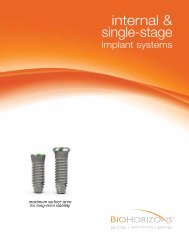
Internal Prosthetic Manual - BioHorizons
Internal Prosthetic Manual - BioHorizons
Internal Prosthetic Manual - BioHorizons
- No tags were found...

You also want an ePaper? Increase the reach of your titles
YUMPU automatically turns print PDFs into web optimized ePapers that Google loves.
TREATMENT PLANNINGFor ideal results in implant dentistry, the treatment team should be in agreement and in communication throughout all stagesof therapy. The patient, the restorative and surgical doctors, as well as the dental laboratory should understand and agree uponthe treatment plan. The treatment plan chosen will determine the design, number and position of the implants.Diagnostic CastsMounted study casts and a diagnostic wax-up are the foundation for determining implant location.Surgical Guide TemplatesOnce the diagnostic wax-up is finalized, the restorative doctor or dental laboratory fabricates the surgical guide template. Thisguide dictates to the surgeon the implant location that offers the best support for the prosthesis, as well as optimal estheticsand hygiene requirements. The surgical guide also provides information about the tooth and supporting structures that havebeen lost.Restoration MatrixA matrix of the diagnostic wax-up may also be utilized by the laboratory when developing the final prosthesis. The matrix actsas a guide for position and contour of the prosthesis.Fabricating a Surgical GuideSeveral methods of fabrication for the surgical template are available. One method is described below.1. Utilizing the diagnostic wax-up, create a duplicate stone model and then fabricate a clear acrylic thermoformguide over the waxed-up area and adjacent teeth. Extend the template to include several teeth on each sideof the implant site for template stability.2. The surgical guide is marked while on the study model in the area where the center of the implant should belocated. Implants are designed for axial load to the implant body. The marks on the surgical guide shouldreflect the optimal long-axis load location. For posterior teeth this would be through the occlusal table. Forcement-retained anterior teeth, the ideal location would be through the incisal edge.3. Drill holes through the template where the marks have been made from the study model. Dependent onimplant location, the facial or lingual walls of the surgical guide may need to be removed for easier surgicalaccess.4. For the totally edentulous arch, the template should extend onto unreflected soft tissue regions, i.e., thepalate and tuberosities in the maxilla or the retromolar pads in the mandible. This assures the template willbe stable after the soft tissues have been reflected from the bone during surgery.shop online at www.biohorizons.com2