

Issue 3 | Aug - Dr. Babette Gladstein, VMD
Issue 3 | Aug - Dr. Babette Gladstein, VMD
Issue 3 | Aug - Dr. Babette Gladstein, VMD

Create successful ePaper yourself
Turn your PDF publications into a flip-book with our unique Google optimized e-Paper software.
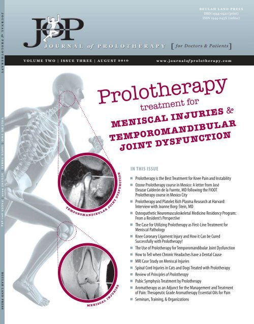
JOURNAL of PROLOTHERAPY V OLUME TWO | ISSUE THREE | AUGUST 2010 | PAGES 401-464 B EULAH LAND PRESS<br />
V OLUME TWO | ISSUE THREE | AUGUST 2010 w ww.journalof prolotherapy.com<br />
T E M P O R O M A N D I B U L A R J O I N T D Y S F U N C T I O N<br />
IN THIS ISSUE<br />
■<br />
■<br />
■<br />
■<br />
■<br />
■<br />
■<br />
■<br />
■<br />
■<br />
■<br />
■<br />
■<br />
■<br />
BEULAH LAND PRESS<br />
ISSN 1944-0421 (print)<br />
ISSN 1944-043X (online)<br />
Prolotherapy<br />
M E N I S C A L I N J U R I E S<br />
treatment for<br />
MENISCAL INJURIES &<br />
TEMPOROMANDIBULAR<br />
JOINT DYSFUNCTION<br />
Prolotherapy is the Best Treatment for Knee Pain and Instability<br />
Ozone Prolotherapy course in Mexico: A letter from José<br />
Eleazar Calderón de la Fuente, MD following the FIOOT<br />
Prolotherapy course in Mexico City<br />
Prolotherapy and Platelet Rich Plasma Research at Harvard:<br />
Interview with Joanne Borg-Stein, MD<br />
Osteopathetic Neuromusculoskeletal Medicine Residency Program:<br />
From a Resident’s Perspective<br />
The Case for Utilizing Prolotherapy as First-Line Treatment for<br />
Meniscal Pathology<br />
Knee Coronary Ligament Injury and How it Can be Cured<br />
Successfully with Prolotherapy!<br />
The Use of Prolotherapy for Temporomandibular Joint Dysfunction<br />
How to Tell when Chronic Headaches have a Dental Cause<br />
MRI Case Study on Meniscal Injuries<br />
Spinal Cord Injuries in Cats and Dogs Treated with Prolotherapy<br />
Review of Principles of Prolotherapy<br />
Pubic Symphysis Treatment by Prolotherapy<br />
Aromatherapy as an Adjunct for the Management and Treatment<br />
of Pain: Therapeutic Grade Aromatherapy Essential Oils for Pain<br />
Seminars, Training, & Organizations
Table of Contents<br />
G R E A T N E W S C O R N E R<br />
406<br />
I N T H I S I S S U E O F T H E J O U R N A L O F P R O L O T H E R A P Y<br />
Prolotherapy is the Best Treatment<br />
for Knee Pain and Instability<br />
Ross A. Hauser, MD<br />
L E T T E R S T O T H E E D I T O R<br />
408<br />
I N T H E S P O T L I G H T<br />
410<br />
413<br />
Ozone Prolotherapy course in<br />
Mexico: A letter from José Eleazar<br />
Calderón de la Fuente, MD<br />
following the FIOOT Prolotherapy<br />
course in Mexico City<br />
Ross A. Hauser, MD<br />
Prolotherapy and Platelet Rich<br />
Plasma Research at Harvard:<br />
Interview with Joanne Borg-Stein, MD<br />
Ross A. Hauser, MD & Joanne Borg-Stein, MD<br />
Osteopathetic Neuromusculoskeletal<br />
Medicine Residency Program: From<br />
a Resident’s Perspective<br />
Peter J. Blakemore, DO<br />
F A N T A S T I C F I N D I N G S<br />
416<br />
The Case for Utilizing Prolotherapy<br />
as First-Line Treatment for Meniscal<br />
Pathology Ross A. Hauser, MD, Hilary J.<br />
Phillips, & Havil S. Maddela<br />
R E M A R K A B L E R E C O V E R I E S<br />
438<br />
Knee Coronary Ligament Injury<br />
and How it Can be Cured<br />
Successfully with Prolotherapy!<br />
Joern Funck, MD<br />
W O N D E R W H Y ?<br />
439<br />
447<br />
454<br />
The Use of Prolotherapy for<br />
Temporomandibular Joint<br />
Dysfunction<br />
J O U R N A L of P R O L O T H E R A P Y | V O L U M E 2 , I S S U E 3 | A U G U S T 2 0 1 0<br />
Roy V. Hakala, DDS & Kim M. Ledermann, DDS<br />
How to Tell when Chronic<br />
Headaches have a Dental Cause<br />
Jeri Coffey, DDS, Ross A. Hauser, MD,<br />
Nicole M. Baird, CHFP, & Doug R. Skinkis<br />
MRI Case Study on Meniscal<br />
Injuries Jack Henry, DC, DACBR<br />
F O U R - L E G G E D P R O L O T H E R A P Y<br />
455<br />
B O O K R E V I E W<br />
457<br />
Spinal Cord Injuries in Cats and<br />
Dogs Treated with Prolotherapy<br />
<strong>Babette</strong> <strong>Gladstein</strong>, <strong>VMD</strong><br />
Review of Principles of Prolotherapy<br />
Ross A. Hauser, MD<br />
T E A C H I N G T E C H N I Q U E S<br />
459<br />
Pubic Symphysis Treatment by<br />
Prolotherapy Rodney S. Van Pelt, MD<br />
I T ’ S A W I D E W I D E W O R L D<br />
461<br />
S K I L L E N H A N C E M E N T<br />
464<br />
Aromatherapy as an Adjunct for the<br />
Management and Treatment of<br />
Pain: Therapeutic Grade<br />
Aromatherapy Essential Oils for Pain<br />
Wanona Wellspring, DN<br />
Seminars, Training, & Organizations
JOP Team Mark DeLaurentis, MD<br />
E D I T O R - I N - C H I E F<br />
Ross A. Hauser, MD<br />
Oak Park, IL<br />
E D I T O R I A L B O A R D<br />
Donna Alderman, DO<br />
Glendale, CA<br />
Gunter Baehnisch, MD<br />
Leipzig, Germany<br />
Robert Banner, MD<br />
London, Ontario, Canada<br />
José Eleazar Calderón, MD<br />
Monclova, Coahuila, Mexico<br />
Gary B. Clark, MD, MPA<br />
Boulder, CO<br />
J O U R N A L O F P R O L O T H E R A P Y T E A M & S U B S C R I B E R I N F O R M A T I O N<br />
Cherry Hill, NJ<br />
Shaun Fauley, DVM<br />
Naperville, IL<br />
Joern Funck, MD<br />
Luebeck, Germany<br />
<strong>Babette</strong> <strong>Gladstein</strong>, <strong>VMD</strong><br />
New York, NY<br />
Mark L. Johnson, MD, FACS<br />
Nashville, TN<br />
George H. Kramer, MD<br />
Minnetonka, MN<br />
John Neustadt, ND<br />
Bozeman, MT<br />
Joan Resk, DO, JD<br />
Roanoke, VA<br />
Subscriber Information<br />
The Journal of Prolotherapy® is unique in that it has a target<br />
audience of both physicians and patients. The purpose of<br />
this journal is to provide the readers with new cutting-edge<br />
information on Prolotherapy, as well as provide a forum for<br />
physicians and patients alike to tell their stories.<br />
The Journal of Prolotherapy® is published quarterly – in February,<br />
May, <strong>Aug</strong>ust, and November by Beulah Land Press, 715 Lake<br />
Street, Suite 600, Oak Park, Illinois, 60301. © Copyright 2010 by<br />
Beulah Land Press. All rights reserved. No portion of the contents<br />
may be reproduced in any form without written permission<br />
from the publisher.<br />
All subscription inquiries, orders, back issues, claims, and<br />
renewals should be addressed to Beulah Land Press, 715 Lake<br />
St. Suite 600, Oak Park, IL 60301; phone: 708.848.5011; fax:<br />
708.848.0978. Email: bairdn@journalofprolotherapy.com;<br />
http://beulahlandpress.com.<br />
P R I C I N G<br />
Annual subscription: $100/year.<br />
Includes 4 paper issues and online access to all<br />
www.journalofprolotherapy.com web content.<br />
C L A I M S<br />
Claims for undelivered copies must be made no later than one<br />
month following the month of publication. The publisher will<br />
supply missing copies when losses have been sustained in<br />
transit and when the reserve stock will permit.<br />
José Hector Salazar, MD<br />
Monterrey Nuevo Leon, Mexico<br />
Garrett Swetlikoff, ND<br />
Kelowna, BC, Canada<br />
Rodney S. Van Pelt, MD<br />
Ukiah, CA<br />
Mark T. Wheaton, MD<br />
Minnetonka, MN<br />
R E S I D E N T E D I T O R S<br />
Peter J. Blakemore, DO<br />
Watertown, NY<br />
Patrick M. Hobbins, DO<br />
South Bend, IN<br />
C H A N G E O F A D D R E S S<br />
Change of address notices should be sent to JOP, 30 days in<br />
advance to: JOP 715 Lake St. Suite 600, Oak Park, IL 60301;<br />
phone: 708.848.5011; fax: 708.848.0978.<br />
C O P Y R I G H T P E R M I S S I O N<br />
Permission requests to photocopy or otherwise reproduce<br />
copyrighted material owned by Beulah Land Press should be<br />
requested by contacting Beulah Land Press at 708.848.5011 or<br />
by emailing info@benuts.com.<br />
D I S C L A I M E R<br />
This publication does not constitute medical or other<br />
professional advice and should not be taken as such. To the<br />
extent the articles published herein may be used to assist in<br />
the care of patients, this is the result of the sole professional<br />
judgment of the health professional. The health care<br />
professional’s judgment is the primary component of quality<br />
health care. The information presented in this journal is<br />
not a substitute for the exercise of such judgment by the<br />
health professional. The opinions expressed in the Journal of<br />
Prolotherapy® are the opinions of the authors of their respective<br />
articles and not necessarily that of the publisher. The decisions<br />
on what to do for a specific medical condition or symptom<br />
should be based on the analysis by the person’s private health<br />
care professional.<br />
J O U R N A L of P R O L O T H E R A P Y | V O L U M E 2 , I S S U E 3 | A U G U S T 2 0 1 0<br />
J O P S T A F F<br />
Marion A. Hauser, MS, RD<br />
Senior Editor<br />
Nicole M. Baird, CHFP<br />
Associate Editor<br />
Travis E. Mitchell<br />
Senior Graphic Designer/Webmaster<br />
Doug R. Skinkis<br />
Assistant to the Editor-in-Chief/<br />
Assistant Graphic Designer<br />
Patricia H. Miller<br />
Assistant to Media Relations<br />
Enid M. Forsyth<br />
Lay Editor<br />
Media for Doctors, Inc.<br />
Media Relations<br />
ISSN 1944-0421 (print)<br />
ISSN 1944-043X (online)
Authors<br />
N I C O L E M . B A I R D , C H F P<br />
J E R I C O F F E Y, D D S<br />
J O U R N A L O F P R O L O T H E R A P Y A U T H O R S<br />
Nicole M. Baird is a Certified Holistic Fitness Practitioner and is the Marketing and Publications Manager for Caring Medical<br />
& Rehabilitation Services, as well as Beulah Land Corporation, in Oak Park, Illinois. Her passion for writing, nutrition, and<br />
food lead her down the path of co-authoring The Hauser Diet: A Fresh Look at Healthy Living! Nicole has an avid interest in<br />
nutrition, alternative medicine, exercise, and medical research and has used her knowledge to become an instrumental<br />
writer for many of the publications associated with Caring Medical/Beulah Land Corporation’s many printed materials,<br />
including patient brochures, website content, case studies, books, e-newsletters, and now the Journal of Prolotherapy®.<br />
Nicole can be reached at Caring Medical & Rehabilitation Services, 715 Lake St. Suite 600, Oak Park, IL 60301;Tel: 708.848.7789;<br />
www.caringmedical.com and www.journalofprolotherapy.com.<br />
P E T E R J . B L A K E M O R E , D O<br />
<strong>Dr</strong>. Peter Blakemore graduated from Bob Jones University in 2004 with a BS in Premedicine. He completed his medical<br />
school training at the University of New England College of Osteopathic Medicine in Biddeford, Maine, graduating<br />
with a Doctorate of Osteopathy in 2008, followed by an internship at Samaritan Medical Center in Watertown, New<br />
York. <strong>Dr</strong>. Blakemore is currently completing a residency in Neuromuscular Medicine and Osteopathic Manipulative<br />
Medicine at Michigan State University College of Osteopathic Medicine in East Lansing, Michigan, where he also<br />
teaches in the department of Manual Medicine. <strong>Dr</strong>. Blakemore has had the privilege of working with and training<br />
under <strong>Dr</strong>. Hauser for ten years and has worked in many medical missionary Prolotherapy clinics. <strong>Dr</strong>. Blakemore has a<br />
love for Prolotherapy and hopes to use it in his medical practice upon completion of his residency. <strong>Dr</strong>. Blakemore is<br />
married to his wife Krista and they have one daughter Petra. In his free time, he enjoys spending time with family, Bible<br />
reading, hiking, cycling, and running, having completed the Ironman Triathlon in 2004.<br />
J O A N N E B O R G - S T E I N , M D<br />
Joanne Borg-Stein, MD is Medical Director of Spaulding-Wellesley and Newton-Wellesley Spine Center. In addition,<br />
she is the team physician for Wellesley College and assistant professor in the Harvard Medical School Department of<br />
Physical Medicine and Rehabilitation. <strong>Dr</strong>. Borg-Stein received her undergraduate degree at Tufts University and her<br />
MD at Albert Einstein College of Medicine. <strong>Dr</strong>. Borg-Stein provides comprehensive musculoskeletal care, including<br />
medical acupuncture, Prolotherapy, and Botox injections. She is involved with gender specific care for women’s<br />
musculoskeletal injuries including conditions related to pregnancy, sports and osteoporosis. <strong>Dr</strong>. Borg-Stein has<br />
published numerous peer-reviewed articles and book chapters in the field of sports medicine, chronic pain, and<br />
rehabilitation. <strong>Dr</strong>. Borg-Stein may be reached at Spaulding-Wellesley Rehab Center, 65 Walnut Street, Wellesley, MA<br />
02481; Tel: 781.431.9144; www.spauldingrehab.org.<br />
Jeri Coffey, DDS received her Bachelor’s degree from North Central College in Naperville, and her DDS from Loyola<br />
University Dental School. She has headed up her General Dentistry private practice since 1981 in Riverside, Illinois. She<br />
was also an Associate Professor of Operative Dentistry at Loyola University Dental School from 1989-1992. <strong>Dr</strong>. Coffey<br />
has a Masters in homeopathic medicine, training in clinical hypnosis, along with training at the Dawson Academy of<br />
Functional Occlusion. <strong>Dr</strong>. Coffey may be reached at 24 Woodside Rd, Riverside, IL 60546; Tel: 708.442.0115.<br />
J O E R N F U N C K , M D<br />
<strong>Dr</strong>. Funck grew up in Luebeck, Germany. After receiving his MD from the University of Hamburg in 1968 he completed<br />
his trauma surgery and orthopedic surgeon training in Hannover and Braunschweig, Germany. After finishing his clinical<br />
education he left the operating branch and switched to muscoloskeletal medicine, as well as manual therapy, opening<br />
his own private office in Luebeck in 1975. In Bremen, Germany, <strong>Dr</strong>. Funck took up studies in orthopedic medicine,<br />
focusing on Cyriax diagnosis and treatment of soft tissues. He studied manual therapy with Prof. Tilscher from Vienna<br />
who first introduced him to Prolotherapy for low back pain patients. <strong>Dr</strong>. Funck may be reached at Fackenburger Allee<br />
22-24 23554 Lübeck, Germany; Tel: 0451.411.20; www.proliferationstherapie.de.<br />
J O U R N A L of P R O L O T H E R A P Y | V O L U M E 2 , I S S U E 3 | A U G U S T 2 0 1 0
B A B E T T E G L A D S T E I N , V M D<br />
R O S S A . H A U S E R , M D<br />
J O U R N A L O F P R O L O T H E R A P Y A U T H O R S<br />
<strong>Babette</strong> <strong>Gladstein</strong>, <strong>VMD</strong> is a graduate of the University of Pennsylvania, School of Veterinary Medicine. Her postdoctoral<br />
work has included veterinary acupuncture at the American Academy of Veterinary Medical Acupuncture<br />
at Colorado State University, as well as pre-veterinary studies at Hunter College in New York City. She is a member of<br />
the American Association of Equine Practitioners, the American Veterinarian Medical Association, and the American<br />
Holistic Veterinary Medical Association. <strong>Dr</strong>. <strong>Gladstein</strong> has also been affiliated with The New York Racing Association,<br />
Meadowlands Raceway, and US Equestrian. As a licensed veterinarian in New York, New Jersey, Connecticut,<br />
Florida, and California, <strong>Dr</strong>. <strong>Gladstein</strong>’s treatment modality expertise includes Prolotherapy, acupuncture, ultrasound,<br />
chiropractic, and massage therapy. Since the mid ‘80s, <strong>Dr</strong>. <strong>Gladstein</strong> observed and studied the benefits of nutrition<br />
and nutritional supplements in animal care and treatments and followed this with investigations into therapeutic<br />
ultrasound, massage and acupuncture, as well as physical therapy for horses. <strong>Dr</strong>. <strong>Gladstein</strong> may be reached at 45 East<br />
89th Street, #31D, NY, NY 10128; Tel: 212.828.5663; www.animalacupuncture.net.<br />
R O Y V . H A K A L A , D D S<br />
Roy V. Hakala, DDS is a graduate of the University of Minnesota School of Dentistry. He received advanced training<br />
at the United States Dental Institute, the American Academy of Craniofacial Pain, and the National Capitol Center for<br />
Craniofacial Pain. He is a diplomate of both the American Board of Craniofacial Pain and the American Board of Dental<br />
Sleep Medicine. He established the Minnesota Craniofacial Center Midway, which is dedicated to the treatment of<br />
TMJ disorders and to oral appliance treatment of obstructive sleep apnea, in 1994. <strong>Dr</strong>. Hakala can be reached at 1690<br />
University Avenue West #390, St. Paul, MN 55104; Tel: 651.642.1013; www.mncranio.com.<br />
Ross A. Hauser, MD received his undergraduate degree from University of Illinois. He graduated from the University<br />
of Illinois College of Medicine in Chicago and did his residency at Loyola/Hines VA in Physical Medicine and<br />
Rehabilitation. <strong>Dr</strong>. Hauser is the Medical Director of Caring Medical and Rehabilitation Services in Oak Park, Illinois and<br />
is passionate about Prolotherapy and natural medicine. <strong>Dr</strong>. Hauser and his wife Marion, have written seven books<br />
on Prolotherapy, including the national best seller “Prolo Your Pain Away! Curing Chronic Pain with Prolotherapy,” now<br />
in its third edition, a four-book mini series of Prolotherapy books, and a 900-page epic sports book on the use of<br />
Prolotherapy for sports injuries. <strong>Dr</strong>. Hauser may be reached at 715 Lake St., Suite 600, Oak Park, IL 60301; Tel: 708.848.7789;<br />
www.caringmedical.com.<br />
J A C K H E N R Y, D C , D A C B R<br />
<strong>Dr</strong>. Henry holds a BS in Human Biology and a BS in Public Health. <strong>Dr</strong>. Henry graduated from National College of<br />
Chiropractic (National University of Health Science) and later completed the residence program in Diagnostic Imaging<br />
(Radiology). <strong>Dr</strong>. Henry is a Diplomate to the American Chiropractic Board of Radiology. He has over 20 years experience<br />
interpreting diagnostic images for chiropractors and medical doctors across the country. <strong>Dr</strong>. Henry is published and<br />
has lectured on topics of radiology for state and local chiropractic societies. At present, <strong>Dr</strong>. Henry makes his home in<br />
Massachusetts with his wife Lisa. Together, they have three adult children, all of whom currently serve in the Armed<br />
Forces. <strong>Dr</strong>. Henry is Radiologist-in-Chief of Radiology Diagnostics, LLC. If you would like to contact <strong>Dr</strong>. Henry e-mail him at<br />
jhenry@radiologydiagnostics.com.<br />
K I M M . L E D E R M A N N , D D S<br />
<strong>Dr</strong>. Ledermann is a graduate of the University of Minnesota School of Dentistry and holds a Master’s degree in oral<br />
function/speech and language pathology. She is certified in the diagnosis and treatment of craniofacial disorders by<br />
the Institute of Craniofacial Pain and is a member of the American Academy of Dental Sleep Medicine. She joined the<br />
Minnesota Craniofacial Center Midway in 2009. <strong>Dr</strong>. Ledermann can be reached at 1690 University Avenue West #390,<br />
St. Paul, MN 55104; Tel: 651.642.1013; www.mncranio.com.<br />
J O U R N A L of P R O L O T H E R A P Y | V O L U M E 2 , I S S U E 3 | A U G U S T 2 0 1 0
H A V I L S . M A D D E L A<br />
H I L A R Y J . P H I L L I P S<br />
J O U R N A L O F P R O L O T H E R A P Y A U T H O R S<br />
Havil S. Maddela currently works as a medical assistant at Caring Medical and Rehabilitation Services in Oak<br />
Park, Illinois. He has had a passion for medicine since he was young and is currently enrolled in the Pre-Med<br />
program at Loyola University, Chicago where he will graduate in May 2010 with a degree in biology. Havil<br />
has worked in a medical missionary clinic during his high school years as well. Havil hopes to attend Medical<br />
School in the fall of 2010. Havil may be reached at 715 Lake St., Suite 600, Oak Park, IL 60301; Tel: 708.848.7789;<br />
www.caringmedical.com.<br />
Hilary J. Phillips received her Associate of Science degree from Triton College, in River Grove, Illinois. Her previous<br />
medical experience includes volunteering at Beulah Land Natural Medicine Clinic and working as medical assistant<br />
in skilled care geriatric nursing, prior to coming to Caring Medical and Rehabilitation Services where she served as a<br />
medical assistant for over a year. Hilary is currently a student at North Park University, where she is pursuing a Bachelor<br />
of Science degree in chemistry.<br />
D O U G R . S K I N K I S<br />
Doug R. Skinkis, a native Chicagoan, graduated from Northern Illinois University with a degree in Communications with<br />
a Radio and Television emphasis, minor in Art. He worked as a recruiter for Western Technical Services placing engineers,<br />
drafters, in contract, part-time, and full-time positions; then for Impact Label as the Plant Manager of a label and sticker<br />
manufacturer; and then for Napco Graphics as the Shipping/Receiving and QA/QC Manager. Doug currently works as<br />
the Director of Operations for Caring Medical and Rehabilitation Services where he assists with many of the graphic<br />
design and videography for their websites and publications. Doug also assists the senior graphic designer of the Journal<br />
of Prolotherapy with artwork. Doug may be reached at 715 Lake St., Suite 600, Oak Park, IL 60301; Tel: 708.848.7789;<br />
www.caringmedical.com.<br />
R O D N E Y S . V A N P E LT , M D<br />
Rodney S. Van Pelt, MD received his medical degree from Loma Linda University Medical School and completed<br />
his family practice residency in Florida. He practiced family medicine for several years until falling in love with the<br />
specialty of Orthopedic Medicine which uses all the different modalities for pain with conservative treatments. <strong>Dr</strong>. Van<br />
Pelt then received specialized training in the Cyriax technique of Orthopedic Medicine, taking some of his training in<br />
Oxford, England. He is one of the few American physicians who is a member of the Society of Orthopedic Medicine<br />
of London, England. <strong>Dr</strong>. Van Pelt practices full time Prolotherapy in northern California. <strong>Dr</strong>. Van Pelt may be reached at<br />
Orthopedic Wellness Center Plaza Del Sol, 776 S State St., Ukiah, CA 95482; Tel: 707.463.1782; www.sfpmg.com.<br />
W A N O N A W E L L S P R I N G , D N<br />
<strong>Dr</strong>. Rev. Wanona Wellspring, DN, Licensed Doctor of Naprapathy received her doctorate in 1999 from the National<br />
College of Naprapathic Medicine. Naprapathy is a manual therapy that specializes in connective tissue disorders. She<br />
is the creator of Wellspring Massage, editor of Oak Leaves, a professional newsletter for the Naprapathic community, as<br />
well as the Educational Chair for the Naprapathic Academy of Science Education and Research, producing educational<br />
seminars for health professionals for continuing education. <strong>Dr</strong>. Wellspring incorporates clinical nutrition and exercise<br />
physiology into a complete healing process that includes Naprapathy, Myofascial Release, Cranio Sacral technique<br />
and Yoga therapeutics. <strong>Dr</strong>. Wellspring can be reached at the Wellspring Family Health Centre at 339 W Palatine Rd,<br />
Palatine, IL 60067; Tel: 847.496.7207; www.wellspringfamily.org.<br />
J O U R N A L of P R O L O T H E R A P Y | V O L U M E 2 , I S S U E 3 | A U G U S T 2 0 1 0
406<br />
G R E A T N E W S C O R N E R : P R O L O T H E R A P Y I S T H E B E S T T R E A T M E N T F O R K N E E P A I N A N D I N S T A B I L I T Y<br />
For many years, data has been mounting that<br />
arthroscopy just doesn’t work any better than<br />
conservative care for most knee conditions,<br />
including degenerative arthritis. This is based on thorough<br />
research published in some of the most prestigious<br />
medical journals in the world. 1-4 This has lead insurance<br />
companies to not cover arthroscopic debridement of the<br />
knee for knee pain, but they will cover it for mechanical<br />
symptoms. This may sound reasonable until one really<br />
looks at the definition of mechanical symptoms of the<br />
knee: any type of locking, popping or giving way of the<br />
knee! Basically, almost every person with a knee has some<br />
type of “popping” or crunching (also called crepitation)<br />
noise in their knees. This means that a patient who sees<br />
an orthopedist who documents mechanical symptoms in<br />
the patient’s knee and/or if the patient’s MRI shows any<br />
type of loose body or meniscal tear, then arthroscopic<br />
surgery can be done and will be covered by insurance.<br />
One problem with the above scenario is that MRIs cannot<br />
reveal the cause of the patient’s pain. Among persons with<br />
radiographic evidence of osteoarthritis, the prevalence of<br />
a meniscal tear was 63% among those with knee pain,<br />
catching, or stiffness on most days, and 60% among those<br />
without symptoms. 5 Let’s think about this some more. A<br />
full 60% of people who have no pain will show a meniscal<br />
tear on MRI! The net result is that the number of knee<br />
arthroscopies continues to rise because everyone with a<br />
knee problem qualifies for it! But there is a better way…<br />
and that is Prolotherapy.<br />
If there is one joint in the body that responds tremendously<br />
well to Prolotherapy, it is the knee! Whether the condition<br />
is degenerative arthritis, pes anserine or patellar<br />
tendinopathy, collateral or cruciate ligament sprains—they<br />
all respond well to Prolotherapy. For decades, Prolotherapy<br />
physicians have treated painful knees, with and without<br />
mechanical symptoms, successfully with Prolotherapy. It’s<br />
really just a matter of how many treatments are needed.<br />
G R E A T N E W S C O R N E R<br />
Prolotherapy is the Best Treatment<br />
for Knee Pain and Instability<br />
Ross A. Hauser, MD<br />
Arthroscopic surgery for mensical issues typically results<br />
in meniscectomy (removal). The thinking is simply that<br />
the meniscal tear didn’t heal on its own, so we need to<br />
remove this tissue and the knee will feel better. This type<br />
of thinking is a bad idea. I urge you to take a look at<br />
the following subheadings excerpted from Prolo Your Sports<br />
Injuries Away! which may further convince you:<br />
“Study Shows Increased Contact Stress Pressure After Meniscectomy.”<br />
“Study Shows Meniscal Surgery Actually Increases Injury.”<br />
“Incomplete Healing And Further Deterioration Result After<br />
Meniscal Repair Surgery.”<br />
“Meniscectomy Causes Arthritis.”<br />
“Partial Meniscectomy: More Arthritic Changes Result.” 6<br />
Prolo Your Sports Injuries Away! was written in 2001 and is<br />
loaded with references to back up its claims regarding<br />
treating mensical injuries. After nearly 10 years, we felt it<br />
was time to revisit the mensicus, therefore Caring Medical<br />
(my private practice) hired a pre-med college student to coauthor<br />
a meniscus paper. During her research, she related<br />
her findings to me during a meeting, “Meniscectomy<br />
surgery results (partial or complete) are terrible! Why<br />
would anyone get that surgery?” Well said, Hilary! The<br />
data clearly reveals that arthroscopic partial meniscectomy<br />
will age a patient’s knee by 20 years! In almost every knee<br />
arthroscopy report I have seen (hundreds and hundreds),<br />
over 90% describe in detail the removal of meniscal tissue<br />
(meniscectomy). (See Figure 1.) Once the meniscal tissue is<br />
gone, there is a tremendous increase in pressure on the<br />
articular cartilage and this quickly starts to break down.<br />
This is what we call the progression from arthroscopy to<br />
arthritis.<br />
Two pre-med college students, Hilary Phillips and Havil<br />
Maddela, collaborated with us on Prolotherapy for MRIdocumented<br />
cases of meniscal degeneration and meniscal<br />
tears on patients from Caring Medical. The results of this<br />
study reveal that Prolotherapy is the best option for knee<br />
pain and instability.<br />
J O U R N A L of P R O L O T H E R A P Y | V O L U M E 2 , I S S U E 3 | A U G U S T 2 0 1 0
G R E A T N E W S C O R N E R : P R O L O T H E R A P Y I S T H E B E S T T R E A T M E N T F O R K N E E P A I N A N D I N S T A B I L I T Y<br />
DESCRIPTION OF THE PROCEDURE: The patient was placed in a<br />
supine position and general anesthesia was administered. The knee<br />
was prepped and draped in usual manner. The tourniquet was raised<br />
to 350 mmHg. The arthroscope was inserted into the suprapatellar<br />
pouch. The underside of the patella was noted to be free of any<br />
pathology. No Loose bodies were noted. The scope was swept from<br />
the medial compartment of the knee. A complex tear of the posterior<br />
two-thirds of the medial meniscus was evident. Chondromalacia of<br />
the medial femoral condyle was quite severe and some areas with<br />
bone were exposed. This was over the entire weightbearing surface<br />
grade 3 to grade 4. The intercondylar notch exhibited an intact cruciate<br />
ligament complex and the lateral meniscus was noted at its free edge<br />
of the middle third and slightly posterior third to have an attritional<br />
tear. The lateral meniscus was trimmed with a basket forceps<br />
taking approximately 10% of the middle third and posterior third.<br />
The basket forceps was utilized to trim away the torn meniscus in<br />
the medial compartment of the knee and this was further smoothed<br />
with a 5.5 mm end cutter and a chondroplasty was performed with<br />
similar instrument over the femoral condyle to remove the unstable<br />
cartilage. The patient had approximately 20% of the posterior middle<br />
thirds balancing it into the anterior third. After this was done to the<br />
satisfaction of the surgeon, the knee was irrigated with a Toomey<br />
syringe, and closed with 4-0 Vicryl. Depo-Medrol and Marcaine<br />
was instilled. A bulky dressing applied. The patient tolerated the<br />
procedure well.<br />
Figure 1. Operative report describing meniscectomy during<br />
a knee arthroscopy. This patient, like most I’ve treated, had<br />
no idea that their meniscus was removed. They would not have<br />
gone through with the arthroscopy, especially if the future<br />
arthritis risks were known ahead of time.<br />
Also in this issue, we welcome new columnist, Jack Henry,<br />
DC who presents a meniscal tear case. We are looking<br />
forward to <strong>Dr</strong>. Henry’s contributions in educating our<br />
readership on radiologic findings. Skill Enhancement<br />
columnist, Rodney Van Pelt, MD demonstrates<br />
Prolotherapy for pubis pain. Veterinary columnist,<br />
<strong>Babette</strong> <strong>Gladstein</strong>, <strong>VMD</strong> discusses Prolotherapy used in<br />
spinal injury cases she has treated.<br />
In the Spotlight first features Peter Blakemore, DO who has<br />
written a piece highlighting the Neuromusculoskeletal<br />
Medicine residency at Michigan State University. Students<br />
can elect to rotate in private Prolotherapy offices, which<br />
continues to gain headway into medical residencies and<br />
hopefully will soon be offered in the school. Also In the<br />
Spotlight is an interview I conducted with Joanne Borg-<br />
Stein, MD, who is practicing PRP Prolotherapy at<br />
Harvard.<br />
From Germany, Joern Funck, MD reports a Remarkable<br />
Recovery case and explanation of how to treat the coronary<br />
ligament of the knee. In this issue, we also have two articles<br />
relating to temporomandibular joint dysfunction and<br />
dentistry. First Roy Hakala, DDS and Kim Ledermann,<br />
DDS wrote a great article on Prolotherapy for TMJ<br />
dysfunction. Second, our JOP team interviewed Jeri<br />
Coffey, DDS in her Riverside, Illinois office and consulted<br />
her on when to consider a dental cause for a patient<br />
presenting with chronic headaches.<br />
Last but not least, in It’s a Wide, Wide World, Wanona<br />
Wellspring, DN has authored an article on using<br />
aromatherapy as an adjunctive pain therapy. In addition,<br />
Jose Eleazar Calderon de la Fuente, MD from Mexico<br />
has written to share the outcome of the first Prolotherapy<br />
conference in Mexico City. He did a tremendous amount<br />
of work, and the results were nothing short of remarkable<br />
in regard to the patients who were treated and the fellow<br />
physicians to whom he taught the Hackett-Hemwall<br />
technique of Prolotherapy.<br />
As always, thanks to all of our readers for your continued<br />
support of Prolotherapy. As the issue shows, Prolotherapy<br />
and its practitioners are continuing to improve patients’<br />
lives around the world! n<br />
Until the next injection,<br />
Ross A. Hauser, MD<br />
1.<br />
2.<br />
3.<br />
4.<br />
5.<br />
6.<br />
B i B l i o g r a p h y<br />
Moseley JB, et al. A controlled trial of arthroscopic surgery<br />
for osteoarthritis of the knee. New England Journal of Medicine.<br />
2002;347:137-139.<br />
Dervin G, et al. Effect of arthroscopic debridement for<br />
osteoarthritis of the knee on health-related quality of life. Journal<br />
of Bone and Joint Surgery American. 2003;85-A(1):10-19.<br />
Kirkley A, et al. A randomized trial of arthroscopic surgery<br />
for osteoarthritis of the knee. New England Journal of Medicine.<br />
2008;359:1097-1107.<br />
Siparksky P, et al. Arthroscopic treatment of osteoarthritis of<br />
the knee: are there any evidence-based indications? Clinical<br />
Orthopaedics and Related Research. 2007;455:107-112.<br />
Englund M, et al. Incidental meniscal findings on knee MRI in<br />
middle-aged and elderly persons. New England Journal of Medicine.<br />
2008;359:1108-1115.<br />
Hauser R, et al. Prolo Your Sports Injuries Away! Oak Park, IL:<br />
Beulah Land Press; 2001:173-176.<br />
J O U R N A L of P R O L O T H E R A P Y | V O L U M E 2 , I S S U E 3 | A U G U S T 2 0 1 0 407
408<br />
L E T T E R S T O T H E E D I T O R<br />
Ozone Prolotherapy course in Mexico:<br />
A letter from José Eleazar Calderón de la<br />
Fuente, MD following the FIOOT<br />
Prolotherapy course in Mexico City<br />
l E T T E r F r o M r E a D E r<br />
Dear <strong>Dr</strong>. Ross Hauser,<br />
I want to inform you that the first course of<br />
Prolotherapy in Mexico City, has been a success. The<br />
course took place on November 27 th and 28 th 2009.<br />
It was organized by the International Federation of<br />
Ozone Oxygen Therapy (FIOOT). Its President,<br />
<strong>Dr</strong>. Fabio Antúnez Guzmán, made the opening<br />
presentation, and the course was taught by myself.<br />
The first day of the course involved a long slide<br />
show, which explained the technical aspects of the<br />
procedure, and was received with much greater<br />
success than I expected. It also reviewed historical<br />
Prolotherapy topics from its founder, <strong>Dr</strong>. George<br />
S. Hackett, along with those of <strong>Dr</strong>. Gustav A.<br />
<strong>Dr</strong>. Calderon with students, marking the low back at the<br />
injection sites.<br />
Ross A. Hauser, MD<br />
Hemwall. The slides also featured theoretical,<br />
clinical topics, physical examination and especially<br />
the histiopathologic studies carried out by different<br />
physicians in the United States, which was an<br />
overwhelming proof of the Prolotherapy science.<br />
The second day was practical application with<br />
patients of different pathologies and different regions<br />
(neck, lumbar, knees, ankles and elbows, even a<br />
temporomandibular). They were first injected by me,<br />
then other doctors were allowed to inject, among<br />
which were mostly orthopedists and chiropractors,<br />
anesthesiologists (who specialize in pain conditions,<br />
ie: phantom pain), and one specialist in physical<br />
medicine and rehabilitation.<br />
We distributed the complimentary “Prolo Your Pain<br />
Away!” books you sent, which were very well received,<br />
as well as the Journal of Prolotherapy issues. It reaffirmed<br />
the credibility and confidence in this technique, due<br />
to the great seriousness and global acceptance of the<br />
journal.<br />
The end of the course was closed by our President,<br />
<strong>Dr</strong>. Antúnez, and was proceeded by the distribution of<br />
diplomas (distributed by myself). Of course, the most<br />
important and wonderful part of this course<br />
was that it was recognized by our Secretary<br />
of Public Education (SEP), which is the body<br />
of educational oversight in our country, and<br />
allows us to perform Prolotherapy freely in<br />
our clinics. Two days after the event, the president<br />
of FIOOT and I spoke. He told me that the treated<br />
patients were extremely happy, because for the first<br />
time they noted a positive change in their chronic<br />
J O U R N A L of P R O L O T H E R A P Y | V O L U M E 2 , I S S U E 3 | A U G U S T 2 0 1 0
pain. <strong>Dr</strong>. Antúnez was so pleased that he wants to<br />
hold the course twice a year by FIOOT. He also<br />
invited me to come to Monclava, Coah. to reaffirm<br />
his training, and to take a personalized course in my<br />
clinic.<br />
With great esteem and affection for having been<br />
taught to see the world of chronic pain from a<br />
different perspective, your student,<br />
<strong>Dr</strong>. José Eleazar Calderón de la Fuente<br />
E D i T o r ’ s c o M M E n T s<br />
Dear <strong>Dr</strong>. Calderon:<br />
That is great news that Prolotherapy has been recognized by<br />
the Secretary of Public Education in Mexico. It is in large<br />
measure because of your tremendous effort to be one of the<br />
first Prolotherapists in all of Mexico. As a representative of<br />
Prolotherapists around the world, I congratulate you on your<br />
work in Mexico! As Prolotherapy gets accepted not only by<br />
patients and the physicians who perform Prolotherapy, but by<br />
the governing bodies overseeing public health and medical care<br />
in the various countries, its availability to eliminate pain across<br />
the globe will surely continue to grow. Let’s hope one day we see<br />
Prolotherapy as one of the main therapies available for people<br />
suffering from disabling pain!<br />
Ross A. Hauser, M.D. n<br />
L E T T E R S T O T H E E D I T O R<br />
<strong>Dr</strong>. Calderon demonstrating Prolotherapy to a knee.<br />
Two students with <strong>Dr</strong>. Calderon and <strong>Dr</strong>. Fabio Antunes<br />
(FIOOT President).<br />
J O U R N A L of P R O L O T H E R A P Y | V O L U M E 2 , I S S U E 3 | A U G U S T 2 0 1 0 409
410<br />
I N T H E S P O T L I G H T : P R O L O T H E R A P Y A N D P L A T E L E T R I C H P L A S M A R E S E A R C H A T H A R V A R D<br />
Prolotherapy and Platelet Rich Plasma Research<br />
at Harvard: Interview with Joanne Borg-Stein, MD<br />
RH: <strong>Dr</strong>. Borg-Stein, please give our readership an<br />
overview of who you are, and your role at Harvard.<br />
JBS: I am on faculty at the Harvard Department of<br />
Physical Medicine and Rehab, and also at the Spaulding<br />
Rehabilitation Hospital. I have roles as the director of the<br />
spine center at Newton Wellesley Hospital and medical<br />
director of the Spaulding Rehab center in Wellesley,<br />
Massachusetts. Also, I am the team physician for Wellesley<br />
College, a division three women’s college, with 12 varsity<br />
teams. I am also the director of our sports medicine<br />
fellowship here at Spaulding and Harvard. I see people<br />
that are older, younger, and adolescent, with acute, subacute,<br />
and chronic musculoskeletal injuries.<br />
RH: In looking at your qualifications and some of the<br />
items on the Spaulding Rehab website, it says your<br />
research interests include Platelet Rich Plasma, women’s<br />
sports injuries and prevention, sports medicine, among<br />
others. Can you go into detail on how that inspired some<br />
of your research interests?<br />
JBS: I’d say about 10-12 years ago, I started getting<br />
interested in regenerative medicine and Prolotherapy. I<br />
did some training in New England, and at other courses,<br />
and started using Prolotherapy as a first regenerative<br />
treatment. We are now in the final stages of publishing<br />
our research comparing Prolotherapy to corticosteroid<br />
injections for chronic lateral elbow pain. We hope to have<br />
that in print within the next few months. Our recruitment<br />
was not as strong as we wished because a lot of the<br />
patients did not want steroids. They did not want to be<br />
randomized. We seemed to have a trend toward better<br />
outcomes with the Prolotherapy group than the steroid<br />
group. That was a few years ago. More recently, over<br />
the past two to three years, as I studied musculoskeletal<br />
ultrasound and became more familiar and comfortable<br />
with that, I started using more Platelet Rich Plasma<br />
for treatment of chronic musculoskeletal injuries. Our<br />
I N T H E S P O T L I G H T<br />
Ross A. Hauser, MD & Joanne Borg-Stein, MD<br />
sports medicine fellow is analyzing our first two years of<br />
data. We looked at patients prospectively, and we have<br />
outcome measures pre- and post-treatment. We’re going<br />
to be analyzing them to see how our patients have done<br />
with Platelet Rich Plasma. Our next step is to set up some<br />
randomized, controlled trials, applying Platelet Rich<br />
Plasma to different musculoskeletal diagnoses.<br />
RH: Having done some Platelet Rich Plasma for the last<br />
several years, are there certain conditions that you have a<br />
sense that this particular type of Prolotherapy works best<br />
with?<br />
JBS: We are in a learning curve to try to sort that out.<br />
Research thus far is strongest for the use of Platelet Rich<br />
Plasma for tendinopathy. This includes the non-insertional<br />
tendinopathy, mid-substance tendinopathy, and small<br />
partial tears. The applications are expanding as we try<br />
to figure out what the physiological effect of PRP may<br />
be on ligaments, muscles, joints, arthritis. Currently, the<br />
strongest indication is tendinopathies.<br />
RH: Please elaborate for readers who may not be<br />
familiar with the term tendinopathy, versus tendonitis,<br />
and tendinosis.<br />
JBS: Sure. Our old thinking was that tendonitis, or<br />
inflammation of the tendon, was the pathologic process.<br />
We’ve learned over the last 10 or 12 years that in chronic<br />
conditions, overuse, or degenerative change in the<br />
substance of the tendon, is not necessarily inflammation<br />
around the tendon. So that is why we’ve had to consider<br />
other treatment options that help to restore, regenerate,<br />
or help heal these areas. Tendinopathy is just the general<br />
term that refers to any disorder of the tendons. There’s a<br />
spectrum from just some mild thickening, to disruptions<br />
of the tendon fibers, to small defects and partial tears<br />
in the tendon fibers. We all have to be careful because<br />
this is part of wear and tear and aging. We have to make<br />
J O U R N A L of P R O L O T H E R A P Y | V O L U M E 2 , I S S U E 3 | A U G U S T 2 0 1 0
I N T H E S P O T L I G H T : P R O L O T H E R A P Y A N D P L A T E L E T R I C H P L A S M A R E S E A R C H A T H A R V A R D<br />
sure we correlate what we see on ultrasound, MRI, with<br />
our physical examination, and the actual pathology. So,<br />
tendinopathy is just the term that’s describing this generic<br />
type of change in the tendon. Tendonitis would be the<br />
term that applies to the inflammatory response in the<br />
tendon or the surrounding sheath around the tendon.<br />
RH: In your discussion on a lot of the chronic conditions<br />
being more degenerative and those situations needing<br />
PRP Prolotherapy or traditional Prolotherapy to try to<br />
regenerate the structures, are there other modalities that<br />
you would prescribe or recommend in those situations?<br />
JBS: Certainly. Those of us who do injections of<br />
Prolotherapy or Platelet Rich Plasma injections have<br />
treated, or are treating, our patients with a precise physical<br />
examination, bio-mechanical assessment and adjustments,<br />
and strengthening. We also consider medical, metabolic<br />
or nutritional factors. You’ve got to look at their structure.<br />
Patients may have some deformity, leg length discrepancy,<br />
weakness, or muscle imbalance. So, injection is just one<br />
minor part of the rehabilitation process to try to heal the<br />
tissue. The most important being exercise, and all the<br />
other factors that we do in rehabilitation.<br />
<strong>Dr</strong>. Borg-Stein performing a Platelet Rich Plasma (PRP)<br />
treatment.<br />
RH: As you discussed, you are a chief of Physical<br />
Medicine & Rehabilitation at a teaching hospital and are<br />
affiliated with Harvard Medical School. Are you sensing<br />
that in the field of Physical Medicine & Rehabilitation,<br />
there is more of an openness toward alternative therapies<br />
such as Prolotherapy?<br />
JBS: I think that there’s a very healthy and appropriate<br />
degree of academic skepticism, but willingness to take<br />
a look and consider that there may be a valid science<br />
behind it. So people are open to hearing about it, but are<br />
compelling those of us in the field who are interested in this,<br />
to be diligent, to do the controlled trials and understand<br />
the basic science and put an evidenced foundation behind<br />
the clinical practice.<br />
RH: Okay. You have done a lot of work related to pain<br />
and musculoskeletal pain as it relates to pelvic disorders.<br />
I was hoping you could elaborate a little on that. What<br />
type of disorders do you see, and what type of treatment<br />
modalities are helpful for those disorders?<br />
JBS: I do not treat gynecological and pelvic floor pain<br />
and incontinence. I really deal more with sports and<br />
musculoskeletal injuries around the pelvis. It can be<br />
often ligamentous or tendinopathy, sacroiliac, hip girdle,<br />
labrum, adductors or groin muscles, lower abdominal<br />
muscles and their attachments, and are often associated<br />
with kicking sport athletes and are very frequently<br />
managed conservatively. Rarely do patients need surgical<br />
intervention. So it’s something that we can approach<br />
like everything else with good analysis of their sport and<br />
their bio-mechanics, their muscles and their strength. I<br />
personally think it is an area that can respond quite well<br />
to regenerative injection treatments, if other treatments<br />
fail. I’ve been most impressed with the research that Topol<br />
and Reeves published both in the Archives of PM&R and<br />
the American Journal of PM&R in terms of their outcome,<br />
using Prolotherapy for treating kicking sport athletes,<br />
rugby soccer players, in South America. They have<br />
excellent outcomes with very chronic conditions.<br />
RH: One condition you mention there was hip or labral<br />
issues. Do you find that is a condition that responds to<br />
regenerative injection therapy?<br />
JBS: I don’t know yet. The hip girdle is complicated.<br />
Often times labral tears are incidental radiographic<br />
finding and not the proximate cause of pain. I think we<br />
J O U R N A L of P R O L O T H E R A P Y | V O L U M E 2 , I S S U E 3 | A U G U S T 2 0 1 0 411
412<br />
I N T H E S P O T L I G H T : P R O L O T H E R A P Y A N D P L A T E L E T R I C H P L A S M A R E S E A R C H A T H A R V A R D<br />
need to be diligent and careful in our physical examination<br />
and assessment of the hip girdle: anteriorly, laterally<br />
and posteriorly. I don’t think there is any data yet, and<br />
certainly no radiographic data and pre- and post- studies,<br />
looking at what happens to the torn labrum if one gets<br />
regenerative injections.<br />
RH: Another condition you seem to have a research<br />
interest in revolves around cervicogenic disorders. You<br />
had co-authored some papers and discussions. I was<br />
hoping you could elaborate on that subject.<br />
JBS: Something I was doing more active research 10-<br />
15 years ago. Although I continue that in clinical care, I<br />
have not really committed any research time to that in a<br />
decade or so. I think local trigger point and regenerativetype<br />
injections can be helpful in some of the soft tissue<br />
musculoskeletal conditions in and around the neck<br />
scapula, and shoulder girdle. But we haven’t really done<br />
any research pertaining to Prolotherapy and vertigo, per<br />
se.<br />
RH: Do you teach residents?<br />
JBS: We have a full time PM&R resident and a PM&R<br />
sports medicine fellow that I teach.<br />
RH: If you were talking to them about how to document<br />
results with regenerative injection therapy, what type of<br />
advice would you give them?<br />
JBS: You want to get important information about a<br />
patient’s functional status. Look at what they were able<br />
to do beforehand and what they were able to do after.<br />
Certainly would like to know their pain rating and pain<br />
score as well. So both functional outcome measures and<br />
pain rating measures would be important.<br />
RH: Is there anything else you want to tell me that you<br />
think would be important about your particular interests<br />
or things that you’ve seen?<br />
JBS: I think that with rehabilitation medicine and the<br />
management of soft tissue injuries that we do, this could<br />
present an opportunity to get people better in ways<br />
that we haven’t before. That said, there is a temptation<br />
when there is a new treatment to make generalizations<br />
and conclusions without supporting it with appropriate<br />
research. I think that’s our call for the future. To think<br />
about and be leaders in the science and clinical outcomes,<br />
to give people studies that can give this a strong academic<br />
basis, so that people will continue to do it, support it, and<br />
add it to their treatment regimen. I had the opportunity<br />
to set up a task force in the American Academy of PM&R<br />
for research in Platelet Rich Plasma in which we’re trying<br />
to get a collaborative effort between different centers<br />
across the country and different practitioners that have<br />
an interest in it. Hopefully we can really do a good job. I<br />
hope soon you will see a review article that we put together<br />
going over all the literature, both in animal and in human<br />
models, summarizing what we know in regenerative<br />
biology as it applies to sports injuries and musculoskeletal<br />
injuries. I’m hoping that will give us all the background<br />
that we need to go forward and do good science. n<br />
J O U R N A L of P R O L O T H E R A P Y | V O L U M E 2 , I S S U E 3 | A U G U S T 2 0 1 0
I N T H E S P O T L I G H T : O S T E O P A T H I C N E U R O M U S C U L O S K E L E T A L M E D I C I N E R E S I D E N C Y P R O G R A M<br />
I N T H E S P O T L I G H T<br />
Osteopathic Neuromusculoskeletal Medicine<br />
Residency Program: From a Resident’s Perspective<br />
I<br />
have been around Prolotherapy for over half my life.<br />
Through high school and before my undergraduate<br />
education, I worked as a lab tech and medical<br />
assistant at Caring Medical in Oak Park, Illinois, and<br />
assisted in many medical missions that incorporated<br />
Prolotherapy into the basic patient care. I also had the<br />
opportunity to work with some excellent osteopathic<br />
doctors who integrated their knowledge of the human<br />
body, palpation and manipulation skills, and their<br />
osteopathic medicine philosophy, with Prolotherapy to<br />
treat many musculoskeletal problems. When I applied to<br />
medical schools, I decided I wanted to have the philosophy<br />
of medicine and training that the osteopathic schools<br />
provided. Through medical school, I continued to enjoy<br />
working with musculoskeletal problems and directed my<br />
focus toward that type of patient, particularly enjoying the<br />
use of manipulation to care for these problems. During<br />
these years of training, I determined that there were at<br />
least a few specific modalities I wanted to become very<br />
proficient in using for the future. Two of those treatment<br />
types were osteopathic manipulation and Prolotherapy.<br />
I also wanted to receive more extensive training in the<br />
anatomy and physiology of the human body and focus on<br />
learning how to improve the function of a person from a<br />
neurological and musculoskeletal perspective. As I looked<br />
into residencies, I wanted something that would help me<br />
get that training, and allow me to continue developing my<br />
skills in manipulation, and also study Prolotherapy. I was<br />
led to, what I feel is the best fit for my specific requirements:<br />
a Neuromusculoskeletal Medicine residency.<br />
Neuromusculoskeletal Medicine (NMM) residency<br />
programs are relatively new and they are uniquely<br />
osteopathic. They are sometimes referred to as Neuromusculoskeletal<br />
Medicine/Osteopathic Manipulative<br />
Medicine (NMM/OMM) programs because of the<br />
extensive training and specialization in manipulation.<br />
There are only a handful of programs, mostly small, with<br />
only a few residents per year at most.<br />
Peter J. Blakemore, DO<br />
There are several other ways to obtain similar training,<br />
including plus-one fellowships that may be performed<br />
after other residencies or a combined Family Practice/<br />
Neuromusculoskeletal Medicine residency (FP/NMM).<br />
NMM/OMM is one of 18 medical specialties recognized<br />
by the American Osteopathic Association (AOA) Bureau<br />
of Osteopathic Specialties (www.osteopathic.org). NMM/<br />
OMM specialists are board certified through the American<br />
Osteopathic Board of Neuromusculoskeletal Medicine<br />
(www.aobnmm.org).<br />
The Neuromusculoskeletal Medicine residency (NMM)<br />
at Michigan State University (MSU) is part of the<br />
Michigan State College of Osteopathic Medicine, and<br />
is under the direction Lisa DeStefano, DO. (See Figure 1.)<br />
It is a two year residency, following a general medicine<br />
rotating internship. The residency prepares well rounded<br />
physicians to practice and teach musculoskeletal medicine,<br />
with emphasis on Osteopathic Manipulative Medicine<br />
(OMM). A normal week is divided into three main parts,<br />
Figure 1. Peter Blakemore, DO with his primary attending<br />
physician and residency director, Lisa DeStefano, DO.<br />
J O U R N A L of P R O L O T H E R A P Y | V O L U M E 2 , I S S U E 3 | A U G U S T 2 0 1 0 413
414<br />
I N T H E S P O T L I G H T : O S T E O P A T H I C N E U R O M U S C U L O S K E L E T A L M E D I C I N E R E S I D E N C Y P R O G R A M<br />
with three to four half-days of direct patient care in the<br />
clinic: two half-days of teaching in the medical school,<br />
and two whole days devoted to the resident’s educational<br />
rotations. As a resident, I treat and follow my own<br />
patient load, usually starting as new patients. I manage<br />
the patient’s care personally. The clinic is an osteopathic<br />
manipulation specialty clinic and most of our patients<br />
come with musculoskeletal or pain complaints. (See Figure<br />
2.) The clinic has two to four residents, and approximately<br />
10 physicians, most of whom also teach in the college of<br />
osteopathic medicine. All the attending physicians are<br />
specialists in manipulation, and some have additional<br />
specialty training. Whenever residents are seeing patients<br />
in the clinic, there is an assigned attending physician<br />
on site to supervise, and give instruction and advice<br />
as needed. All patients are presented to the attending<br />
physician, and as a new resident, there is involvement with<br />
each patient by the attending. This personal involvement<br />
from the attending physician gradually changes through<br />
the residency as the resident becomes more competent,<br />
so that by the end of the program the resident will be<br />
Figure 2. <strong>Dr</strong>. Blakemore examining a patient.<br />
comfortable and competent to see all their patients<br />
without supervision. There are some injection treatments<br />
performed at the clinic, including trigger point injections,<br />
but at present, patients who require Prolotherapy are<br />
usually referred out to another Prolotherapist. Through<br />
the residency program, though, we are planning to begin<br />
treating our own patients with Prolotherapy in the very<br />
near future.<br />
The environment is very conducive to learning by<br />
requiring the resident to personally take the responsibility<br />
for the patient while incorporating a teaching atmosphere<br />
for instruction as needed. The situation is also extremely<br />
beneficial to the patient because the resident usually has<br />
more time to devote to each patient than the attending<br />
physician would be able to give, and the patient’s care<br />
is directed through a combination of the resident’s fresh<br />
perspective and the attending physician’s advanced<br />
experience and counsel.<br />
The second main division of time in a normal week<br />
is devoted to teaching in the college of osteopathic<br />
medicine. (See Figure 3.) The medical students spend their<br />
first two years of school on site at the campus during their<br />
preclerkship portion before doing their clinical years. In<br />
addition to the complete medical school curriculum of<br />
basic sciences and applied medical sciences, osteopathic<br />
physicians are trained to use their hands for diagnosis<br />
and treatment with osteopathic manipulation. Through<br />
the NMM residency at MSU, the resident’s are involved<br />
in teaching Osteopathic manipulation to the students.<br />
This teaching ranges from being a small group instructor<br />
for 10-15 students to personally teach the students the<br />
practical portion of the manipulation curriculum to<br />
giving complete lectures to the entire 250 student class.<br />
During two full days of a normal week, the NMM resident<br />
is also involved in a rotation schedule to continue his/her<br />
own training in allied fields to better understand disease<br />
processes and produce a competent, well-rounded doctor.<br />
These rotations include specialties like rheumatology,<br />
physical medicine and rehabilitation, radiology, sports<br />
medicine and neurology. The research portion of the<br />
residency is also performed using the rotation schedule.<br />
There are also elective rotation blocks that can be used<br />
to personalize the resident’s education in specific areas of<br />
interest. The electives may be performed in the normal<br />
rotation schedule of two days per week or there are<br />
allowances to do devoted rotations where an entire month<br />
J O U R N A L of P R O L O T H E R A P Y | V O L U M E 2 , I S S U E 3 | A U G U S T 2 0 1 0
I N T H E S P O T L I G H T : O S T E O P A T H I C N E U R O M U S C U L O S K E L E T A L M E D I C I N E R E S I D E N C Y P R O G R A M<br />
Figure 3. <strong>Dr</strong>. Blakemore teaching a group of medical<br />
students.<br />
is spent on a rotation. In that case, the patient and teaching<br />
responsibilities may be waived so the resident can spend<br />
a significant amount of time in that area of study and, if<br />
necessary, travel to a distant site for the rotation.<br />
In my case, I have used one of my electives to do a devoted<br />
Prolotherapy rotation. I spent a month working with Mark<br />
Cantieri, DO in Mishawaka, Indiana. <strong>Dr</strong> Cantieri is a<br />
specialist in Osteopathic manipulation and Prolotherapy<br />
and treats primarily musculoskeletal problems. During<br />
that month I was able to apply myself in depth to the study<br />
of muscle firing patterns, biomechanics and ligament<br />
and tendon injuries and their relationships to the overall<br />
function of the patient. It was very helpful to study a<br />
practice type that uses an integration of manipulation,<br />
therapy and injections, including Prolotherapy, to treat<br />
musculoskeletal pain and dysfunction. <strong>Dr</strong> Cantieri is<br />
an excellent physician and teacher. His training was<br />
invaluable.<br />
The residency at MSU also hosts other NMM and<br />
FP/NMM residencies, approximately once per month,<br />
to take part in a journal club and didactics that have a<br />
musculoskeletal and osteopathic manipulation focus.<br />
Other responsibilities include assisting in a student run<br />
free clinic where medical students see patients from the<br />
community and treat them using osteopathic manipulation<br />
at no charge and oversight of a research study using<br />
osteopathic manipulation to prevent the occurrence of<br />
stress fractures in athletes. In the stress fracture study,<br />
we have been following the MSU women’s cross country<br />
team for a number of years, under the direction of Lynn<br />
Brumm, DO, and have been developing a program of<br />
interventional manipulation to evaluate for and treat<br />
incorrect biomechanics to decrease the stress that is<br />
applied through the athletics. Thereby decreasing the<br />
incidence of stress fractures.<br />
Overall, the Neuromusculoskeletal Medicine residency<br />
gives a good preparatory base to practice musculoskeletal<br />
medicine with a focus on improved function and decreased<br />
pain and gives the opportunity for an osteopathic<br />
physician to improve his diagnostic and manipulation<br />
skills and integrate the use of valuable procedures like<br />
Prolotherapy. n<br />
For more information about NMM/OMM residencies, visit the<br />
American Academy of Osteopathy (www.academyofosteopathy.org).<br />
J O U R N A L of P R O L O T H E R A P Y | V O L U M E 2 , I S S U E 3 | A U G U S T 2 0 1 0 415
416<br />
F A N T A S T I C F I N D I N G S : T H E C A S E F O R U T I L I Z I N G P R O L O A S F I R S T - L I N E T R E A T M E N T F O R M E N I S C A L P A T H O L O G Y<br />
The Case for Utilizing Prolotherapy as First-Line<br />
Treatment for Meniscal Pathology:<br />
A Retrospective Study Shows Prolotherapy is Effective in the<br />
Treatment of MRI-Documented Meniscal Tears and Degeneration<br />
a B s T r a c T<br />
F A N T A S T I C F I N D I N G S<br />
Ross A. Hauser, MD, Hilary J. Phillips, and Havil S. Maddela<br />
Meniscus injuries are a common cause of knee pain, accounting<br />
for one sixth of knee surgeries. Tears are the most common form of<br />
meniscal injuries, and have poor healing ability primarily because<br />
less than 25% of the menisci receive a direct blood supply. While<br />
surgical treatments have ranged from total to partial meniscectomy,<br />
meniscal repair and even meniscus transplantation, all have a high<br />
long-term failure rate with the recurrence of symptoms including<br />
pain, instability, locking, and re-injury. The most serious of the longterm<br />
consequences is an acceleration of joint degeneration. This poor<br />
healing potential of meniscus tears and degeneration has led to the<br />
investigation of methods to stimulate biological meniscal repair.<br />
Research has shown that damaged menisci lack the growth factors<br />
to heal. In vitro studies have found that growth factors, including<br />
platelet derived growth factor (PDGF), transforming growth factor<br />
(TGF), and others, augment menisci cell proliferation and collagen<br />
growth manifold. Animal studies with these same growth factors<br />
have confirmed that meniscal tears and degeneration can be<br />
stimulated to repair with various growth factors or solutions that<br />
stimulate growth factor production. The injection technique whereby<br />
the proliferation of cells is stimulated via growth factor production<br />
is called Prolotherapy. Prolotherapy solutions can include dextrose,<br />
human growth hormone, platelet rich plasma, and others, all of<br />
which stimulate connective tissue cells to proliferate.<br />
A retrospective study was done involving 24 patients, representing<br />
28 knees, whose primary knee complaints were due to meniscal<br />
pathology documented by MRI. The average number of Prolotherapy<br />
visits was six and the patients were followed on average 18 months<br />
after their last Prolotherapy visit. Prolotherapy caused a statistically<br />
significant decline in the patients’ knee pain and stiffness. Starting<br />
and ending knee pain declined from 7.2 to 1.6, while stiffness went<br />
from 6.0 to 1.8. Prolotherapy caused large improvements in other<br />
clinically relevant areas such as range of motion, crepitation, exercise,<br />
and walking ability. Patients stated that the response to Prolotherapy<br />
met their expectations in 27 out of the 28 knees (96%). Only one out<br />
of the 28 patients ended up getting surgery after Prolotherapy. Based<br />
on the results of this study, Prolotherapy appears to be an effective<br />
treatment for meniscal pathology. While this is only a pilot study,<br />
the results are so overwhelmingly positive that it warrants using<br />
Prolotherapy as first-line therapy for meniscal pathology including<br />
meniscal tears and degeneration.<br />
Journal of Prolotherapy. 2010;2(3):416-437.<br />
KEyWorDs: human growth hormone, meniscal degeneration, meniscal tear,<br />
meniscus, platelet rich plasma, prolotherapy.<br />
E p i D E M i o l o g y o F M E n i s c a l i n j u r i E s<br />
Knee injuries are a common concern resulting in<br />
over 1 million surgeries performed to the knee<br />
in the United States every year. 1-3 According to<br />
the National Athletic Trainers’ Association, knee injuries<br />
account for 10% to 19% of high school sports injuries<br />
and 60.3% of all high school athletic-related surgeries. 4<br />
Similar studies of collegiate sports have shown that knee<br />
injuries make up 7% to 54% of athletic injuries, varying<br />
by the nature of the sport. 5-9 The leading injuries to the<br />
knee, in both adults and children alike, are primarily<br />
patellofemoral derangements or ligament strains and<br />
tears. 10-12 Secondary to these injuries are meniscal tears,<br />
which have generated particular interest in both the<br />
young and elderly population as studies over the past<br />
several decades have revealed a rise in both degenerative<br />
and traumatic meniscal injuries. Meniscal tears occur<br />
as early as childhood, where they serve as the leading<br />
cause of pediatric arthroscopy, and increase with age and<br />
activity. 13, 14 An estimated one sixth of knee surgeries are<br />
J O U R N A L of P R O L O T H E R A P Y | V O L U M E 2 , I S S U E 3 | A U G U S T 2 0 1 0
F A N T A S T I C F I N D I N G S : T H E C A S E F O R U T I L I Z I N G P R O L O A S F I R S T - L I N E T R E A T M E N T F O R M E N I S C A L P A T H O L O G Y<br />
performed for lesions of the meniscus, and it is likely that<br />
many more remain untreated every year. 15, 16 In one study<br />
of cadaver knees, untreated meniscal lesions were found<br />
in 34% of the autopsied subjects. 17<br />
A significant percentage of meniscal injuries result from<br />
athletic injury. On a professional level, meniscal tears<br />
accounted for 0.7% of all injuries sustained in the National<br />
Basketball Association, totaling 3,819 days missed by NBA<br />
athletes over a 10 year span. 18 In college sports, studies<br />
conducted over a 16 year span by the National Collegiate<br />
Athletic Association Injury Surveillance System found<br />
internal knee derangement was second only to ankle<br />
sprains in both men’s and women’s college basketball and<br />
men’s and women’s soccer. 5-8 An independent study of<br />
college football had equally devastating statistics, reporting<br />
injuries to the meniscus in roughly one in five elite college<br />
football athletes. 9 With participation in college sports on<br />
the rise, the number of meniscal injuries and subsequent<br />
surgeries are consequently rising at an alarming rate. 19<br />
Although athletes appear to have the highest instance of<br />
injury, meniscus injuries can happen anywhere, regardless<br />
of a person’s level of activity. A research study conducted<br />
in Greece showed that meniscal tears developed equally<br />
from traumatic and non-traumatic causes with 72% of<br />
all meniscal tears occurring during normal activities of<br />
daily living. 20<br />
a n a T o M y & F u n c T i o n<br />
The menisci (plural of meniscus) are a pair of C-shaped<br />
fibrocartilages which lie between the femur and tibia in<br />
each knee, extending peripherally along each medial and<br />
lateral aspect of the knee. (See Figure 1.) The anatomy<br />
of both menisci is essentially the same, with the only<br />
exception being that the medial meniscus is slightly more<br />
circular than its hemispherical lateral counterpart. Each<br />
meniscus has a flat underside to match the smooth top<br />
of the tibial surface, and a concave superior shape to<br />
provide congruency with the convex femoral condyle.<br />
Anterior and posterior horns from each meniscus then<br />
attach to the tibia to hold them in place. The meniscus is<br />
comprised of approximately 70% water and 30% organic<br />
matter. This organic matter is primarily a fibrous collagen<br />
matrix consisting of type I collagen, fibrochondrocytes,<br />
proteoglycans, and a small amount of dry noncollagenous<br />
matter. 21, 27 There has been a great deal of speculation<br />
and research dedicated to what exact function the<br />
meniscus serves, but today there is general consensus that<br />
the menisci provide stability in the joint, nutrition and<br />
Lateral<br />
Aspect<br />
Lateral & Medial<br />
Femoral Condyles<br />
Synovial Fluid<br />
Anterior Cruciate<br />
Ligament<br />
Lateral<br />
Meniscus<br />
Femoral<br />
Collateral<br />
Ligament<br />
Superior Aspect<br />
Inferior Aspect<br />
Figure 1. Anterior aspect of the right knee.<br />
Articular Cartilage<br />
Posterior Cruciate<br />
Ligament<br />
Medial<br />
Meniscus<br />
Medial<br />
Aspect<br />
Tibal Collateral<br />
Ligament<br />
lubrication to articular cartilage, and shock absorption<br />
during movement. 21-25<br />
The menisci provide stability to the knee joint by both<br />
restricting motion and providing a contour surface for<br />
tibiofemoral bone tracking. The function of stability is<br />
shared with several ligaments which work together to<br />
prevent overextension of any motion. The transverse<br />
ligament connects the two menisci in the front of each<br />
knee and prevents them from being pushed outside of the<br />
joint at any point. Hypermobility is avoided through the<br />
connection of the medial collateral ligament (MCL) to<br />
the medial tibial condyle, femoral condyle, and medial<br />
meniscus, and the connection of the lateral collateral<br />
ligament (LCL) to the lateral femoral epicondyle and the<br />
head of the fibula; these ligaments provide tension and<br />
limit motion during full flexion and extension, respectively.<br />
The anterior and posterior meniscofemoral ligaments<br />
form an attachment between the lateral meniscus and the<br />
femur and remain taut during complete flexion. Lastly, the<br />
anterior cruciate ligament (ACL) and posterior cruciate<br />
ligament (PCL) are responsible for preventing too much<br />
23, 24<br />
backward or forward motion of the tibia.<br />
The menisci also provide shock absorption and stability<br />
by equally distributing weight across the joint. It is<br />
J O U R N A L of P R O L O T H E R A P Y | V O L U M E 2 , I S S U E 3 | A U G U S T 2 0 1 0 417<br />
Tibia
418<br />
F A N T A S T I C F I N D I N G S : T H E C A S E F O R U T I L I Z I N G P R O L O A S F I R S T - L I N E T R E A T M E N T F O R M E N I S C A L P A T H O L O G Y<br />
estimated that 45% to 70% of the weight-bearing load<br />
is transmitted through the menisci in a completely intact<br />
joint. 21 By channeling the majority of this weight evenly,<br />
the meniscus is able to avoid placing too much direct<br />
stress at any one point of the knee. In turn, proper weight<br />
transmission in the knee reduces stress on any other joints<br />
in the body affected by load bearing. 25<br />
One of the most vital roles of the meniscus is to provide<br />
lubrication to the knee, which it accomplishes through<br />
diffusing synovial fluid across the joint. Synovial fluid<br />
provides nutrition and acts as a protective measure for<br />
articular cartilages in the knee. 26 The femoral condyle in<br />
the knee is covered in a thin layer of articular cartilage,<br />
which serves to reduce motional friction and to withstand<br />
weight bearing. This cartilage is very susceptible to injury<br />
both because of its lack of proximity to blood supply and<br />
the high level of stress placed on it by excessive motion.<br />
27, 28<br />
The meniscus, therefore, is able to provide a much-needed<br />
source of nutrition to the femoral and tibial articular<br />
cartilage by spreading fluid to that avascular area.<br />
By acting as a spacer between the femur and tibia, the<br />
meniscus eliminates any direct contact between the bones,<br />
preventing any contact wear. 29 To see what effect the<br />
presence of the meniscus has on degeneration within the<br />
knee, researchers from the UK at the Institute of Medical<br />
and Biological Engineering conducted an in vitro study<br />
by mounting dissected bovine knee joints in a pendulum<br />
friction simulator and monitoring wear on knee cartilage<br />
both with and without a meniscus. Their results showed<br />
no change in surface integrity or loss of cartilage with<br />
Fibular<br />
Collateral<br />
Ligament<br />
Bursa<br />
Lateral<br />
Meniscus<br />
Lateral<br />
Aspect<br />
Transverse<br />
Ligament<br />
Popliteus<br />
Tendon<br />
Posterior Aspect<br />
PCL<br />
Anterior Aspect<br />
ACL<br />
Patellar<br />
Ligament<br />
Tibal Collateral<br />
Ligament<br />
Medial<br />
Meniscus<br />
Medial<br />
Aspect<br />
Red Zone<br />
White Zone<br />
Figure 2. Superior aspect of right knee showing red and<br />
white zones.<br />
an intact meniscus, but removal of the meniscus resulted in<br />
immediate surface wear and cartilage deterioration. 30<br />
The ability to preserve the meniscus, unfortunately, is<br />
somewhat hampered by the fact that only a very small<br />
percentage (10% to 25% peripherally) of the meniscus<br />
receives direct blood supply. 31 This area is often referred<br />
to as the red zone, and the inner portion of the meniscus<br />
which does not receive blood supply is referred to as<br />
the white zone. (See Figure 2.) While the red zone has a<br />
moderate chance of healing from injury, the white zone<br />
is almost completely incapable of healing itself in the<br />
event of injury. 32<br />
i n j u r y<br />
Tears are the most common form of meniscal injury, and<br />
are generally classified by appearance into four categories:<br />
longitudal tears (also referred to as bucket handle tears),<br />
radial tears, horizontal tears, and oblique tears. 33 (See<br />
Figure 3.) Research indicates that radial or horizontal tears<br />
are more likely to occur in the elderly population while<br />
younger patients have a higher incidence of longitudal<br />
tears. 34-36 Each can be further described as partial<br />
thickness tears or complete thickness tears, depending on<br />
the vertical depth of the tear. (See Figure 4.)<br />
Meniscal damage can be caused by either trauma or<br />
gradual degeneration. Traumatic injury is most often<br />
a result of a twisting motion in the knee or the motion<br />
of rising from a squatting position, both of which place<br />
particular strain and pressure on the meniscus. More<br />
often than not, traumatic injuries occur during athletic<br />
activity. The ratio of degenerative to traumatic tears<br />
increases from equal incidence in those under 20 years of<br />
age to a ratio of 7:8<br />
Medial<br />
Aspect<br />
Red Zone<br />
White Zone<br />
J O U R N A L of P R O L O T H E R A P Y | V O L U M E 2 , I S S U E 3 | A U G U S T 2 0 1 0<br />
in the 30 to 39 age<br />
group, to nearly 4:1<br />
in individuals over<br />
the age of 40. 20 This<br />
pattern of increased<br />
d e g e n e r a t i v e<br />
breakdown is to be<br />
expected with age, as<br />
joint wear will result<br />
from years of mechanical stress. Unlike the anatomy of<br />
younger and more active patients, however, the fibers<br />
in older patients are less capable of healing themselves,<br />
due to decreased diffusion of synovial fluid with lessened<br />
motion. 37
F A N T A S T I C F I N D I N G S : T H E C A S E F O R U T I L I Z I N G P R O L O A S F I R S T - L I N E T R E A T M E N T F O R M E N I S C A L P A T H O L O G Y<br />
Longitudinal (bucket handle) Tears – vertical tear around the long axis of the meniscus often with<br />
displacement of the inner margin (bucket handle).<br />
Radial Tears – extend from the medial rim toward the lateral rim of the meniscus.<br />
Horizontal Tears – tears that are in the same horizontal axis as the meniscus tissue.<br />
Oblique Tears – full thickness tears running obliquely from the inner edge of the meniscus out into<br />
the body of the meniscus.<br />
Complex Tears – more than one of the above patterns.<br />
Lateral<br />
Aspect<br />
Radial Tear<br />
Posterior Aspect<br />
Anterior Aspect<br />
Figure 3. Common types of meniscal tears.<br />
Figure 4. Depths of tears in the meniscus.<br />
Longitudal Tear<br />
Partial Thickness Tear:<br />
Any tear which extends through<br />
only a portion of the vertical depth<br />
of the meniscus.<br />
Full Thickness Tear:<br />
Any tear which extends through<br />
the entire vertical depth of the<br />
meniscus.<br />
Medial<br />
Aspect<br />
A basic ability to identify meniscal tear symptoms is<br />
essential for diagnosis and treatment of injury. (See Figure<br />
5.) The first symptom typically indicative of a meniscal<br />
tear is pain. In the case of a traumatic tear, pain may<br />
present immediately at the time of injury and is often<br />
accompanied by an audible pop. In a degenerative tear,<br />
Lateral<br />
Aspect<br />
Horizontal Tear<br />
Posterior Aspect<br />
Anterior Aspect<br />
Oblique Tear<br />
Medial<br />
Aspect<br />
the onset of pain may be more gradual, with no definite<br />
moment of injury. In both cases, pain may be accompanied<br />
by swelling and subsequent limitation in range of motion.<br />
Another hallmark of meniscal tears is clicking, popping, or<br />
locking in the knee joint. These symptoms are most likely<br />
a result of a torn flap of meniscal tissue which catches<br />
in the joint during movement. Instability and weakness<br />
are also both common symptoms because a damaged<br />
meniscus, as well as damaged ligaments and tendons,<br />
inhibits normal mechanical function.<br />
The severity of initiating trauma, as well as the nature<br />
and characteristics of the tear, plays an important role<br />
in the meniscus’ ability to heal. (See Figure 6.) Tears that<br />
are shorter, partial thickness, and located in the vascular<br />
red zone have a much better chance of healing than<br />
extensive, complete thickness tears located in the white<br />
zone. 38, 39 When other cartilages and ligaments are<br />
injured in the knee, this can also have a detrimental effect<br />
on the meniscus’ ability to heal on its own. Because of<br />
the interdependence of each of the knee’s mechanisms,<br />
J O U R N A L of P R O L O T H E R A P Y | V O L U M E 2 , I S S U E 3 | A U G U S T 2 0 1 0 419
420<br />
F A N T A S T I C F I N D I N G S : T H E C A S E F O R U T I L I Z I N G P R O L O A S F I R S T - L I N E T R E A T M E N T F O R M E N I S C A L P A T H O L O G Y<br />
Symptoms of Meniscus Tears<br />
• clicking or popping<br />
• Decreased knee range of motion<br />
• instability<br />
• joint line tenderness<br />
Figure 5. Symptoms of meniscus tears.<br />
More Likely to Heal<br />
• Males<br />
• patients 40<br />
• Degenerative tears<br />
• White zone tears<br />
• horizontal or longitudal tears<br />
• complete thickness tears<br />
• longer tears<br />
• Medial meniscus<br />
• Tears with associated injuries<br />
Figure 6. Factors affecting the healing of a torn meniscus.<br />
meniscal injuries often occur in conjunction with other<br />
internal ligament damage; the most common example<br />
of this is O’Donoghue’s “unhappy triad,” the correlated<br />
injury of the meniscus (debatably either medial or lateral),<br />
tibial collateral ligament, and ACL. 40-42 The severity of<br />
meniscal lesions has been found to increase in direct<br />
proportion to ACL injury and/or laxity, and create less<br />
favorable conditions for repair. 43 Furthermore, previous<br />
injury to either the meniscus or any other ligament inside<br />
the knee can increase the risk of future injury to the<br />
meniscus, even if the injury has healed or been surgically<br />
repaired.<br />
Another condition which can be both a cause and<br />
complication of meniscal tears is a discoid meniscus.<br />
(See Figure 7.) A discoid meniscus occurs when the lateral<br />
meniscus takes on the shape of a disc, rather than a<br />
crescent, and is most often manifested in adolescence. 44<br />
Although the cause has never been officially determined,<br />
the repercussions of a discoid meniscus have been<br />
widely documented. Often referred to as “snapping<br />
knee syndrome,” this condition is identified with its only<br />
symptom, snapping on extension. The “snap” is caused<br />
when the femur and the meniscus are not able to move<br />
in sync with each other and the femur either slips over a<br />
ridge in the meniscus or off of the meniscus altogether. 45<br />
Unlike the normal meniscus, which is shaped to fit the<br />
condyle of the femur, a discoid meniscus lacks the<br />
configuration to serve as a stable surface for motion. This<br />
abnormal tracking adds stress on the meniscus, increasing<br />
the probability of lateral meniscus tears. 46 Unfortunately,<br />
discoid menisci often remain undetected when no<br />
symptoms present prior to injury, and the only other way<br />
to identify a discoid meniscus is by magnetic resonance<br />
imaging (MRI).<br />
i M a g i n g<br />
For decades, MR imaging has been used as a primary<br />
determinant for meniscal injuries. MR imaging uses<br />
magnetic frequency to read radio waves given off by<br />
protons in the body; through these waves, the MRI is<br />
able to identify different tissues in the body and produce<br />
a semi-accurate picture of these tissues. The fact that<br />
MR imaging is more sensitive to some tissues than others,<br />
however, can prevent it from producing a completely<br />
accurate picture of an injured area. This can cause injured<br />
tissues to remain undetected, or other “abnormalities” on<br />
the MRI to be misread as actual injuries. These errors<br />
include shadows, truncation artifacts, and even foreign<br />
tissues, such as scar tissue, that can have the appearance<br />
of an injury on an MRI film. As a result, relying on<br />
MR imaging alone, especially as it relates to meniscal<br />
tears, will very often lead to an improper diagnosis and,<br />
subsequently, improper treatment.<br />
Discoid<br />
Lateral<br />
Meniscus<br />
Fibula<br />
J O U R N A L of P R O L O T H E R A P Y | V O L U M E 2 , I S S U E 3 | A U G U S T 2 0 1 0<br />
Tibia<br />
Figure 7. Discoid meniscus of right knee.<br />
Femur<br />
Medial<br />
Meniscus
F A N T A S T I C F I N D I N G S : T H E C A S E F O R U T I L I Z I N G P R O L O A S F I R S T - L I N E T R E A T M E N T F O R M E N I S C A L P A T H O L O G Y<br />
One study that brought these issues into the spotlight<br />
was performed on college basketball players at Duke<br />
University who displayed no clinical symptoms of knee<br />
abnormality. Internal irregularities of the knee including<br />
cartilage defects, joint effusions, bone marrow edema,<br />
and even discoid menisci were found on the MRIs of<br />
75% of subjects, who never displayed any symptoms<br />
of meniscal abnormality. 47 When an MRI shows a<br />
tear or meniscal degeneration when the person has no<br />
symptoms, this is called a false positive. The MRI is<br />
falsely positive. Kornick and associates investigated 64<br />
volunteers, between the ages of 10 and 74, and found that<br />
over 25% had abnormal signals in their menisci, despite<br />
being totally asymptomatic. 48 More distressing is the fact<br />
that in another study on children, mean age 12.2 years,<br />
66% showed a high signal intensity within the menisci. 49<br />
A high signal intensity is one of the criterion to diagnose<br />
degenerative menisci. (See Figure 8.) Perhaps the best study<br />
to date to document abnormal meniscal MRI findings<br />
in asymptomatic individuals was published in the New<br />
England Journal of Medicine in 2008. 50 In this study, MRI<br />
Figure 8. False-positive MRIs of the knee in teenagers.<br />
Because significant abnormalities show up in the menisci on<br />
MRI in teenagers, when no true injury exists, relying on this<br />
modality to make a diagnosis is a scary proposition, especially if<br />
surgery is contemplated.<br />
Used with permission of Beulah Land Press © 2001 Oak Park, IL. Prolo Your Sports<br />
Injuries Away!, fig. 16-10.<br />
scans on 991 knees were taken and compared to clients<br />
responses about pain and disability in those knees. The<br />
prevalence of meniscal tear or of meniscal destruction in<br />
the knee as detected on MRI ranged from 19% among<br />
women 50 to 59 years of age to 56% among men 70 to<br />
90 years of age. The MRIs in these patients ages 50 to 90<br />
showed that over 60% had meniscal tears documented on<br />
MRI and that 61% of subjects who had meniscal tears did not<br />
have any pain, aching, or stiffness in their knees.<br />
Not only do MRI findings often fail to correlate with the<br />
associated symptoms of meniscal injury, they are also<br />
frequently found to be inaccurate in correctly predicting<br />
meniscal pathology found on arthroscopy. 51-54 This was<br />
the case in a study in which clinical examination correctly<br />
identified 97% of medial meniscal tears and 85% of<br />
lateral meniscal tears found on arthroscopy, as opposed<br />
to MRI predictions, which were 10% less accurate in<br />
each category. 55 Gelb et al. found that, when compared<br />
to arthroscopic findings, clinical examination was 100%<br />
accurate in the diagnosis of ACL injuries, 91% accurate<br />
for meniscal tears, and 100% accurate for articular<br />
cartilage damage. MR imaging, on the other hand, was<br />
95% sensitive for ACL injuries, 82% sensitive for meniscal<br />
tears, and only 33% accurate in predicting articular<br />
cartilage injuries. 56 Other published studies by Liodakis<br />
and his colleagues found similar results when studying<br />
the preoperative MRI scans of 2,000 arthroscopic<br />
meniscectomy patients. Their patients’ MRI findings only<br />
correlated with the intraoperative arthroscopic findings a<br />
mean of 52% of the time. 57 One study published in the<br />
Journal of Arthroscopic Surgery reported that 35% of their<br />
patients would have undergone unnecessary surgery if<br />
the examiner had relied on just MRI findings of meniscal<br />
tear alone, leading the researchers to conclude that MRIs<br />
are “an expensive, unnecessary procedure.” 58 (See Figure 9.)<br />
Just as MRIs can lead to false-positive readings, they<br />
may also produce false-negative findings by failing to<br />
detect an actual meniscal injury. This was the case in<br />
one study of 254 human knees, where the researchers<br />
found that 13% of their patients presented with normal<br />
MRIs, despite exhibiting symptoms of meniscal injury<br />
confirmable on arthroscopy. 59 In studying the correlation<br />
between arthroscopy, clinical examination, and MR<br />
imaging, Stanitski found that 71% of his patients were<br />
given inaccurate MRI readings, with 24% showing falsepositive<br />
evidence of meniscal tears, while actual ACL,<br />
meniscal, and cartilage injuries went undetected in half<br />
of the patients. 60<br />
J O U R N A L of P R O L O T H E R A P Y | V O L U M E 2 , I S S U E 3 | A U G U S T 2 0 1 0 421
422<br />
F A N T A S T I C F I N D I N G S : T H E C A S E F O R U T I L I Z I N G P R O L O A S F I R S T - L I N E T R E A T M E N T F O R M E N I S C A L P A T H O L O G Y<br />
Excerpt from radiologist’s MRI report.<br />
FINDINGS:<br />
post surgical changes are demonstrated in medial meniscus with smaller<br />
than expected size of body of medial meniscus. altered signal intensity<br />
in body and posterior horn of medial meniscus extending to inferior<br />
articular surface demonstrates similar appearance to previous outside<br />
Mr. This either represents residual changes from prior surgery and<br />
meniscal tear or recurrent tear persistent from prior exam.<br />
Figure 9. MRI of the right knee without contrast. Noted are<br />
changes in the medial meniscus. See how even the radiologist<br />
cannot determine whether this represents a recurrent meniscal<br />
tear or is just post surgical changes.<br />
Part of the reason there are so many “abnormalities” in the<br />
menisci in asymptomatic individuals is because structures<br />
that attach to the menisci can cause an increased signal,<br />
which produces the false appearance of a meniscal tear.<br />
This was demonstrated in a study in which 109 patients<br />
had both arthroscopy and MR imaging of the knee, and<br />
the two were correlated. 61 It was found that 42 people<br />
(39%) had a normal meniscofemoral ligament attaching<br />
onto the lateral meniscus that was appearing on the MR<br />
scan as a lateral meniscal tear (high-signal intensity). As<br />
seen by these and numerous other studies, MR imaging<br />
often disagrees with patients’ clinical symptoms or<br />
arthroscopic findings, making it a poor tool for diagnosis.<br />
At an average cost of $2,500 per scan, MR imaging<br />
is an unnecessary expense, especially when incorrect<br />
results initiate unnecessary surgeries. It is clear from the<br />
knowledge that we have that MRIs are not helping solve<br />
the problem of knee pain but can be part of the problem.<br />
T r E a T M E n T s<br />
The traditional management of a torn meniscus most<br />
often involves some measure of surgical treatment, such<br />
as partial or total meniscectomy, meniscal repair, or<br />
meniscal transplant. There are an estimated 650,000<br />
arthroscopic meniscal procedures and a total number<br />
of 850,000 meniscal surgeries performed in the United<br />
States every year. 1-3<br />
The most aggressive surgical treatment is meniscectomy,<br />
which involves either complete or partial removal of<br />
the meniscus depending on the horizontal extent of<br />
the tear. Guided by arthroscopy, the damaged portion<br />
of the meniscus is surgically debrided and removed. In<br />
either operation, a peripheral rim of the meniscus must<br />
be kept to preserve any form of normal function within<br />
the knee. The decision of whether to remove all or part<br />
of the meniscus is based on the severity of the tear, the<br />
restriction of activity caused by the tear, and the age of<br />
the tear. Total meniscectomy is generally performed on<br />
the most severe and avascular tears which cannot be<br />
62, 63<br />
otherwise repaired.<br />
Short-term follow-up of meniscectomy has generated<br />
some positive results. For example, a meniscectomy can<br />
provide temporary pain relief in early stages following<br />
the operation, especially when an acute tear had caused<br />
excessive pain or popping preoperatively. Another<br />
immediate result may be a greater feeling of stability, if<br />
the tear had previously been a source of instability. On<br />
long-term follow-up, however, these initial improvements<br />
have rarely been shown to last. 63-65 Complete pain relief<br />
from meniscectomy is nearly unheard of after more than<br />
10 years and, at that point, more complex issues including<br />
limited range of motion, radiographic degeneration,<br />
crepitation, and severe functional impairment have usually<br />
begun to surface. In many cases, a simple meniscus tear,<br />
if operated on, can become a career-ending injury. 63-66<br />
In long-term follow-up studies, four to 14 years after a<br />
meniscectomy, nearly 50% of patients had to decrease<br />
or stop their typical sporting activities. 63, 64 This included<br />
the adolescents who underwent total meniscectomy. The<br />
X-ray progression of the degenerative change paralleled<br />
the reduction in activity. Some 17 years after follow-up<br />
after total meniscectomy, the incidence of degenerative<br />
arthritis as documented by X-ray was 300% more likely<br />
in the knee that had the meniscectomy versus the nonoperated<br />
knee. 64<br />
J O U R N A L of P R O L O T H E R A P Y | V O L U M E 2 , I S S U E 3 | A U G U S T 2 0 1 0
F A N T A S T I C F I N D I N G S : T H E C A S E F O R U T I L I Z I N G P R O L O A S F I R S T - L I N E T R E A T M E N T F O R M E N I S C A L P A T H O L O G Y<br />
Joint instability is a common result of meniscectomy,<br />
which is not surprising considering that the meniscus<br />
is a primary stabilizing component of the knee. One<br />
of the principle reasons for meniscal operation is to<br />
improve joint stability, yet meniscectomy often appears<br />
to have the opposite effect, eliciting even more instability,<br />
crepitation, and degeneration than the injury produced<br />
prior to operation. This is why reoperation rates after<br />
meniscectomy can be as high as 29% to improve the joint<br />
instability that the meniscectomy caused. 65-67 A knee joint<br />
becomes unstable when ligaments, cartilages, or bone<br />
structures are weakened and unable to carry out the level<br />
of function of a healthy knee. Such is the case when the<br />
meniscus is removed from the knee and unable to perform<br />
the usual weight-bearing and tracking functions, placing<br />
additional stress on the rest of the knee. 68, 69 Common<br />
physical symptoms of instability after meniscectomy are<br />
crepitation, such as cracking or popping, and locking in<br />
the joint. On radiographic examination, this postoperative<br />
deterioration of the joint is evidenced by narrowing of<br />
joint space and flattening of the tibiofemoral surfaces.<br />
Because the knee is a joint designed for rotational motion,<br />
the shape of the bone structures is a vital part of mobility,<br />
and when those rotational mechanisms are altered, proper<br />
motion is not possible, causing the crepitation in the joint.<br />
For example, one study following over 1,000 meniscectomy<br />
patients found that 10 to 20 years after the surgery,<br />
27% had more crepitus in the knees which underwent<br />
meniscectomy than they had in the untreated knees. 66<br />
In this same group of patients, degenerative changes<br />
ranging from flattened tibial and femoral bone surfaces to<br />
significant joint space narrowing were found in 62.5% of<br />
the patients with X-ray evaluation of their knees. These<br />
researchers concluded that there was a direct correlation<br />
between the degeneration of these tibiofemoral surfaces<br />
and complaints on clinical examination. 65 A similar study<br />
found that 10 years after undergoing meniscectomy, 65%<br />
of patients had radiographic evidence of joint space<br />
narrowing greater that 50%. 69<br />
The greatest risk of partial and total meniscectomy is in<br />
the development of long term degenerative osteoarthritis.<br />
Numerous studies have confirmed that a large percentage<br />
of the meniscectomy population experience joint<br />
osteoarthritis later in life. 70-76 One study found that 15<br />
to 22 years after having a meniscectomy, the odds ratio<br />
of knee degenerative arthritis was 2.6 after medial<br />
meniscectomy and 5.3 after lateral meniscectomy, using<br />
the non-operated knee as the control. 71 In one study, 20<br />
to 29 years after meniscectomy,<br />
X-rays showed 53% had significant<br />
progression of degenerative arthritis<br />
compared to 13% of the non-<br />
Joint instability is<br />
a common result of<br />
meniscectomy.<br />
operated knees. 72 Another group of researchers found<br />
that 21 years after meniscectomy, 71% of operated knees<br />
showed signs of at least mild degeneration and 48%<br />
showed signs of moderate or severe joint degeneration. 74<br />
Another study found that 40% of meniscectomies<br />
resulted in degenerative osteoarthritis, and many were<br />
accompanied by other injuries, including a large number<br />
of ligament tears. 75 One study noted, that “although risk<br />
factors for post-traumatic osteoarthritis are multifactorial,<br />
the primary risk factor that stood out in this study was<br />
if a meniscectomy had been performed.” In this study<br />
the risk of developing osteoarthritis in the knee after<br />
meniscectomy was 100%. 76<br />
Biomechanically, the development of osteoarthritis can<br />
be explained, in part, by the increased stress placed on<br />
the tibia and femur post meniscectomy. It is a known<br />
fact that reducing the size of contact area on a surface<br />
increases pressure in the remaining area. Therefore, by<br />
removing all or part of the meniscus from the knee, the<br />
area through which weight is transmitted in the joint is<br />
reduced, thus increasing the pressure on both the tibia<br />
and the femur, and their articular cartilage. The amount<br />
of contact stress on the tibiofemoral joint can increase<br />
by 65% with only a 10% reduction in contact area, and<br />
this percentage increases in proportion to the amount of<br />
meniscus removed. Complete removal of the meniscus<br />
can increase contact stress by as much as 700%. 74-77<br />
(See Figure 10.) What this means for any knee without a<br />
meniscus is that it now bears the pressure proportional to<br />
carrying seven extra people on one knee.<br />
An additional aspect contributing to the acceleration of<br />
the osteoarthritic process is through structural wear of the<br />
articular cartilage. (See Figure 11.) By depriving the joint of<br />
the ability to lubricate the articular cartilage, the motion<br />
of the femur against the tibia will begin to break down<br />
the cartilage. When these articular cartilage cells, which<br />
are metabolically active, degenerate faster than they<br />
can regenerate, the result is the accelerated breakdown<br />
(degeneration) within the joint. 76 One study which<br />
followed rabbits in three-month intervals after varying<br />
levels of injury, found that the amount of cartilage damage<br />
sustained was greatest in the meniscectomy subjects,<br />
proving this treatment to be even more damaging than<br />
J O U R N A L of P R O L O T H E R A P Y | V O L U M E 2 , I S S U E 3 | A U G U S T 2 0 1 0 423
PERCENT INCREASE IN JOINT CONTAC T STRESS<br />
424<br />
F A N T A S T I C F I N D I N G S : T H E C A S E F O R U T I L I Z I N G P R O L O A S F I R S T - L I N E T R E A T M E N T F O R M E N I S C A L P A T H O L O G Y<br />
Figure 10. Increase in joint contact stress versus percent of<br />
meniscus removed. As the percentage of meniscus removed<br />
during surgery increases, joint contract stress increases<br />
exponentially. Thus arthroscopic meniscectomy dramatically<br />
increases the incidence of future degenerative knee arthritis.<br />
Source: 1. Baratz ME, et al. Meniscal tears: the effect of meniscectomy and of repair<br />
on intraarticular contact areas and stress in the human knee. A preliminary report.<br />
Am J Sports Med. 1986;14:270-275.<br />
2. Lee SJ, et al. Tibiofemoral contact mechanics after serial medial meniscectomies<br />
in the human cadaveric knee. Am J Sports Med. 2006;34(8):1334-1344.<br />
Intact Meniscus<br />
PERCENT OF MENISCUS REMOVED<br />
Femoral<br />
Condyle<br />
Damage to<br />
Articular Cartilage<br />
& Bone Surfaces<br />
Tibial<br />
Plateau<br />
Fibula<br />
Degenerated Meniscus<br />
Figure 11. Healthy knee joint with intact meniscus and<br />
degenerated knee joint without meniscus. The removal<br />
of part or all of the meniscus during arthroscopic surgery<br />
accelerates the degeneration of the articular cartilage.<br />
non-treatment. 77 Although osteoarthritis (OA) may have<br />
a reputation as a slowly developing disease only prevalent<br />
in the elderly, this is clearly not the case. Cartilage loss can<br />
develop from adolescent injuries and appear as early as a<br />
few years after a meniscectomy. 78-81 Precursors to OA, such<br />
as evidence of biological cartilage alterations, can appear<br />
in as little as three months post meniscectomy. 82, 83 Because<br />
OA develops steadily with time, this can have devastating<br />
effects by just five to 10 years after the procedure. The<br />
articular cartilage in a knee deteriorates at an average rate<br />
of 4.1% per year after meniscectomy. 84 This rate is about<br />
twice the rate of normal cartilage loss with aging. 85 After<br />
OA forms, the articular cartilage continues to deteriorate<br />
in the knee joint at a rate of about four to five percent per<br />
86, 87 year.<br />
The results of total meniscectomy have led to a more<br />
cautious approach to meniscal excision, particularly with<br />
surgical techniques removing only the damaged portion<br />
of the meniscus. 85-88 The thought is that if a portion of the<br />
meniscus is preserved, then meniscal function will be more<br />
normal as well. Studies have confirmed that removing<br />
only the torn portion of a meniscus lowers the severity<br />
of postoperative complications, shortens the length of<br />
hospital recovery and therapy, and reduces overall pain<br />
levels, but the nature of postoperative complications<br />
remains the same. These risks include degenerative<br />
osteoarthritis, joint instability, femoral and tibial surface<br />
damage, and risk of re-injury requiring re-operation.<br />
J O U R N A L of P R O L O T H E R A P Y | V O L U M E 2 , I S S U E 3 | A U G U S T 2 0 1 0<br />
87, 88<br />
Partial meniscectomy, like total meniscectomy, was<br />
found via MRI volume measurement to cause cartilage<br />
loss at a rate of 4.1% per year; a rate that is 78%<br />
faster than controls. 89 Other researchers noted when<br />
meniscal integrity is compromised, such as with partial<br />
meniscectomy, the likelihood of developing degenerative<br />
arthritis is large. 90, 91 One of the main reasons for this,<br />
is that partial meniscectomy by definition puts additional<br />
strain on the ligamentous support of the knee to provide<br />
stability. Follow-up studies, show that ligament laxity in<br />
the medial and lateral collateral ligaments and anterior<br />
cruciate ligaments is increased with meniscectomies. 92-94<br />
In regard to degenerative symptoms, one study reported<br />
that one year after undergoing an operation, 9% of<br />
partial meniscectomy patients experienced functional<br />
impairment, versus 28% of total meniscectomy patients.<br />
Almost seven years later, these numbers had increased to<br />
62% in partial meniscectomy patients and 52% in total<br />
meniscectomy patients. 95 McGinity et al. documented in<br />
his study of post surgical function that “athletes who have<br />
undergone partial meniscectomy and total meniscectomy<br />
were equally likely to give up sports altogether as a direct<br />
result of the operation.” 96
F A N T A S T I C F I N D I N G S : T H E C A S E F O R U T I L I Z I N G P R O L O A S F I R S T - L I N E T R E A T M E N T F O R M E N I S C A L P A T H O L O G Y<br />
As the importance of maintaining complete intact<br />
menisci has become more widely recognized, the desire<br />
for a less invasive and more curative treatment has been<br />
sought out for meniscal injuries. For this reason, many<br />
have turned to meniscal repair as their treatment of<br />
choice. Meniscal repair utilizes one of several suturing<br />
techniques to reattach a torn flap of the meniscus, rather<br />
than removing it. In preparation for meniscal repair, the<br />
meniscus is generally debrided to remove any tissue that<br />
is rendered too loose or “contaminated” to heal, and then<br />
the procedure is performed either open or through incision<br />
under arthroscopy. 97 Meniscal repair is generally reserved<br />
for peripheral tears that extend into the red zone, because<br />
97, 98<br />
the likelihood of healing is greater in that region.<br />
The short-term results of meniscal repair have varied<br />
significantly, with a range of both promising and<br />
disappointing outcomes. 99, 100 As the ability to track<br />
long-term results has become possible, repair failures<br />
and associated symptoms have been observed in large<br />
numbers, proving the effectiveness of this treatment to<br />
be questionable at best. Preoperative symptoms have<br />
been shown to resurface as early as six months following<br />
meniscal repair, and can lead to long-term joint damage<br />
prevalent decades later. 101, 102 Specifically, as documented<br />
by CT arthrogram, completely healing from meniscal<br />
repair was found in only 58% of the menisci. 101 After a<br />
13 year follow-up the failure rate in one Swedish study<br />
was 29%. 102 In this same study, knee function showed a<br />
statistically significant decline in the meniscal-repaired<br />
knee compared to the non-operated knee. The authors<br />
noted, “We conclude that 13 years after repair, knee<br />
function is good but not better than after meniscectomy<br />
and not as good as in an uninjured knee.”<br />
A failed meniscal repair is generally defined as lack of<br />
improvement after operation or, more specifically, any<br />
re-injury and subsequent re-operation. Re-injury after<br />
meniscal repair is not uncommon, affecting the original<br />
site of injury as well as new areas of the meniscus. Six<br />
independently performed studies, conducted an average<br />
of eight years after a repair, found that 10% to 38% of all<br />
meniscal repairs were considered failures. 103-108 The 38%<br />
failure rate was in patients under the age of 18. It is also<br />
worth noting that these injuries were not isolated, but in<br />
many cases occurred in addition to the return of multiple<br />
preoperative symptoms, such as joint pain, instability,<br />
weakness, and swelling. In summary, it can be concluded<br />
that about 25% of all meniscal repairs are failures.<br />
In more practical terms, for 25% of all patients undergoing<br />
meniscal repair, the surgery will either not relieve their<br />
symptoms or the repair will fail and their symptoms will<br />
again return and need another operation or some other<br />
form of therapy. For instance, in one study, meniscectomy<br />
was needed in 10% of the patients after meniscal repair. 109<br />
Considering the average follow-up for these groups of<br />
studies was eight years, imagine what the failure rate of<br />
meniscal repairs would be at 16 or 24 years!<br />
Another concern associated with meniscal repair is, not<br />
surprisingly, long-term degenerative osteoarthritis. 109-112<br />
This makes sense since a great percentage of meniscal<br />
repairs do not heal completely. One study found that only<br />
30% of patients after meniscal repair showed no signs<br />
of osteoarthritis, whereas 83% of patients exhibited no<br />
signs of osteoarthritis before the treatment. 109 Although<br />
meniscal repair is most frequently performed in regions<br />
of greater vascularity, the treatment does not actually<br />
stimulate meniscal healing through vascular supply. 110<br />
There was a 12% re-injury rate after meniscal repair in<br />
this study.<br />
The most recent contribution to surgical treatment of<br />
meniscus injuries has been the advent of the meniscal<br />
transplant. Transplantation can be performed either with<br />
human allograft or artificial collagen implants, with the<br />
majority utilizing deep-frozen cryopreserved allografts<br />
extracted from human cadaver knees. Before a transplant<br />
can be conducted, the patient must undergo arthroscopic<br />
removal of any remaining meniscal tissue to prepare for<br />
the new implant. Using one of two techniques, a bone<br />
plug or a bridge, the implant is then placed inside the knee<br />
in alignment with the femur and tibia, and then sutured<br />
into place. This procedure requires careful measurement<br />
of the meniscus and precision in matching the size and<br />
placement of a new meniscus, as even the slightest error<br />
in measurement could cause improper tracking and<br />
damage to the knee. 113<br />
This method has been monitored closely for short-term<br />
results, but because it is a relatively new treatment and<br />
methods between studies have varied, long-term results<br />
are difficult to assess. Based on what information we<br />
do have, however, hope for long-term relief remains<br />
questionable. In a number of studies spanning from<br />
two to seven years after allograft transplantation, failure<br />
rates ranged from 28% to 58%, where symptoms such<br />
as allograft deterioration, new tears, and unresolved pain<br />
J O U R N A L of P R O L O T H E R A P Y | V O L U M E 2 , I S S U E 3 | A U G U S T 2 0 1 0 425
426<br />
F A N T A S T I C F I N D I N G S : T H E C A S E F O R U T I L I Z I N G P R O L O A S F I R S T - L I N E T R E A T M E N T F O R M E N I S C A L P A T H O L O G Y<br />
symptoms resulted in premature removal of allografts<br />
or additional arthroscopic surgeries. 113-115 As one study<br />
states, “[patients] should be advised that the procedure is<br />
not curative in the long term, and additional surgery will<br />
most likely be required.” 113 (See Figure 12.) Recovery time<br />
is another important issue in assessing any treatment, and<br />
transplants have a longer rehabilitation time than other<br />
meniscal operations. In documented transplantation<br />
cases, patients did not begin physical rehabilitation<br />
until eight weeks post operation, at which time they<br />
were started on non-strenuous activities such as cycling,<br />
followed by swimming and walking, between nine and 12<br />
weeks post operation. Even in the most successful knees,<br />
patients were informed that they should never return to<br />
arduous physical activity, including athletics. 114 Meniscal<br />
transplantation with such a high failure rate, diminishes<br />
the hope that anyone, especially athletes, would have for<br />
maintaining an active lifestyle.<br />
Although there is some short term improvement in<br />
aspects such as pain control, the long term effects of<br />
meniscectomy, meniscal repair, and meniscal allograft<br />
transplantation reveal that symptoms, such as pain and<br />
instability, will persist for years afterward. The main<br />
reason that these and other treatments are ineffective in<br />
healing the meniscus can simply be attributed to the fact<br />
that, regardless of what is done to structurally repair the<br />
meniscus, it is still primarily an avascular cartilaginous<br />
structure which cannot heal without a sufficient supply<br />
of nutrition. The poor healing potential of meniscal tears<br />
has led to the investigation of methods to provide blood<br />
supply to the injured area. The methods include vascular<br />
access channels and synovial pedicale flaps. Unfortunately,<br />
Figure 12. Arthroscopy of the knee. Arthroscopy of the knee<br />
for meniscal injury is often non-curative and can lead to long<br />
term problems, including degenerative arthritis.<br />
no surgical treatment to date has been shown to stimulate<br />
healing of the meniscus. On the contrary, surgeries often<br />
have the opposite effect. They initiate additional damage<br />
to the joint, further decreasing the probability of healing.<br />
Current surgical techniques for meniscal injuries<br />
accelerate menisci and joint degeneration. Perhaps<br />
Lohmander et al. in their comprehensive review of<br />
surgical procedures for meniscal pathology said it best,<br />
“there is a lack of evidence to support a protective<br />
role of repair or reconstructive surgery of the anterior<br />
cruciate ligament or meniscus against osteoarthritis<br />
development…Osteoarthritis developed in the injured<br />
joints is caused by intraarticular pathogenic processes<br />
initiated at the time of injury, combined with long-term<br />
changes in dynamic joint loading.” 116 The bottom line is<br />
surgical procedures do not initiate the regenerative process<br />
needed in these traumatized knee joints. Left alone or<br />
treated by the surgery, the degenerative process initiated<br />
by the initial trauma continues, unless something is done<br />
to initiate regeneration. The reverse of degeneration<br />
is simply regeneration. In other words, a degenerative<br />
process can only be reversed when stimulated to repair<br />
itself. Degeneration of the meniscus is initiated by a<br />
damaged meniscus’ inability to repair itself, and the<br />
surgical procedures themselves accelerate the degenerative<br />
process. The ideal treatment for the damaged meniscus<br />
is one that can stimulate regeneration of the degenerated<br />
or torn meniscus. The injection technique whereby<br />
the proliferation of cells is stimulated via growth factor<br />
production is called Prolotherapy. (See Figure 13.)<br />
p r o l o T h E r a p y F o r M E n i s c a l p a T h o l o g y<br />
In order to understand how growth factors affect the<br />
treatment of meniscus injuries, it is first important to<br />
understand the role that they play in the natural process<br />
of healing. The preliminary steps of healing begin with<br />
the attraction of blood cells to the site of an injured tissue.<br />
When a tissue is injured, bleeding will naturally occur in<br />
that area. A specialized type of blood cell called platelets,<br />
rush to the area to cause coagulation, or the clotting of<br />
blood cells, to prevent excessive bleeding from an injury.<br />
In addition, platelets also release growth factors which<br />
are an integral part of the healing process. Each platelet<br />
is made up of an alpha granule and a dense granule<br />
which contain a number of proteins and growth factors;<br />
the growth factors contained in the alpha-granule are<br />
an especially important component to healing. When<br />
activated by an injury, the platelets will change shape<br />
and develop branches to spread over injured tissue to<br />
J O U R N A L of P R O L O T H E R A P Y | V O L U M E 2 , I S S U E 3 | A U G U S T 2 0 1 0
F A N T A S T I C F I N D I N G S : T H E C A S E F O R U T I L I Z I N G P R O L O A S F I R S T - L I N E T R E A T M E N T F O R M E N I S C A L P A T H O L O G Y<br />
Figure 13. Effects of treatments for meniscal tears. Only Prolotherapy stimulates the repair<br />
of injured meniscal tissue.<br />
Effects of treatment<br />
Meniscal<br />
removal<br />
Meniscal<br />
repair<br />
Meniscal<br />
transplant<br />
help stop the bleeding in a process called aggregation,<br />
and then release growth factors, primarily from the alpha<br />
granules.<br />
At this point, the healing process then proceeds in three<br />
simple stages: inflammatory, fibroblastic, and maturation.<br />
After growth factors are released from the platelets,<br />
they stimulate the inflammatory stage, each growth<br />
factor playing a key role. (See Figure 14.) This stage is<br />
marked by the appearance of monocytes which are<br />
white blood cells that respond quickly to inflammatory<br />
signals and elicit an immune response. Growth factor<br />
production is at its highest level immediately following<br />
the inflammatory stage. Fibroblasts begin to enter the site<br />
within the first 48 hours after an injury and become the<br />
most abundant cells in that area by the seventh day. The<br />
fibroblasts deposit collagen, the main material of tissues<br />
such as the meniscus, for up to many weeks afterward.<br />
The maturation of collagen may then continue for up<br />
to one to two years after the initial inflammatory event.<br />
It is important to understand that each of these stages<br />
stimulates the next. If the inflammatory stage does not<br />
occur, neither will the fibroblastic stage, and so on. If there<br />
is not a significant enough immune response to completely regenerate<br />
the damaged tissue in any of these stages, the injury will be unable to<br />
heal completely, leaving the person with a chronic degenerated knee.<br />
In the case of the injured meniscus, it is clear that the<br />
damaged tissue can not repair itself. Healing in the<br />
meniscus depends on the having enough of a blood supply<br />
and/or growth factors at the site of the injury. Since less<br />
than 20% of the meniscus is vascularized by the time a<br />
Untreated<br />
injury<br />
Prolotherapy<br />
Articular cartilage deterioration YES YES YES YES NO<br />
Bone deformity YES YES YES YES NO<br />
Chronic pain YES YES YES YES NO<br />
Continuing instability YES YES YES YES NO<br />
Joint space narrowing on MRI YES YES YES YES NO<br />
Likely to be re-injured YES YES YES YES NO<br />
Long term osteoarthritis YES YES YES YES NO<br />
Restricted motion YES YES YES YES NO<br />
Weakened ligaments YES YES YES YES NO<br />
Stimulates meniscus repair NO NO NO NO YES<br />
person reaches the age of<br />
40 years, meniscal healing is<br />
generally incomplete. 117 Once<br />
torn, the menisci, because of its<br />
low cellularity and incomplete<br />
healing response, is unable to<br />
fully repair itself. 118, 119 In one<br />
study, upon a five-year follow<br />
up after meniscal allograft<br />
transplantation, transplanted<br />
menisci were found to have<br />
decreased growth factor pro-<br />
duction indicating decreased<br />
biological function. Furthermore,<br />
the transplanted menisci<br />
were repopulated with fewer<br />
cells than even an untreated<br />
torn meniscus. 120, 121 It has also been shown that the<br />
number of cells in the meniscus decreases with age. 122<br />
i n V i V o a n D i n V i T r o s T u D i E s o n g r o W T h F a c T o r s<br />
i n s T i M u l a T i n g M E n i s c a l r E p a i r<br />
Because growth factors are known to be a basic<br />
component of healing, the adjunct use of growth factors<br />
to stimulate connective tissue repair has been studied<br />
as a potential for the treatment of injured soft tissues,<br />
including the meniscus. Direct exposure of connective<br />
tissues to fibroblastic growth factors can indeed cause<br />
new cell growth and formation of collagen. Therefore,<br />
injecting growth factors at the site of a soft tissue injury<br />
allows the damaged tissue to heal itself.<br />
Before any treatment is tested on humans, it is common<br />
practice to investigate the effect of that treatment, in this<br />
case growth factors, on cells (see side bar), as well as on<br />
animal models with similar pathology to humans. The<br />
Figure 14. Various growth factors found in platelets and<br />
their actions.<br />
Platelet-Derived<br />
Growth Factor (PDGF)<br />
Transforming Growth<br />
Factor- β (TGF- β)<br />
Vascular Endothelial<br />
Growth Factor (VEGF)<br />
Fibroblast Growth<br />
Factor (FGF)<br />
Attracts immune system cells to the area<br />
and stimulates them to proliferate. Has<br />
been shown to enhance ligament and<br />
tendon healing.<br />
Secreted by and affects all major cell types<br />
involved in healing. Similar affects as PDGF.<br />
Helps new blood vessel formation, thereby<br />
increasing vascularity in injured areas.<br />
Promotes the growth of the cells involved<br />
in collagen and cartilage formation.<br />
J O U R N A L of P R O L O T H E R A P Y | V O L U M E 2 , I S S U E 3 | A U G U S T 2 0 1 0 427
428<br />
F A N T A S T I C F I N D I N G S : T H E C A S E F O R U T I L I Z I N G P R O L O A S F I R S T - L I N E T R E A T M E N T F O R M E N I S C A L P A T H O L O G Y<br />
primary objective of these<br />
studies is to determine if and<br />
how a poorly vascularized<br />
tissue, such as the meniscus,<br />
can be stimulated for reliable<br />
cellular and tissue repair. In<br />
such studies, growth factors,<br />
such as the ones extracted and<br />
secreted from the platelets<br />
in vivo - experimentation<br />
done on live isolated cells.<br />
in vitro - experimentation<br />
done in an artificial<br />
environment outside the<br />
living organism.<br />
are incubated with meniscal cells and then injected<br />
into injured meniscal tissue to see if cellular repair and<br />
regeneration occurs. Many studies demonstrate that<br />
injection of various growth factors can increase meniscal<br />
cell activity and stimulate repair, in this tissue and other<br />
connective tissues. 123-140 As with other tissues that have a<br />
poor blood supply, like cartilage, meniscal cells are sparse.<br />
They are best categorized as fibrochondrocytes, as they<br />
have cellular characteristics of chondrocytes, cartilage<br />
cells, and fibroblasts that synthesize connective tissues<br />
such as ligaments. 141 The meniscal cells are responsible<br />
for maintaining the extracellular matrix. The ideal mode<br />
of treatment for meniscal tears and degeneration would<br />
stimulate the production of meniscal fibrochondrocytes<br />
and its synthesis of extracellular matrix (ECM). Increased<br />
ECM synthesis would render the generated meniscal tissue<br />
more able to withstand the forces placed on the knee. For<br />
it is the collagen, proteoglycans and glycoproteins in the<br />
ECM which give the meniscus its compressive properties<br />
to withstand tensile loads. 142<br />
Platelet-derived growth factor (PDGF) is one growth<br />
factor commonly used in animal meniscus studies.<br />
One recent study measured both cell proliferation and<br />
extracellular collagen matrix formation in each of the<br />
inner, middle, and outer regions of sheep menisci, in the<br />
presence of PDGF-AB. After one week, meniscal cell<br />
proliferation was apparent in all three meniscal zones,<br />
reaching an 800% increase in the inner vascular zone<br />
compared to control. The formation of the collagen<br />
matrix had increased by 450% in the middle zone and by<br />
300% in the outer zone. (See Figure 15.) An increase in the<br />
production of glycosaminoglycans, a main component of<br />
synovial fluid, in each of the three zones was observed. 132<br />
Meniscal cell migration was also stimulated. A similar in<br />
vitro study found that cell production of sheep menisci<br />
increased with proportion to the increased concentration<br />
of PDGF-AB used. This study observed a 2.5-fold increase<br />
in cell production. 133 Another in vitro study placed bovine<br />
meniscal cells in different solutions containing cytokines<br />
and measured the effect of each on the synthesis of new<br />
cells in each of the three meniscal zones. The authors<br />
reported that significant DNA synthesis occurred in<br />
meniscal cells treated with PDGF-AB, hepatocye growth<br />
factor, and bone morphogenic protein-2, in all three<br />
regions. 134 Similar results were found when analyzing<br />
the effect of basic fibroblastic growth factor (bFGF) on<br />
meniscal cells from sheep. When cultured in the bFGF,<br />
the formation of DNA increased by as much as sevenfold,<br />
and protein synthesis increased by as much as 15fold<br />
in the inner (avascular) zone of the meniscus. The<br />
results of the outer and middle zones likewise yielded<br />
statistically significant cell growth. 135, 138 The synthesis of<br />
proteoglycans, the principle component of the extracellular<br />
collagen matrix, was specifically measured in another<br />
study on sheep menisci. In all meniscal zones, transforming<br />
growth factor beta (TGF-β) stimulated proteoglycan<br />
production by up to 100% and the proteoglycans were<br />
larger than controls. TGF-β also stimulated cell division<br />
in the fibrochondrocyte cultures. 137 Other authors have<br />
also confirmed that meniscal fibrochondrocytes from all<br />
three zones, including the avascular zone, can proliferate<br />
and generate new extracellular matrix given the proper<br />
stimuli. 128, 129, 139, 140 Such findings have been the basis of<br />
the integration of growth factors in the treatment of<br />
meniscal pathology.<br />
J O U R N A L of P R O L O T H E R A P Y | V O L U M E 2 , I S S U E 3 | A U G U S T 2 0 1 0<br />
H 3 – THYMIDINE INCORPORATION (CPM/WELL)<br />
1400<br />
1200<br />
1000<br />
800<br />
600<br />
400<br />
200<br />
0<br />
PDGF<br />
OUTER<br />
ZONE<br />
MIDDLE<br />
ZONE<br />
INNER<br />
ZONE<br />
control<br />
0.1 ng/ml<br />
1.0 ng/ml<br />
10 ng/ml<br />
Figure 15. Effect of platelet-derived growth factor-AB on<br />
DNA synthesis in cells from the three zones of the meniscus.<br />
Results are mean + SEM (N = 12). Platelet-derived growth factor<br />
at 1 and 10 ng/ml increased DNA synthesis (in both the middle<br />
and inner zones) by over 400% compared with control.<br />
Source: Bhargava MM, et al. The effect of cytokines on the proliferation and<br />
migration of bovine meniscal cells. Am J Sports Med. 1999;27:636-643.
F A N T A S T I C F I N D I N G S : T H E C A S E F O R U T I L I Z I N G P R O L O A S F I R S T - L I N E T R E A T M E N T F O R M E N I S C A L P A T H O L O G Y<br />
One study involved the use of growth factors TGF-β1<br />
and insulin-like growth factor (IGF-1) as an aid in the<br />
insertion of meniscal plugs into the avascular portion of<br />
the meniscus. This study found that TGF-β1 was effective<br />
in forming an attachment between the actual meniscus<br />
and the plugs, and IGF-1 was effective in cell proliferation.<br />
Both growth factors also significantly increased the cell<br />
density of the plugs. 127 Canine menisci with a defect in<br />
the avascular portion documented a 10-fold increase in<br />
healing by the addition of a fibrin sealant and endothelial<br />
cell growth factor. 137 In this study, the ingrowth of new<br />
blood vessels (neovascularization) and granulation tissue<br />
(connective tissue) to the avascular portion of the meniscus<br />
was noted. Growth factors have even been introduced into<br />
surgical treatments, particularly meniscal transplantation,<br />
143, 144<br />
to preserve and enhance joint tissue.<br />
The evidence that avascular cells are capable of<br />
regeneration, when properly stimulated to do so, serves as<br />
the basis and rationale for Prolotherapy in the treatment<br />
of meniscal pathology.<br />
p r o l o T h E r a p y s T i M u l a T E s g r o W T h<br />
F a c T o r F o r M a T i o n<br />
The primary objective of Prolotherapy injections is to<br />
initiate or recreate the inflammatory stage of the healing<br />
process. It does so by raising the levels of growth factors to<br />
resume or initiate a repair sequence that has prematurely<br />
aborted or never started. 145 Cells in the area of exposure,<br />
such as fibroblasts, chondrocytes and fibrochondrocytes,<br />
can also be expected to respond if the growth factors<br />
are those that proliferate such cells. 146 By triggering this<br />
cascade of anabolic events, Prolotherapy stimulates the<br />
new growth of cells and is indirectly responsible for<br />
rebuilding depleted tissues.<br />
Typical Prolotherapy solutions use a hypertonic solution<br />
of dextrose (glucose) as its base. Studies have shown<br />
that even a brief exposure to small amounts of glucose<br />
molecules causes an elevation in growth factors such<br />
as IGF-1, TGF-β, TFG-β, bFGF, and PDGF-B. 145-150<br />
Another substance used in Prolotherapy, especially for<br />
degenerative knee conditions, is human growth hormone<br />
(HGH). 151 HGH stimulates the production of IGF-1 in the<br />
liver, but it can also have an important role in the localized<br />
treatment of degenerate cartilage cells. Circulating and<br />
locally produced IGF-1 can stimulate DNA synthesis,<br />
cell replication, and proteoglycan and glycosaminoglycan<br />
synthesis in articular chondocytes. 152 Additional studies<br />
show that HGH and IGF-1 have both been shown to cause<br />
growth and repair of articular cartilage cells. 153-155 One<br />
reason for this cartilage growth can be that cartilage cells<br />
have HGH receptors. 156 Anecdotal radiographic evidence<br />
of the regeneration of articular cartilage has been seen<br />
with and without the use of HGH with Prolotherapy to<br />
the knee. 157, 158 Other published studies have documented<br />
symptomatic improvement in patients with degenerative<br />
knee arthritis with Prolotherapy. 159-161<br />
Another emerging technique in the field of Prolotherapy is<br />
Platelet Rich Plasma Prolotherapy (PRPP), which utilizes<br />
the injection of human autologous blood components<br />
to facilitate healing of degenerative tissue injuries. In<br />
this technique a small amount of whole blood is drawn<br />
and is separated into platelet poor plasma and platelet<br />
rich plasma. The latter is used in PRPP and consists<br />
of plasma, which is the liquid component of blood,<br />
containing a high concentration of platelets. Because<br />
platelets are the storehouses of growth factors, platelet<br />
rich plasma is abundant in growth factors. PRPP though<br />
not only provides a higher concentration of growth factors<br />
to the tissue than is provided by normal blood supply,<br />
it stimulates the injured tissues to increase their own<br />
innate growth factor production. 162-164 PRPP has shown<br />
in one controlled study to give statistically significantly<br />
better results for knee osteoarthritis then hyaluronan<br />
injection. 165<br />
In actual clinical practice, there are a host of solutions<br />
available to the doctor performing Prolotherapy. Although<br />
there have been no studies to date directly addressing the<br />
use of Prolotherapy for meniscal injuries, Prolotherapy<br />
has a long history of treating degenerative knee conditions<br />
including meniscal pathology. 166-169 For seventeen years<br />
the primary author (R.H.) has treated meniscal pathology<br />
successfully with Prolotherapy. This retrospective study<br />
was done to document the degree of improvement in<br />
meniscal tears and degeneration with Prolotherapy.<br />
p a T i E n T s a n D M E T h o D s<br />
All patients were treated at the primary author’s private<br />
practice, Caring Medical and Rehabilitation Services in<br />
Oak Park, Illinois. A premedical student (H.M.) reviewed<br />
in-house medical charts of patients who had completed<br />
their last Prolotherapy treatment at least one year ago<br />
and had MRI documented meniscal pathology. This<br />
criterion was chosen to give adequate time to determine<br />
if the positive effects of the Prolotherapy treatments<br />
J O U R N A L of P R O L O T H E R A P Y | V O L U M E 2 , I S S U E 3 | A U G U S T 2 0 1 0 429
430<br />
F A N T A S T I C F I N D I N G S : T H E C A S E F O R U T I L I Z I N G P R O L O A S F I R S T - L I N E T R E A T M E N T F O R M E N I S C A L P A T H O L O G Y<br />
continued once they were finished. H.M. completed<br />
phone interviews asking the patients a series of questions<br />
with an emphasis on the effect Prolotherapy had on their<br />
knee pain, stiffness, and quality of life.<br />
All patients received the Hackett-Hemwall technique of<br />
dextrose Prolotherapy to the knee. This included a 15%<br />
dextrose, 10% Sarapin and 0.2% lidocaine solution as the<br />
base solution. Each patient was given an intraarticular<br />
injection of 5-10cc of solution. Twenty-four of 28 knees<br />
received 2IU of human growth hormone added to this<br />
base solution. Two patients received 3.5cc of Platelet Rich<br />
Plasma Prolotherapy (PRPP) injected inside the joint.<br />
One patient received only base solution and one patient<br />
received manganese as an additive. Injections were also<br />
given along the tender points about the knee, including<br />
the medial collateral ligament. Patients typically received<br />
a total of 30-40cc of solution per treatment visit. Patients<br />
were seen every four to six weeks. (See Figure 16.)<br />
All data was analyzed by an independent computer<br />
consultant (D.G.), who had no previous knowledge of<br />
Prolotherapy. A matched sample paired t-test was used<br />
to determine statistically significant improvements in the<br />
before and after Prolotherapy measurements for pain and<br />
stiffness.<br />
P a t i e n t C h a r a C t e r i s t i C s<br />
Data was tabulated on 28 knees in 24 patients. Of the 24<br />
patients, 71% (17) were male and 29% (7) were female<br />
with an average age of 45 years. Eighteen knees had MRI<br />
Figure 16. Typical injection sites for Hackett-Hemwall<br />
dextrose Prolotherapy of the knee.<br />
Source: Hauser R, et al. A retrospective study on dextrose Prolotherapy for<br />
unresolved knee pain. Journal of Prolotherapy. 2009;1:11-21.<br />
documented meniscal tears of which eight were acute<br />
and ten were chronic (over six months since the tear). The<br />
medial meniscus was torn in sixteen of the knees and the<br />
lateral meniscus in two. According to the radiographic<br />
reports, four of the tears were complex, three were vertical,<br />
three were horizontal, two were bucket handle, and one<br />
was complete. The other five were nondescript meniscal<br />
tears. Arthroscopy had been previously performed on ten<br />
of the knees. Ten of the knees had MRI documented<br />
meniscal degeneration without tears. (See Table 1.)<br />
Table 1. Patient characteristics prior to Prolotherapy.<br />
Injured knees n=28<br />
Percentage of male patients 71%<br />
Percentage of female patients 29%<br />
Average age of patients 45<br />
Left knees treated 14<br />
Right knees treated 14<br />
Knees with MRI documented meniscal tears 18<br />
Number of complex tears 4<br />
Number of vertical tears 3<br />
Number of horizontal tears 3<br />
Number of bucket handle tears 2<br />
Number of complete tears 1<br />
Number of non-descript tears 5<br />
Arthroscopy previously performed 10<br />
MRI documented meniscal degeneration 10<br />
t r e a t m e n t O u t C O m e s<br />
Patients received an average of 6.2 Prolotherapy<br />
treatments per knee. The average time since their last<br />
Prolotherapy treatment was 18.6 months.<br />
Patients were asked to rate their knee pain, stiffness,<br />
and crepitation (crunching sensation) levels on a scale<br />
of 0 (none) to 10 (severe crippling). For the 28 knees<br />
the average starting pain level was 7.2, stiffness 5.8, and<br />
crepitation 4.9 which improved to levels of 1.6, 2.0, and<br />
2.5 respectively, after Prolotherapy. (See Figure 17.) Before<br />
Prolotherapy, 17 patients were taking one or more pain<br />
medications, but after Prolotherapy only three were taking<br />
one pain medication.<br />
J O U R N A L of P R O L O T H E R A P Y | V O L U M E 2 , I S S U E 3 | A U G U S T 2 0 1 0
F A N T A S T I C F I N D I N G S : T H E C A S E F O R U T I L I Z I N G P R O L O A S F I R S T - L I N E T R E A T M E N T F O R M E N I S C A L P A T H O L O G Y<br />
Pain Levels Before & After Prolotherapy<br />
NUMBER OF PATIENTS<br />
12<br />
10<br />
8<br />
6<br />
4<br />
2<br />
0<br />
0 1 2 3 4 5 6 7 8 9 10<br />
BEFORE PROLO<br />
PAIN LEVEL<br />
AFTER PROLO<br />
Stiffness Levels Before & After Prolotherapy<br />
NUMBER OF PATIENTS<br />
10<br />
8<br />
6<br />
4<br />
2<br />
0<br />
0 1 2 3 4 5 6 7 8 9 10<br />
BEFORE PROLO<br />
STIFFNESS LEVEL<br />
AFTER PROLO<br />
Crepitation Levels Before & After Prolotherapy<br />
NUMBER OF PATIENTS<br />
12<br />
10<br />
8<br />
6<br />
4<br />
2<br />
0<br />
0 1 2 3 4 5 6 7 8 9 10<br />
CREPITATION LEVEL<br />
BEFORE PROLO<br />
AFTER PROLO<br />
Figure 17. Pain, stiffness, and crepitation levels before and<br />
after Hackett-Hemwall Prolotherapy in 28 patients with<br />
unresolved knee pain due to meniscal injuries.<br />
Patients were also asked to rank their knee range of<br />
motion with 0 being normal motion and 10 representing<br />
no motion. The average range of motion improved from<br />
a level of 4.1 to 1.1 after Prolotherapy. Only 25.9% of the<br />
patients had normal motion (0 or 1) before Prolotherapy,<br />
but after Prolotherapy this increased to 75%. Before<br />
Prolotherapy only four (14.3%) patients had normal<br />
walking ability (0% compromised), but this increased<br />
to 20 (71.4%) after Prolotherapy. (See Figure 18.) In<br />
regard to exercise ability, before Prolotherapy 14.2% of<br />
patients could exercise greater than 60 minutes, but after<br />
Prolotherapy this increased to 85.7%. (See Figure 19.)<br />
In regard to the question “Did the improvements with<br />
Prolotherapy last?” 96.4% of the patients stated they still<br />
have lasting pain relief in their knees as a result of<br />
the Prolotherapy. Eighty-six percent reported lasting<br />
improvement in stiffness, while 100% of the patients<br />
noted their improvement in walking ability continues to<br />
this day.<br />
To the question “Did Prolotherapy meet your expectations?”<br />
96.4% (27 out of 28) of the patients treated answered<br />
“yes.” In regard to the question “Did you end up getting surgery<br />
on your knee?” only one patient answered “yes.”<br />
s T a T i s T i c a l a n a l y s i s<br />
A matched sample paired t-test was used to calculate<br />
the difference in responses between the before and<br />
after measures for pain and stiffness for the 24 patients<br />
(representing 28 knees). Using the paired t-test, the p<br />
values for pain and stiffness for the two groups reached<br />
statistical significance to the p
432<br />
F A N T A S T I C F I N D I N G S : T H E C A S E F O R U T I L I Z I N G P R O L O A S F I R S T - L I N E T R E A T M E N T F O R M E N I S C A L P A T H O L O G Y<br />
Starting Walking Ability<br />
14.3%<br />
3.6%<br />
7.1%<br />
28.6%<br />
46.4%<br />
0% compromised,<br />
able to walk as much as I want<br />
Figure 18. Walking ability before and after Hackett-Hemwall Prolotherapy in 28 patients with unresolved knee pain due to<br />
meniscal injuries.<br />
Starting Exercise Ability<br />
7.1%<br />
7.1%<br />
25.0%<br />
28.6%<br />
32.1%<br />
third visit he said his overall improvement was 70% and<br />
at his last visit his overall improvement was 80%. He<br />
was not seen after 7/26/07. While this patient ended<br />
up needing a total knee replacement in February 2010,<br />
our review found that the patient did not comply with<br />
the recommended Prolotherapy treatment course, to be<br />
received every four to six weeks until his pain resolved or<br />
he was satisfied with the improvements. It is not known<br />
why this patient did not continue to follow-up and receive<br />
more Prolotherapy after the July 2007 visit, if his pain<br />
returned, as reported.<br />
While this study cannot be compared to a clinical trial<br />
in which an intervention is investigated under controlled<br />
Mildly compromised, able to walk<br />
> 3 blocks, but not as far as desired<br />
Moderately compromised,<br />
able to walk 1–2 blocks<br />
Severely compromised,<br />
able to walk < 1 block<br />
Totally compromised, 0%<br />
unable to walk without<br />
assistance or a wheelchair<br />
0% compromised, able to<br />
exercise as much as I want<br />
conditions, clearly the findings are extremely positive.<br />
The level of improvement with pain and stiffness met a<br />
highly statistically significant level and more importantly<br />
the Prolotherapy met the expectations of the patient<br />
in 27 of 28 knees to the point that surgery was not<br />
required. The marked decrease in pain and stiffness was<br />
accompanied by similar improvements in walking and<br />
exercise ability, suggestive that the Prolotherapy was<br />
indeed repairing the meniscus tear. Another plausible<br />
explanation for the extremely high success rate of this<br />
study is that the treatment given was Hackett-Hemwall<br />
Prolotherapy. In this type of Prolotherapy, not only are<br />
the meniscal injuries treated, but also any joint instability,<br />
ligament weakness or tear, as well as any tendinopathy.<br />
J O U R N A L of P R O L O T H E R A P Y | V O L U M E 2 , I S S U E 3 | A U G U S T 2 0 1 0<br />
0%<br />
Mildly compromised, able to<br />
exercise > 60 minutes, but not<br />
as much as desired<br />
Moderately compromised,<br />
able to exercise 30–60 minutes<br />
Severely compromised,<br />
able to exercise 0–30 minutes<br />
Totally compromised,<br />
unable to exercise<br />
Ending Walking Ability<br />
Figure 19. Exercise ability before and after Hackett-Hemwall Prolotherapy in 28 patients with unresolved knee pain due to<br />
meniscal injuries.<br />
0%<br />
17.9%<br />
10.7%<br />
71.4%<br />
Ending Exercise Ability<br />
28.6%<br />
10.7%<br />
3.6%<br />
57.1%
F A N T A S T I C F I N D I N G S : T H E C A S E F O R U T I L I Z I N G P R O L O A S F I R S T - L I N E T R E A T M E N T F O R M E N I S C A L P A T H O L O G Y<br />
In regard to the patients with degeneration of the<br />
menisci, Prolotherapy strengthened the menisci to the<br />
point that they could function properly under load. As<br />
previously mentioned, various types of tears were treated<br />
and showed improvement with Prolotherapy, including<br />
the tears of the white zone (with little or no blood supply).<br />
One reason to do this study was to determine if a certain<br />
type of meniscal tear did not respond to Prolotherapy.<br />
But in this pilot study, all type of tears (including vertical,<br />
horizontal, complex and oblique) and locations (lateral,<br />
medial, posterior, and anterior) responded. In the future,<br />
MRI documentation of meniscal repair with Prolotherapy<br />
would confirm these conclusions.<br />
c o n c l u s i o n s<br />
The Hackett-Hemwall technique of dextrose Prolotherapy<br />
used on patients with MRI documented meniscal<br />
pathology including tears and degeneration, interviewed<br />
an average of 18 months after their last Prolotherapy<br />
treatment, was shown in this retrospective pilot study to<br />
improve patients’ quality of life. Most patients reported<br />
statistically significantly less pain and stiffness and major<br />
improvements in range of motion, crepitation of the knee,<br />
medication usage, walking ability, and exercise ability. The<br />
improvements with Prolotherapy met the expectations of<br />
the patients in over 96% of the knees to the point where<br />
surgery was not needed. Prolotherapy improved knee<br />
pain and function regardless of the type or location of<br />
the meniscal tear or degeneration. The improvements<br />
were so overwhelmingly positive that Hackett-Hemwall<br />
Prolotherapy should be considered as a first-line treatment<br />
for pain and disability caused by meniscal tears and<br />
degeneration. If these results are confirmed by further<br />
studies under more controlled circumstances, with larger<br />
patient populations, and with MRI confirmation, surely<br />
Hackett-Hemwall Prolotherapy will become a first-line<br />
treatment for meniscal tears and degeneration. n<br />
1.<br />
2.<br />
3.<br />
B i B l i o g r a p h y<br />
Baker BE, et al. Review of meniscal injury and associated<br />
sports. Am J Sports Med. 1985;13(1):1-4.<br />
DeFrances CJ, et al. 2006 National Hospital Discharge Survey.<br />
National Center for Health Statistics. National Health Statistics<br />
Reports No. 5. 2008.<br />
Owings MF, et al. Ambulatory and inpatient procedures in the<br />
United States, 1996. Vital and health statistics. Series 13. No<br />
139. Hyattsville, Md.: National Center for Health Statistics,<br />
November 1998. (DHHS publication no (PHS) 99-1710.)<br />
4.<br />
5.<br />
6.<br />
7.<br />
8.<br />
9.<br />
10.<br />
11.<br />
12.<br />
13.<br />
14.<br />
15.<br />
16.<br />
17.<br />
18.<br />
19.<br />
20.<br />
21.<br />
Powell JW, et al. Injury patterns in selected high school sports:<br />
a review of the 1995-1997 Seasons. Journal of Athletic Training.<br />
1999;34(3):277-284.<br />
Dick R, et al. Descriptive epidemiology of collegiate men’s<br />
basketball injuries: National Collegiate Athletic Association<br />
Injury Surveillance System, 1988–1989 through 2003–2004.<br />
Journal of Athletic Training. 2007;42(2):194–201.<br />
Agel J, et al. Descriptive epidemiology of collegiate women’s<br />
basketball injuries: National Collegiate Athletic Association<br />
Injury Surveillance System, 1988-1989 through 2003-2004.<br />
Journal of Athletic Training. 2007;42(2):202-210.<br />
Agel J, et al. Descriptive epidemiology of collegiate men’s<br />
soccer injuries: National Collegiate Athletic Association Injury<br />
Surveillance System, 1988-1989 through 2002-2003. Journal of<br />
Athletic Training. 2007;42(2):270-277.<br />
Dick R, et al. Descriptive epidemiology of collegiate women’s<br />
soccer injuries: National Collegiate Athletic Association Injury<br />
Surveillance System, 1988-1989 through 2002-2003. Journal of<br />
Athletic Training. 2007;42(2)278-85.<br />
Bradley J, et al. Incidence and variance of knee injuries in<br />
elite college football players. American Journal of Orthopaedics.<br />
2008;37(6):310-314.<br />
Adirim TA, et al. Overview of injuries in the young athlete.<br />
Sports Medicine. 2003;33(1):75-81.<br />
Luhmann SJ. Acute traumatic knee effusions in children and<br />
adolescents. Journal of Pediatric Orthopedics. 2003;23(2):199-202.<br />
Arendt EA, et al. Anterior cruciate ligament injury patterns<br />
among collegiate men and women. Journal of Athletic Training.<br />
1999;34(2):86-92.<br />
Hope PG. Arthroscopy in children. Journal of the Royal Society of<br />
Medicine. 1991;84:29-31.<br />
Clark CR, et al. Development of the menisci of the human<br />
knee joint; morphological changes and their potential role<br />
in childhood meniscal injury. Journal of Bone and Joint Surgery<br />
America. 1983;65(4):538-547.<br />
Majewski M, et al. Epidemiology of athletic knee injuries: a 10year<br />
study. The Knee Journal. 2006;13(3):184-188.<br />
Hede A, et al. Epidemiology of meniscal lesions in the knee:<br />
1,215 open operations in Copenhagen 1982-84. Acta Orthop<br />
Scand. 1990;61(5):435-437.<br />
Noble J. Lesions of the menisci: autopsy incidence in adults less<br />
than fifty-five years old. Journal of Bone and Joint Surgery. 1977;59-<br />
A(4):480-483.<br />
Starkey C. Injuries and illnesses in the National Basketball<br />
Association: a 10-year perspective. Journal of Athletic Training.<br />
2000;35(2):161-167.<br />
Hootman J, et al. Epidemiology of collegiate injuries for 15<br />
sports: summary and recommendations for injury prevention<br />
and initiatives. Journal of Athletic Training. 2007;42(2):311-319.<br />
<strong>Dr</strong>osos GI, et al. The causes and mechanism of meniscal<br />
injuries in the sporting and non-sporting environment in an<br />
unselected population. The Knee. 2003;11:143-149.<br />
Brindle T, et al. The meniscus: review of basic principles with<br />
application to surgery and rehabilitation. Journal of Athletic<br />
Training. 2001;36(2):160-169.<br />
J O U R N A L of P R O L O T H E R A P Y | V O L U M E 2 , I S S U E 3 | A U G U S T 2 0 1 0 433
22.<br />
23.<br />
24.<br />
25.<br />
26.<br />
27.<br />
28.<br />
29.<br />
30.<br />
31.<br />
32.<br />
33.<br />
34.<br />
35.<br />
36.<br />
37.<br />
38.<br />
39.<br />
40.<br />
41.<br />
434<br />
F A N T A S T I C F I N D I N G S : T H E C A S E F O R U T I L I Z I N G P R O L O A S F I R S T - L I N E T R E A T M E N T F O R M E N I S C A L P A T H O L O G Y<br />
King D. The function of semilunar cartilages. Journal of Bone and<br />
Joint Surgery America. 1936;18(4):1069-1076.<br />
Brantigan OC, et al. The mechanics of the ligaments and<br />
menisci of the knee joint. Journal of Bone and Joint Surgery America.<br />
1941;23:44-66.<br />
Last RJ. Some anatomical details of the knee joint. Journal of<br />
Bone and Joint Surgery. 1948;30(4):683-688.<br />
Bourne RB, et al. The effect of medial meniscectomy on strain<br />
distribution in the proximal part of the tibia. J Bone Joint Surg<br />
Am. 1984;66-A(9):1431-1437.<br />
Davies DV, et al. The blood supply of the synovial membrane<br />
and intra-articular structures. Ann R Coll Surg Engl.<br />
1948;2(3):142-156.<br />
Trias A. Effect of persistent pressure on articular cartilage: an<br />
experimental study. J Bone Joint Surg. 1961;43-B(2):376-386.<br />
Mankin HJ. The response of articular cartilage to mechanical<br />
injury. J Bone Joint Surg. 1982;64-A(3):460-466.<br />
Messner K, et al. The menisci of the knee joint: anatomical and<br />
functional characteristics, and a rationale for clinical treatment.<br />
J Anat. 1998;193:161-178.<br />
McCann L, et al. Influence of the meniscus on friction and<br />
degradation of cartilage in the knee joint. Available at:<br />
http://www.ncbi.nlm.nih.gov/pubmed/19328878.<br />
Accessed June 4, 2009.<br />
Arnoczky SP, et al. Microvasculature of the human meniscus.<br />
Am J Sports Med. 1982;10(2): 90-95.<br />
King D. The healing of semilunar cartilages. J Bone Joint Surg<br />
Am. 1936;18(2):333-342.<br />
Greis PE, et al. Meniscal injury I: basic science and evaluation.<br />
J Am Acad Orthop Surg. 2002;10(3):168-176.<br />
Shakespeare DT, et al. The bucket-handle tear of the meniscus:<br />
a clinical and arthrographic study. J Bone Joint Surg Br. 1983;65-<br />
B(4):383-387.<br />
Andrish H. Meniscal injuries in children and adolescents:<br />
diagnosis and management. J Am Acad Orthop Surg.<br />
1996;4(5):231-237.<br />
Englund M, et al. Incidental meniscal findings on knee<br />
MRI in middle-aged and elderly persons. N Engl J Med.<br />
2008;359(11):1108-1115.<br />
Petersen W, et al. Age-related blood and lymph supply of the<br />
knee menisci. Acta Orthop Scand. 1995;66(4):308-312.<br />
Belzer JP, et al. Meniscus tears: treatment in the stable and<br />
unstable knee. J Am Acad Orthop Surg. 1993;1(1):41-47.<br />
Scott GA, et al. Combined posterior incision and arthroscopic<br />
intra-articular repair of the meniscus: an examination of factor<br />
affecting healing. J Bone Joint Surg Am. 1986;68(6):847-861.<br />
O’Donoghue DH. Surgical treatments of fresh injuries to the<br />
major ligaments of the knee. J Bone Joint Surg Am. 1950;32:721-<br />
738.<br />
Shelbourne KD, et al. The O’Donoghue triad revisited:<br />
combined knee injuries involving anterior cruciate and medial<br />
collateral ligament tears. Am J Sports Med. 1991;19:474-477.<br />
De Pablos Fernandez J, et al. The O’Donoghue terrible triad<br />
observations on 34 anterior cruciate ligament (ACL) injuries in<br />
adolescents and pre-adolescents. J Bone Joint Surg Br. 2006;88-<br />
B:118.<br />
Cipolla M, et al. Different patterns of meniscal tears in acute<br />
anterior cruciate ligament (ACL) ruptures and in chronic<br />
ACL-deficient knees. Knee Surg, Sports Traumatol, Arthroscopy.<br />
1995;3:130-134.<br />
Yaniv M, et al. The discoid meniscus. J Child Orthop.<br />
2007;1(2):89-96.<br />
Smillie IS. The congenital discoid meniscus. J Bone Joint Surg.<br />
1948;30-B(4):671-682.<br />
Rohren EM, et al. Discoid lateral meniscus and the frequency<br />
of meniscal tears. Skeletal Radiology. 2001;30(6):316-320.<br />
Major NA, et al. MR imaging of the knee: findings in<br />
asymptomatic collegiate basketball players. AJR. 2002;179:641-<br />
644.<br />
Kornick J. Meniscal abnormalities in the asymptomatic<br />
population at MR imaging. Radiology. 1990;177:463-465.<br />
Takeda Y. MRI high-signal intensity in the menisci of<br />
asymptomatic children. The Journal of Bone and Joint Surgery.<br />
1998;80B:463-467.<br />
Englund M, et al. Incidental meniscal findings on knee MRI<br />
in middle-aged and elderly persons. The New England Journal of<br />
Medicine. 2008;359P:1108-1115.<br />
Rose NE, et al. A comparison of accuracy between clinical<br />
examination and magnetic resonance imaging in the diagnosis<br />
of meniscal and anterior cruciate ligament tears. Arthroscopy: The<br />
Journal of Arthroscopic and Related Surgery. 1996;12(4):389-405.<br />
Zanetti M, et al. Patients with suspected meniscal tears:<br />
prevalence of abnormalities seen on MRI of 100 symptomatic<br />
and 100 asymptomatic contralateral asymptomatic knees. AJR.<br />
2003;181:635-641.<br />
Madhusudhan TR, et al. Clinical examination, MRI and<br />
arthroscopy in meniscal and ligamentous knee injuries- a<br />
prospective study. Journal of Orthopaedic Surgery and Research.<br />
2008;3(19).<br />
Jah AAE, et al. Accuracy of MRI in compassion with clinical<br />
and arthroscopic findings in ligamentous and meniscal injuries<br />
of the knee. Acta Orthop Belg. 2005;71(2):189-196.<br />
Gelb HJ, et al. Magnetic resonance imaging of knee disorders.<br />
Clinical value and cost effectiveness in a sports medicine<br />
practice. Am J Sports Med. 1996;24(1):99-103.<br />
Liodakis E, et al. The role of preoperative MRI in knee<br />
arthroscopy: a retrospective analysis of 2,000 patients. Knee Surg<br />
Sports Traumatol Arthrosc. 2009;17(9):1102-1106.<br />
Miller GK. A prospective study comparing the accuracy of the<br />
clinical diagnosis of meniscus tear with magnetic resonance<br />
imagine and its effect on clinical outcome. Arthroscopy: The<br />
Journal of Arthroscopic and Related Surgery. 1996;12(4):406-413.<br />
Quinn SF, et al. Meniscal tears diagnosed with MR imaging<br />
versus arthroscopy: how reliable a standard is arthroscopy?<br />
Radiology. 1991;181(3):843-847.<br />
J O U R N A L of P R O L O T H E R A P Y | V O L U M E 2 , I S S U E 3 | A U G U S T 2 0 1 0<br />
42.<br />
43.<br />
44.<br />
45.<br />
46.<br />
47.<br />
48.<br />
49.<br />
50.<br />
51.<br />
52.<br />
53.<br />
54.<br />
55.<br />
56.<br />
57.<br />
58.
59.<br />
60.<br />
61.<br />
62.<br />
63.<br />
64.<br />
65.<br />
66.<br />
67.<br />
68.<br />
69.<br />
70.<br />
71.<br />
72.<br />
73.<br />
74.<br />
75.<br />
76.<br />
77.<br />
F A N T A S T I C F I N D I N G S : T H E C A S E F O R U T I L I Z I N G P R O L O A S F I R S T - L I N E T R E A T M E N T F O R M E N I S C A L P A T H O L O G Y<br />
Stanitski CL. Correlation of arthroscopy and clinical<br />
examination with magnetic resonance imaging findings in<br />
injured knees in children and adolescents. Am J Sports Med.<br />
1998;26(1):2-6.<br />
MedNews. X-ray often better and cheaper than MRI in knee<br />
diagnostics. 2008. http://mednews.com/mri-xray-kneediagnostics.<br />
Accessed May 12, 2010.<br />
Vahey T. MR imaging of the knee: pseudotear of the lateral<br />
meniscus caused by the meniscofemoral ligament. American<br />
Journal of Radiology. 1990;154:1233-1235.<br />
McCarty EC, et al. Meniscal tears in the athlete: operative<br />
and non-operative management. Phys Med Rehabil Clin N Am.<br />
2000;11(4):867-880.<br />
Jorgensen U, et al. Long-term follow-up of meniscectomy in<br />
athletes. J Bone Joint Surg. 1987;69-B(1):80-83.<br />
McNicholas MJ, et al. Total meniscectomy in adolescents: a<br />
thirty-year follow-up. J Bone Joint Surg Br. 2000;82-B(2):217-221.<br />
Hoser C, et al. Long-term results of arthroscopic partial lateral<br />
meniscectomy in knees without associated damage. J Bone Joint<br />
Surg Br. 2001;83-B(4):513-516.<br />
Tapper EM, et al. Late results after meniscectomy. J Bone Joint<br />
Surg Am. 1969;51-A(3):517-603.<br />
Sharma L, et al. Relationship of meniscal damage, meniscal<br />
extrusion, malalignment, and joint laxity to subsequent<br />
cartilage loss in osteoarthritic knees. Arthritis & Rheumatism.<br />
2008;58(6):1716-1726.<br />
Roos E. Joint injury causes knee osteoarthritis in young adults.<br />
Curr Opin Rheumatol. 2005;17(2):195-200.<br />
Aglietti P, et al. Arthroscopic meniscectomy for discoid lateral<br />
meniscus in children and adolescents: a 10-year follow-up. Am J<br />
Knee Surg. 1999;12(2)83-87.<br />
Allen PR, et al. Late degenerative changes after meniscectomy:<br />
factors affecting the knee after operation. J Bone Joint Surg.<br />
1984;66-B(5):666-671.<br />
Englund M, et al. Patellofemoral osteoarthritis coexistent with<br />
tibiofemoral osteoarthritis in a meniscectomy population. Ann<br />
Rheum Dis. 2005;64:1721-1726.<br />
Jackson JP. Degenerative changes in the knee after<br />
meniscectomy. Brit Med J. 1968;2:525-527.<br />
Rangger C. Osteoarthritis after arthroscopic partial<br />
meniscectomy. Am J Sports Med. 1995;23(2):240-241.<br />
Roos H, et al. Knee osteoarthritis after meniscectomy:<br />
prevalence of radiographic changes after twenty-one years,<br />
compared with matched controls. Arthritis & Rheumatism.<br />
1998;41(4):687-693.<br />
Dandy DJ, et al. The diagnosis of problems after meniscectomy.<br />
J Bone Joint Surg. 1975;57-B(3):349-352.<br />
Neuman P, et al. Prevalence of tibiofemoral osteoarthritis 15<br />
years after nonoperative treatment of anterior cruciate ligament<br />
injury: a prospective cohort study. American Journal of Sports<br />
Medicine. 2008; 36:1717-1725.<br />
Hoshino A, et al. Impact-absorbing properties of the human<br />
knee. J Bone Joint Surg. 1987;69-B(5):807-811.<br />
78.<br />
79.<br />
80.<br />
81.<br />
82.<br />
83.<br />
84.<br />
85.<br />
86.<br />
87.<br />
88.<br />
89.<br />
90.<br />
91.<br />
92.<br />
93.<br />
94.<br />
95.<br />
Wilson W, et al. Pathways of load-induced cartilage damage<br />
causing cartilage degeneration in the knee after meniscectomy.<br />
Journal of Biomechanics. 2003;36:825-851.<br />
Baratz MA, et al. Meniscal tears: the effect of meniscectomy<br />
and of repair on intraarticular contact areas and stress in<br />
the human knee: a preliminary report. Am J Sports Med.<br />
1986;14:270-275.<br />
Lee SJ, et al. Tibiofemoral contact mechanics after serial medial<br />
meniscectomies in the human cadaveric knee. Am J Sports Med.<br />
2006;34(8):1334-1344.<br />
Hunter DJ, et al. The association of meniscal pathologic<br />
changes with cartilage loss in symptomatic knee osteoarthritis.<br />
Arthritis & Rheumatism. 2006;54(3):795-801.<br />
Sandell L, et al. Articular cartilage and changes in arthritis:<br />
cell biology of osteoarthritis. Arthritis Research & Therapy.<br />
2001;3(2):107-113.<br />
Hede A, et al. Articular cartilage changes following meniscal<br />
lesions: repair and meniscectomy studied in the rabbit knee. Acta<br />
Orthop Scand. 1991;62(4):319-322.<br />
Krause WR, et al. Mechanical changes in the knee after<br />
meniscectomy. J Bone Joint Surg Am. 1976;58-A(5):599-604.<br />
Hanna F. Factors influencing longitudinal change in knee<br />
cartilage volume measured from magnetic resonance imaging in<br />
healthy men. Annals of Rheumatic Diseases. 2005;64:1038-1042.<br />
Fairbank TJ. Knee joint changes after meniscectomy. J Bone Joint<br />
Surg. 1948;30-B(4):664-670.<br />
Gelber AC, et al. Joint injury in young adults and risk for<br />
subsequent knee and hip osteoarthritis. Annals of Internal<br />
Medicine. 2000;133(5):321-328.<br />
Appleyard RC, et al. Biomechanical, histological, and<br />
immunohistologial studies of patellar cartilage in an ovine<br />
model of osteoarthritis induced by later meniscectomy.<br />
Osteoarthritis and Cartilage. 1999;7(3):281-294.<br />
Cincuttini FM, et al. Rate of knee cartilage loss after partial<br />
meniscectomy. Journal of Rheumatology. 2002;29(9):1954-1956.<br />
Raynauld JP, et al. Long term evaluation of disease progression<br />
through the quantitative magnetic resonance imaging of<br />
symptomatic knee osteoarthritis patients: correlation with<br />
clinical symptoms and radiographic changes. Arthritis Research &<br />
Therapy. 2006;8(1):R21.<br />
Wluka A, et al. Knee cartilage loss in symptomatic knee<br />
osteoarthritis over 4.5 years. Arthritis & Research Therapy.<br />
2006;8(4):R90.<br />
Johnson RJ, et al. Factors affecting late results after<br />
meniscectomy. J Bone Joint Surg. 1974;56-A(4):719-729.<br />
Tapper EM, et al. Late results after meniscectomy. J Bone Joint<br />
Surg Am. 1969;51-A(3):517-603.<br />
Williams RJ, et al. MRI evaluation of isolated arthroscopic<br />
partial meniscectomy patients at minimum five-year follow up.<br />
HSS J. 2007;3(1):35-43.<br />
Hede A, et al. Partial versus total meniscectomy: a prospective,<br />
randomized study with long-term follow-up. J Bone Joint Surg.<br />
1992;74-B(1):118-121.<br />
J O U R N A L of P R O L O T H E R A P Y | V O L U M E 2 , I S S U E 3 | A U G U S T 2 0 1 0 435
96.<br />
97.<br />
98.<br />
99.<br />
100.<br />
101.<br />
102.<br />
103.<br />
104.<br />
105.<br />
106.<br />
107.<br />
108.<br />
109.<br />
110.<br />
111.<br />
112.<br />
113.<br />
114.<br />
115.<br />
116.<br />
436<br />
F A N T A S T I C F I N D I N G S : T H E C A S E F O R U T I L I Z I N G P R O L O A S F I R S T - L I N E T R E A T M E N T F O R M E N I S C A L P A T H O L O G Y<br />
McGinity JB, et al. Partial or total meniscectomy: a comparative<br />
analysis. J Bone Joint Surg Am. 1977;59-A(6):763-766.<br />
Greis PE, et al. Meniscal injury II: management. J Am Acad<br />
Orthop Surg. 2002;10(3):177-187.<br />
Cannon WD, et al. Meniscal repair: part II: arthroscopic repair<br />
techniques. J Bone Joint Surg Am. 1994;76:294-311.<br />
O’Shea JJ, et al. Repair of locked bucket-handle meniscal tears<br />
in knees with chronic anterior cruciate ligament deficiency. Am J<br />
Sports Med. 2003;31(2):216-219.<br />
Bach BR, et al. Arthroscopic meniscal repair: analysis of<br />
treatment failures. Journal of Knee Surgery. 2005;18(4).<br />
Pujol N, et al. Meniscal healing after meniscus repair: a CT<br />
arthrography assessment. Am J Sports Med. 2008;36(8):1489-<br />
1495.<br />
Rockborn P, et al. Results of open meniscus repair: long-term<br />
follow-up study with a matched uninjured control group. J Bone<br />
Joint Surg Br. 2000;82-B(4):494-498.<br />
DeHaven KE, et al. Open meniscus repair. Am J Sports Med.<br />
1989;17(6):788-795.<br />
DeHaven KE, et al. Long-term results of open meniscal repair.<br />
Am J Sports Med. 1995;23:524-530.<br />
Eggli S, et al. Long-term results of arthroscopic meniscal repair:<br />
an analysis of isolated tears. Am J Sports Med. 2008;36:1489-1495.<br />
Morgan CD, et al. Arthroscopic meniscal repair evaluated by<br />
second-look arthroscopy. Am J Sports Med. 1991;19:632-638.<br />
Rubman MH, et al. Arthroscopic repair of meniscal tears<br />
that extend into the avascular zone: a review of 98 single and<br />
complex tears. Am J Sports Med. 1998;26:87-95.<br />
Krych AJ, et al. Arthroscopic repair of isolated meniscal tears in<br />
patients 18 years and younger. Am J Sports Med. 2008;36(7):1283-<br />
1289.<br />
Abdelkafy A, et al. Two to nineteen years follow-up of<br />
arthroscopic meniscal repair using the outside-in technique:<br />
a retrospective study. Archives of Orthopaedic and Trauma Surgery.<br />
2007;127(4):245-252.<br />
Hamberg P, et al. Suture of new and old peripheral meniscus<br />
tears. J Bone Joint Surg Am. 1983;65-A(2):193-197.<br />
Kurosaka M, et al. Repeat tears of repaired menisci after<br />
arthroscopic confirmation of healing. J Bone Joint Surg Br.<br />
2002;84-B(1):34-37.<br />
Falazare JJ, et al. Meniscal repair of the knee. Orthopaedics.<br />
2002;32:199-206.<br />
Farr J, et al. Current meniscal allograft transplantation. Sports<br />
Med Arthroscop Rev. 2004;12(1):69-82.<br />
Noyes FR, et al. Meniscal transplantation in symptomatic<br />
patients less than fifty years old. J Bone Joint Surg. 2004;86-<br />
A(7):1392-1404.<br />
Cole B, et al. Allograft meniscal transplantation: background<br />
techniques and results. Journal of Bone and Joint Surgery.<br />
2002;84A:1236-1250.<br />
Lohmander LS, et al. The long-term consequences of anterior<br />
cruciate ligament and meniscal injuries: osteoarthritis. American<br />
Journal of Sports Medicine. 2007;35:1756-1769.<br />
117.<br />
118.<br />
119.<br />
120.<br />
121.<br />
122.<br />
123.<br />
124.<br />
125.<br />
126.<br />
127.<br />
128.<br />
129.<br />
130.<br />
131.<br />
132.<br />
133.<br />
134.<br />
135.<br />
Peters TJ, et al. Studies on the chemical composition of the<br />
menisci of the knee joint with special reference to the horizontal<br />
cleavage lesion. Clinical Orthopaedics. 1972;86:245-252.<br />
Rodkey WG. Basic biology of the meniscus and response to<br />
injury. Instructional Course Lecture. 2000;49:189-193.<br />
Koski JA, et al. Meniscal injury and repair: clinical status. Orthop<br />
Clin North America. 2000; 31:419-436.<br />
Rath E, et al. Meniscal allograft transplantation: Two-to eightyear<br />
results. Am J Sports Med. 2001;29(4):410-414.<br />
Van Arkel ERA, et al. Survival analysis of human meniscal<br />
transplantation. J Bone Joint Surg Br. 2002;84-B(2):227-231.<br />
Merkel KH. The surface of human menisci and its aging<br />
alterations during age. A combined scanning and transmission<br />
electron microscopic examination. Archives of Orthopaedic and<br />
Trauma Surgery. 1980;97:185-191.<br />
Crane D, et al. Platelet rich plasma (PRP) matrix grafts. Practical<br />
Pain Management. 2008;12-26,<br />
Marx RE. Platelet-rich plasma: Evidence to support its use.<br />
Journal of Oral Maxillofacial Surgery. 2004;62:489-496.<br />
Sampson S, et al. Platelet-rich plasma injection grafts<br />
for musculoskeletal injuries: a review. Current Reviews in<br />
Musculoskeletal Medicine. 2008;1:165-174.<br />
Mcnulty AL, et al. Integrative repair of the meniscus: lessons<br />
from in vitro. Biorheology. 2008;45:487-500.<br />
Izal I, et al. In vitro healing of avascular meniscal injuries with<br />
fresh and frozen plugs treated with TGF-BA and IGF-1 in<br />
sheep. International Journal of Experimental Pathology.<br />
2008;1:426-434.<br />
Pangborn CA, et al. Growth factors and fibrochondrocytes in<br />
scaffolds. Journal of Orthopaedic Research. 2010;23:1184-1190.<br />
Pangborn CA, et al. Effects of growth factors on meniscal<br />
fibrochondrocytes. Tissue Engineering. 2005;11:1141-1148.<br />
Cook JL. The current status of treatment of large meniscal<br />
defects. Clinical Orthopaedics and Related Research. 2005;435:88-95.<br />
Angel MJ. Meniscus repair and future directions. In: Knee<br />
Arthroscopy. New York: Springer; 2009. p. 25-52.<br />
Bhargava MM, et al. The effect of cytokines on the proliferation<br />
and migration of bovine meniscal cells. The American Journal of<br />
Sports Medicine. 1999;27:636-643.<br />
Spindler KP, et al. Regional mitogenic response of the meniscus<br />
to platelet-derived growth factor (PDGF-AB). Journal of<br />
Orthopaedic Research. 2005;13:201-207.<br />
Bhargava MM, et al. Effect of hepatocyte growth factor<br />
and platelet-derived growth factor on the repair of meniscal<br />
defects. In Vitro Cellular & Developmental Biology- Animal.<br />
2005;41(8/9):305-310.<br />
Murray MM, et al. Enhanced histologic repair in a central<br />
wound in the anterior cruciate ligament with a collagen-plateletrich<br />
plasma scaffold. Journal of Orthopedic Research.<br />
2007;25:1007-1017.<br />
136.<br />
Collier S, et al. Effects of transforming growth factor beta on<br />
proteoglycan synthesis by cell and explant cultures derived from<br />
knee joint meniscus. Osteoarthritis and Cartilage. 1995;3:127-138.<br />
J O U R N A L of P R O L O T H E R A P Y | V O L U M E 2 , I S S U E 3 | A U G U S T 2 0 1 0
137.<br />
138.<br />
139.<br />
140.<br />
141.<br />
142.<br />
143.<br />
144.<br />
145.<br />
146.<br />
147.<br />
148.<br />
149.<br />
150.<br />
151.<br />
152.<br />
153.<br />
154.<br />
F A N T A S T I C F I N D I N G S : T H E C A S E F O R U T I L I Z I N G P R O L O A S F I R S T - L I N E T R E A T M E N T F O R M E N I S C A L P A T H O L O G Y<br />
Hashimoto J, et al. Meniscal repair using fibrin sealant and<br />
endothelial growth factor. The American Journal of Sports Medicine.<br />
1992;20:537-541.<br />
Tumia NS, et al. Promoting the proliferative and synthetic<br />
activity of knee meniscal fibrochondrocytes using basic<br />
fibroblast growth factor in vitro. The American Journal of Sports<br />
Medicine. 2004;32:915-920.<br />
Tumia NS, et al. Platelet derived growth factor-AB enhances<br />
knee meniscal cell activity in vitro. The Knee. 2009;16:73-76.<br />
Webber RJ, et al. Cell culture of rabbit meniscal<br />
fibrochondrocytes: proliferative and synthetic response to<br />
growth factors and ascorbate. Journal of Orthopaedic Research.<br />
2005;3:36-42.<br />
Ghadially FN, et al. Ultrastructure of rabbit seminlunar<br />
cartilages. Journal of Anatomy. 1978;125:499-517.<br />
McDevitt CA, et al. The ultrastructure and biochemistry of<br />
meniscal cartilage. Clinical Orthopaedics. 1990;Mar(252):8-18.<br />
Martinek V, et al. Second generation of meniscus<br />
transplantation: in vivo study with tissue engineered meniscus<br />
replacement. Archives of Orthopaedic and Trauma Surgery.<br />
2006;126:228-234.<br />
Sclafani AP, et al. Modulation of wound response and soft<br />
tissue ingrowth in synthetic and allogenic implants with platelet<br />
concentrate. Arch facial plast surg. 2005;7:163-169.<br />
Reeves KD. Technique of Prolotherapy. In Lennard T. Physiatric<br />
Procedures in Clinical Practice. Philadelphia, PA: Hanely & Belfus;<br />
1995. p. 57-70.<br />
Reeves KD. Prolotherapy: Basic Science, Clinical Studies, and<br />
Technique. In Lennard, T. Pain Procedures in Clinical Practice,<br />
Second Edition. Philadelphia, PA: Hanley & Belfus; 2000. p. 172-<br />
190.<br />
Okuda Y, et al. Increased production of PDGH by angiotensin<br />
and high glucose in human vascular endothelium. Life Sciences.<br />
1996;59:455-461.<br />
Pugliese G, et al. Increased activity of the insulin-like growth<br />
factor system in mesangial cells cultured in high glucose<br />
conditions. Relation to glucose-enhanced extracellular matrix<br />
production. Diabetologia. 1996;39:775-784.<br />
Kolm-Litty V, et al. High glucose-induced transforming growth<br />
factor beta 1 production is mediated by the hexosamine<br />
pathway in porcine glomerular mesangial cells. Journal of Clinical<br />
Investigation. 1998;101:160-169.<br />
Liu X, et al. High glucose upregulates connective tissue growth<br />
factor expression in human vascular smooth muscle cells. BMC<br />
Cell Biology. 2007;8:1-14.<br />
Hauser R. The regeneration of articular cartilage with<br />
Prolotherapy. Journal of Prolotherapy. 2009:1(1):39-44.<br />
Barkan A. Acromegalic arthropathy and sleep apnea. Journal of<br />
Endocrinology. 1997;155:S41-S44.<br />
Chrisman OD. The effect of growth hormone on established<br />
cartilage legions. Clinical Orthopedics. 1975;107:232-238.<br />
Sledge CB. Growth hormone and articular cartilage. Federation<br />
Procedings. 1973;32:2503-1505.<br />
155.<br />
156.<br />
157.<br />
158.<br />
159.<br />
160.<br />
161.<br />
162.<br />
163.<br />
164.<br />
165.<br />
166.<br />
167.<br />
168.<br />
Smith R. Growth hormone stimulates insulin-like growth factor<br />
action on adult articular chondrocytes. Journal of Orthopaedic<br />
Research. 1989;7:198-207.<br />
Werther G. Visual demonstration of growth hormone receptors<br />
on human growth plate chondrocytes. Journal of Clinical<br />
Endocrinology & Metabolism. 1990;70:1725-1731.<br />
Hauser R, et al. Standard clinical X-ray studies document<br />
cartilage regeneration. Journal of Prolotherapy. 2009;1:22-28.<br />
Reeves K. Randomized, prospective, double-blind placebocontrolled<br />
study of dextrose Prolotherapy for knee osteoarthritis<br />
with or without ACL laxity. Alternative Therapies. 2000;6:311-320.<br />
Jo DH, et al. The effects of Prolotherapy on knee joint pain due<br />
to ligament laxity. Korean Pain Society. 2004;17(1):47-50.<br />
Kim JM. The effect of Prolotherapy for osteoarthritis of the<br />
knee. J Korean Acad Rehabil Med. 2002;26(4):445-448.<br />
Jacks A, et al. A retrospective audit of patients with osteoarthritic<br />
knees treated with Prolotherapy in a general practice.<br />
International Musculoskeletal Medicine. 2008;30(2):61-66.<br />
Pietrzak W, et al. Scientific foundations platelet rich plasma:<br />
biology and new technology. Journal of Craniofacial Surgery.<br />
2005;16:1043-1054.<br />
Sampson S, et al. Platelet rich plasma injection grafts for<br />
musculoskeletal injuries: a review. Current Review in Musculoskeletal<br />
Medicine. 2008;1:165-174.<br />
Antigua E, et al. Autologous preparations rich in growth factors<br />
promote proliferation and induce VEGF and HGF productions<br />
by human tendon cells in culture. Journal of Orthopaedic Research.<br />
2005;23:281-286.<br />
Sanchez, et al. Intra-articular injection of an autologous<br />
preparation rich in growth factors for the treatment of<br />
knee OA: a retrospective cohort study. Clinical Experimental<br />
Rheumatology. 2008;26:910-913.<br />
Hackett GS. Ligament and tendon relaxation treated by Prolotherapy,<br />
Third Edition. Springfield, IL: Charles C Thomas; 1956.<br />
Hauser R, et al. Prolo Your Pain Away! Third Edition. Oak Park,<br />
IL: Beulah Land Press; 2007.<br />
Hauser R, et al. Prolo Your Sports Injuries Away! Oak Park, IL:<br />
Beulah Land Press; 2001.<br />
169.<br />
Hauser R, et al. A retrospective study on dextrose Prolotherapy<br />
for unresolved knee pain. Journal of Prolotherapy. 2009;1(1):11-21.<br />
J O U R N A L of P R O L O T H E R A P Y | V O L U M E 2 , I S S U E 3 | A U G U S T 2 0 1 0 437
438<br />
R E M A R K A B L E R E C O V E R I E S : K N E E C O R O N A R Y L I G A M E N T I N J U R Y & H O W I T C A N B E C U R E D W I T H P R O L O T H E R A P Y<br />
R E M A R K A B L E R E C O V E R I E S<br />
Knee Coronary Ligament Injury and How it Can<br />
be Cured Successfully with Prolotherapy!<br />
A<br />
very common knee problem, especially with<br />
sports men and women, is a lesion of the coronary<br />
ligament. Nevertheless, many doctors and even<br />
knee experts have never heard of this specific ligament<br />
and its problems. The coronary ligament runs from the<br />
meniscus to the tibial plateau edge (medial and lateral),<br />
on the inside and outside of the knee, and the symptoms<br />
can be very similar to a meniscus lesion or meniscus tear.<br />
(See Figure 1.) In this case, the doctor has to find the tender<br />
point right beneath the meniscus and secure his/her<br />
diagnosis with a test injection of anaesthetic. If the pain<br />
disappears the diagnosis is correct and Prolotherapy can<br />
be administered very successfully.<br />
a T y p i c a l c a s E<br />
Patient RB, a woman friend of mine, was suffering from<br />
severe knee problems since the year 2000. At this time,<br />
a first X-ray was taken. The pain steadily got worse. In<br />
2005, at the age of 71, she was not able to go on hiking<br />
trips in the hill country anymore. Her family doctor<br />
prescribed NSAIDs. Due to her ongoing pain, she decided<br />
to seek help at the University of Luebeck medical school<br />
in that year. She thought that maybe she was suffering<br />
Femur<br />
Patella<br />
Patellar<br />
ligament<br />
Meniscus<br />
Anterior cruciate<br />
ligament<br />
Tibial collateral<br />
ligament<br />
Coronary ligament<br />
Posterior cruciate<br />
ligament<br />
Tibia<br />
Figure 1. Illustration of knee showing the coronary<br />
ligament.<br />
Joern Funck, MD<br />
from a rheumatic disease. However, the doctors at the<br />
clinic found only small signs of arthrosis, no arthritis, and<br />
were unable to offer any real solutions.<br />
When RB came into my office, I found a very small<br />
tender point on the inside of both knees where the medial<br />
(inside) coronary ligament is situated. The coronary<br />
ligament produces pain at the junction of the meniscus<br />
and the tibial plateau and a painful lateral rotatory<br />
movement can mislead you, because this test could also<br />
be a sign of a torn meniscus. Pain distal to the joint line<br />
indicates ligament injury, while pain more cephalad can<br />
be of meniscus origin. The test injection with a local<br />
anaesthetic secured the true diagnosis on both knees in<br />
this single case, because after 20 minutes the patient was<br />
able to climb stairs without pain. Right after this test, she<br />
got the first Prolotherapy injection on both knees.<br />
The Prolotherapy needle was directly aimed at the<br />
tender point, just under the skin, and done in the typical<br />
peppering style of <strong>Dr</strong>. Cyriax, the father of Orthopedic<br />
Medicine. I used 2mL of proliferant at each side. (See Figure<br />
2.) As the proliferant, I always use 40% glucose, mixed<br />
down to a 15% solution with lidocaine. Three months<br />
after five sessions of Prolotherapy, received at two week<br />
intervals, she told me that she could walk again without<br />
pain. Now in 2010, five years after this event, I called her<br />
by phone and she informed me that she continues to walk<br />
without pain.<br />
From the year 2000 until<br />
2006, I counted 120 knee<br />
treatments in my private<br />
office in Luebeck. Three<br />
months after the treatment<br />
with Prolotherapy, 102<br />
patients told me that it had<br />
been successful, 12 patients<br />
failed, and eight patients were<br />
not able to be reached. n<br />
J O U R N A L of P R O L O T H E R A P Y | V O L U M E 2 , I S S U E 3 | A U G U S T 2 0 1 0<br />
Figure 2. Injection sites<br />
for Prolotherapy to the<br />
coronary ligament.
W O N D E R W H Y ? : T H E U S E O F P R O L O T H E R A P Y F O R T E M P O R O M A N D I B U L A R J O I N T D Y S F U N C T I O N<br />
The Use of Prolotherapy<br />
for Temporomandibular<br />
Joint Dysfunction<br />
Roy V. Hakala, DDS & Kim M. Ledermann, DDS<br />
a B s T r a c T<br />
Prolotherapy was first described in the scientific literature in 1937<br />
for the treatment of TMJ disorders. This article describes basic TMJ<br />
anatomy and the common components of TMJ dysfunction (TMD).<br />
Diagnostic methods used to identify TMD are listed and the technique<br />
for injecting the TMJ is detailed. Three case studies are included. A<br />
clinical survey of 30 patients treated with Prolotherapy is presented<br />
and documents the effectiveness of TMJ Prolotherapy, even in cases<br />
refractive to conventional treatment with an intraoral orthosis,<br />
physical therapy, home exercises, and dietary restrictions.<br />
Journal of Prolotherapy. 2010;2(3):439-446.<br />
KEyWorDs: prolotherapy, temporomandiublar joint dysfunction, TMD, TMj.<br />
Temporomandibular joint disorders, commonly<br />
known as TMJD or TMD, are endemic in the<br />
American population. The National Institute of<br />
Health estimates that 10 million Americans are afflicted<br />
with some form of TMD that is capable of developing<br />
into significant, long-term problems. 1 TMD is frequently<br />
a causative agent or a significant aggravating factor in<br />
such common symptoms as tension headache, migraine,<br />
facial pain, ear pain, and tinnitus; but effective treatment<br />
may be difficult to obtain. Even the largest academy<br />
dedicated to the diagnosis and treatment of TMD, the<br />
American Academy of Craniofacial Pain, has only about<br />
900 members out of the 150,000 currently licensed<br />
dentists in the U.S., and only about 15 percent of these<br />
members limit their practices to TMD. 2 Prolotherapy<br />
is an effective treatment choice for TMD sufferers who<br />
do not have access to other forms of treatment, to those<br />
patients who cannot or will not tolerate other treatment<br />
plans, and to patients whose conditions do not respond to<br />
other treatment modalities.<br />
W O N D E R W H Y ?<br />
s y M p T o M s o F T M D<br />
Headache is the most common TMD symptom. 3 The<br />
temporalis muscle is thickest immediately behind the orbit<br />
and contracts each time the teeth are brought together.<br />
Tension or spasm in the temporalis muscle frequently is<br />
the cause of temple-area headaches and headaches that<br />
feel like a piercing pain in the back of the eye.<br />
Migraine, with or without aura, is frequently associated<br />
with TMD. Postural compensations for jaw misalignment<br />
often produce head and neck protraction. Such faulty<br />
posture combined with the unbalanced weight of the<br />
head, often ten to fifteen pounds, places a heavy load on<br />
the posterior cervical muscles, where many migraines<br />
begin.<br />
Facial pain is a common result of tension or trigger point<br />
development in the masseter muscles that extend from the<br />
zygomatic arch (“cheekbone”) to the lower and posterior<br />
border of the jaw. The masseter muscles are the primary<br />
elevators of the jaw when eating and bring the teeth into<br />
contact before the temporalis muscles contract. TM<strong>Dr</strong>elated<br />
jaw misalignment often results in parafunction of<br />
the masseters, with pain extending over the lower half of<br />
the face.<br />
Temporomandibular joint (TMJ) pain is an obvious sign<br />
of TMD but is usually found on palpation, rather than<br />
a chief complaint. TMD sufferers quickly learn that<br />
chewing certain types of food is painful, and modify their<br />
diets so as to minimize frank joint pain. Conversely, some<br />
TMD sufferers often discover that it feels better to keep<br />
something such as chewing gum between their teeth,<br />
minimizing compression of painful structures within the<br />
TMJs. Without a physical examination that includes TMJ<br />
palpation, TMD diagnosis is easily missed.<br />
Restricted jaw movement, irregular jaw movement, and<br />
TMJ sounds such as clicking, popping, or crepitation are<br />
obvious symptoms of TMD, but patients may mask these<br />
symptoms. Eating only soft foods and limiting mouth<br />
opening by taking only small bites can prevent noticeable<br />
joint noise. Also, patients with minor joint sounds may<br />
not perceive these noises to be abnormal unless the noise<br />
is accompanied by pain.<br />
J O U R N A L of P R O L O T H E R A P Y | V O L U M E 2 , I S S U E 3 | A U G U S T 2 0 1 0 439
440<br />
W O N D E R W H Y ? : T H E U S E O F P R O L O T H E R A P Y F O R T E M P O R O M A N D I B U L A R J O I N T D Y S F U N C T I O N<br />
Otalgia and tinnitus are reported by as many as 65<br />
percent of TMJ sufferers 4 , due to the close proximity<br />
of the external auditory meatus to the TMJ and to<br />
tension on structures such as the discomallear and<br />
malleomandibular ligaments. 5 However, patients with ear<br />
symptoms rarely perceive a connection between their ear<br />
symptoms and symptoms of TMD. Such patients often<br />
go through repeated courses of antibiotics for suspected<br />
ear infections and may have had extensive ear imaging<br />
before being referred for a TMJ evaluation.<br />
T M j E X a M i n a T i o n<br />
A few basic diagnostic procedures can identify most cases<br />
of TMD. These include:<br />
•<br />
•<br />
•<br />
•<br />
•<br />
•<br />
Firm bilateral palpation of temporalis muscles in the<br />
anterior, middle, and posterior segments.<br />
Firm bilateral palpation of the masseter muscles at<br />
their zygomatic origins, at their mid-bellies, and at their<br />
insertions on the mandible.<br />
Light palpation over both TMJs during maximal opening<br />
and closing to detect joint noises and irregularity in<br />
movement.<br />
Firm palpation with the small fingers in the patient’s<br />
ears as the patient opens and closes the mouth.<br />
Measurement of maximum interincisal opening, which<br />
is normally in the range of 48-52 millimeters, sometimes<br />
approximated as “three finger widths.”<br />
Observation to determine if jaw movements are smooth,<br />
linear, and free of pain.<br />
When checking for joint sounds, avoid using a stethoscope.<br />
Stethoscopes are of a specific length to optimize detection<br />
of heart sounds, rarely pick up significant TMJ sounds,<br />
and can lead to false negative conclusions. If palpation<br />
does not provide definitive results such as clicking or<br />
crepitation, closer examination with either an obstetric or<br />
vascular Doppler may be helpful.<br />
T M D T r E a T M E n T<br />
Parafunctional jaw habits, such as chewing pencils or<br />
fingernails or excessive gum chewing, can aggravate<br />
TMD. Stress often is blamed as a causative agent.<br />
Encouraging the patient to discontinue non-functional<br />
jaw activities and work on stress reduction may provide<br />
some benefit, especially in cases with smooth joint<br />
function and only minor facial muscle pain; 6 however, jaw<br />
parafunction and stress have never been proven to cause<br />
internal TMJ derangement. Orthodontic treatment often<br />
is recommended for TMD sufferers, but the literature<br />
shows no relationship between malocclusion and TMD. 7<br />
Effective treatment of all but the most minor cases of<br />
TMD requires some understanding of the joint anatomy<br />
and function.<br />
T M j a n a T o M y<br />
The TMJ fossa (“glenoid fossa”) is located in the temporal<br />
bone, immediately anterior to the ear. The tympanic<br />
plate of bone between the ear and the TMJ fossa is very<br />
thin, almost to the point of being transparent when<br />
observed surgically. This thin plate of bone is not suited<br />
to bearing any significant load and, in a healthy joint, the<br />
mandibular condyle remains a few millimeters anterior<br />
to it, even during forceful chewing. The retrodiscal tissues<br />
are uncompressed and the retrodiscal ligaments stabilize<br />
the posterior aspect of the disc.<br />
The anterior slope of the glenoid fossa is thick and capable<br />
of supporting masticatory pressure on the condyle. The<br />
mandibular condyle normally is separated from this part<br />
of the fossa by the fibrous (not cartilaginous) disc, which<br />
moves with the condyle during all jaw movements. This<br />
disc is loosely attached and remains properly positioned<br />
between the condyle and fossa due to its biconcave shape<br />
that closely adapts to the convexity of the condyle and<br />
the eminence, and by peripheral ligaments that normally<br />
limit its movement. (See Figure 1.)<br />
Figure 1. Schematic representation of normal TMJ. The disc<br />
(gray) maintains alignment between the articular slope and<br />
eminence (A) and the condyle (C). The condyle remains wellseparated<br />
from the external auditory meatus (EAM) with no<br />
significant pressure on the retrodiscal tissues (R).<br />
J O U R N A L of P R O L O T H E R A P Y | V O L U M E 2 , I S S U E 3 | A U G U S T 2 0 1 0
W O N D E R W H Y ? : T H E U S E O F P R O L O T H E R A P Y F O R T E M P O R O M A N D I B U L A R J O I N T D Y S F U N C T I O N<br />
T M j i n T E r n a l D E r a n g E M E n T<br />
Since the disc is not adherent to either the condyle or<br />
fossa, it can be displaced by various factors including<br />
trauma. Low-grade, long-term trauma such as a significant<br />
malocclusion can thin the margins of the disc and cause<br />
it to lose stability. Direct trauma such as a blow to the<br />
jaw can cause joint misalignment and disc displacement.<br />
So-called “indirect trauma” involving violent acceleration<br />
and deceleration of the head and jaw has been shown<br />
to damage the restraining ligaments and allow disc<br />
displacement and misalignment of the joint. (See Figures<br />
2 & 3.) One magnetic resonance imaging (MRI) study<br />
suggested that as many as two-thirds of patients who<br />
receive a significant cervical acceleration/deceleration<br />
neck injury (“whiplash”) suffer concurrent TMJ injuries. 8<br />
Disc displacement is often in the anterior or anteromedial<br />
direction with reciprocal posterior displacement of<br />
the mandibular condyle into the retrodiscal tissues,<br />
adjacent to the external auditory meatus. (See Figure 4.)<br />
During mouth opening, the condyle first rotates and then<br />
translates forward, often with a perceptible click as the<br />
condyle passes over the posterior border of the disc.<br />
After an unpredictable period of jaw clicking on opening,<br />
the ligaments may become so injured and lax that the<br />
disc remains out of place, obstructing the condyle and<br />
limiting jaw movement. This joint condition is commonly<br />
referred to as “closed lock.” The joint becomes quieter<br />
Figure 2. This MRI reveals normal “bow tie” disc anatomy<br />
and placement from the twelve o’clock to three o’clock<br />
position (between arrows), anterosuperior to the condyle.<br />
Magnetic resonance images courtesy of Imaging Systems, Inc., of Peachtree City, GA.<br />
Figure 3. This MRI shows a distorted disc (between arrows)<br />
that is displaced anteriorly and inferiorly to the condyle.<br />
Reciprocally, the condyle is displaced posteriorly into the<br />
retrodiscal tissues adjacent to the external auditory meatus.<br />
Magnetic resonance images courtesy of Imaging Systems, Inc., of Peachtree City, GA.<br />
Figure 4. The disc has displaced anteriorly and become<br />
distorted. The condyle is beginning to remodel and develop<br />
a “beak” on its anterior surface. The extended and disrupted<br />
retrodiscal ligaments (D) can no longer stabilize the disc.<br />
at this point but the posteriorly displaced condyle may<br />
eventually perforate the retrodiscal tissues, resulting in<br />
bone-on-bone contact and degenerative condylar changes.<br />
(See Figure 5.) Pain levels vacillate with the progression of<br />
this internal joint derangement.<br />
T r E a T M E n T s T r a T E g i E s<br />
The most common treatment approach to TMD is<br />
construction of an intraoral nightguard. Such appliances<br />
protect the teeth but do not necessarily realign the<br />
displaced structures within the TMJ. Even if alignment is<br />
J O U R N A L of P R O L O T H E R A P Y | V O L U M E 2 , I S S U E 3 | A U G U S T 2 0 1 0 441
442<br />
W O N D E R W H Y ? : T H E U S E O F P R O L O T H E R A P Y F O R T E M P O R O M A N D I B U L A R J O I N T D Y S F U N C T I O N<br />
Figure 5. The condyle has perforated the retrodiscal tissues<br />
and begun to place pathologic pressure on the tissues<br />
between the condyle and the ear, often producing various<br />
types of otalgia and/or tinnitus. The disc and anterior surface<br />
of the condyle have undergone severe degenerative change.<br />
achieved overnight, leaving the appliance out during the<br />
day allows joint misalignment during waking hours and<br />
the degenerative process within the joint may continue.<br />
Furthermore, bulky appliances may intrude on tongue<br />
space and compromise both comfort and breathing, and<br />
rubbery appliances often encourage bruxism, which can<br />
aggravate the myogenous pain of the disorder. 9<br />
TMJ surgery is indicated only in cases with no other<br />
practical alternative and is generally considered as a last<br />
resort. No fully functional total TMJ replacement joint is<br />
available and synthetic TMJ discs were removed from the<br />
market in 1991. Discectomy may relieve pressure on the<br />
painful structures within the joints but, without discs, the<br />
joints and occlusion are unstable and these patients often<br />
have recurrent pain and dysfunction. 10<br />
Orthodontic treatment or prosthodontic (crown and<br />
bridge dentistry) treatment is sometimes used to establish<br />
functional dental occlusion, but this treatment is not<br />
normally done until the TMJs have been stabilized in a<br />
11, 12<br />
comfortable and functional position by other means.<br />
Physical therapy by a practitioner knowledgeable about<br />
TMJ anatomy and function can be very helpful, but<br />
such improvement tends to be limited and short-term if<br />
the intracapsular TMJ structures cannot be aligned and<br />
stabilized.<br />
Occlusal orthotics that are worn over the teeth 24 hours a<br />
day and have an occlusal surface that realigns the condyles<br />
and discs within their fossae can be very effective, but not<br />
all patients are willing or able to wear such appliances on<br />
a full-time basis. Also, some discs remain unstable even<br />
after many months of use of such an appliance. 13<br />
p r o l o T h E r a p y a s T M j T h E r a p y<br />
The first published article on Prolotherapy, short for<br />
“proliferation injection therapy” and now also known as<br />
regenerative injection therapy (RIT), focused on treating<br />
the TMJ. 14 As readers of this journal undoubtedly<br />
know, the basic principle of Prolotherapy is to inject a<br />
substance that will cause a low-grade inflammatory<br />
process within the joint, drawing in fibroblasts that<br />
strengthen the attachments of tendons and ligaments.<br />
The process stabilizes the joint, improves the range of<br />
motion in a hypomobile joint, helps prevent dislocation<br />
in a hypermobile joint, and relieves pain.<br />
Prolotherapy has been an important adjunct to TMD<br />
treatment in our office. While we work extensively with<br />
intraoral orthoses, physical therapy, home exercises, and<br />
dietary restrictions, not all patients respond optimally.<br />
Some patients are reluctant or unable to wear an intraoral<br />
appliance full-time because such appliances can interfere<br />
with speech. This means that many teachers, salespeople,<br />
receptionists, and others who speak for a living are<br />
reluctant to wear an orthosis during the day, or simply<br />
refuse to do so. Physical or mental handicaps can prevent<br />
some patients from being able to care for, or function with,<br />
an intraoral appliance. Some disc displacements are so<br />
severe that an intraoral appliance cannot reasonably be<br />
made to the dimensions necessary to recapture the discs.<br />
Some patients with particularly lax or damaged ligaments<br />
experience pain relief while wearing the orthosis, but<br />
pain and joint noise returns as soon as the appliance is<br />
removed. Prolotherapy can be effective in all such cases.<br />
T E c h n i Q u E s F o r T M j p r o l o T h E r a p y<br />
The face and TMJ are highly innervated and sensitive<br />
areas. Injections in this area must be as atraumatic as<br />
possible. To this end, we routinely use a 30-gauge, oneinch<br />
needle. We also use a dextrose solution whenever<br />
possible, as it causes less post-injection soreness than fish<br />
oil or pumice, and pumice is difficult to express through a<br />
30-gauge needle. Compounding pharmacies can provide<br />
pre-mixed solutions, but we mix our solutions directly<br />
in the syringe. This consists of drawing up 0.75mL of<br />
50% dextrose, 0.75mL of bacteriostatic water, and 1.5mL<br />
of 2% lidocaine into a 3-mL syringe for each TMJ.<br />
J O U R N A L of P R O L O T H E R A P Y | V O L U M E 2 , I S S U E 3 | A U G U S T 2 0 1 0
W O N D E R W H Y ? : T H E U S E O F P R O L O T H E R A P Y F O R T E M P O R O M A N D I B U L A R J O I N T D Y S F U N C T I O N<br />
Using a 25-gauge needle to draw up the solutions speeds<br />
the process, then the needle is changed to 30-gauge and<br />
the syringe is shaken and the air expressed. The result is<br />
a dextrose concentration of approximately 12.5%. The<br />
precise concentration of dextrose is not critical so long as<br />
it is strongly hypertonic and causes adequate cell wall lysis<br />
to attract fibroblasts and begin the regenerative process.<br />
Since TMJ disc displacement usually is anterior, our<br />
priority is to accomplish repair of the extended or torn<br />
posterior disc attachment. We locate the posterior joint<br />
space by cleansing the skin immediately anterior to the ear<br />
with alcohol and palpating the lateral pole of the condyle<br />
as the patient opens and closes. (See Figure 6.) The target<br />
is the depth of the depression that forms immediately<br />
anterior to the tragus of the ear as the condyle translates<br />
forward and down. This can be marked with a washable<br />
felt-tip pen, if desired. (See Figure 7.) Then, a disposable<br />
bite block is placed between the patient’s anterior teeth<br />
to keep the patient from closing the condyle back into<br />
the fossa and onto the needle. The injection needle<br />
penetrates the skin at the marked point and is directed<br />
medially and slightly anteriorly to avoid penetration into<br />
the ear. Surface skin and connective tissue is deceptively<br />
thick in this location and the needle usually penetrates to,<br />
or nearly to, its full one-inch length before encountering<br />
the medial wall of the fossa. Slight negative pressure is<br />
exerted on the plunger to confirm that the needle tip is<br />
not in a vessel, even though no vessels of any size are<br />
expected to be encountered within the fossa. One mL of<br />
Prolotherapy solution is deposited here. (See Figure 8.)<br />
Figure 6. Palpation of the TMJ fossa posterior to the<br />
translated condyle with mouth wide open.<br />
Figure 7. Marked injection sites.<br />
Figure 8. Angle and depth for injection of posterior joint<br />
space with a one-inch needle, behind the translated condyle<br />
and into the depth of the fossa.<br />
The second target is the anterior disc attachment, where<br />
the disc connects to the superior portion of the lateral<br />
pterygoid muscle. This muscle often is foreshortened or in<br />
spasm in cases of chronic disc displacement. Injecting the<br />
Prolotherapy solution here can strengthen the tendinous<br />
attachment of this muscle to the disc at the same time<br />
the anesthetic component anesthetizes and elongates the<br />
muscle, which can allow the disc to reposition itself over<br />
the condyle and often produces an immediate reduction<br />
in TMJ clicking. We locate this target area at the same<br />
time we palpate the location of the posterior joint space,<br />
note the location of the slight depression just anterior to<br />
the condyle when the mouth is closed, and mark this point<br />
with washable ink. Marking this point before injecting the<br />
posterior aspect of the joint is advisable, as it becomes<br />
much more difficult to palpate this depression after the<br />
posterior joint recess has been injected. For this injection,<br />
the bite block is removed and the patient is instructed to<br />
J O U R N A L of P R O L O T H E R A P Y | V O L U M E 2 , I S S U E 3 | A U G U S T 2 0 1 0 443
444<br />
W O N D E R W H Y ? : T H E U S E O F P R O L O T H E R A P Y F O R T E M P O R O M A N D I B U L A R J O I N T D Y S F U N C T I O N<br />
close gently, moving the condyle back into the fossa. We<br />
insert the needle at the marked point, again directing the<br />
tip medially and angulated slightly anteriorly to, or nearly<br />
to, its full one-inch length. Aspiration is performed and<br />
another 1mL of Prolotherapy solution is injected here.<br />
Most TMD patients have some chronic masseter tension<br />
and pain with resultant strain on its attachment to the<br />
zygomatic arch. The third mL of Prolotherapy solution<br />
is used to address this problem. We palpate the masseter<br />
attachment along the inferior border of the zygomatic<br />
arch at the same time that we palpate and mark the<br />
posterior and anterior aspects of the condyle, and mark<br />
the area of the masseter that is most tender to palpation.<br />
Asking the patient to clench the teeth makes the masseter<br />
stand out, and the area that is most rigid to palpation is<br />
usually the most tender as well. The patient is told to relax<br />
the jaw, and the final mL is injected directly into this area,<br />
again at or near the full one-inch length of the needle.<br />
The injection sites are wiped with alcohol, which removes<br />
the washable ink as well, and a pulse is taken for the<br />
medical record and to confirm that the patient has relaxed<br />
and is ready for discharge.<br />
Our standard program is to repeat the injections three<br />
times, at two-week, four-week, and six-week intervals.<br />
This totals four injection appointments over twelve weeks.<br />
We palpate the joints for pain and noise, and palpate<br />
the affected muscles for pain, at each appointment. We<br />
also measure the range of jaw motion interincisally and<br />
record all these findings. Patients typically report some<br />
improvement after the first injection appointment but<br />
often have some increased discomfort shortly before<br />
the second appointment. The following appointments<br />
generally produce more benefit, quieter joints, and<br />
symptom relief without rebound. We expect the healing<br />
process to continue for at least twelve more weeks and<br />
schedule a final recall three months out.<br />
c o M p l i c a T i o n s<br />
Dextrose is a corn product and must not be used in patients<br />
with a corn allergy. Also, an alternative local anesthetic<br />
must be used in patients who are allergic to lidocaine.<br />
At the one-inch depth of needle penetration, the areas<br />
described above have no major blood vessels and<br />
intravascular injections are not a significant risk with this<br />
technique, especially if aspiration is performed before<br />
each injection. Some authorities have stated that the<br />
lumen of a 30-gauge needle is too small to admit red<br />
blood cells, but clinical experience in injecting other, more<br />
vascular areas has shown that blood can easily be drawn<br />
up through this small needle. On rare occasion, the local<br />
anesthetic will diffuse forward and partially paralyze<br />
the lower eyelid. When this happens, it is immediately<br />
apparent and the patient is told to make a conscious effort<br />
to blink that eye frequently for the next hour or so, until<br />
the anesthetic effect diminishes, to lubricate the eye and<br />
prevent a corneal abrasion. Fair-skinned patients may<br />
display some minor bruising for a day or two but this<br />
is more common if the operator has difficulty locating<br />
landmarks and moves the needle laterally after insertion.<br />
The most common side effect is a temporary change in<br />
the dental occlusion. Until the 2mL of injection solutions<br />
dissipates from the joint, which may take one to four days,<br />
the condyle will rest lower in the fossa and the posterior<br />
teeth may not fully occlude. It is important to warn the<br />
patient that this is likely, and to be careful to chew food<br />
carefully and thoroughly before swallowing.<br />
c a s E s<br />
Case #1 is a 49-year-old female. Her original chief<br />
complaints included temporalis-area headaches one to<br />
two times per week, bilateral facial pain that was worse<br />
on the left side, and bilateral TMJ clicking. Symptoms<br />
were not known to be related to any particular incident<br />
or injury and had been present for about four years<br />
prior to her initial examination in our office. She had a<br />
good range of mandibular motion at baseline. She was<br />
treated with a home care program, therapy, and wore<br />
an intraoral orthosis for four months with some success.<br />
The most significant improvement was reduction of her<br />
headaches, but TMJ clicking indicative of persistent disc<br />
displacement, and intermittent facial pain remained.<br />
A series of four Prolotherapy appointments was carried<br />
out over twelve weeks, as previously described above. On<br />
follow-up 11 weeks later, her mandibular range of motion<br />
remained good and her maximum mandibular protrusion<br />
had increased from five millimeters to 10 millimeters. The<br />
patient reported that she was feeling much better as her<br />
facial pain and TMJ clicking had dissipated. Very minimal<br />
masseter discomfort was elicited on firm palpation. Mild<br />
intermittent clicking was palpable in the left TMJ but the<br />
patient reported that this was steadily diminishing.<br />
J O U R N A L of P R O L O T H E R A P Y | V O L U M E 2 , I S S U E 3 | A U G U S T 2 0 1 0
W O N D E R W H Y ? : T H E U S E O F P R O L O T H E R A P Y F O R T E M P O R O M A N D I B U L A R J O I N T D Y S F U N C T I O N<br />
Case #2 is a 56-year-old female who presented to our clinic<br />
in chronic closed lock. She had TMJ clicking for several<br />
years and her jaw became locked four months prior to her<br />
initial examination at our clinic. Chief complaints include<br />
limited ROM and throbbing pain in the left TMJ, which<br />
had begun spreading over her lower face and into both<br />
temples. Her maximum opening at the initial exam was<br />
about half of normal at only 29 millimeters with deflection<br />
of her jaw to the left on opening. Use of an intraoral<br />
orthosis along with home care and physical therapy over<br />
three months produced only minor improvement with<br />
myalgia and slight improvement in range of movement.<br />
A series of Prolotherapy appointments was carried out<br />
as described above but only to the left TMJ and masseter<br />
muscle. By the fourth visit, maximum opening had<br />
improved to 38 millimeters and lateral and protrusive<br />
range of movement had improved as well. Headache and<br />
facial pain had resolved and no significant tenderness was<br />
reported on palpation of the left TMJ or temporalis or<br />
masseter muscles.<br />
Case #3 is a 23-year-old male who presented with audible<br />
and palpable clicking in the left TMJ, accompanied by<br />
pain that he rated at 3-4 on a scale of 5. Treatment<br />
alternatives were presented and treatment with an intraoral<br />
orthosis to be worn full-time for four to six months was<br />
recommended. The patient declined this treatment plan<br />
as he worked as a professional model and was concerned<br />
that wearing a dental appliance would adversely affect<br />
his facial appearance. He opted for Prolotherapy, which<br />
was done at four separate appointments over a 12-week<br />
interval. At three-month recall, both the TMJ clicking<br />
and the joint pain were completely gone.<br />
s T a T i s T i c s<br />
Chart review of the most recent 30 patients treated<br />
with Prolotherapy demonstrates significant therapeutic<br />
benefit. All 30 of these patients had unilateral or bilateral<br />
TMJ clicking in a total of 55 joints. All clicking was easily<br />
palpable and reported pain on firm preauricular palpation<br />
of these joints averaged a level of 2.8 on a scale of 5, and<br />
these signs and symptoms had persisted despite at least<br />
5 months of treatment with an intraoral orthosis, home<br />
exercises, and dietary restrictions. It should be noted that<br />
these patients were essentially refractory to such typical<br />
nonsurgical care.<br />
All of these patients in this audit received Prolotherapy<br />
injections in the affected joints and ipsilateral masseter<br />
muscle origins at four separate appointments over an<br />
average of 14.2 weeks. At 12-week recall, 43 joints (78%)<br />
had substantial improvement in clicking (no longer<br />
detectable by clinical palpation and perceptible only<br />
as reported by the patient) and 32 had completely quit<br />
clicking (no clicking perceptible by palpation or by patient<br />
report). The palpation pain report had improved to a<br />
level of 1 or less on a scale of 5, in 39 joints (71%), and<br />
had reached a level of zero in 23 joints (42%). None of<br />
these patients had any significant bruising, and only one<br />
had an event of paresis of the lower eyelid, which lasted<br />
approximately 90 minutes.<br />
D i s c u s s i o n<br />
In our office, Prolotherapy is used most often for patients<br />
who have been refractory to treatment with an intraoral<br />
orthosis, physical therapy, dietary restrictions, and home<br />
care. Our clinical results in our office indicate that<br />
Prolotherapy can be very effective, even in these difficult<br />
patients. We speculate that Prolotherapy success would be<br />
even greater when used in milder cases of TMD but we<br />
do not yet have the statistics to support this hypothesis.<br />
Continued research into Prolotherapy effectiveness in<br />
patient populations with varying histories, symptom sets,<br />
and levels of TMD severity is needed.<br />
s u M M a r y<br />
TMJ Prolotherapy has been performed in various clinical<br />
settings for more than 70 years. This technique continues<br />
to demonstrate its safety and effectiveness, even in<br />
patients who do not respond adequately to other forms of<br />
nonsurgical therapy. n<br />
1.<br />
2.<br />
3.<br />
4.<br />
5.<br />
B i B l i o g r a p h y<br />
http://www.nidcr.nih.gov/OralHealth/Topics/TMJ/.<br />
http://www.aacfp.org/referral.html.<br />
Abrahansson C, et al. TMD in consecutive patients referred for<br />
orthognathic surgery. Angle Orthodontist. 2009;79(4):621-7.<br />
de Felício CM, et al. Otologic symptoms of temporomandibular<br />
disorder and effect of orofacial myofunctional therapy. Cranio.<br />
2008;26(2):118-25.<br />
Sencimen M, et al. Anatomical and functional aspects of<br />
ligaments beween the malleus and the temporomandibular<br />
joint. International Journal of Oral and Maxillofacial Surgery. 2008<br />
37(10):943-947.<br />
J O U R N A L of P R O L O T H E R A P Y | V O L U M E 2 , I S S U E 3 | A U G U S T 2 0 1 0 445
6.<br />
7.<br />
8.<br />
9.<br />
10.<br />
11.<br />
12.<br />
13.<br />
14.<br />
446<br />
W O N D E R W H Y ? : T H E U S E O F P R O L O T H E R A P Y F O R T E M P O R O M A N D I B U L A R J O I N T D Y S F U N C T I O N<br />
Glaros A, et al. The role of parafunctions, emotions and stress<br />
in predicting facial pain. Journal of the American Dental Association.<br />
2006;136(4):451-458.<br />
McNamara J. Orthodontic treatment and temporomandibular<br />
disorders. Oral Surgery, Oral Medicine, Oral Pathology, Oral Radiology<br />
and Endodontology. 1997;83(1):107-117.<br />
Pressman B, et al. MR imaging of temporomandibular joint<br />
abnormalities with cervical hyperextension/hyperflexion<br />
(whiplash) injuries. Journal of MRI. 1992;2(5):569-574.<br />
Narita N, et al. Effects of jaw clenching while wearing an occlusal<br />
splint on awareness of tiredness, bite force, and EEG power<br />
spectrum. Journal of Prosthodontic Research. 2009;53(3):120-5.<br />
Trumpy IG, et al. Surgical treatment of internal derangement<br />
of the temporomandibular joint: long-term evaluation of three<br />
techniques. Journal of Oral and Maxillofacial Surgery. 1995;53(7):740-<br />
6; discussion 746-7.<br />
Greene CS. Orthodontics and the temporomandibular joint.<br />
Angle Orthodontist. 1982;52:66-172.<br />
Okeson J, ed. Orofacial Pain: Guidelines for Assessment, Diagnosis and<br />
Management. Carol Stream, IL, Quintessence; 1996:154-155.<br />
Badel T, et al. A quantitative analysis of splint therapy of<br />
displaced temporomandibular joint disc. Annals of Anatomy.<br />
2009;191(3):280-7.<br />
Schultz L. A treatment for subluxation of the<br />
temporomandibular joint. JAMA. 1937;25:1035-7.<br />
p u r c h a s E T h E p r o l o T h E r a p y i n s T r u c T i o n a l D V D :<br />
Prolotherapy: Anatomy<br />
& Injection Techniques<br />
By Jeffrey J. Patterson, DO with Mark G. Timmerman, MD<br />
DVD Set: $300 + $7.50 US Shipping = $307.50<br />
Buy online at: www.hacketthemwall.org<br />
or<br />
Mail a check in US funds to: Hackett Hemwall Foundation,<br />
2532 Balden Street, Madison, WI 53713. Include your shipping address.<br />
T h E h a c K E T T h E M W a l l F o u n D a T i o n<br />
J O U R N A L of P R O L O T H E R A P Y | V O L U M E 2 , I S S U E 3 | A U G U S T 2 0 1 0
W O N D E R W H Y ? : H O W T O T E L L W H E N C H R O N I C H E A D A C H E S H A V E A D E N T A L C A U S E<br />
How to Tell when<br />
Chronic Headaches<br />
have a Dental Cause:<br />
Functional occlusion in patients<br />
with chronic headaches:<br />
Interview with Jeri Coffey, DDS<br />
Jeri Coffey, DDS, Ross A. Hauser, MD,<br />
Nicole M. Baird, CHFP, & Doug R. Skinkis<br />
a B s T r a c T<br />
The Journal of Prolotherapy team of Ross Hauser, MD, Nicole Baird,<br />
and Doug Skinkis, met with Jeri Coffey, DDS in her office in Riverside,<br />
Illinois. <strong>Dr</strong>. Coffey is often referred patients whose headache and neck<br />
pain may be due, in part, to dental occlusion problems. She is trained<br />
in the Dawson Technique of dentistry, which stresses the importance<br />
of functional occlusion. In this interview, <strong>Dr</strong>. Coffey discusses how<br />
headaches, migraines, and neck pain are often related to a bite<br />
problem, and how correcting this can result in lessened intensity<br />
and frequency of headaches. She explains the specific questions she<br />
asks patients to determine if there is the likelihood of malocclusion<br />
being one cause of their headaches. In addition, she explains the<br />
key differences in how a patient without headaches will generally<br />
present compared to a patient with chronic headache and neck pain<br />
complaints.<br />
Journal of Prolotherapy. 2010;2(3):447-453.<br />
KEyWorDs: Dawson technique, equilibration, functional occlusion, headache,<br />
malocclusion, mandibular condyle, migraine, neck pain, temporomandibular joint,<br />
TMj.<br />
The Journal of Prolotherapy team of Ross Hauser, MD,<br />
Nicole Baird, and Doug Skinkis, met with Jeri<br />
Coffey, DDS in her office in Riverside, Illinois. <strong>Dr</strong>.<br />
Coffey is often referred patients whose headache and neck<br />
pain may be due, in part, to dental occlusion problems.<br />
She is trained in the Dawson Technique of dentistry,<br />
which stresses the importance of functional occlusion. As<br />
<strong>Dr</strong>. Coffey demonstrated during our interview, headache,<br />
neck pain, and migraine patients have key differences in<br />
their bite that are indicative of a dental component to<br />
their condition.<br />
W O N D E R W H Y ?<br />
JOP: When do you believe a migraine is related to a<br />
person’s bite?<br />
<strong>Dr</strong>. Coffey: I think most migraines have some element<br />
of a bite issue. If you have headache patients that you<br />
have ruled out organic causes for, and your only effective<br />
treatment is pain medication, you need to consider a<br />
dental bite problem as a possible cause. When I see<br />
patients, the first thing I ask them is where exactly do<br />
they feel the migraine. If the patient has a one-sided<br />
headache/migraine, I feel there is a high probability their<br />
bite is involved. Or if someone has one-sided neck pain<br />
or shoulder pain, I’ll check their bite. The typical biterelated<br />
headaches are felt in the suboccipitals, temporalis,<br />
and/or migraine.<br />
I explain to a patient why I feel that is important. You<br />
see, your teeth can bite together and chew and chew and<br />
chew. When you open and close just a little bit, you have<br />
a rotational movement between the upper and lower jaw.<br />
When you start to open your mouth wide, the joint unseats<br />
and then comes back together when you close. Ideally,<br />
when the teeth are fully closed together, the mandibular<br />
condyles are fully seated. In headache sufferers, those two<br />
things cannot occur together. Either the joint can fully<br />
seat, or the teeth can bite together. When they do not<br />
occur together, the four major muscle groups that control<br />
the jaw can NEVER relax. It would be as if I asked you<br />
to pick up a bag of groceries and never let you put it<br />
down. Eventually, your arm would get fatigued and begin<br />
to recruit the muscles in the shoulder, then the neck and<br />
the upper back. Same goes for a patient whose condyle is<br />
never properly seated. Nearby muscle groups need to be<br />
continuously recruited for jaw function. That recruitment<br />
causes stiff necks and headaches. (See Figure 1.)<br />
JOP: How do you explain the ligament and other<br />
structural involvement to a patient?<br />
<strong>Dr</strong>. Coffey: In dentistry we used to just put the blue<br />
paper in a patient’s mouth and have them close it. Well,<br />
that proves that the teeth come together, but how does<br />
that relate to the joint? We want the teeth and the joint to<br />
be able to close at the same time. Some places we want the<br />
teeth to come together and touch, and other places we do<br />
not want them to touch. When you look at the teeth and<br />
where they are “marking” or touching, we want nothing<br />
to touch on an incline because then it moves your jaw. We<br />
J O U R N A L of P R O L O T H E R A P Y | V O L U M E 2 , I S S U E 3 | A U G U S T 2 0 1 0 447
448<br />
W O N D E R W H Y ? : H O W T O T E L L W H E N C H R O N I C H E A D A C H E S H A V E A D E N T A L C A U S E<br />
Fully Seated Condyle Unseated Condyle<br />
Disc<br />
Ear Canal<br />
also want nothing to touch in the back of a tooth, because<br />
it will shove your jaw forward or backward. In the perfect<br />
bite, every tooth is hitting, at the same time and the<br />
same intensity all around the entire mouth. Now, in<br />
some bites it is impossible, and you cannot get that 100%,<br />
but it’s what you’re shooting for.<br />
When I’m talking with a patient and I see there is a spot<br />
hitting on an incline, I explain that we need to erase that<br />
because the incline is interfering with that joint seating.<br />
It’s more important that the joint seat, than the teeth<br />
seat. There are ligaments connecting the lower jaw to the<br />
back of the skull and those are the ones we don’t want to<br />
stretch out. If you dislocate over and over again, you are<br />
really going to over stretch the TMJ ligaments. These will<br />
totally fatigue and then you do not have a guard from that<br />
joint unseating. You also begin wearing down the disc in<br />
the condyle.<br />
Teeth are designed to take pressure along their axis on<br />
a vertical plane. When teeth hit opposing teeth on a<br />
diagonal plane, versus vertical, it unseats the TMJ. When<br />
teeth are together, there are certain parts of the teeth that<br />
should touch, and other parts that absolutely should not<br />
touch. Headache patients have a lot of “not touch” areas<br />
touching. The dental procedure of equilibration relieves<br />
the “not touch” areas. Hence, it redirects the forces into a<br />
vertical plane, thereby allowing the joints to fully seat and<br />
the musculature to finally relax.<br />
JOP: So, to a patient sitting in front of you, what do you<br />
ask them during the exam?<br />
<strong>Dr</strong>. Coffey: First thing I ask them to do is “Bite your teeth<br />
together a few times fast.” Then I ask this exact question,<br />
Lateral<br />
Pterygoid<br />
Muscle<br />
Figure 1. In headache patients, very often the condyle is never fully seated which, in turn, never allows the surrounding<br />
musculature to relax.<br />
and the phrasing is important: “Which side hits first: the<br />
left, right, or front?” The right answer is nothing should hit<br />
first. All the teeth should all hit at the same time. When<br />
biting teeth together fast, nothing should touch first. If one<br />
or several teeth hit first, this is an immediate sign that<br />
there is a bite problem. When you make a patient choose<br />
a side (right, left, or front), the right answer is actually<br />
“neither, everything hits at the same time.” But I do not<br />
ask them “Does anything hit first?” because the answer<br />
will be “No.” So phrasing the question the first way makes<br />
the patient concentrate on what is really happening when<br />
they open and close.<br />
For headache patients, I will manually seat their condyle<br />
and I will then tap their teeth together a few times fast.<br />
I want them to answer the same question to see if they<br />
notice a difference when the condyle is properly seated.<br />
I’m looking for their point of interference. Headache<br />
patients will tend to hit on one side first when I’ve seated<br />
the joint versus when they tap their teeth together on<br />
their own. Patients may also begin to open up and tell<br />
me things that happen when they bite, often confirming<br />
the recruitment of additional musculature to eat or when<br />
using their jaw. For instance, some people who tell me they<br />
hit everywhere at the same time are actually dislocating<br />
and recruiting muscle groups in order to do that. It still<br />
means the joint is not seated properly and those jaw<br />
muscles can never relax.<br />
Upon examination, some patients will also present more<br />
classic problems that indicate a bite problem. Often this<br />
can include a large difference between the upper teeth<br />
and lower teeth, which is called an overjet. It can be the<br />
presence of tori, which are bony bumps on the roof or<br />
floor of the mouth. Or it may be a noticeable abfraction<br />
J O U R N A L of P R O L O T H E R A P Y | V O L U M E 2 , I S S U E 3 | A U G U S T 2 0 1 0<br />
Retrodiscal<br />
Tissues
W O N D E R W H Y ? : H O W T O T E L L W H E N C H R O N I C H E A D A C H E S H A V E A D E N T A L C A U S E<br />
<strong>Dr</strong>. Coffey manually seating a patient’s condyle to see which<br />
teeth hit first, compared to when the patient taps their own<br />
teeth together.<br />
lesion, which is a ditched out area of the tooth, near the<br />
gum line, which you can feel with your finger or fingernail.<br />
(See Figure 2.) For example, with tori, the patient may be<br />
really working like a maniac on their teeth, much more<br />
than the average person. So, the body is going to try and<br />
build up the area around the teeth to support it.<br />
JOP: You also use a questionnaire for your patients to<br />
rule out a bite problem. Can we go through that?<br />
<strong>Dr</strong>. Coffey: Sure. Generally three or more “Yes” answers<br />
to these questions indicate an occlusion problem. (See Figure 3.)<br />
JOP: On this questionnaire, one question is if the patient<br />
can chew gum?<br />
<strong>Dr</strong>. Coffey: Yes, and not “do you” but “can you.”<br />
Generally there are two extremes. The first is the person<br />
who cannot chew gum because it almost immediately<br />
causes aching and stress in the jaw. The other extreme is<br />
the person who chews gum all day long because their jaw<br />
is so fatigued that is the only thing that brings some relief<br />
in the jaw muscles. Otherwise, they will clench their jaw<br />
all day.<br />
JOP: Ringing in the ears is an interesting one.<br />
<strong>Dr</strong>. Coffey: Since I began asking that question, I’ve had<br />
three people who had complained about tinnitus and it<br />
cleared up after we fixed their bite. The thought is if your<br />
jaw is unseating, or pounding up and back down, all day<br />
long, you can get ringing in your ears.<br />
Abfraction<br />
Overjet<br />
Tori<br />
Figure 2. Examples of some additional classic dental<br />
problems that indicate a bite problem.<br />
Another one “Do you hear your jaw pop?” sometimes<br />
they won’t hear it, but others can. Or you may be able to<br />
feel it. If you feel it pop out, there is a 100% chance the<br />
patient has a problem with subluxating.<br />
But one of the most interesting questions for most people<br />
is the location of their tongue through the day. Realize that<br />
95% of the day is considered “at rest” for most people,<br />
meaning they are not chewing, speaking, or swallowing.<br />
Your teeth should be slightly apart and your tongue should<br />
be in the valley of your lower teeth. Again, you should<br />
only bring your teeth together when you chew, speak, or<br />
swallow. But when your tongue is glued to the roof of your<br />
mouth it creates suction. It’s a splinting technique.<br />
When people answer “no” to this question about their<br />
tongue, but I can tell they have a problem with subluxating,<br />
I ask them to check and get back with me. I tell them<br />
“For the next two days check in with yourself once per<br />
hour.” Ask yourself two questions. First, “Are my teeth<br />
slightly apart, or clenched together?” Second question is<br />
J O U R N A L of P R O L O T H E R A P Y | V O L U M E 2 , I S S U E 3 | A U G U S T 2 0 1 0 449
450<br />
W O N D E R W H Y ? : H O W T O T E L L W H E N C H R O N I C H E A D A C H E S H A V E A D E N T A L C A U S E<br />
Signs and symptoms that the condyle<br />
and teeth do not seat at the same time<br />
1. Do you have teeth that are sensitive to cold?<br />
2. Do you have any teeth that have fractured, chipped, or cracked?<br />
3. Do you have any crowns that have fractured, chipped, or cracked?<br />
4. Do any of your teeth show wear?<br />
5. are any of your teeth shorter than they used to be?<br />
6. have your gums receded around any teeth?<br />
7. Do you have abfraction lesions? (Ditched out spots near gum)<br />
8. have you had fillings break?<br />
9. Do you have any loose teeth?<br />
10. are any of your teeth tender to biting or chewing?<br />
11. Do your jaws get tired when chewing?<br />
12. Do your jaws get sore?<br />
13. can you chew gum?<br />
14. Do you chew gum for more than an hour per day?<br />
15. Do you have extra bony bumps on the roof or floor of your mouth? (tori)<br />
16. Do you get more than two headaches per year?<br />
17. Do you suffer from stiff necks?<br />
18. Do you suffer from sore/tight shoulders?<br />
19. Do you get frequent ear pain?<br />
20. Do you have tinnitus?<br />
21. Do you hear your jaw pop when you open or chew?<br />
22. Do you hear joint noises when you open or chew? (scratching or squish)<br />
23. Do you clench your teeth?<br />
24. Do you grind your teeth?<br />
25. is your tongue frequently pushed against the roof of your mouth?<br />
26. Do you have jaw joint pain?<br />
27. are any of the four muscles of chewing tender to touch?<br />
Figure 3. Patient questionnaire to recognize when<br />
headaches and facial pain have a dental cause. Three or more<br />
“Yes” answers are indicative of a bite problem.<br />
“Where is my tongue? On the roof of my mouth, or in<br />
the valley of my lower teeth?” Most people are surprised<br />
to learn that they spend most of the day with their tongue<br />
plastered to the roof of their mouth! Creating this suction<br />
takes off about 50% of the stress on the muscles.<br />
JOP: What are some symptoms a physician can look for?<br />
<strong>Dr</strong>. Coffey: There are some key factors in their medical<br />
history that may point to a bite problem. (See Table 1.)<br />
The last question of the questionnaire is an easy thing<br />
a physician can do. Palpate the mastication, or chewing,<br />
muscles to check for tenderness. So, you can palpate<br />
the masseter, the temporalis, and the medial and lateral<br />
pterygoids, and ask the patient if there is soreness when<br />
you press on these areas. (See Figure 4.) This can indicate<br />
areas that would need to be addressed due to a bite<br />
problem. Also, one of the most obvious physical signs<br />
to look and feel for is the patient’s jaw dislocating upon<br />
opening their mouth wide.<br />
One last trick is, with a gloved hand, put your finger in<br />
their mouth and see where the tongue goes. Most often<br />
in cases where people unknowingly keep their tongue<br />
suctioned against the roof of their mouth, you will see<br />
them move their tongue back as you enter their mouth.<br />
If you hold your finger toward the roof of their mouth,<br />
and ask them to just leave their tongue in the valley of<br />
their lower teeth, see if they are even able to do it or if<br />
they are still pressing against your finger. Many people<br />
just don’t know how to keep their tongue down, because<br />
it is always at the roof of their mouth and that they do not<br />
even notice it being there anymore.<br />
Table 1. Normal patient versus headache patient.<br />
Normal Abnormal<br />
Experiences less than two<br />
headaches per year.<br />
Extremely infrequently<br />
experiences a stiff neck.<br />
When biting teeth together fast,<br />
nothing should touch first. When<br />
you make a patient choose a<br />
side (right, left, or front), the<br />
right answer is actually “Neither,<br />
everything hits at the same time.”<br />
At rest (while not chewing,<br />
speaking, or swallowing), the<br />
teeth should not touch at all.<br />
They should only come together<br />
to chew, swallow, or speak.<br />
At rest, the tongue should be in<br />
the valley of the mouth.<br />
There is not subluxation of the<br />
mandible upon opening the<br />
mouth wide. This can be felt by<br />
placing both hands on cheeks<br />
with fingertips right at the ear,<br />
upon opening and closing the<br />
mouth.<br />
No cracked or fractured teeth,<br />
unless the reason is related to<br />
trauma.<br />
No history of cracked, chipped,<br />
or fractured crowns.<br />
J O U R N A L of P R O L O T H E R A P Y | V O L U M E 2 , I S S U E 3 | A U G U S T 2 0 1 0<br />
Experiences two or more<br />
headaches per year.<br />
Frequently experiences a stiff<br />
neck.<br />
When biting teeth together fast,<br />
one side is hitting first (right,<br />
left, or front).<br />
Top and bottom teeth are<br />
touching all the time, only<br />
coming apart when they chew,<br />
swallow, or speak.<br />
The tongue is plastered<br />
against the roof of their mouth<br />
constantly. This is the body’s<br />
effort to create a splint for the<br />
musculature through suction.<br />
There is a lateral pop out of<br />
the heads of the mandibular<br />
condyle upon opening the<br />
mouth wide. This proves<br />
the joints and the teeth do<br />
not match, often causing<br />
headaches.<br />
Multiple cracks or fractures<br />
indicate a probable bite<br />
discrepancy.<br />
History of cracked, chipped,<br />
or fractured crowns is almost<br />
100% due to dental occlusion<br />
problems.<br />
No tinnitus. Often patients suffering from<br />
tinnitus have a bite discrepancy.
W O N D E R W H Y ? : H O W T O T E L L W H E N C H R O N I C H E A D A C H E S H A V E A D E N T A L C A U S E<br />
JOP: Very interesting, you know this is not something that<br />
most physicians would be looking for or were trained in.<br />
<strong>Dr</strong>. Coffey: You know, this is relatively new over the<br />
last seven to ten years that the way we adjust teeth has<br />
changed. We used to seat the jaw by going up and back<br />
and adjust the teeth that way, and it was horrible. But<br />
the more cadaver research that was done really showed<br />
that the mandible seats forward and up. Once we realized<br />
that it has a definite stop and it’s reproducible, there is<br />
something called an articulator where I can make models<br />
and take records on a patient and it is 100% reproducible,<br />
provided we go up and forward. So, we used to think the<br />
jaw seated back and up, but it’s really forward and up.<br />
Pete Dawson, DDS who did a lot of the research on this<br />
is considered the father of modern dentistry.<br />
JOP: What is the equilibration procedure like when<br />
someone wants to correct their occlusion problem?<br />
<strong>Dr</strong>. Coffey: Equilibration is just a light sanding so teeth<br />
can seat evenly. Basically, I will mark their teeth to see<br />
what is not lined up correctly and I will “erase” those,<br />
which means I very selectively and minimally grind down<br />
the areas that should not be touching. Then people will<br />
often say “Oh my goodness, you’re grinding my teeth?! ”<br />
I explain that actually you are grinding down your own<br />
teeth, and you are doing it indiscriminately. So, I’m going<br />
to do it less than you are, and then you’re going to be able to<br />
stop. So, instead of your teeth becoming flattened because<br />
you’re grinding down everything, I’m going to find a tiny<br />
little spot and only take off about a tenth of a millimeter.<br />
Pterygoid Muscle<br />
Masseter<br />
Temporalis<br />
Figure 4. Palpating the mastication, or chewing, muscles for<br />
tenderness may help indicate a bite problem.<br />
One case I have here is a girl who is 35 years-old. At midthirties,<br />
the normal length of these front teeth should<br />
be 10mm. She has ground hers down to 6.5mm. It’s an<br />
excessive amount of wear. So, on the questionnaire, where<br />
it says “Do your teeth look shorter?” She would say “yes.”<br />
She’s worn away over a third of her natural teeth. For<br />
years, I thought she was biting her fingernails or would just<br />
assume that she was nervous and grinding her teeth down<br />
in the front. But it was actually because she had so many<br />
interference issues in the back teeth that it was making her<br />
do that. She wasn’t doing it because she was nervous, there<br />
were so many things happening in the back teeth that it<br />
was driving her jaw forward to give her relief.<br />
JOP: What do you tell someone who says that they’ve<br />
already had their teeth shaved, or they wear a splint and<br />
have had a lot of dental work done already?<br />
<strong>Dr</strong>. Coffey: I tell the patient that you’re obviously still<br />
symptomatic even though you’ve worn all these different<br />
splints. So, why don’t we do some of these tests and check<br />
if this is a possibility for you. I often share my own story<br />
with patients. I had a headache every day for 90 days,<br />
and I’ve worn every splint known to man. I had a lot of<br />
occipital headaches that turned into migraines. I knew it<br />
was my bite, I just could not prove it. Because everything<br />
we’ve read and studied, and everything that we’re taught<br />
in dental school does not go into this. I took the course a<br />
couple years ago and I was able to get substantial relief<br />
from my headaches. I went from having a headache a<br />
minimum of 20 days out of the month to only a couple<br />
very minor, manageable headaches a month. So, for<br />
me, this was a huge improvement. The splint that I<br />
wear now is different than any splint I wore in the past.<br />
<strong>Dr</strong>. Coffey demonstrating the cast of a 35 year-old woman<br />
whose teeth are over a third shorter than normal, due to<br />
abnormal grinding.<br />
J O U R N A L of P R O L O T H E R A P Y | V O L U M E 2 , I S S U E 3 | A U G U S T 2 0 1 0 451
452<br />
W O N D E R W H Y ? : H O W T O T E L L W H E N C H R O N I C H E A D A C H E S H A V E A D E N T A L C A U S E<br />
What we realize now with splinting is we have to cover<br />
all of the teeth and keep your back teeth from touching<br />
with the splint. It reduces the compressive force and I can<br />
generally alleviate a patients symptoms this way.<br />
JOP: Do you find that people still need to wear a splint<br />
afterward?<br />
<strong>Dr</strong>. Coffey: About 20% of people, no matter if their<br />
equilibration is perfect, still need a splint at night. The<br />
majority of people do not. But 20% of people will sleep<br />
with a splint at night, but will wear nothing during the<br />
day.<br />
JOP: What types of results are typical after an equilibration<br />
procedure?<br />
<strong>Dr</strong>. Coffey: Most headaches have multiple triggers, and<br />
the aspect I am involved in is correcting the bite. When<br />
the joint is seated properly and the muscles all start doing<br />
the jobs they were intended for, and for the lengths of<br />
time they were intended to do them, the headaches lessen<br />
considerably. If the bite is one of the primary causes of the<br />
headaches, you can expect a minimum of 40% reduction<br />
in headache numbers and intensity from this aspect.<br />
First I will do a spot adjustment, which acts as a test. When<br />
we know the bite is off and the patient has headaches, I<br />
will do a spot adjustment to see if the whole procedure is<br />
likely to be beneficial. Often patients will have relief right<br />
afterward for four to five days. Then after those days, the<br />
headache will come back. I tell them this is normal and<br />
not to be disappointed. It is only to see if the test works,<br />
we then move ahead with the remaining adjustments. I’d<br />
say 80% are good after the full procedure, and 20% may<br />
need additional work as the musculature relaxes.<br />
C a s e s t u d i e s<br />
For a number of years, Jeri Coffey, DDS has assisted Ross<br />
Hauser, MD, in the care of some of his Prolotherapy<br />
patients. Temporal and occipital headaches, neck pain,<br />
and TMJ pain are the conditions most often helped by<br />
equilibration. Following are three of these cases…<br />
B J W i t H t M J<br />
BJ came to Caring Medical at the age of 38 for care of<br />
his temporomandibular joint syndrome. He stated that he<br />
had suffered with it for most of his life. Despite going to<br />
several dentists, getting his “teeth shaved,” and wearing<br />
many different appliances, he continued to suffer facial<br />
pain with loss of motion in his jaw. He woke up with facial<br />
pain (bilateral, though worse on the right). He received<br />
numerous Prolotherapy injections to his TMJ’s, over the<br />
course of several years. BJ would get almost complete pain<br />
relief with Prolotherapy, but the pain would eventually<br />
come back. A referral was made for BJ to see <strong>Dr</strong>. Coffey.<br />
<strong>Dr</strong>. Coffey’s assessment: This patient presented with<br />
nearly flat posterior teeth and completely flat, straight<br />
edges to his anterior teeth, demonstrating excessive wear.<br />
At first, we made appliances to keep his teeth apart at night<br />
and during sports. He also reported TMJ pain that had<br />
persisted after other treatment attempts. The appliances<br />
had some positive benefits initially. The patient’s upper<br />
right first molar became increasingly painful, without an<br />
apparent cause. My impression was that it was bite related,<br />
but minor adjustments did not bring sustained relief.<br />
That tooth eventually abscessed and was treated with root<br />
canal therapy. Around that time, I was taking substantial<br />
additional training in Occlusion. (How teeth fit together<br />
with respect to the TMJ, not independent of it.) I checked<br />
to see if his ideal joint closure position matched his actual<br />
full tooth closure position. It did not, and was substantially<br />
off. I believe the nerve in the tooth died as a result of this<br />
discrepancy, because that tooth was a significant offender<br />
in the shifting of his mandible. We did an equilibration<br />
to get the joints and the teeth in sync, and the patient<br />
was delighted to have significant relief (95%) from his<br />
TMJ pain. He also attributes it with improvement in his<br />
athletic skills, which can be substantiated by a study that<br />
was done with Olympic weight lifters. When wearing<br />
dental appliances that balanced their TMJ’s, they were<br />
able to lift much more weight.<br />
M V W i t H H e a d a C H e s a n d n e C k p a i n<br />
Though MV was in her mid 70s, she was extremely active.<br />
Her only complaint was terrible neck pain with tension<br />
headaches. The headaches were primarily occipital in<br />
location. Typically her headaches were associated with<br />
neck pain. She was initially treated with Prolotherapy and<br />
had about 90% relief of her neck pain and headaches,<br />
but the pain returned after a year. While another course<br />
of Prolotherapy relieved the majority of her pain, it<br />
could not relieve it all. She was referred to <strong>Dr</strong>. Coffey for<br />
a dental assessment.<br />
J O U R N A L of P R O L O T H E R A P Y | V O L U M E 2 , I S S U E 3 | A U G U S T 2 0 1 0
W O N D E R W H Y ? : H O W T O T E L L W H E N C H R O N I C H E A D A C H E S H A V E A D E N T A L C A U S E<br />
<strong>Dr</strong>. Coffey’s assessment: This 74 year-old patient<br />
presented with a chief complaint of neck pain that woke<br />
her from sleep and 15-16 headaches per month. It is<br />
rare for a 74 year-old patient to have all of her natural<br />
teeth. This patient did. When I positioned her jaw with<br />
my hands, and mechanically tapped her teeth with my<br />
motion alone, her right side touched first. That almost<br />
always indicates a shifting of the mandible during closure,<br />
which can cause neck pain and headaches. Because<br />
this patient did not live within an easy commute of my<br />
office, I chose to try reversible procedures first. I had the<br />
patient sleep with an “Aqualizer” appliance (a very soft<br />
temporary appliance that keeps the posterior teeth apart).<br />
If she had no relief from that, I was reluctant to treat.<br />
(This patient lives in another state). The patient phoned<br />
in 10 days with all neck pain completely resolved. She<br />
returned to have me make permanent splints to sleep<br />
with, and is very pleased with the results. Note: When<br />
I do an equilibration, I like to have the patient available<br />
during the next few days in case we stir up a hornet’s nest.<br />
I want to be able to “fine tune” the adjustment in case the<br />
patient is bruxing. Because this patient did not live close,<br />
had a dentist in her state who would not be able to fit<br />
new dental work into her adjusted bite satisfactorily, had<br />
worked with her dentition all of her life, and was satisfied<br />
with her comfort level, I stopped there.<br />
B c W i T h a T y p i c a l F a c i a l p a i n<br />
BC is a 40 year-old, long-time Prolotherapy patient. Over<br />
the course of several years, BC had been treated for<br />
numerous injures to various body areas, each with a very<br />
good response to the treatment. In 2005, she developed<br />
what was diagnosed as trigeminal neuralgia (primarily on<br />
the right side). This caused severe and disabling lanciating<br />
pain in her face, mouth, scalp, and chest. She was<br />
treated with numerous Prolotherapy and neural therapy<br />
treatments to her face, which would temporarily relieve<br />
her symptoms. However, the pain would eventually return.<br />
In addition, BC suspected a dental cause, and had work<br />
by other dentists which included a root canal, followed by<br />
the pulling of the root canal, and having a bridge put in.<br />
She has also seen a multitude of specialists and clinicians<br />
including an ENT doctor and neurologist, because of the<br />
incapacitating pain, which was not relieved, even by strong<br />
medications, including anticonvulsants and pain relievers.<br />
BC was referred to <strong>Dr</strong>. Coffey for an assessment.<br />
<strong>Dr</strong>. Coffey’s assessment: This patient has had severe<br />
shooting facial pain that has been disrupting her life for<br />
about five years. Although she initially felt that it was<br />
coincidental, she had significant unsuccessful dental<br />
work on her left posterior teeth right before the onset<br />
of symptoms. She eventually had a tooth extracted and<br />
a very large, very wide permanent bridge placed in the<br />
lower left quadrant. She had major bite discrepancies, too<br />
numerous to list. We tried a spot, limited adjustment to<br />
see how it affected her. She had initial improvement, and<br />
then a return to symptoms. That is viewed as a positive<br />
result, and we continued on with the full equilibration.<br />
The patient had initial improvement again, and then a<br />
return of symptoms, but misunderstood the directions to<br />
call and advise us if she had problems. In a major bite<br />
adjustment case, the mandible will “settle” several times<br />
and need slight fine tuning. We had not performed the<br />
fine tunes on this patient. When I was able to speak with<br />
this patient, I remembered how bulky and ill fitting her<br />
lower left bridge was, (I remembered this off the top of my<br />
head, so imagine how bad it was, considering I see teeth<br />
all day long!). I requested that she return to my office. We<br />
both agreed to remove the existing bridge and to make a<br />
temporary bridge that could be easily adjusted repeatedly.<br />
The patient had noticed a positive change, which is what<br />
we are looking for. For long standing pain patients with<br />
significant bite issues, we are looking for positive changes<br />
with each fine tune, which this patient is achieving. I have<br />
advised this patient that I strongly believe we are on the<br />
right path. First, is it coincidence that she gets quadrant<br />
dentistry done, and then develops severe pain? I think not.<br />
Second, patients with long standing pain often develop<br />
parafunctional jaw habits as a way to tolerate pain. Those<br />
patients are sensitive to one hundredth of a millimeter bite<br />
discrepancies, and take longer to treat. As of the writing<br />
of this article, the patient reports waking up with less jaw,<br />
chest and facial tightness/soreness in the morning. The<br />
patient is “cautiously optimistic.”<br />
D a W s o n - T r a i n E D D E n T i s T s<br />
Physicians can call 800-952-2178 to reach the Dawson<br />
Center, for the name and number of a Dawson-trained<br />
dentist, based on the patient’s location. This is a number<br />
for medical professionals only. n<br />
J O U R N A L of P R O L O T H E R A P Y | V O L U M E 2 , I S S U E 3 | A U G U S T 2 0 1 0 453
454<br />
a B s T r a c T<br />
Case Study on<br />
Meniscal Injuries<br />
Jack Henry, DC, DACBR<br />
Case study of a 55 year-old female with right medial knee pain.<br />
Meniscal injuries are relatively common and may present clinically<br />
different due to mechanism of injury, patient’s age, and orientation<br />
of tear.<br />
Journal of Prolotherapy. 2010;2(3):454.<br />
KEyWorDs: Knee pain, normal and abnormal menisci.<br />
The menisci are fibrocartilaginous crescent-shaped<br />
structures. The menisci are thickest peripherally<br />
and narrow centrally. This shape allows<br />
compensation to transmitted forces uniformly throughout<br />
the medial and lateral joint compartments, reducing<br />
isolated or focal compression to the articular cartilage.<br />
The normal medial meniscus is slightly larger than the<br />
lateral meniscus. The medial and anterior horns attach at<br />
the anterior intercondylar area of the tibia. The medial<br />
meniscus is intimately attached to the deep layer of the<br />
medial collateral ligament.<br />
Meniscal tears can be described by signal intensities<br />
on MR that can be directly related histologically.<br />
Sagittal slices demonstrate the meniscus as elongated<br />
signal void rectangles or triangles depending on<br />
relationship to the sagittal cut. Normal adult menisci are<br />
devoid of signal due to poor vascularization. Children<br />
and young adults may demonstrate intermediate to high<br />
signal depending on persistent vascularity. This may be<br />
confused with meniscus degeneration.<br />
Tears are further classified by morphological characteristics<br />
generally: vertical, horizontal, and complex tears.<br />
Each classification is further subdivided: vertical tears,<br />
peripheral tears, capsulomeniscal separation, and bucket<br />
handle tears, for vertical type meniscal injuries. Horizontal<br />
tears may be termed horizontal clefts, redial tears, oblique<br />
W O N D E R W H Y ? : C A S E S T U D Y O N M E N I S C A L I N J U R I E S<br />
W O N D E R W H Y ?<br />
and parrot beak tears. Complex tears are a combination of<br />
variable tears and fragments. Intrameniscal tears may or<br />
may not communicate with the articular surface. Meniscal<br />
degeneration results from axial loading with chronic shear<br />
stresses. Meniscal cysts may develop secondary to synovial<br />
fluid forced through a meniscal tear.<br />
What does all this mean to the clinician? The patient’s<br />
age, mechanism of injury, type and orientation of tear will<br />
drive the treatment protocol. The physician’s experience<br />
and training will also be critical to the treatment option.<br />
This non-contrast MR case study demonstrates pan<br />
compartmental degenerative joint disease. There are<br />
complex tears of the medial and lateral menisci with<br />
resultant subchondral reactive bone marrow changes. In<br />
addition, there is increased signal of the lateral collateral<br />
ligament suggesting sprain. A suprapatellar joint effusion<br />
is present. (See Figures 1-4.) n<br />
Figure 1. Complex tears<br />
of the medial and lateral<br />
mensci.<br />
Figure 3. Moderate<br />
suprapatellar effusion.<br />
J O U R N A L of P R O L O T H E R A P Y | V O L U M E 2 , I S S U E 3 | A U G U S T 2 0 1 0<br />
Figure 2. Lateral collateral<br />
ligament sprain.<br />
Figure 4. Prepatellar<br />
bursitis.
F O U R - L E G G E D P R O L O T H E R A P Y : S P I N A L C O R D I N J U R I E S I N C A T S A N D D O G S<br />
F O U R - L E G G E D P R O L O T H E R A P Y<br />
Spinal Cord Injuries in<br />
Cats and Dogs Treated<br />
with Prolotherapy<br />
a B s T r a c T<br />
<strong>Babette</strong> <strong>Gladstein</strong>, <strong>VMD</strong><br />
Acute Spinal Cord Injuries (SCI) in both canines and felines remain<br />
an ongoing area of controversial and invasive surgical options.<br />
Prolotherapy serves to offer a viable treatment modality. Prolotherapy<br />
is both a cost effective and humane solution for (SCI) in our animal<br />
population. This article reviews cases of SCI treated with<br />
Prolotherapy.<br />
Journal of Prolotherapy. 2010;2(3):455-456.<br />
KEyWorDs: prolotherapy, spinal cord injuries.<br />
Cases pertaining to spinal issues in the canine<br />
and feline are rather problematic and often<br />
difficult to diagnose without benefit of an MRI.<br />
Myelograms are often used with radiography to diagnose<br />
spinal compression. Myelography is invasive because<br />
of a spinal tap and infusion of radio-opaque material<br />
into the subarachnoid space. An MRI is costly and this<br />
equipment is usually only available at large veterinary<br />
medical hospitals. Many clients face the unfortunate<br />
decision that back injuries in both the canine and feline<br />
are often too expensive to properly diagnose, and animals<br />
are euthanized. Steroids are used to stop spinal cord<br />
congestion, but they are not without side effects. NSAID<br />
(Non-steroidal anti-inflammatory drugs) narcotics or<br />
opioids are used for pain management. These too have<br />
numerous side effects. In the September 2008 issue of<br />
the Compendium, a well respected Veterinary journal, their<br />
conclusion in a Continuing Education article “Managing<br />
Acute Spinal Cord Injuries,” stated, “At this point, there<br />
is no proven treatment for SCI (Spinal Cord Injuries)<br />
other than decompression or stabilization surgery when<br />
indicated.”<br />
Over the years, I have found that treating back problems<br />
with a combination of acupuncture with electric<br />
stimulation, ultrasound and laser therapy, with added<br />
supplements, has saved at least 75% of my back cases<br />
from needing surgery. This usually is an ongoing therapy<br />
treatment that, in many cases, continues for the remainder<br />
of the animal’s life. In the past several years, I have<br />
combined these modalities with Prolotherapy. In many<br />
cases, I have seen the cessation of clinical signs, including<br />
pain, on a more conclusive basis—over several months,<br />
rather than multiple treatments encompassing years.<br />
This article cites two cases I have treated. But first, I will<br />
mention one case that, in fact, was not successful. The<br />
case was a 9-year-old male, neutered boxer with severe<br />
spondylosis. He improved dramatically with ultrasound<br />
and Prolotherapy initially, but within several weeks<br />
reverted back to the initial presented pain. I suspect that<br />
the severity of the spondylosis is an issue, since many<br />
other back cases have responded. I would like to see more<br />
Veterinary comments on this particular subject.<br />
c a s E o n E<br />
Bella, a 3½-year-old female, spayed shih tzu came to the<br />
Humane Society for euthanasia. The owner had been<br />
told she had only a 10% chance of walking again. She<br />
was paralyzed (hind limbs) and in pain along the back and<br />
neck areas. The owner reported that there had been prior<br />
incidences of back problems and that had progressively<br />
worsened over time.<br />
Radiographs indicated and read by <strong>Dr</strong> Van Knox<br />
of Susquehanna Veterinary Imaging indicated “C6<br />
vertebral body tipping and C4–C5–C6–C7 intervertebral<br />
spaces slightly wedged. C4–C5 is narrower than C3–C4<br />
and C5–C6. Mineralized disc material occupies the C3–<br />
C4–C6–C7 spaces.” And his diagnosis read as “possible<br />
cervical spine intervertebral disc disease C4–C5 > C5–<br />
C6 > C6–C7.” His recommendation; “Myelography,<br />
CT and /or MRI would be needed to evaluate the spinal<br />
cord compression from the ligamentous hypertrophy or<br />
intervertebral disc extrusion.” (See Figure 1. )<br />
Bella’s family was not in a position to afford further<br />
diagnostics much less the estimated $10,000 dollars<br />
for back surgery. On November 29, 2009, we started<br />
treatment of laser therapy and Prolotherapy under<br />
light sedation (.3cc Telazol ® ). Small amounts ( 1 /8cc)<br />
of Prolotherapy solution (¼ dextrose, ¼ lidocaine, ¼<br />
traumel, ¼ Vit B12), was injected in the intervertebral<br />
spaces from C3–L3 on either side of the spine. This was<br />
followed by acupuncture and electric stimulation for 30<br />
minutes while Bella was still waking up. This procedure<br />
J O U R N A L of P R O L O T H E R A P Y | V O L U M E 2 , I S S U E 3 | A U G U S T 2 0 1 0 455
456<br />
F O U R - L E G G E D P R O L O T H E R A P Y : S P I N A L C O R D I N J U R I E S I N C A T S A N D D O G S<br />
Figure 1. C6 vertebral body tipping and C4–C5–C6–C7<br />
intervertebral spaces slightly wedged. C4–C5 is narrower<br />
than C3–C4 and C5–C6. Mineralized disc material occupies the<br />
C3–C4–C6–C7 spaces.<br />
was followed up by a supplement regimen of bromelain,<br />
omega 3, and probiotics, along with physical therapy<br />
(hand held neuromuscular stimulation and exercises).<br />
We repeated this procedure four times at weekly intervals,<br />
adding Prolotherapy of the right knee on three of those<br />
sessions. We then waited three weeks and did sessions five<br />
and six supplemented by ACell’s Matristem ® injections.<br />
The dog’s progression was quick. By the second treatment<br />
session she was standing, albeit wobbly. By the third<br />
session she was walking a few steps on her own and by<br />
session four, it was evident she was well on her way to total<br />
repair. She was cage confined all of this time. By session<br />
six, which was two months later ( January 29, 2010), we<br />
confined her one week after the last treatment and then<br />
let her loose to resume her life.<br />
c a s E T W o<br />
Mattie, an 11-year-old male, neutered maltese. In this case,<br />
the dog fell off a luggage cart when going to the airport<br />
to board the private plane to Cabo, Mexico. He presented<br />
the next day to me in severe pain. He was moving very<br />
slowly, limping right hind and the pain was palpable on<br />
the lower cervical area. Radiographic findings read by <strong>Dr</strong>.<br />
Van Knox at Susquehanna Veterinary Imaging indicated<br />
“C5–C6 and C6–C7 intervertebral spaces repeatedly<br />
narrow. Spondylosis deformans present at C7–T1.” His<br />
diagnosis was narrow C5–C6 and C6–C7 intervertebral<br />
spaces and chronic instability at C7–T1 intervertebral<br />
space.” His recommendation: “To evaluate the above<br />
spaces further to see if there is spinal cord or nerve root<br />
compression – CT, MRI or myelography is needed.”<br />
(See Figure 2.)<br />
Figure 2. C5–C6 and C6–C7 intervertebral spaces repeatedly<br />
narrow. Spondylosis deformans present at C7–T1. Narrow<br />
C5–C6 and C6–C7 intervertebral spaces and chronic instability<br />
at the C7–T1 intervertebral space.<br />
The owner refused further diagnostics because of the<br />
dog’s age, and proceeded with Prolotherapy within several<br />
days after the injury. We did four sessions, each a week<br />
apart. We started each treatment with laser therapy, then<br />
Prolotherapy was administered. Small amounts ( 1 /8cc) of<br />
Prolotherapy solution (¼ dextrose, ¼ lidocaine, ¼ traumel,<br />
¼ Vit B12), was injected in the intervertebral spaces from<br />
C3–T3 on either side of the spine. The fourth session we<br />
also administered Prolotherapy in his right knee because<br />
of patella luxation (common in the breed). All sessions<br />
were followed by acupuncture and electric stimulation.<br />
This procedure was followed up by supplementation of<br />
weekly Adequan ® injections alone. The owner refused to<br />
cage and confine the dog in between sessions. By the third<br />
session, Mattie was walking and playing normally without<br />
pain. (See Figure 3.)<br />
Figure 3. Mattie (13-year-old Maltese) and his female<br />
companion on the beach at Cabo, enjoying his “Bark<br />
Mitzvah” (13-year-old boy dog) thanks to Prolotherapy.<br />
In Conclusion, Prolotherapy has its place in a wide range<br />
of Veterinary applications. As these cases prove, it is a<br />
viable treatment in Acute Spinal Cord Injuries for fourlegged<br />
creatures. n<br />
J O U R N A L of P R O L O T H E R A P Y | V O L U M E 2 , I S S U E 3 | A U G U S T 2 0 1 0
Review of<br />
Principles of Prolotherapy<br />
By Thomas H. Ravin, MD,<br />
Mark S. Cantieri, DO, FAAO,<br />
& George J. Pasquarello, DO, FAAO<br />
Ross A. Hauser, MD<br />
One of the most frequently asked questions from<br />
a physician learning Prolotherapy is “how<br />
do you know what and where to inject?” The<br />
book Principles of Prolotherapy by Thomas H. Ravin, MD,<br />
Mark S. Cantieri, DO, FAAO, and George J. Pasquarello,<br />
DO, FAAO provides this information! The book is<br />
extremely well written and can be easily understood by<br />
both the expert physician and the lay person interested<br />
in Prolotherapy. The book is produced in four color and<br />
most points are wonderfully illustrated.<br />
The book begins with an introduction to Prolotherapy<br />
and explains in great detail the wound healing and<br />
tissue repair cascades. The authors explain clearly that<br />
Prolotherapy is the purposeful iatrogenic stimulation of<br />
the body’s capacity to heal wounds and repair injured<br />
tissues. Prolotherapy stimulates healing of ligamentous<br />
laxity, chronic enthesopathy, or tendinosis, by initiating<br />
an acute inflammatory response. This is then illustrated<br />
by light microscopy photographs of a sacroiliac ligament<br />
pre and post Prolotherapy.<br />
The authors of Principles of Prolotherapy all have clinical<br />
practices in which they also perform manipulation. They<br />
are well-known instructors and possess an immense<br />
knowledge of anatomy, which is evident in the book. The<br />
chapter on postural models makes the reader realize how<br />
the whole body is influenced by its various parts. They<br />
explain that “in Prolotherapy, the use of the degenerative<br />
postural cascade model can help predict when and where<br />
ligaments will commonly fail.” Injury to one ligament,<br />
such as the iliolumbar ligament in the lower back, they<br />
illustrate, can lead to all kinds of muscles tightening in<br />
the lower back and pelvis leading to increased thoracic<br />
kyphois, increased cervical lordosis, and rotation at<br />
B O O K R E V I E W : P R I N C I P L E S O F P R O L O T H E R A P Y<br />
B O O K R E V I E W<br />
various transitional junctions in the spine. Based on this<br />
model, the treatment of ligament laxity is as critical to<br />
restoring function as the reestablishment of muscle tone,<br />
fascial balance, and biomechanical integrity.<br />
The majority of the chapters in the book are divided into<br />
the various body parts, including the cervical spine and<br />
temporomandibular joint, thorax and rib cage, shoulder,<br />
elbow, wrist and hand, lumbosacral spine, pelvis and<br />
hip, knee, and ending with ankle and foot. The book<br />
is filled with extremely detailed and colorful anatomic<br />
illustrations showing all the pertinent anatomy, along<br />
with where to inject and where not to inject. The book<br />
does a nice job of illustrating where the various nerves are<br />
located in relation to the ligaments and structures where<br />
the Prolotherapy injections are reaching, so as to avoid<br />
them. For the doctor just beginning to add Prolotherapy<br />
to his/her practice, the sections on anatomical danger<br />
zones should be well-appreciated!<br />
In Principles of Prolotherapy, the pertinent clinical history<br />
is discussed. Following this is a section discussing how to<br />
J O U R N A L of P R O L O T H E R A P Y | V O L U M E 2 , I S S U E 3 | A U G U S T 2 0 1 0 457
Figure 1. Photograph demonstrates<br />
the FABERE test of the right hip.<br />
Figure 10-18 from Principles of<br />
Prolotherapy. Used with author<br />
permission, 2009.<br />
clearly outline how the patient and<br />
clinician can know exactly which<br />
structure is injured on physical<br />
examination. (See Figures 1 & 2.)<br />
Principles of Prolotherapy clearly<br />
illustrates the various ligaments and<br />
their referral pain patterns which<br />
can be seen in Figure 3.<br />
For those clinicians who utilize<br />
manipulation in your practices,<br />
osteopathic manipulative treatment<br />
is well documented for each<br />
body part. One of the main<br />
highlights for everyone reading<br />
this book will certainly be the<br />
tremendous illustrations demonstrating Prolotherapy<br />
to the various injured body parts superimposed<br />
on clear anatomic drawings. (See Figures 4 & 5.)<br />
These illustrations are the best I have ever seen to show<br />
the patient, as well as both the experienced and novice<br />
Prolotherapy physician, what structures are injected with<br />
each of the various Prolotherapy injections.<br />
For the physician desiring to truly know how to accurately<br />
diagnose the cause of their patients’ pain and how to<br />
document that by physical examination, Principles of<br />
Prolotherapy offers a truly comprehensive and illustrative<br />
explanation. For both novice and expert physicians<br />
458<br />
B O O K R E V I E W : P R I N C I P L E S O F P R O L O T H E R A P Y<br />
Figure 2. Photograph shows<br />
palpation of the proximal adductor<br />
tendons. Figure 10-19 from Principles<br />
of Prolotherapy. Used with author<br />
permission, 2009.<br />
Figure 4. Photograph shows the<br />
injection technique for the posterior<br />
hip capsule. Figure 10-35 from<br />
Principles of Prolotherapy. Used with<br />
author permission, 2009.<br />
J O U R N A L of P R O L O T H E R A P Y | V O L U M E 2 , I S S U E 3 | A U G U S T 2 0 1 0<br />
Figure 3. Image illustrates referred<br />
pain patterns from the anterior (a) and<br />
posterior (b) hip capsule ligaments.<br />
Figure 10-10 from Principles of Prolotherapy.<br />
Used with author permission, 2009.<br />
Figure 5. Photograph shows the<br />
injection technique for the lateral hip<br />
capsule. Figure 10-36 from Principles<br />
of Prolotherapy. Used with author<br />
permission, 2009.<br />
utilizing Prolotherapy in their practices, Principles of<br />
Prolotherapy is a much-needed guide illustrating the<br />
injected structures, as well as how to safely perform these<br />
injections. For anyone involved in Prolotherapy, especially<br />
the physicians using the procedure in their practice, the<br />
book Principles of Prolotherapy is a must-have. It is a book<br />
that will stand the test of time and be used for generations<br />
to come. n
T E A C H I N G T E C H N I Q U E S : P U B I C S Y M P H Y S I S T R E A T M E N T B Y P R O L O T H E R A P Y<br />
Pubic Symphysis<br />
Treatment by<br />
Prolotherapy<br />
Rodney S. Van Pelt, MD<br />
Injuries to the pubic symphysis and pubic ramus<br />
are common. They occur in many sports and<br />
can be associated with pregnancy and following<br />
delivery. These injuries often cause severe pubic/groin<br />
pain and profound tenderness around the injured area.<br />
These injuries account for a great deal of disability and<br />
discomfort and are generally overlooked. Many athletes<br />
resign themselves to a permanent retirement, after several<br />
rounds of physical therapy are unsuccessful, but shouldn’t<br />
have to. Pubic symphysis injury is quite treatable, and<br />
typically resolved with Prolotherapy!<br />
Pain and injury at the pubic ramus and symphysis, while<br />
debilitating, are readily treated by Prolotherapy. The<br />
exam of the patient reveals marked tenderness at the<br />
injured site, either at the symphysis centrally or at the<br />
pubic ramus more laterally (and not uncommonly both).<br />
In fact, the tenderness is so substantial that many patients<br />
dislike the identification of the structures by palpation<br />
more than they dislike the injections!<br />
In regard to anatomy, centrally located are the pubic<br />
symphysis and the pubic symphysis ligament. More<br />
laterally are tendon attachments of the abdominal<br />
muscles (superiorly) and thigh adductor muscles<br />
(gracilus and adductor longus muscles). (See Figure 1.)<br />
To begin treatment, discuss with the patient that you will<br />
be giving them injections in the “very low abdomen.” Also<br />
discuss that these shots are important to the resolution of<br />
their pain and restoring their activities (this part is very<br />
important because of what you are about to tell them<br />
next!). Shots into this area hurt a lot!<br />
The patient is then positioned supine with a pillow under<br />
the knees and the knees near each other. The drape is<br />
lowered to just below the pubic bone. The skin is cleansed.<br />
A few blebs of 1% lidocaine are infiltrated over the injured<br />
areas.<br />
T E A C H I N G T E C H N I Q U E S<br />
Figure 1. Common injection sites for pubalgia.<br />
The attachments of the thigh adductors and abdominal muscles<br />
at the pubic symphysis and ramus are treated with Prolotherapy<br />
when tender.<br />
Used with permission of Beulah Land Press © 2001 Oak Park, IL. Prolo Your Sports<br />
Injuries Away!, fig. 21-14.<br />
For treatment of the pubic symphysis, I will use standard<br />
Prolotherapy solution. In selected cases, this may be<br />
augmented with stronger proliferants as the patient<br />
tolerates. I will straddle the pubic symphysis with my<br />
index and long fingers. Using a 10cc luerlock syringe<br />
with a 25G 2-inch (these injections are shallow and will<br />
not need all 2 inches.) needle we will then “pepper”<br />
(See A.) 5cc of the Prolotherapy solution at the fibro-osseous<br />
junction just laterally to the symphysis on each side (four<br />
shots on each side). (See Figure 2.) We will finish the 5cc<br />
by peppering two injections into the symphysis itself.<br />
(See Figure 3.)<br />
A. Peppering is a technique where an area is peppered with injections<br />
of 0.5cc of solution. The technique is begun with an injection of 0.5cc<br />
into the injured structure then the needle is partially withdrawn<br />
and redirected slightly and reinserted around the injured area and<br />
another 0.5cc are injected there. This is repeated multiple times thus<br />
“peppering” the fibro-osseous insertion of the tendon or ligament.<br />
J O U R N A L of P R O L O T H E R A P Y | V O L U M E 2 , I S S U E 3 | A U G U S T 2 0 1 0 459
460<br />
T E A C H I N G T E C H N I Q U E S : P U B I C S Y M P H Y S I S T R E A T M E N T B Y P R O L O T H E R A P Y<br />
Treating the pubic ramus calls for another 6cc of<br />
Prolotherapy solution for each involved side. Palpation can<br />
reveal injured tendon insertion sites. (See Figure 4.) While<br />
straddling the tender pubic ramus between my index and<br />
long fingers, the injections are administered by peppering<br />
six to eight injections to the pubic ramus at the points of<br />
tenderness. I will use appropriate caution for the femoral<br />
nerve, artery and vein which lie laterally at the inguinal<br />
canal. With knowledge of the involved anatomy and good<br />
technique, this is a very safe and effective treatment that<br />
routinely restores athletes and non-athletes alike to full<br />
activities without pain! n<br />
Figure 2. Injection of the pubic symphysis.<br />
Figure 3. Prolotherapy of the pubic symphysis. This is a very<br />
effective treatment for sports injuries causing groin pain.<br />
Used with permission of Beulah Land Press © 2001 Oak Park, IL. Prolo Your Sports<br />
Injuries Away!, fig. 21-10.<br />
Figure 4. Model showing pubic symphysis and needles at<br />
superior pubic ramus.<br />
J O U R N A L of P R O L O T H E R A P Y | V O L U M E 2 , I S S U E 3 | A U G U S T 2 0 1 0
I T ’ S A W I D E W I D E W O R L D : A R O M A T H E R A P Y A S A N A D J U N C T F O R T H E M A N A G E M E N T A N D T R E A T M E N T O F P A I N<br />
Aromatherapy as an Adjunct for the<br />
Management and Treatment of Pain: Therapeutic<br />
Grade Aromatherapy Essential Oils for Pain<br />
a B s T r a c T<br />
Essential oils of Peppermint, Eucalyptus, Lavender and Myrrh contain<br />
neuro-depressive qualities for blocking pain as suggested by several<br />
studies in recent years. Also unique in their pain-relieving properties<br />
are Helichrysum, White Fir, and Douglas Fir. There are several factors in<br />
pain sensation resulting from trauma (both physical and emotional),<br />
nerve damage, joint dysfunction, arthritis, and surgery. Most of the<br />
research has been done in France and has not been translated into<br />
English. However, there is substantial documentation to support the<br />
safe and effective use of therapeutic grade essential oils for many<br />
health related maladies, including pain.<br />
Journal of Prolotherapy. 2010;(2)3:461-463.<br />
KEyWorDs: aromatherapy, essential oil, pain.<br />
i n T r o D u c T i o n<br />
Of the many nerve fibers of the human body,<br />
the nociceptors comprise a large number and<br />
are located primarily within moving joints,<br />
and innervate various tissues within the body. There is a<br />
protective nature of the sensation of pain to help prevent<br />
injury to ourselves, staying within our pain “threshold,”<br />
and to signal the brain to compensate for a previous<br />
injury. Nature has supplied us with a natural plant<br />
aromatic oil that has the ability to suppress substance<br />
P, a neurotransmitter that signals pain within the brain.<br />
Other essential oils from plants help muscles relax, heal<br />
bone, repair tendon, and calm the otherwise stressed<br />
sympathetic nervous system that keeps us in the painspasm-pain<br />
feedback loop with no relief.<br />
I T ’ S A W I D E W I D E W O R L D<br />
Wanona Wellspring, DN<br />
F o r M u l a s<br />
The primary pain formulas used varies from person to<br />
person although the same basic formula keeps repeating.<br />
Pre-Prolotherapy Pain Control Formula:<br />
10 drops Valerian<br />
10 drops Vetiver<br />
5 drops Clove<br />
5 drops Helichrysum<br />
Post-Prolotherapy Pain Control Formula:<br />
20 drops Valerian<br />
20 drops Vetiver<br />
10 drops Helichrysum<br />
5 drops Clove<br />
5 drops Idaho Balsam Fir<br />
5 drops Peppermint<br />
Instructions: Make the formula and place into 00<br />
capsules (00 is the size of the capsules.). Take as often as<br />
needed, or three to four hours apart.<br />
Pain Formula for Trauma:<br />
19 drops Valerian<br />
2 to 3 drops Clove<br />
2 to 3 drops Peppermint<br />
Instructions: Take internally in an 00 capsule, two<br />
capsules at night are good for about five hours of pain<br />
relief.<br />
J O U R N A L of P R O L O T H E R A P Y | V O L U M E 2 , I S S U E 3 | A U G U S T 2 0 1 0 461
462<br />
I T ’ S A W I D E W I D E W O R L D : A R O M A T H E R A P Y A S A N A D J U N C T F O R T H E M A N A G E M E N T A N D T R E A T M E N T O F P A I N<br />
Placing essential oil drops into 00 capsules.<br />
B a c K g r o u n D<br />
When plants are selected for essential oils they undergo<br />
a stringent process to verify their purity and potency.<br />
They are grown in the best of conditions verifying their<br />
organic and wild-crafted nature. Many are grown in areas<br />
where the soil is extremely rich and have never received<br />
chemicals of any kind. They are harvested at their peak<br />
to insure that the quality of the oil in their leaves is the<br />
highest. Then they are diffused at a temperature that<br />
will allow the oils to be extracted into the steam, but not<br />
so high a temperature that the healing components are<br />
degraded. This is often the case with casual perfume<br />
companies who adjust the oils with chemicals to make<br />
them appear to be pleasant to the nose, but have no true<br />
therapeutic properties.<br />
Select few companies have taken it upon themselves to<br />
provide pure therapeutic grade essential oils that are<br />
potent, organic and contain the highest quality ingredients<br />
according to scientific standards set by professional<br />
aromatherapy laboratories.<br />
The chemistry of essential oils consists of simple<br />
hydrocarbons, oxygenated hydrocarbons, and their<br />
isomers. The oxygenated compounds of essential oils<br />
give them their most satisfying aroma and quality.<br />
Monoterpenes and sesquiterpenes fall into the category<br />
of Terpenes and contain groups of hydrocarbons<br />
responsible for their therapeutic effect in cells. There are<br />
an estimated 1,000 different monoterpenes and as many<br />
as 3,000 sesquiterpenes found in essential oils.<br />
Both Valerian and Helichrysum contain sesquiterpenes:<br />
gamma-Curcumene in Helichrysum (helichrysum<br />
italicum) and alpha-Farnescene in Valerian (Valeriana<br />
officinalis). Sesquiterpenes are a fundamental class of plant<br />
compounds containing three isoprene units per molecule<br />
and are found in about 50% of all plant oils. Myrrh<br />
(Commiphora myrrha) contains 90% sesquiterpenes and<br />
is well known for its fixing property.<br />
Ketones are composed of a carbonyl group with a<br />
hydrocarbon attached. They emit a strong aroma and aid<br />
in nerve sedation, as well as promoting healing. Valerian<br />
(vetiveria zizanioides) contains a ketone known as alpha-<br />
Vetivone.<br />
Both Valerian and Vetiver contain small amounts of<br />
carboxylic acids. The presence of acids in essential oils is<br />
so rare and unusual that many of them are named for their<br />
parent plant, valerinic acid and vetiveric acid are found in<br />
valerian and vetiver respectively. The acid molecules carry<br />
a benzene ring giving them a higher resonance energy<br />
giving them electromagnetic frequencies. Valerinic acid<br />
when combined with the aldehyde valerianal gives valerian<br />
its unpleasant smell. The sedative properties of valerian<br />
however comes from the sesquiterpenes present in the oil.<br />
It has been used as a tranquilizer in Germany and to treat<br />
insomnia. The aldehydes in oils provide an antiviral and<br />
anti-inflammatory quality as well as calming effect to the<br />
nervous system. Working on the parasympathetic system<br />
they reduce blood pressure by dilating blood vessels, and<br />
relieve emotional stress.<br />
Essential oils being applied topically.<br />
J O U R N A L of P R O L O T H E R A P Y | V O L U M E 2 , I S S U E 3 | A U G U S T 2 0 1 0
I T ’ S A W I D E W I D E W O R L D : A R O M A T H E R A P Y A S A N A D J U N C T F O R T H E M A N A G E M E N T A N D T R E A T M E N T O F P A I N<br />
Lavender is one of the more well-known essential oils used<br />
in aromatherapy for relaxation.<br />
Helichrysum (helichrysum italicum) is also known as<br />
Everlasting. It contains less than 0.1% essential oil content<br />
and is costly to obtain. It contains the sesquiterpene<br />
gamma-Curcumene. Sesquiterpenes delete faulty<br />
information by correcting transcriptase function in DNA<br />
replication. The largest element found in helichrysum<br />
is the ester Neryl Acetate, up to 38%. Esters are known<br />
for their relaxing effect, analgesic and strong affinity for<br />
acetylcholine receptor sites. This may explain why muscle<br />
relaxation occurs as the ester occupies the space of the<br />
muscle receptor site and slows down the contraction<br />
of muscle fibers. Other well known esters are found in<br />
Lavender, Roman Chamomile, Onycha, Cardamom,<br />
Jasmine and Spruce.<br />
Peppermint (mentha piperita) contains a high<br />
concentration of Menthol (34-44%) and is never<br />
mistaken for other oils of the mint family. The flavor of<br />
peppermint will dominate the blend. It will fight against<br />
invading microbes and fight off bacteria and viruses<br />
while offering pain relieving qualities. Peppermint will<br />
suppress the production of substance-P neurotransmitter<br />
in response to pain. It is a popular ingredient to freshen<br />
mouth, soothe digestive difficulties, and relieve colic and<br />
indigestion.<br />
In addition to its effectiveness in pain relief, clove<br />
(syzygium aromatica) has also demonstrated itself as an<br />
anti-inflammatory and a blood thinner. It has a higher<br />
ORAC (Oxygen Radical Absorbance Capacity) value<br />
of over 10 million. By comparison the ORAC score<br />
of blueberries is only 2,400, 1,260 for spinach, 750 for<br />
oranges, and 210 for carrots. Clove has been used as<br />
an oral anesthetic by dentists for more than 500 years.<br />
The phenolic compounds found in clove clean receptor<br />
sites, eat up free radicals and protect us from viruses and<br />
harmful bacteria. Clove contains more eugenol (75-80%)<br />
than any other essential oil, giving it the anesthetic quality<br />
when used topically. Jean Valnet, MD, a French physician,<br />
reportedly found clove beneficial for many purposes<br />
including nausea, bronchitis, arthritis, acne, halitosis,<br />
headaches, insect bites, mouth sores, warts, lymphoma,<br />
and prevention of contagious disease.<br />
c o n c l u s i o n<br />
Using therapeutic grade essential oils when there are<br />
symptoms of pain or as a protecting agent, and to prevent<br />
pain from a medical procedure, has been reported<br />
successful and warrants investigation. The scientific data<br />
justifies the use of specific oils which have been tested<br />
by health practitioners and patients with great success.<br />
Whether a person is preparing for Prolotherapy, a simple<br />
dental cleaning, or other medical procedure, the use of<br />
even a few essential oils may help reduce the pain and<br />
inflammation, as well as protect against infection and<br />
emotional stress a patient may be experiencing. n<br />
1.<br />
2.<br />
3.<br />
4.<br />
5.<br />
6.<br />
7.<br />
8.<br />
r E s o u r c E s<br />
Young G. Essential Oils Integrative Medical Guide. Essential Science<br />
Publishing, April 2003. Second Edition. ISBN 0-943685-34-6.<br />
Delaveau P, et al. Neuro-depressive properties of essential oil of<br />
lavender. C R Seances Soc Biol Fil. 1989;183(4):342-8.<br />
Dolara P, et al. Analgesic effects of myrrh. Nature. 1996 Jan<br />
4;379(6560):29.<br />
Gobel H, et al. Effect of peppermint and eucalyptus oil<br />
preparations on neurophysiological and experimental algesimetric<br />
headache parameters. Cephalalgia. 1994; 14(3):228-34.<br />
Higley C, et al. (2001) Reference Guide for Essential Oils, 11th<br />
Edition. Abundant Health, Oren Utah. ISBN 0-97-06583-6-2.<br />
Manwaring B. Essential Oils Desk Reference. compiled by Essential<br />
Science Publishing, Orem, Utah. Third Edition 2006. ISBN<br />
0-943685-39-7.<br />
Pharmacological and Toxicological Aspects of Essential Oils.<br />
Integrated Aromatic Medicine 2001, proceedings from the<br />
International Symposium Grasse, France, March 2-5, 2001.<br />
Stewart D. Chemistry of Essential Oils Made Simple, God’s Love<br />
Manifest in Molecules. Care Publications, 2006 Second Edition.<br />
ISBN 0-934426-99-6.<br />
J O U R N A L of P R O L O T H E R A P Y | V O L U M E 2 , I S S U E 3 | A U G U S T 2 0 1 0 463
464<br />
S K I L L E N H A N C E M E N T : S E M I N A R S , T R A I N I N G , & O R G A N I Z A T I O N S<br />
S K I L L E N H A N C E M E N T<br />
Seminars, Training,<br />
& Organizations<br />
The British Institute of Musculoskeletal Medicine offers<br />
educational courses in musculoskeletal medicine<br />
targeted at GPs who wish to develop a special interest<br />
in musculoskeletal problems, SpR’s in rheumatology,<br />
orthopaedics and pain medicine, occupational physicians<br />
and sports medicine practitioners. For more information:<br />
http://www.bimm.org.uk<br />
o c T o B E r 8 – 1 0 , 2 0 1 0 | F E r r a r a , i T a l y<br />
Neural Prolotherapy Course with John Lyftogt, MD. Jointly<br />
sponsored by the Hackett Hemwall Foundation and the<br />
Italian Prolotherapy Association, and organized by <strong>Dr</strong>.<br />
Stephen Cavallino.The workshop will correlate 1) structure<br />
2) function 3) clinical presentation and 4) treatment of<br />
neuropathic pain arising from the Peripheral Nervous<br />
System. For more information: s.cavallino@gmail.com<br />
or call 011.39.392.0581185.<br />
o r g a n i z a T i o n s<br />
American Association of<br />
Orthopedic Medicine (AAOM)<br />
600 Pembrook <strong>Dr</strong>ive,<br />
Woodland Park, CO 80863<br />
Phone: 888.687.1920<br />
Fax: 719.687.5184<br />
www.aaomed.org<br />
GetProlo.com<br />
Beulah Land Corporation<br />
715 Lake St. Suite 400<br />
Oak Park, IL 60301<br />
Phone: 708.848.5011<br />
Fax: 708.848.8053<br />
www.getprolo.com<br />
The American Academy<br />
of Osteopathy<br />
3500 DePauw Blvd, Suite 1080<br />
Indianapolis, IN 46268<br />
Phone: 317.879.1881<br />
Fax: 317.879.0563<br />
www.academyofosteopathy.org<br />
The Hackett Hemwall Foundation<br />
2532 Balden Street,<br />
Madison, WI 53705 USA<br />
www.HackettHemwall.org<br />
American College of Osteopathic<br />
Sclerotherapeutic Pain<br />
Management, Inc.<br />
303 S. Ingram Ct.<br />
Middletown, DE 19709<br />
Phone: 302.376.8080<br />
Toll Free: 800.471.6114<br />
Fax: 302.376.8081<br />
www.acopms.com<br />
American Osteopathic Academy<br />
of Sports Medicine (AOASM)<br />
2810 Crossroads <strong>Dr</strong>ive, Suite 3800<br />
Madison, WI 53718<br />
Phone: 608.443.2477<br />
Fax: 608.443.2474<br />
www.aoasm.org<br />
n o V E M B E r 6 – 1 3 , 2 0 1 0 | g u a D a l a j a r a , M E X i c o<br />
The American Association of Orthopaedic Medicine is<br />
offering a Hands-on Prolotherapy Course in Ciudad<br />
Guzman, Guadalajara in Mexico. This course will<br />
include daily lectures, instruction in patient evaluation<br />
and diagnosis, solutions, needle placement, injection<br />
technique and hands-on patient treatment.<br />
For more information: http://www.aaomed.org<br />
Notice to meeting organizers: If you are sponsoring a<br />
Prolotherapy meeting or training session, please email:<br />
info@journalofprolotherapy.com for a free listing of your<br />
meeting.<br />
Do you offer Prolotherapy<br />
Physician Training in your office?<br />
Contact the Journal of Prolotherapy for a free listing today!<br />
info@journalofprolotherapy.com<br />
J O U R N A L of P R O L O T H E R A P Y | V O L U M E 2 , I S S U E 3 | A U G U S T 2 0 1 0<br />
American Holistic Veterinary<br />
Medical Association<br />
2218 Old Emmorton Road<br />
Bel Air, MD 21015<br />
Phone: 410.569.0795<br />
Fax: 410.569.2346<br />
www.ahvma.org<br />
The International Veterinary<br />
Acupuncture Society<br />
2625 Redwing Rd. Suite 160<br />
Fort Collins, CO 80526<br />
Phone: 970.266.0666<br />
Fax: 970.266.0777<br />
www.ivas.org<br />
British Institute of<br />
Musculoskeletal Medicine<br />
PO Box 1116<br />
Bushey, WD23 9BY<br />
Phone: 0208.421.9910<br />
Fax: 0208.386.4183<br />
www.bimm.org.uk
CURING SPORTS INJURIES<br />
and Enhancing Athletic Performance<br />
WITH PROLOTHERAPY<br />
Just as the original book Prolo Your Pain<br />
Away! a�ected the pain management<br />
�eld, Prolo Your Sports Injuries Away! has<br />
rattled the sports world.<br />
Learn the twenty myths of sports<br />
medicine including the myths of:<br />
• anti-inflammatory medications<br />
• why cortisone shots actually<br />
weaken tissue<br />
• how ice, rest, & immobilization<br />
may actually hurt the athlete<br />
• why the common practice of<br />
taping and bracing does not<br />
stabilize injured areas<br />
• & why the arthroscope is one<br />
of athletes’ worst nightmares!<br />
AVAILABLE AT<br />
www.amazon.com<br />
www.beulahlandpress.com<br />
&
Doctors<br />
SHARE YOUR EXPERIENCE<br />
Calling all Prolotherapists! Do you have a Prolotherapy article<br />
you would like published in the Journal of Prolotherapy?<br />
We would love to review it and help you share it with<br />
the world! For information, including submission<br />
guidelines, please log on to the authors’ section<br />
of www.journalofprolotherapy.com.<br />
[ JOURNAL of PROLOTHERAPY.COM]<br />
[ 708-84 8-5011]<br />
Patients<br />
TELL US YOUR STORIES<br />
The Journal of Prolotherapy is unique in that it has a target audience of<br />
both physicians and patients. Help spread the word to other people like<br />
yourself who may benefit from learning about your struggle with<br />
chronic pain, and first-hand experience with Prolotherapy.<br />
For information on how to tell your story in the Journal of<br />
Prolotherapy, please log on to the contact section of<br />
www.journalofprolotherapy.com.<br />
[ for Doctors & Patients]
