

Journal of Prolotherapy Volume 1 | Issue 4 | Nov - Dr. Babette ...
Journal of Prolotherapy Volume 1 | Issue 4 | Nov - Dr. Babette ...
Journal of Prolotherapy Volume 1 | Issue 4 | Nov - Dr. Babette ...

Create successful ePaper yourself
Turn your PDF publications into a flip-book with our unique Google optimized e-Paper software.
JOURNAL <strong>of</strong> PROLOTHERAPY V OLUME ONE | ISSUE FOUR | NOVEMBER 2009 | PAGES 193-256 B EULAH LAND PRESS<br />
V OLUME ONE | ISSUE FOUR | NOVEMBER 2009 w ww.journal<strong>of</strong> prolotherapy.com<br />
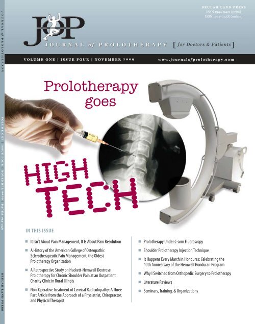
IN THIS ISSUE<br />
■<br />
■<br />
■<br />
■<br />
<strong>Prolotherapy</strong><br />
goes<br />
High<br />
Tech<br />
It Isn’t About Pain Management, It Is About Pain Resolution<br />
A History <strong>of</strong> the American College <strong>of</strong> Osteopathic<br />
Sclerotherapeutic Pain Management, the Oldest<br />
<strong>Prolotherapy</strong> Organization<br />
A Retrospective Study on Hackett-Hemwall Dextrose<br />
<strong>Prolotherapy</strong> for Chronic Shoulder Pain at an Outpatient<br />
Charity Clinic in Rural Illinois<br />
Non-Operative Treatment <strong>of</strong> Cervical Radiculopathy: A Three<br />
Part Article from the Approach <strong>of</strong> a Physiatrist, Chiropractor,<br />
and Physical Therapist<br />
■<br />
■<br />
■<br />
■<br />
■<br />
■<br />
<strong>Prolotherapy</strong> Under C-arm Fluoroscopy<br />
Shoulder <strong>Prolotherapy</strong> Injection Technique<br />
BEULAH LAND PRESS<br />
ISSN 1944-0421 (print)<br />
ISSN 1944-043X (online)<br />
It Happens Every March in Honduras: Celebrating the<br />
40th Anniversary <strong>of</strong> the Hemwall Honduran Program<br />
Why I Switched from Orthopedic Surgery to <strong>Prolotherapy</strong><br />
Literature Reviews<br />
Seminars, Training, & Organizations
Table <strong>of</strong> Contents<br />
G R E A T N E W S C O R N E R<br />
197<br />
I N T H E S P O T L I G H T<br />
200<br />
I N T H I S I S S U E O F T H E J O U R N A L O F P R O L O T H E R A P Y <br />
It Isn’t About Pain Management,<br />
It Is About Pain Resolution<br />
Ross A. Hauser, MD<br />
A History <strong>of</strong> the American College<br />
<strong>of</strong> Osteopathic Sclerotherapeutic<br />
Pain Management, the Oldest<br />
<strong>Prolotherapy</strong> Organization<br />
Donna Alderman, DO<br />
F A N T A S T I C F I N D I N G S<br />
205<br />
A Retrospective Study on<br />
Hackett-Hemwall Dextrose<br />
<strong>Prolotherapy</strong> for Chronic Shoulder<br />
Pain at an Outpatient Charity Clinic<br />
in Rural Illinois<br />
Ross A. Hauser, MD & Marion A. Hauser, MS, RD<br />
R E M A R K A B L E R E C O V E R I E S<br />
217<br />
Non-Operative Treatment <strong>of</strong> Cervical<br />
Radiculopathy: A Three Part Article<br />
from the Approach <strong>of</strong> a Physiatrist,<br />
Chiropractor, and Physical Therapist.<br />
Ross A. Hauser, MD, Glen M. Batson, DC,<br />
& Chris Ferrigno, MS, PT<br />
T E A C H I N G T E C H N I Q U E S<br />
232<br />
243<br />
<strong>Prolotherapy</strong> Under<br />
C-arm Fluoroscopy<br />
Christopher J. Centeno, MD<br />
Shoulder <strong>Prolotherapy</strong><br />
Injection Technique<br />
Rodney S. Van Pelt, MD<br />
I T ’ S A W I D E W I D E W O R L D<br />
246<br />
249<br />
250<br />
It Happens Every March in<br />
Honduras: Celebrating the<br />
40th Anniversary <strong>of</strong> the<br />
Hemwall Honduran Program<br />
Gary B. Clark, MD, MPA<br />
Why I Switched<br />
from Orthopedic<br />
Surgery to<br />
<strong>Prolotherapy</strong><br />
Joern Funck, MD<br />
S K I L L E N H A N C E M E N T<br />
256<br />
Literature Reviews<br />
Gary B. Clark, MD, MPA<br />
J O U R N A L <strong>of</strong> P R O L O T H E R A P Y | V O L U M E 1 , I S S U E 4 | N O V E M B E R 2 0 0 9<br />
Seminars, Training, & Organizations
E D I T O R - I N - C H I E F<br />
Ross A. Hauser, MD<br />
Oak Park, IL<br />
E D I T O R I A L B O A R D<br />
Donna Alderman, DO<br />
Glendale, CA<br />
Gunter Baehnisch, MD<br />
Leipzig, Germany<br />
Robert Banner, MD<br />
London, Ontario, Canada<br />
José Eleazar Calderón, MD<br />
Monclova, Coahuila, Mexico<br />
Gary B. Clark, MD, MPA<br />
Boulder, CO<br />
J O U R N A L O F P R O L O T H E R A P Y T E A M & S U B S C R I B E R I N F O R M A T I O N<br />
JOP Team Mark DeLaurentis, MD<br />
Cherry Hill, NJ<br />
Shaun Fauley, DVM<br />
Naperville, IL<br />
Joern Funck, MD<br />
Luebeck, Germany<br />
<strong>Babette</strong> Gladstein, DVM<br />
New York, NY<br />
Mark L. Johnson, MD, FACS<br />
Nashville, TN<br />
George H. Kramer, MD<br />
Minnetonka, MN<br />
John Neustadt, ND<br />
Bozeman, MT<br />
Joan Resk, DO, JD<br />
Roanoke, VA<br />
Subscriber Information<br />
The <strong>Journal</strong> <strong>of</strong> <strong>Prolotherapy</strong> is unique in that it has a target<br />
audience <strong>of</strong> both physicians and patients. The purpose <strong>of</strong><br />
this journal is to provide the readers with new cutting-edge<br />
information on <strong>Prolotherapy</strong>, as well as provide a forum for<br />
physicians and patients alike to tell their stories.<br />
<strong>Journal</strong> <strong>of</strong> <strong>Prolotherapy</strong> is published quarterly – in February, May,<br />
August, and <strong>Nov</strong>ember by Beulah Land Press, 715 Lake Street,<br />
Suite 600, Oak Park, Illinois, 60301. © Copyright 2009 by Beulah<br />
Land Press. All rights reserved. No portion <strong>of</strong> the contents may<br />
be reproduced in any form without written permission from the<br />
publisher.<br />
All subscription inquiries, orders, back issues, claims, and<br />
renewals should be addressed to Beulah Land Press, 715 Lake<br />
St. Suite 600, Oak Park, IL 60301; phone: 708-848-5011; fax:<br />
708-848-0978. Email: bairdn@journal<strong>of</strong><strong>Prolotherapy</strong>.com;<br />
http://beulahlandpress.com.<br />
P R I C I N G<br />
Annual subscription: $100/year.<br />
Includes 4 paper issues and online access to all<br />
www.<strong>Journal</strong><strong>of</strong><strong>Prolotherapy</strong>.com web content.<br />
C L A I M S<br />
Claims for undelivered copies must be made no later than one<br />
month following the month <strong>of</strong> publication. The publisher will<br />
supply missing copies when losses have been sustained in<br />
transit and when the reserve stock will permit.<br />
José Hector Salazar, MD<br />
Monterrey Nuevo Leon, Mexico<br />
Judith Shoemaker, DVM<br />
Nottingham, PA<br />
Garrett Swetlik<strong>of</strong>f, MD<br />
Kelowna, BC, Canada<br />
Rodney S. Van Pelt, MD<br />
Ukiah, CA<br />
Mark T. Wheaton, MD<br />
Minnetonka, MN<br />
R E S I D E N T E D I T O R S<br />
Peter J. Blakemore, DO<br />
Watertown, NY<br />
Patrick M. Hobbins, DO<br />
South Bend, IN<br />
C H A N G E O F A D D R E S S<br />
Change <strong>of</strong> address notices should be sent to JOP, 30 days in<br />
advance to: JOP 715 Lake St. Suite 600, Oak Park, IL 60301;<br />
phone: 708-848-5011; fax: 708-848-0978.<br />
C O P Y R I G H T P E R M I S S I O N<br />
Permission requests to photocopy or otherwise reproduce<br />
copyrighted material owned by Beulah Land Press should be<br />
requested by contacting Beulah Land Press at 708-848-5011 or<br />
by emailing info@benuts.com.<br />
D I S C L A I M E R<br />
This publication does not constitute medical or other<br />
pr<strong>of</strong>essional advice and should not be taken as such. To the<br />
extent the articles published herein may be used to assist in<br />
the care <strong>of</strong> patients, this is the result <strong>of</strong> the sole pr<strong>of</strong>essional<br />
judgment <strong>of</strong> the health pr<strong>of</strong>essional. The health care<br />
pr<strong>of</strong>essional’s judgment is the primary component <strong>of</strong> quality<br />
health care. The information presented in this journal is<br />
not a substitute for the exercise <strong>of</strong> such judgment by the<br />
health pr<strong>of</strong>essional. The opinions expressed in the <strong>Journal</strong> <strong>of</strong><br />
<strong>Prolotherapy</strong> are the opinions <strong>of</strong> the authors <strong>of</strong> their respective<br />
articles and not necessarily that <strong>of</strong> the publisher. The decisions<br />
on what to do for a specific medical condition or symptom<br />
should be based on the analysis by the person’s private health<br />
care pr<strong>of</strong>essional.<br />
J O U R N A L <strong>of</strong> P R O L O T H E R A P Y | V O L U M E 1 , I S S U E 4 | N O V E M B E R 2 0 0 9<br />
J O P S T A F F<br />
Marion A. Hauser, MS, RD<br />
Senior Editor<br />
Nicole M. Baird, CHFP<br />
Associate Editor<br />
Travis E. Mitchell<br />
Senior Graphic Designer/Webmaster<br />
Doug R. Skinkis<br />
Assistant to the Editor-in-Chief/<br />
Assistant Graphic Designer<br />
Patricia H. Miller<br />
Assistant to Media Relations<br />
Enid M. Forsyth<br />
Lay Editor<br />
Media for Doctors, Inc.<br />
Media Relations<br />
ISSN 1944-0421 (print)<br />
ISSN 1944-043X (online)
Authors<br />
D O N N A A L D E R M A N , D O<br />
G L E N M . B A T S O N , D C<br />
J O U R N A L O F P R O L O T H E R A P Y A U T H O R S<br />
Donna Alderman, DO is a graduate <strong>of</strong> Cornell University in Ithaca, New York, where she received her<br />
Bachelor <strong>of</strong> Science degree in Nutrition. She completed her pre-med requirements at the University <strong>of</strong><br />
California, San Diego in La Jolla and attended medical school at the Western University <strong>of</strong> Health Science<br />
in Pomona, California where she received the Doctor <strong>of</strong> Osteopathic Medicine degree. <strong>Dr</strong>. Alderman<br />
completed her internship in family medicine at Pacific Hospital <strong>of</strong> Long Beach, and is a member <strong>of</strong><br />
the American Academy <strong>of</strong> Orthopedic Medicine. She has been a clinical instructor for students at the<br />
Family Practice clinic residency program for Pacific Hospital <strong>of</strong> Long Beach, and does clinical training<br />
for premedical students from University <strong>of</strong> California, Berkeley. <strong>Dr</strong>. Alderman has also done numerous<br />
postdoctorate courses in <strong>Prolotherapy</strong> and uses it daily in her practice at Hemwall Family Medical Center,<br />
1740 Broadview <strong>Dr</strong>., Glendale, CA 91208; Tel: 818.957.3000; www.thefamilydr.com.<br />
Glen M. Batson, DC received his Doctorate in Chiropractic from Palmer College <strong>of</strong> Chiropractic in 1988.<br />
Upon graduation, <strong>Dr</strong>. Batson has lectured and taught Chiropractic procedures and video-flouroscopic<br />
imaging through the continuing education department <strong>of</strong> Palmer College. <strong>Dr</strong>. Batson developed and<br />
sponsored the video fluoroscopic imaging certification course through Palmer College certifying Doctors<br />
<strong>of</strong> Chiropractic in the protocol and interpretation <strong>of</strong> video fluoroscopic spinal motion imaging. <strong>Dr</strong>. Batson<br />
is certified in video fluoroscopy imaging, radiographic imaging, chiropractic manipulative procedures,<br />
and physiotherapy. <strong>Dr</strong>. Batson has been in private practice in Illinois since 1988 specializing in specific<br />
spinal analysis and manipulation. <strong>Dr</strong>. Batson may be reached at his <strong>of</strong>fice in Willowbrook, Illinois at<br />
630.323.1181 or by e-mail at backbonedr@aol.com.<br />
C H R I S T O P H E R J . C E N T E N O , M D<br />
Christopher J. Centeno, MD is a double boarded certified physician in both Physical Medicine and<br />
Rehabilitation as well Pain Medicine. He is the medical director <strong>of</strong> The Centeno-Schultz Clinic. <strong>Dr</strong>. Centeno<br />
earned his MD from the University <strong>of</strong> South Florida and completed his residency at the Institute for<br />
Rehabilitation Research, Physical Medicine and Rehabilitation at the Baylor College <strong>of</strong> Medicine. He is a<br />
founding member and medical director <strong>of</strong> the International Cellular Medicine Society. Through ICMS, <strong>Dr</strong>.<br />
Centeno has worked with other stem cell experts to produce International Guidelines for stem cell use as<br />
well as an International Re-implantation Registry. <strong>Dr</strong>. Centeno published the world’s first clinical case reports<br />
on the use <strong>of</strong> mesenchymal stem cells in orthopedic patients. He has cared for thousands <strong>of</strong> patients in<br />
chronic pain and has authored over 20 medical publications that are indexed in the US national library <strong>of</strong><br />
medicine. He has also treated hundreds <strong>of</strong> patients with <strong>Prolotherapy</strong>. <strong>Dr</strong>. Centeno may be reached at 403<br />
Summit Blvd, Suite 201, Broomfield, CO 80021; Tel: 303.429.6448; www.centenoschultzclinic.com.<br />
G A R Y B . C L A R K , M D , M P A<br />
Gary B. Clark, MD, MPA is currently the Medical Director <strong>of</strong> the Center for Healing Injury and Pain and<br />
Boulder <strong>Prolotherapy</strong> in Boulder, Colorado. <strong>Dr</strong>. Clark earned a BA and MD from University <strong>of</strong> Colorado,<br />
interned in Pediatrics at Yale-New Haven Hospital; and completed his residency and board certification<br />
in Pathology and Neuropathology at Walter Reed Army Medical Center and Armed Forces Institute<br />
<strong>of</strong> Pathology. <strong>Dr</strong>. Clark also earned a Masters Degree in Public Administration with Special Study in<br />
Organizational Development at George Washington University. He retired from the US Army in 1991 after<br />
23 years <strong>of</strong> active duty. To contact <strong>Dr</strong>. Clark: 1790 30th Street, Suite 230, Boulder, Colorado 80301; Tel:<br />
303.444.5131; www.doctorclark.com.<br />
J O U R N A L <strong>of</strong> P R O L O T H E R A P Y | V O L U M E 1 , I S S U E 4 | N O V E M B E R 2 0 0 9
C H R I S F E R R I G N O , M S , P T<br />
R O S S A . H A U S E R , M D<br />
J O U R N A L O F P R O L O T H E R A P Y A U T H O R S<br />
Chris Ferrigno is a physical therapist in Chicago, Illinois where he works in outpatient orthopedics. He<br />
received his Masters <strong>of</strong> Physical Therapy in 1999 from the Medical College <strong>of</strong> Georgia and his Bachelors<br />
<strong>of</strong> Science in Education in 1995 from the University <strong>of</strong> Georgia. Chris is an avid athlete. His athletic<br />
accomplishments include finishing the Ironman Triathlon and winning Team Gauntlet’s “Athlete <strong>of</strong> the<br />
Year” in 2007.<br />
J O E R N F U N C K , M D<br />
<strong>Dr</strong>. Funck grew up in Luebeck, Germany. After receiving his M.D. from the University <strong>of</strong> Hamburg in 1968 he<br />
completed his trauma surgery and orthopedic surgeon training in Hannover and Braunschweig, Germany.<br />
After finishing his clinical education he left the operating branch and switched to muscoloskeletal medicine,<br />
as well as manual therapy, opening his own private <strong>of</strong>fice in Luebeck in 1975. In Bremen, Germany, <strong>Dr</strong>.<br />
Funck took up studies in orthopedic medicine, focusing on Cyriax diagnosis and treatment <strong>of</strong> s<strong>of</strong>t tissues.<br />
He studied manual therapy with Pr<strong>of</strong>. Tilscher from Vienna who first introduced him to <strong>Prolotherapy</strong><br />
for low back pain patients. From 2000 to 2006 <strong>Dr</strong>. Funck has treated 1500 cases with <strong>Prolotherapy</strong>. He<br />
was successful in 88.3 % <strong>of</strong> them. <strong>Dr</strong>. Funck may be reached at Fackenburger Allee 22-24 23554 Lübeck,<br />
Germany; Tel: 0451.411.20; www.proliferationstherapie.de.<br />
M A R I O N A . H A U S E R , M S , R D<br />
Marion A. Hauser, MS, RD received her Bachelor <strong>of</strong> Science in Nutrition from University <strong>of</strong> Illinois and her<br />
Master <strong>of</strong> Science in Nutrition and dietetic internship from Eastern Illinois University. Marion is the CEO<br />
<strong>of</strong> Caring Medical and Rehabilitation Services in Oak Park, Illinois and owner <strong>of</strong> Beulah Land Nutritionals.<br />
As a registered dietitian, Marion is also a well-known speaker and writer on a variety <strong>of</strong> topics related<br />
to natural medicine and nutrition. Marion has recently released “The Hauser Diet: A Fresh Look at Healthy<br />
Living.” Marion co-authored the national best seller entitled “Prolo Your Pain Away! Curing Chronic Pain with<br />
<strong>Prolotherapy</strong>” along with a four-book mini series <strong>of</strong> <strong>Prolotherapy</strong> books, as well as a comprehensive sports<br />
book discussing the use <strong>of</strong> <strong>Prolotherapy</strong> for sports injuries. Marion Hauser may be reached at 715 Lake St.<br />
Suite 600, Oak Park, IL 60301; Tel: 708.848.7789; www.caringmedical.com.<br />
Ross A. Hauser, MD received his undergraduate degree from University <strong>of</strong> Illinois. He graduated from the<br />
University <strong>of</strong> Illinois College <strong>of</strong> Medicine in Chicago and did his residency at Loyola/Hines VA in Physical<br />
Medicine and Rehabilitation. <strong>Dr</strong>. Hauser is the Medical Director <strong>of</strong> Caring Medical and Rehabilitation<br />
Services in Oak Park, Illinois and is passionate about <strong>Prolotherapy</strong> and natural medicine. <strong>Dr</strong>. Hauser and<br />
his wife Marion, have written seven books on <strong>Prolotherapy</strong>, including the national best seller “Prolo Your<br />
Pain Away! Curing Chronic Pain with <strong>Prolotherapy</strong>,” now in its third edition, along with a four-book topical<br />
mini series <strong>of</strong> <strong>Prolotherapy</strong> books. He also spearheaded the writing <strong>of</strong> a 900-page epic sports book that<br />
discusses the use <strong>of</strong> <strong>Prolotherapy</strong> for sports injuries, “Prolo Your Sports Injuries Away! Curing Sports Injuries<br />
and Enhancing Athletic Performance with <strong>Prolotherapy</strong>.” <strong>Dr</strong>. Hauser may be reached at 715 Lake St., Suite<br />
600, Oak Park, IL 60301; Tel: 708.848.7789; www.caringmedical.com.<br />
R O D N E Y S . V A N P E LT , M D<br />
Rodney S. Van Pelt, MD received his medical degree from Loma Linda University Medical School and<br />
completed his family practice residency in Florida. He practiced family medicine for several years until<br />
falling in love with the specialty <strong>of</strong> Orthopedic Medicine which uses all the different modalities for pain<br />
with conservative treatments. <strong>Dr</strong>. Van Pelt then received specialized training in the Cyriax technique <strong>of</strong><br />
Orthopedic Medicine, taking some <strong>of</strong> his training in Oxford, England. He is one <strong>of</strong> the few American<br />
physicians who is a member <strong>of</strong> the Society <strong>of</strong> Orthopedic Medicine <strong>of</strong> London, England. <strong>Dr</strong>. Van Pelt<br />
practices full time <strong>Prolotherapy</strong> in northern California. <strong>Dr</strong>. Van Pelt may be contacted at Orthopedic<br />
Wellness Center Plaza Del Sol, 776 S State St., Ukiah, CA 95482; Tel: 707.463.1782; www.sfpmg.com.<br />
J O U R N A L <strong>of</strong> P R O L O T H E R A P Y | V O L U M E 1 , I S S U E 4 | N O V E M B E R 2 0 0 9
G R E A T N E W S C O R N E R : I T I S N ' T A B O U T P A I N M A N A G E M E N T , I T I S A B O U T P A I N R E S O L U T I O N<br />
Welcome to our fourth <strong>Journal</strong> <strong>of</strong> <strong>Prolotherapy</strong><br />
issue! Wow, it is packed! We have received a<br />
wonderful array <strong>of</strong> great comments from<br />
our readers. I wanted to share a few <strong>of</strong> these emails/<br />
correspondences. Scott Greenberg, MD, was nice enough<br />
to send us this:<br />
“Why can’t conventional medicine find your pain? Pain is <strong>of</strong>ten<br />
misunderstood and mismanaged in traditional medical settings.<br />
While many <strong>of</strong> us hurt or have hurt to various degrees during our<br />
lifetime, there is no traditional test to ‘quantify’ our pain, nor does the<br />
series <strong>of</strong> happy and sad faces to describe our pain level aid in finding<br />
adequate relief from our symptoms.<br />
What we have lost in medicine is our ability to examine the patient,<br />
correlate the examination with the patient’s symptoms, and lastly<br />
consider the diagnostic tests. Instead, we as patients enter the system<br />
<strong>of</strong> pain treatment, done almost as a mass production protocol<br />
involving first a trial <strong>of</strong> anti-inflammatory medication and then<br />
physical therapy. If these ‘conservative’ measures fail to provide<br />
relief, it’s <strong>of</strong>f to see the surgeon, where the decision is made to have<br />
either surgery or pain management.<br />
As a physician, I never wanted to manage pain, nor would want,<br />
as a patient, to have my pain managed. Having suffered with pain<br />
myself, I could not even imagine living the rest <strong>of</strong> my life in chronic<br />
pain. So why are we so far <strong>of</strong>f the mark with treatment <strong>of</strong> pain? I<br />
think that the answer lies in two important factors. First, we are<br />
overly reliant on diagnostic tests. Secondly, we have lost the art <strong>of</strong><br />
physical examination.<br />
Take, for instance, the case <strong>of</strong> lower back pain. It is one <strong>of</strong> the<br />
most common causes <strong>of</strong> pain and disability in the world, but <strong>of</strong>ten<br />
misunderstood. Why? Because most cases are due to musculoskeletal<br />
conditions such as sacroiliac joint dysfunction, pyriformis syndrome,<br />
or facet joint arthropathy. Such problems are not seen on MRI, CT,<br />
or X-rays, thus a clinician without expertise in curing these conditions<br />
will not be able to effectively manage them.<br />
G R E A T N E W S C O R N E R<br />
It Isn’t<br />
About Pain<br />
Management,<br />
It Is About<br />
Pain Resolution<br />
Ross A. Hauser, MD<br />
Even though we have access to the greatest diagnostic tests in the<br />
world, we as physicians need to use our clinical judgment to determine<br />
their significance. For example, the majority <strong>of</strong> healthy people who<br />
do not have any back pain at all will have degenerative, bulging,<br />
or herniated discs in their lumbar spine. But if you do have pain,<br />
the job <strong>of</strong> your physician is to determine the relevance <strong>of</strong> your test<br />
results. It is not a black and white issue in what may be causing<br />
your pain.<br />
So how do we determine what the best treatment courses are for<br />
our patients? First we must listen to our patients and ask the right<br />
questions—where is the pain, where does it travel, is there any<br />
numbness or weakness? What makes it better and what makes it<br />
worse? Are there any ominous signs like loss <strong>of</strong> bowel and bladder<br />
function, fever, chills, weight loss, and so on. From our questions<br />
alone, the skilled physician should be able to determine 85% <strong>of</strong> the<br />
diagnosis, and then confirm it with physical examination.<br />
The examination is key to determine and confirm the root cause <strong>of</strong><br />
pain, and unfortunately it is becoming a lost art. Many <strong>of</strong> my patients<br />
have told me they were recommended to undergo surgery with either<br />
a very brief exam or no exam at all. I find this to be a disservice to<br />
patient care that can only lead to bad outcomes. The physical exam<br />
is not without its faults, and to be reliable must be performed with<br />
experienced hands. Palpation <strong>of</strong> ligaments, tendons, and joints is a<br />
skill and an innate gift to those that possess the ability to acquire its<br />
skill. Skilled hands have the ability to determine damaged, weak,<br />
and painful joints from those that are normal. This critical tool<br />
allows us to incorporate all <strong>of</strong> the information about a patient’s<br />
condition and formulate a treatment plan.<br />
There is no one size fits all formula to treat a pain condition. However,<br />
most pain and sports injury conditions are curable, in the right hands,<br />
with reconstructive and regenerative treatments such as <strong>Prolotherapy</strong>.<br />
I found my way to a complete cure after suffering for over 10 years,<br />
and I wish you the best in finding your solution, as it exists. If not<br />
then hold on tight as we are working on new solutions and treatment<br />
options to cure pain and arthritis, all without ever going under the<br />
knife.” Well said <strong>Dr</strong>. Greenberg!<br />
Obviously, one <strong>of</strong> the messages we are trying to promote<br />
here at JOP is that pain can be resolved, whereas just<br />
managing the pain by other methods will leave the<br />
underlying disease process untouched, free to continue<br />
to worsen. <strong>Prolotherapy</strong> is one method <strong>of</strong> treatment<br />
that has the potential to stop and reverse the underlying<br />
degenerative process. The net result is pain resolution,<br />
not pain management!<br />
One recent story Marion (my wife) received was the<br />
testimony <strong>of</strong> Ken Allen regarding the power <strong>of</strong> the<br />
human body to heal itself ! We are reprinting his<br />
correspondences with his permission:<br />
J O U R N A L <strong>of</strong> P R O L O T H E R A P Y | V O L U M E 1 , I S S U E 4 | N O V E M B E R 2 0 0 9 197
198<br />
G R E A T N E W S C O R N E R : I T I S N ' T A B O U T P A I N M A N A G E M E N T , I T I S A B O U T P A I N R E S O L U T I O N<br />
First email: I just wanted to say thanks for your article online<br />
about the detrimental effects <strong>of</strong> RICE treatment and NSAIDs on<br />
ligament and tendon healing. I came across your article after suffering<br />
terrible extensor tendonitis in my right foot, while ramping up mileage<br />
too quickly in marathon training. I followed your advice, skipped the<br />
ice and ibupr<strong>of</strong>en, healed up 100% in 5 weeks, and just finished<br />
my first half-marathon 3 weeks ago with ZERO PAIN in my foot<br />
whatsoever! I just let my foot heal naturally and didn’t interfere. If I<br />
followed current standard advice, at best I’d have a weaker foot and<br />
at worst I’d never run again without pain. I’m telling everyone who<br />
will listen, don’t use ice and NSAIDs if you want to heal! –Best<br />
wishes, Ken<br />
Second email: I finished the Kaiser 1/2 Marathon in San<br />
Francisco in 1 hour 26 minutes. Not too bad for a guy that couldn’t<br />
even walk 3 months earlier due to a sports injury. I learned a lot from<br />
the experience—especially to listen and be nicer to my body. And<br />
I really credit your article for helping me heal completely. Thanks<br />
again and good luck in your next race! –Sincerely, Ken<br />
Third email: My age group is 35-39. I was something like<br />
25 in that group, and I placed 143 out <strong>of</strong> about 5000 overall.<br />
I’ve really only been training for several months, and I’d like to get<br />
quicker over time. Please feel free to use my email however you like.<br />
The more people that get your message the better. I’m so happy my<br />
foot healed as well as it did! I messed it up really bad by trying to<br />
run through serious pain and I just kept pushing. Unfortunately, I<br />
didn’t know better.<br />
I was having ankle pain in both legs from running too much too soon,<br />
and a guy at a local shoe store recommended stability shoes for me. I<br />
don’t have pronation issues though, so the shoes rotated my feet out.<br />
Gradually I started getting pain on the top <strong>of</strong> my right foot. Pushing<br />
things further, I tried a long 18 mile run and had to limp home<br />
after 14 miles. I couldn’t bear weight for several days, and it was a<br />
month before I could even consider light running again. But I learned<br />
from my mistakes, went back to neutral shoes, and now I listen very<br />
closely for any hint <strong>of</strong> pain during and after runs. My foot has been<br />
completely pain free, which is awesome! –Ken<br />
Thank God for the power <strong>of</strong> the internet! What a great<br />
way for people like Ken and others to receive information<br />
on how to heal themselves!<br />
We also received four letters from JOP reader, Clive<br />
Sin<strong>of</strong>f, MD:<br />
Letter #1: <strong>Dr</strong>. Hauser and the entire publication staff should be<br />
congratulated on achieving the publication <strong>of</strong> this important journal.<br />
For reasons which I cannot comprehend, <strong>Prolotherapy</strong> has been ignored<br />
and greeted with hostility. This publication takes an important step<br />
in furthering the knowledge and use <strong>of</strong> this highly effective therapy. In<br />
the article by Hauser and Cukla 1 the X-ray changes are dramatic. It<br />
would be useful if the authors could provide more detail as to how the<br />
injections were done. What was injected and was the target directly<br />
into the subchondral area, ligaments and/or into the joint space?<br />
–Clive Sin<strong>of</strong>f M.D.<br />
Hauser RA and Cukla JJ. Standard clinical X-ray studies document<br />
cartilage regeneration in five degenerated knees after <strong>Prolotherapy</strong>. J Prolo<br />
2009;1:22-28.<br />
Editor’s Comments: Dear <strong>Dr</strong>. Sin<strong>of</strong>f, We at JOP<br />
appreciate your comments and questions. To answer your<br />
questions: 2IU <strong>of</strong> HGH was injected into the joint space.<br />
With each treatment the medial and lateral collateral<br />
ligaments were also injected with normal <strong>Prolotherapy</strong><br />
solution.<br />
Letter #2: What a tour de force! <strong>Dr</strong>. Hauser’s review <strong>of</strong> the effects<br />
<strong>of</strong> corticosteroids was comprehensive and thoroughly documented. 1<br />
Hauser RA. The deterioration <strong>of</strong> articular cartilage in osteoarthritis by<br />
corticosteroid injections. J Prolo 2009;2:107-123.<br />
Editor’s Comments: Thank you for your comments.<br />
The treatment <strong>of</strong> osteoarthritis with corticosteroid<br />
injections has to stop! Clearly one <strong>of</strong> the main causes <strong>of</strong><br />
the “bone-on-bone” phenomenon leading to hip and knee<br />
replacements is the corticosteroid injections the patients<br />
are receiving.<br />
Letter #3: I have two questions to ask the <strong>Prolotherapy</strong><br />
community. Many authors, including <strong>Dr</strong>. Van Pelt 1 , recommend the<br />
use <strong>of</strong> human growth hormone (HGH) as a growth factor. My<br />
understanding is that HGH is released in the pituitary and acts on<br />
the liver to produce somatomedin. Is there any evidence for a direct<br />
effect locally? It would seem more logical to use a cytokines such as<br />
granulocyte stimulating factor (G-CSF) or fibroblast growth factor<br />
(FGF) which have been shown to attract inflammatory cells. Does<br />
anyone know <strong>of</strong> scientific or clinical evidence to support such growth<br />
factors? – Clive Sin<strong>of</strong>f M.D.<br />
Van Pelt RS. Hip arthritis <strong>Prolotherapy</strong> injection technique.<br />
J Prolo 2009;1:101-103.<br />
Editor’s Comments: Wow, what a topic, growth<br />
factors and <strong>Prolotherapy</strong>! As you know the day will arrive<br />
where doctors will inject fibroblastic growth factor or<br />
granulocyte stimulating factor into injured structures, but<br />
unfortunately that day is not here. Here are some items<br />
for you to ponder:<br />
There are growth hormone receptors on mesenchymal<br />
cells including human growth plate chondrocytes. 1<br />
Pituitary growth hormone acts directly on many cells<br />
in the body. As a matter <strong>of</strong> fact, most <strong>of</strong> the effects<br />
J O U R N A L <strong>of</strong> P R O L O T H E R A P Y | V O L U M E 1 , I S S U E 4 | N O V E M B E R 2 0 0 9<br />
1.<br />
1.<br />
1.<br />
1.<br />
2.
3.<br />
4.<br />
5.<br />
G R E A T N E W S C O R N E R : I T I S N ' T A B O U T P A I N M A N A G E M E N T , I T I S A B O U T P A I N R E S O L U T I O N<br />
attributed to Growth Hormone action appear to be<br />
the result <strong>of</strong> a direct effect <strong>of</strong> GH on cells in different<br />
peripheral tissues, including cartilage. Not on IGF-1. 2<br />
Growth Hormone has direct anabolic effects on<br />
“old”cartilage cells. 3<br />
Yes, there are estrogen receptors on cartilage cells also! 4<br />
Chondrocytes (cartilage cells) can produce their own<br />
sex hormones! 5<br />
What it all means is that cartilage cells are somewhat under<br />
the control <strong>of</strong> hormones. From a <strong>Prolotherapy</strong> standpoint<br />
if we can make cartilage physiology more anabolic there<br />
will be a good chance that the chondrocytes will make<br />
more cartilage which will ultimately help the patient!<br />
Letter #4: Does anyone have experience with the use <strong>of</strong><br />
<strong>Prolotherapy</strong> in true rheumatoid arthritis (as opposed to osteoarthritis<br />
misdiagnosed as rheumatoid arthritis)? –Thank you, Clive Sin<strong>of</strong>f<br />
M.D., 22200 Halburton Rd, Beachwood, OH 44122<br />
Editor’s Comments: As you know, not every joint pain<br />
in a rheumatoid arthritis (RA) patient is due to RA. From<br />
a <strong>Prolotherapy</strong> standpoint in treating the RA patient, you<br />
should do the following: assess the condition <strong>of</strong> their RA<br />
and evaluate the painful area like you would with any other<br />
patient. If someone has active synovitis at the time <strong>of</strong> the<br />
<strong>Prolotherapy</strong> evaluation, we (Caring Medical) would inject<br />
a solution <strong>of</strong> sterile water and procaine (anywhere from<br />
a total <strong>of</strong> 0.4% to 1.0% procaine) into the painful areas<br />
to cool it <strong>of</strong>f (versus steroids) and treat the rheumatoid<br />
arthritis with a natural medicine program. Once the RA<br />
is under control, meaning no heat in the joint, hands,<br />
wrists, or feet, then <strong>Prolotherapy</strong> could be done to the<br />
joint or structures involved assuming they have injuries<br />
that typically respond to <strong>Prolotherapy</strong>. As you know,<br />
rheumatoid arthritis by definition destroys joints. What is<br />
one <strong>of</strong> the best treatments to repair joints? <strong>Prolotherapy</strong>.<br />
So yes, <strong>Prolotherapy</strong> can be done in folks with RA, but<br />
just make sure the RA is under good control. If you inject<br />
the typical <strong>Prolotherapy</strong> solutions into joints with active<br />
synovitis you run the risk <strong>of</strong> increasing the pain quite a bit,<br />
but the good news is, the increase in pain is temporary.<br />
Some <strong>of</strong> the highlights <strong>of</strong> this fourth issue <strong>of</strong> JOP include<br />
articles focused on the cervical spine, and on shoulder<br />
pain. From personal experience, I can tell you one <strong>of</strong> the<br />
most horrific cervical conditions is cervical radiculopathy.<br />
Glen Batson, DC and Chris Ferrigno, PT join me in a<br />
three part article on treating cervical radiculopathy<br />
from the experience <strong>of</strong> a Physiatrist, a Chiropractor,<br />
and a Physical Therapist. In Teaching Techniques we begin<br />
to explore the high-tech world <strong>of</strong> <strong>Prolotherapy</strong> with<br />
Christopher Centeno, MD, as he discusses the use <strong>of</strong><br />
C-arm fluoroscopy in his <strong>Prolotherapy</strong> practice. Our<br />
Teaching Techniques columnist, Rodney Van Pelt, MD,<br />
teaches the shoulder injection technique.<br />
The American College <strong>of</strong> Osteopathic Sclerotherapeutic<br />
Pain Management is In the Spotlight. Donna Alderman, DO<br />
takes us on a trip through time with the oldest <strong>Prolotherapy</strong><br />
organization. It really is a Wide Wide World when it comes<br />
to <strong>Prolotherapy</strong>. We hear from Joern Funck, MD, from<br />
Germany, on his pr<strong>of</strong>essional switch from orthopedic<br />
surgery to <strong>Prolotherapy</strong>. Our Literature Review columnist,<br />
Gary Clark, MD, reviews whiplash literature and presents<br />
the intriguing case study <strong>of</strong> General George S. Patton. He<br />
also reports on the fortieth anniversary <strong>of</strong> the Hemwall<br />
Honduran program, which hosts the largest <strong>Prolotherapy</strong><br />
training course <strong>of</strong> its kind. It is great to see <strong>Dr</strong>. Hemwall’s<br />
legacy continuing with such gusto!<br />
Last, but not least, Marion and I present a retrospective<br />
study on chronic shoulder pain. We are always pleased to<br />
see the final statistics in these reports that we’ve presented<br />
in the journal because it continues to show that <strong>Prolotherapy</strong><br />
works! By stimulating the natural healing mechanisms<br />
<strong>of</strong> the body, via injecting simple, safe solutions into and<br />
around damaged structures, <strong>Prolotherapy</strong> re-ignites the<br />
body to heal itself. The net result is not pain management,<br />
but pain resolution. Ultimately, that should be the goal<br />
<strong>of</strong> all clinicians who see patients in chronic pain. n<br />
Until the next injection,<br />
1.<br />
2.<br />
3.<br />
4.<br />
5.<br />
B i B l i o g r a p h y<br />
Werther GA, et al. Visual demonstration <strong>of</strong> growth hormone<br />
receptors <strong>of</strong> human growth plate chondrocytes. The <strong>Journal</strong> <strong>of</strong><br />
Clinical Endocrinology and Metabolism. 1990;70:1725-1731.<br />
Isaksson OG, et al. Mode <strong>of</strong> action <strong>of</strong> puituitary growth<br />
hormone on target cells. Annu Rev Physiol. 1985;47:483-499.<br />
Livne E, et al. Comparison <strong>of</strong> in vitro response to growth<br />
hormone by chondrocytes from mandibular condyle cartilage <strong>of</strong><br />
young and old mice. Calcified Tissue International. 1997;61:62-67.<br />
Ushiyama T, et al. Expression <strong>of</strong> genes for estrogen receptors<br />
a and b in human articular chondrocytes. Osteoarthritis and<br />
Cartilage. 1999;7:560-566.<br />
Takeuchi S, et al. Production <strong>of</strong> sex steroid hormones from<br />
DHEA in articular chondrocyte <strong>of</strong> rats. American <strong>Journal</strong> <strong>of</strong><br />
Endocrinol Metab. 2007;293:E410-E415.<br />
J O U R N A L <strong>of</strong> P R O L O T H E R A P Y | V O L U M E 1 , I S S U E 4 | N O V E M B E R 2 0 0 9 199
200<br />
I N T H E S P O T L I G H T : H I S T O R Y O F T H E A M E R I C A N C O L L E G E O F O S T E O P A T H I C S C L E R O T H E R A P E U T I C P A I N M A N A G E M E N T<br />
A History <strong>of</strong> the American<br />
College <strong>of</strong> Osteopathic<br />
Sclerotherapeutic Pain<br />
Management, the Oldest<br />
<strong>Prolotherapy</strong> Organization<br />
Donna Alderman, DO<br />
a B S T r a C T<br />
Modern <strong>Prolotherapy</strong> evolved from the insights and<br />
courage <strong>of</strong> a few doctors in the early part <strong>of</strong> the 1900s.<br />
These pioneers would then form groups to teach<br />
others, share knowledge, and improve techniques. The<br />
earliest record <strong>of</strong> such a group started as the American<br />
Society <strong>of</strong> Herniologists in 1926, now known as the<br />
American College <strong>of</strong> Osteopathic Sclerotherapeutic Pain<br />
Management. This fascinating article reviews the history<br />
<strong>of</strong> these courageous men and how their hard work and<br />
efforts developed into an organization that not only<br />
promotes and teaches state-<strong>of</strong>-the-art <strong>Prolotherapy</strong>, but<br />
is on the cutting edge <strong>of</strong> musculoskeletal medicine.<br />
<strong>Journal</strong> <strong>of</strong> <strong>Prolotherapy</strong>. 2009;4:200-204.<br />
KEyWorDS: american College <strong>of</strong> osteopathic Sclerotherapeutic pain Management,<br />
prolotherapy, Sclerotherapy, herniologists, osteopathy.<br />
The day was like any other for <strong>Dr</strong>. Earl Gedney, an<br />
osteopathic surgeon at the Osteopathic Hospital<br />
in Philadelphia. It was 1936. <strong>Dr</strong>. Gedney prepped<br />
for surgery, as usual, but this time when he went through<br />
the electric doors to the operating room, the doors closed<br />
prematurely on his hand, pulling his thumb joint so far<br />
that it hung limp <strong>of</strong>f his hand. After checking with several<br />
colleagues and getting X-rays, <strong>Dr</strong>. Gedney learned there<br />
was no fracture, no correctable dislocation, only a very<br />
severe stretching <strong>of</strong> the ligaments and tendons at the<br />
thumb joint. He suffered in pain and could barely move<br />
his thumb. Far worse, he was told by the best surgeons<br />
he knew that there was nothing that could be done for<br />
him and that he would have to retire from doing what<br />
he loved most, being a surgeon. (See Figure 1.) <strong>Dr</strong>. Gedney<br />
was not the type <strong>of</strong> person to give up. He was resourceful<br />
I N T H E S P O T L I G H T<br />
and intelligent. Gedney had been pondering the problem<br />
<strong>of</strong> the hypermobile joint since 1925 when, as a medical<br />
student, he heard a lecture on restricted motion in<br />
lumbar segments. At that time the young Gedney asked<br />
the speaker, the late <strong>Dr</strong>. Charles Muttart, the question:<br />
“What about treatment <strong>of</strong> the vertebra that is too freely<br />
mobile?” <strong>Dr</strong>. Muttart answered: “That, my young man,<br />
is the problem for your generation to solve.” Gedney took<br />
this to heart.<br />
Faced with the situation <strong>of</strong> his own hypermobile joint,<br />
Gedney put his thoughts together. He had recently attended<br />
a lecture discussing sclerosing (irritating) solutions for<br />
abdominal hernias (muscle weakness or tears) and knew<br />
<strong>of</strong> a group <strong>of</strong> physicians who had been doing this for<br />
years. These physicians were known as “herniologists.”<br />
The idea behind the herniologists’ method <strong>of</strong> treatment<br />
was that irritating injections would stimulate repair and<br />
scar tissue formation, making muscular tissue at the<br />
hernia site thicker and stronger. This was in the days<br />
before modern surgery when surgical risk was quite high,<br />
so a non-surgical approach was popular for hernia repair.<br />
The first organization for hernia sclerosing methods was<br />
formed in 1923 and became the American Society <strong>of</strong><br />
Herniologists in 1926. By the early 1930s, such procedures<br />
were declared a success in the treatment <strong>of</strong> hernias. The<br />
American Osteopathic Society <strong>of</strong> Herniologists was<br />
formed in 1938 for the injection <strong>of</strong><br />
hernias, veins and hemorrhoids. One<br />
<strong>of</strong> its early Presidents was <strong>Dr</strong>. Harry<br />
Earl Stahlman, a 1918 graduate<br />
<strong>of</strong> the internship program at the<br />
Philadelphia College <strong>of</strong> Osteopathy.<br />
(See Figures 2-4.)<br />
What happened next set the stage for<br />
modern <strong>Prolotherapy</strong>. <strong>Dr</strong>. Gedney<br />
extrapolated his knowledge <strong>of</strong> nonsurgical<br />
hernia repair to the nonsurgical<br />
repair <strong>of</strong> joints, ligaments<br />
and tendons. Reasoning he had little<br />
to lose by being a guinea pig for his<br />
theory, he started injecting his thumb<br />
with the sclerosing solutions and<br />
had a dramatically successful result.<br />
Before long, he was back working as<br />
a surgeon. However, Gedney took it a<br />
step further. Excited about his result,<br />
J O U R N A L <strong>of</strong> P R O L O T H E R A P Y | V O L U M E 1 , I S S U E 4 | N O V E M B E R 2 0 0 9<br />
Figure 1. Earl<br />
Gedney, DO.<br />
Figure 2. Harry<br />
Stahlman, DO.
I N T H E S P O T L I G H T : H I S T O R Y O F T H E A M E R I C A N C O L L E G E O F O S T E O P A T H I C S C L E R O T H E R A P E U T I C P A I N M A N A G E M E N T<br />
Figure 3. Diploma <strong>of</strong> Harry Earl Stahlman, 1918 internship<br />
class Philadelphia College <strong>of</strong> Ostepathy.<br />
Figure 4. Board members listed on the letterhead <strong>of</strong> the<br />
American Society <strong>of</strong> Herniologists.<br />
he started on a lifelong career <strong>of</strong> research and in June 1937,<br />
he published his first article Special Technic: Hypermobile<br />
Joint: a preliminary report (Osteopathic Pr<strong>of</strong>ession. 1937; 9:30-<br />
31), followed by a presentation: The hypermobile joint – further<br />
reports on injection method at the February 13, 1938 meeting <strong>of</strong><br />
the Osteopathic Clinical Society <strong>of</strong> Philadelphia. Gedney<br />
outlined the theory <strong>of</strong> using sclerosing solutions for joints<br />
which had become stretched and were causing pain. The<br />
1937 article gave a preliminary protocol and two case<br />
reports, one <strong>of</strong> a patient with knee pain and another with<br />
low back pain, both successfully treated by addressing the<br />
hypermobile joint with irritating solutions. He had also<br />
recently fathered the Gedney Osteopathic Hospital in<br />
Philadelphia where he continued his research. (See Figure 5.)<br />
Figure 5. January 1937 Osteopathic Digest Announcement<br />
for Gedney Osteopathic Hospital.<br />
Gedney began experimenting with different irritating<br />
solutions and perfecting his technique for joint injections,<br />
along with his colleague, David Shuman, DO, a 1931<br />
graduate <strong>of</strong> the Philadelphia College <strong>of</strong> Osteopathy and<br />
who was then an instructor there. (See Figure 6.) Both men<br />
began studying and using this technique on unstable<br />
joints, especially knees, lumbar spines and sacroiliac joints.<br />
In 1949, an article by Shuman appeared in the medical<br />
literature: Sclerotherapy – Injections may be the best way to<br />
restrengthen ligaments in case <strong>of</strong> slipped knee cartilage (Osteopathic<br />
Pr<strong>of</strong>ession, 1949). Both<br />
Gedney and Shuman<br />
continued to do research<br />
and publish reports<br />
throughout the 1950s.<br />
Skepticism, however,<br />
among orthopedic<br />
surgeons existed and<br />
more evidence and<br />
studies were needed.<br />
Now practicing in<br />
Maine, <strong>Dr</strong>. Gedney was<br />
determined to provide<br />
this evidence, using<br />
his own money to fund<br />
research if needed. In<br />
March, 1950, a Bangor,<br />
Maine paper headlined:<br />
Figure 6. David Shuman, from<br />
1931 yearbook, Philadelphia<br />
College <strong>of</strong> Osteopathy.<br />
“Bangor Doctor Seeks New Approach To Spinal Fusion Of Lower<br />
Back In His Experiments With Monkeys.”<br />
J O U R N A L <strong>of</strong> P R O L O T H E R A P Y | V O L U M E 1 , I S S U E 4 | N O V E M B E R 2 0 0 9 201
202<br />
I N T H E S P O T L I G H T : H I S T O R Y O F T H E A M E R I C A N C O L L E G E O F O S T E O P A T H I C S C L E R O T H E R A P E U T I C P A I N M A N A G E M E N T<br />
The article goes on to explain that Gedney had been<br />
contemplating this type <strong>of</strong> “needle surgery” study for<br />
20 years, since his graduation from the Philadelphia<br />
College <strong>of</strong> Osteopathy, in order to address the alternative<br />
to surgical bone fusion “to the complete satisfaction <strong>of</strong><br />
bone surgeons.” (See Figure 7.) The paper reported that<br />
<strong>Dr</strong>. Gedney expected to pay out more than $3,000 from<br />
his own pocket to finish this study. The article also states,<br />
“The doctor says this experiment is a follow-up to another<br />
completed in 1940 which resulted in a new internationally<br />
recognized technique in treating hypermobile or<br />
loose joints. This method involves needle surgery.”<br />
Unfortunately there does not appear to be any conclusion<br />
published to the monkey study, however there are three<br />
Gedney publications from 1951-1954 addressing disc,<br />
low back or sacroiliac issues and injection treatment, but<br />
all with regard to human subjects. (Disk syndrome: New<br />
approach in the treatment <strong>of</strong> symptomatic intervertebral<br />
disk, Osteopathic Pr<strong>of</strong>ession, September 1951, 11-15; Technic<br />
Figure 7. <strong>Dr</strong>. Gedney with one <strong>of</strong> his monkeys “Pretty,” The<br />
Bangor Daily News, Bangor, Maine, Friday March 10, 1950,<br />
p. 22.<br />
for sclerotherapy in the management <strong>of</strong> hypermobile<br />
sacroiliac, Osteopathic Pr<strong>of</strong>ession, August 1952, 16-19; 37-<br />
38; Progress report on use <strong>of</strong> sclerosing solutions in low<br />
back syndromes, Osteopathic Pr<strong>of</strong>ession, August 1954, 18-21,<br />
40-44.)<br />
By the 1950s, hernia surgical techniques had progressed so<br />
well that there was less demand for hernia sclerotherapy,<br />
and because <strong>of</strong> the interest generated by Gedney and<br />
Shuman, the herniologists began paying more attention<br />
to joint injections. Two groups formed out <strong>of</strong> the original<br />
group <strong>of</strong> herniologists: The Sclerotherapy College<br />
and, in Philadelphia, the Osteopathic College <strong>of</strong> Joint<br />
Sclerotherapy. In 1954 the two groups combined, forming<br />
the American Osteopathic College <strong>of</strong> Sclerotherapy,<br />
which became recognized and chartered by the AOA in<br />
1956. David Shuman went on to be Secretary-Treasurer<br />
<strong>of</strong> the American Osteopathic College <strong>of</strong> Sclerotherapy<br />
from 1968-1977 and also served as President <strong>of</strong> the<br />
Organization, as well as holding distinguished positions<br />
as President <strong>of</strong> the Philadelphia County Osteopathic<br />
Society, head <strong>of</strong> Department <strong>of</strong> Osteopathic Therapeutics<br />
(Juanita Park Medical Center), and Member <strong>of</strong> the Board<br />
<strong>of</strong> Directors <strong>of</strong> Blue Cross. In 1960 Shuman published<br />
the first layperson’s book on joint sclerotherapy, entitled<br />
Your Aching Back and What You Can Do About It. The book<br />
writes:<br />
“…application <strong>of</strong> sclerotherapy to weakened sacroiliacs was<br />
easy. No operation, no blood, no bone grafts. Just a little weekly<br />
hypodermic injection, repeated ten or perhaps a dozen times, and<br />
the sacroiliac stayed put. But when it came to problems like spondy<br />
[spondylolisthesis], beyond the reach <strong>of</strong> all operative techniques, and<br />
ruptured discs, where operations could promise only a little more than<br />
half a chance at complete recovery, sclerotherapy assumed its greatest<br />
significance. No myelograms, no nucleograms, no arthrodesis, no<br />
fusions, not even hospitalization. Just simple injections, easily made<br />
at an <strong>of</strong>fice visit the patient could make on his way to the movies.”<br />
(Gedney and Staab, Your Aching Back and What You Can Do<br />
About It, Gramercy Publishing Co, NY, 1960, p. 104).<br />
The book goes on to give numerous case reports,<br />
diagrams and examples. It also discusses the work <strong>of</strong><br />
George Hackett, MD, another surgeon interested in<br />
joint sclerotherapy. About the time Gedney was starting<br />
to use joint sclerotherapy, George Hackett, MD, made<br />
an observation while doing hernia repair on patients<br />
previously treated for hernias with sclerosants. He is<br />
quoted as saying, “Injections made (usually in error) at<br />
J O U R N A L <strong>of</strong> P R O L O T H E R A P Y | V O L U M E 1 , I S S U E 4 | N O V E M B E R 2 0 0 9
I N T H E S P O T L I G H T : H I S T O R Y O F T H E A M E R I C A N C O L L E G E O F O S T E O P A T H I C S C L E R O T H E R A P E U T I C P A I N M A N A G E M E N T<br />
the junction <strong>of</strong> ligament and bone resulted in pr<strong>of</strong>use<br />
proliferation <strong>of</strong> new tissue at this union.” Although<br />
there is no evidence <strong>of</strong> direct collaboration between<br />
Hackett, and either Gedney or Shuman, the studies done<br />
independently support each other’s conclusions, that<br />
sclerotherapy (<strong>Prolotherapy</strong>) for joint pain and disability<br />
worked.<br />
<strong>Dr</strong>s. Gedney and Shuman and others spent years in their<br />
research efforts. They looked at the microscopic effects<br />
on ligaments and tendons <strong>of</strong> various formulas to establish<br />
workable protocols for various treatment areas. They<br />
formed a lecture team and traveled to various osteopathic<br />
medical centers to lecture and demonstrate injections<br />
until 1963. In 1967, Shuman wrote a journal article<br />
stressing the importance <strong>of</strong> combining the osteopathic<br />
principles <strong>of</strong> mobilization and ambulation, along with<br />
joint sclerotherapy, for low back disorders, a novel<br />
concept because at the time most low back pain patients<br />
were being confined to bed rest for long periods <strong>of</strong> time.<br />
(Ambulation, osteopathic manipulative therapy, and joint<br />
sclerotherapy in the management <strong>of</strong> common low-back<br />
disorders, <strong>Journal</strong> <strong>of</strong> the American Osteopathic Association,<br />
1967 67:52-59).<br />
In 1986, the name <strong>of</strong> the American Osteopathic College<br />
<strong>of</strong> Sclerotherapy was changed to reflect its evolution<br />
into predominantly injections for joint pain. The new<br />
name was The American College <strong>of</strong> Osteopathic Pain<br />
Management and Sclerotherapy. In 1996 the group was<br />
granted full status as a college by the AOA and became<br />
the American College <strong>of</strong> Osteopathic Pain Management<br />
and Sclerotherapy. Because <strong>of</strong> a conflict over the use <strong>of</strong><br />
the term “sclerotherapy” which was also being used by<br />
the osteopathic dermatology group to denote varicose<br />
veins injections, the AOA changed our name to The<br />
American College <strong>of</strong> Osteopathic Sclerotherapeutic Pain<br />
Management (ACOSPM), which is its current name.<br />
After twenty years this name has recently come under<br />
discussion as needing updating. Studies and biopsies<br />
over the last two decades have shown that <strong>Prolotherapy</strong><br />
solutions in use today stimulate the proliferation <strong>of</strong> new<br />
normal ligament and tendon tissue, not scar tissue which<br />
was originally believed and which is reflected in the name<br />
“sclero” (scar) therapy. To further confuse the issue, the<br />
word “sclerotherapy” has become almost exclusively<br />
identified by the general public as meaning varicose vein<br />
injections. Thus “sclerotherapy” has become somewhat<br />
<strong>of</strong> a misnomer as it relates to regenerative joint injections<br />
such as <strong>Prolotherapy</strong>. At the most recent ACOSPM<br />
board meeting last spring, discussion began on the idea<br />
<strong>of</strong> changing the name to more accurately represent<br />
modern terminology while also preserving the unique<br />
historical background <strong>of</strong> the group. The new name<br />
proposed is: The American Osteopathic College <strong>of</strong><br />
<strong>Prolotherapy</strong> and Sclerotherapeutic Pain Management.<br />
This preserves the historical background <strong>of</strong> sclerotherapy,<br />
and allows continued adjunct teaching <strong>of</strong> hernia, vein and<br />
hemorrhoid injections, but also puts forward the current<br />
main emphasis and purpose <strong>of</strong> our group: <strong>Prolotherapy</strong><br />
joint injections to stimulate the repair and regeneration<br />
<strong>of</strong> injured joints, ligaments and tendons.<br />
Meetings <strong>of</strong> the ACOSPM continue, with membership<br />
open to both DOs and MDs. Over the past forty years these<br />
meetings have taken place at least once or twice yearly,<br />
<strong>of</strong>fering instruction to both novice and veteran physicians<br />
interested in learning about <strong>Prolotherapy</strong> and other<br />
pain injection techniques. At those conferences speakers<br />
demonstrate and share their knowledge. Topics include<br />
training in <strong>Prolotherapy</strong> technique as well as hands on<br />
workshops and introduction to current developments in<br />
the field such as platelet rich plasma (PRP) <strong>Prolotherapy</strong><br />
injections. Also taught at the meetings are complementary<br />
pain injection techniques such as neural therapy and<br />
mesotherapy, providing additional tools for the pain<br />
practitioner to help his/her patient. <strong>Dr</strong>. Aline Fournier, a<br />
leader in the field <strong>of</strong> mesotherapy, and <strong>Dr</strong>. Gerald Harris,<br />
a leader in the field <strong>of</strong> neural therapy, are both regular<br />
speakers at the conferences. Other topics for upcoming<br />
meetings include the use <strong>of</strong> stem cells in <strong>Prolotherapy</strong> and<br />
current research. The ACOSPM’s journal, “GET THE<br />
POINT” has recently been reinstated and is available on<br />
the group’s website: www.acopms.com.<br />
Also in the works at the ACOSPM is the creation <strong>of</strong> a<br />
residency program. This has already been presented at a<br />
meeting <strong>of</strong> the American Osteopathic Association (AOA)<br />
and is pending approval at the next AOA meeting. The<br />
residency program will be open to applications from<br />
members and will <strong>of</strong>fer extensive training and practice in<br />
<strong>Prolotherapy</strong> and adjunct treatments. The development <strong>of</strong><br />
an AOA sanctioned residency program is quite exciting as<br />
it allows for the creation <strong>of</strong> standards, protocols and board<br />
certification, and opens the door for the more consistent<br />
acceptance <strong>of</strong> medical insurance reimbursement for these<br />
procedures.<br />
J O U R N A L <strong>of</strong> P R O L O T H E R A P Y | V O L U M E 1 , I S S U E 4 | N O V E M B E R 2 0 0 9 203
204<br />
I N T H E S P O T L I G H T : H I S T O R Y O F T H E A M E R I C A N C O L L E G E O F O S T E O P A T H I C S C L E R O T H E R A P E U T I C P A I N M A N A G E M E N T<br />
Video recordings <strong>of</strong> ACOSPM meetings from the 1980s<br />
onward document instruction and lectures. (See Figures 8-12.)<br />
In conclusion, as an osteopathic physician I cannot<br />
help but appreciate the striking correlation between<br />
<strong>Prolotherapy</strong> and the osteopathic principles that must<br />
have appealed to the early pioneers in the field: that the<br />
body has the inherent capacity to repair itself, with the<br />
physician as an assistant in this process. <strong>Prolotherapy</strong><br />
also encompasses the Hippocratic notion <strong>of</strong> “First, do no<br />
harm.” While complications can occur, they are rare as<br />
compared to surgical risk, another appealing element for<br />
the early pioneers. The American College <strong>of</strong> Osteopathic<br />
Sclerotherapeutic Pain Management has come a long<br />
way in terms <strong>of</strong> medical and technical advances and the<br />
forwarding <strong>of</strong> the principles <strong>of</strong> <strong>Prolotherapy</strong>. I am sure if<br />
<strong>Dr</strong>s. Earl Gedney, David Shuman, Harry Stahlman and<br />
the others were still with us they would be proud <strong>of</strong> how<br />
far we have come. n<br />
I would like to express special thanks to the following individuals<br />
who helped me in their historical knowledge, research assistance<br />
and/or sending photos and information:<br />
Kent Pomeroy, MD, for his detailed historical chronology and support<br />
<strong>of</strong> our group. Mark Stahlman, NYC, grandson <strong>of</strong> <strong>Dr</strong>. Harry<br />
Stahlman, for taking the time to pull out his grandfather’s photo<br />
albums and memorabilia, and also to Steve Stahlman, whose detailed<br />
family tree helped me to find one <strong>of</strong> the original herniologists. Linda<br />
Pavina, Executive Director <strong>of</strong> the American College <strong>of</strong> Osteopathic<br />
Sclerotherapeutic Pain Management, for her assistance in answering<br />
questions and transferring historical videos to DVD. Gerald Harris,<br />
DO, for locating the out <strong>of</strong> print book by David Shuman, and<br />
providing other useful information. Mitzi Killeen, Cataloger &<br />
Special Collections, Philadelphia College <strong>of</strong> Osteopathic Medicine<br />
medical library, for pulling, scanning, and emailing the archived<br />
information on Earl Gedney and David Shuman. Also thanks to<br />
Stephanie Ferretti and Randall Blackwell at the library for sending<br />
needed reference articles.<br />
Figure 8. <strong>Dr</strong>. Rodney Chase<br />
demonstrating low back<br />
injections.<br />
Figure 10. <strong>Dr</strong>. John<br />
Sessions demonstrating<br />
low back injections.<br />
J O U R N A L <strong>of</strong> P R O L O T H E R A P Y | V O L U M E 1 , I S S U E 4 | N O V E M B E R 2 0 0 9<br />
Figure 9. <strong>Dr</strong>. F. Curtis<br />
Hudgins demonstrating<br />
low back injections.<br />
Figure 11. <strong>Dr</strong>. John<br />
Sessions discussing neck<br />
ligament referral patterns.<br />
Figure 12. <strong>Dr</strong>. John Sessions, first graduating class, Texas<br />
College <strong>of</strong> Osteopathic Medicine, 1974 (back row).<br />
<strong>Dr</strong>. Sessions is a current ACOSPM Board Member.
F A N T A S T I C F I N D I N G S : A R E T R O S P E C T I V E S T U D Y O N P R O L O T H E R A P Y F O R C H R O N I C S H O U L D E R P A I N<br />
A Retrospective Study on Hackett-Hemwall<br />
Dextrose <strong>Prolotherapy</strong> for Chronic Shoulder Pain<br />
at an Outpatient Charity Clinic in Rural Illinois<br />
a B S T r a C T<br />
The optimal long-term, symptomatic therapy for chronic<br />
shoulder pain has not been established. Accordingly,<br />
we investigated the outcomes <strong>of</strong> patients undergoing<br />
Hackett-Hemwall dextrose <strong>Prolotherapy</strong> treatment<br />
for unresolved shoulder pain at a charity clinic in rural<br />
Illinois. We studied a sample <strong>of</strong> 94 patients with an<br />
average <strong>of</strong> 53 months <strong>of</strong> unresolved shoulder pain that<br />
were treated quarterly with <strong>Prolotherapy</strong>. An average<br />
<strong>of</strong> 20 months following their last <strong>Prolotherapy</strong> session,<br />
patients were contacted and asked numerous questions<br />
in regard to their levels <strong>of</strong> pain and a variety <strong>of</strong> physical<br />
and psychological symptoms, as well as activities <strong>of</strong><br />
daily living, before and after their last <strong>Prolotherapy</strong><br />
treatment. The results <strong>of</strong> this study showed that patients<br />
had a statistically significant decline in their level <strong>of</strong><br />
pain, stiffness, and crunching sensations (crepitation),<br />
to the p
206<br />
F A N T A S T I C F I N D I N G S : A R E T R O S P E C T I V E S T U D Y O N P R O L O T H E R A P Y F O R C H R O N I C S H O U L D E R P A I N<br />
George S. Hackett, MD, coined the term <strong>Prolotherapy</strong>. 26<br />
As he described it, “The treatment consists <strong>of</strong> the injection<br />
<strong>of</strong> a solution within the relaxed ligament and tendon which<br />
will stimulate the production <strong>of</strong> new fibrous tissue and<br />
bone cells that will strengthen the ‘weld’ <strong>of</strong> fibrous tissue<br />
and bone to stabilize the articulation and permanently<br />
eliminate the disability.” 27 Animal studies have shown that<br />
<strong>Prolotherapy</strong> induces the production <strong>of</strong> new collagen by<br />
stimulating the normal inflammatory reaction. 28,29 In<br />
addition, animal studies have shown improvements in<br />
ligament and tendon diameter and strength. 30,31 Human<br />
studies have shown improvements in pain symptoms<br />
including those with chronic low back pain. 32-35 Studies<br />
on the effectiveness <strong>of</strong> <strong>Prolotherapy</strong> on knee pain have<br />
been promising. 36,37 Though Prolotherapists routinely<br />
treat shoulder problems with <strong>Prolotherapy</strong> 38 , no studies<br />
have been published to date. To evaluate the effectiveness<br />
<strong>of</strong> Hackett-Hemwall dextrose <strong>Prolotherapy</strong>, not just<br />
on shoulder pain but on quality <strong>of</strong> life measures, this<br />
observational retrospective study was undertaken.<br />
Objective: To investigate the outcomes <strong>of</strong> patients<br />
undergoing Hackett-Hemwall dextrose <strong>Prolotherapy</strong><br />
treatment for unresolved shoulder pain at a charity clinic<br />
in rural Illinois.<br />
Patients and Methods: Patients with unresolved<br />
shoulder pain treated with dextrose <strong>Prolotherapy</strong> every<br />
three months were included into an observational study.<br />
The patients were called on the phone and asked to<br />
answer detailed questions on the level <strong>of</strong> their shoulder<br />
pain, stiffness, range <strong>of</strong> motion, medication usage,<br />
anxiety, depression, activities <strong>of</strong> daily living, and other<br />
quality <strong>of</strong> life measures before and after receiving dextrose<br />
<strong>Prolotherapy</strong>.<br />
Results: Complete data was available on 94 shoulders<br />
who were treated during the years 2001-2005. The<br />
average starting shoulder pain level was 7.1 and ending<br />
shoulder pain level was 2.3. A matched sample paired<br />
t-test was used to calculate the difference in responses<br />
between the before and after measures for pain and<br />
stiffness for the 94 shoulder patients. The paired t-ratios<br />
for both pain and stiffness on the 94 shoulders were<br />
highly significant, using N pairs minus one as the degrees<br />
<strong>of</strong> freedom. For the entire 94 shoulder study participants<br />
the paired t-ratio was significant for pain relief at t(93)=-<br />
13.3 p
F A N T A S T I C F I N D I N G S : A R E T R O S P E C T I V E S T U D Y O N P R O L O T H E R A P Y F O R C H R O N I C S H O U L D E R P A I N<br />
in southern Illinois. The primary modality <strong>of</strong> treatment<br />
<strong>of</strong>fered was <strong>Prolotherapy</strong> for pain control. Dextrose<br />
was selected as the main ingredient in the <strong>Prolotherapy</strong><br />
solution because <strong>of</strong> it being readily available, inexpensive<br />
(compared to other proliferants), and having a high safety<br />
pr<strong>of</strong>ile. The clinic met every three months until July 2005.<br />
All treatments were given free <strong>of</strong> charge.<br />
p a T i E n T S<br />
Patients who received <strong>Prolotherapy</strong> for their unresolved<br />
shoulder pain in the years 2001 to 2005 were interviewed<br />
via telephone by an independent data collector (D.P.)<br />
who had no prior knowledge <strong>of</strong> <strong>Prolotherapy</strong>. General<br />
inclusion criteria were an age <strong>of</strong> at least 18 years, having<br />
an unresolved shoulder condition more than six months<br />
that typically responds to <strong>Prolotherapy</strong>, and a willingness<br />
to undergo at least four <strong>Prolotherapy</strong> sessions, unless the<br />
pain remitted with fewer <strong>Prolotherapy</strong> sessions.<br />
i n T E r v E n T i o n S<br />
The Hackett-Hemwall technique <strong>of</strong> <strong>Prolotherapy</strong> was<br />
used. Each patient received 20 to 40 injections with a<br />
15% dextrose, 0.2% lidocaine solution for a total <strong>of</strong> 20<br />
to 30cc <strong>of</strong> solution used per shoulder. Each patient was<br />
given an intraarticular injection <strong>of</strong> 5 to 10cc <strong>of</strong> solution.<br />
Around the shoulder, tender areas were also injected,<br />
and 0.5 to 1cc <strong>of</strong> solution was used per extra-articular<br />
injection. (See Figure 1.) Tender areas injected on the<br />
anterior and superior portions <strong>of</strong> the shoulder could<br />
include the acromioclavicular joint and ligaments, rotator<br />
cuff tendon attachments, coracoacromial ligaments, as<br />
well as the biceps tendons and glenohumeral ligament<br />
attachments. No other therapies were used. As much as<br />
the pain would allow, the patients were asked to reduce<br />
or stop nonsteroidal anti-inflammatory and narcotic<br />
medications.<br />
o u T C o M E S<br />
The independent data collector (D.P.) was the sole person<br />
obtaining the patient information during the telephone<br />
interviews. The patients were asked a series <strong>of</strong> questions<br />
about their pain and previous treatments before starting<br />
<strong>Prolotherapy</strong>. Their response to <strong>Prolotherapy</strong> was also<br />
detailed with an emphasis on the effect <strong>Prolotherapy</strong><br />
had on their need for subsequent treatments and their<br />
quality <strong>of</strong> life. Specifically, patients were asked questions<br />
concerning years <strong>of</strong> pain, pain intensity, overall disability,<br />
Figure 1. <strong>Prolotherapy</strong> to the shoulder. Injection sites to the<br />
shoulder are demonstrated, including the coracoid process,<br />
subscapularis tendon, and the greater tuberosity.<br />
number <strong>of</strong> physicians seen and medications taken, quality<br />
<strong>of</strong> life concerns, psychological factors, and whether the<br />
response to <strong>Prolotherapy</strong> continued after the <strong>Prolotherapy</strong><br />
sessions were finished.<br />
a n a l y S i S<br />
For the analysis, patient percentages <strong>of</strong> the various<br />
responses were calculated by another independent person<br />
(D.G.) who had no prior knowledge <strong>of</strong> <strong>Prolotherapy</strong>.<br />
These responses gathered from clients before <strong>Prolotherapy</strong><br />
were then compared with the responses to the same<br />
questions after <strong>Prolotherapy</strong>. A matched sample t-test was<br />
used to determine if there were statistically significant<br />
improvements in the before and after <strong>Prolotherapy</strong><br />
measurements for pain, stiffness, and crunching sensations.<br />
Further analyses were done with those patients who<br />
stated their medical doctors said that surgery was their<br />
only option or that there were no other treatment options<br />
for their pain.<br />
J O U R N A L <strong>of</strong> P R O L O T H E R A P Y | V O L U M E 1 , I S S U E 4 | N O V E M B E R 2 0 0 9 207
Results<br />
208<br />
F A N T A S T I C F I N D I N G S : A R E T R O S P E C T I V E S T U D Y O N P R O L O T H E R A P Y F O R C H R O N I C S H O U L D E R P A I N<br />
p a T i E n T C h a r a C T E r i S T i C S<br />
From a total <strong>of</strong> 122 patients with unresolved shoulder<br />
pain whose charts were analyzed and who were<br />
interviewed via telephone, 94 met the inclusion criteria.<br />
The main reasons for exclusion were inability to come<br />
for treatments primarily due to travel/distance (42%),<br />
stopped treatments because <strong>of</strong> their medical doctor’s<br />
recommendation (i.e. needed treatments more frequently<br />
or other medical problems) or on their own (31%), inability<br />
or unwillingness to answer survey (16%), and other (11%).<br />
A total <strong>of</strong> 94 shoulders from 90 people met the inclusion<br />
criteria. Of these, 60% were female and 40% were male.<br />
The average age <strong>of</strong> the patients was 54 years-old. Patients<br />
had an average <strong>of</strong> fifty-three months <strong>of</strong> pain, 33% had<br />
pain for greater than six years, and 19% had pain for<br />
between four and six years. Seventy-six percent received<br />
their first <strong>Prolotherapy</strong> treatment primarily because <strong>of</strong><br />
the recommendation <strong>of</strong> a friend. The average patient<br />
saw 2.9 MD’s before receiving <strong>Prolotherapy</strong>. Twenty-one<br />
percent were told by one <strong>of</strong> their physicians that surgery<br />
was the only answer to their pain problem, and 39% <strong>of</strong><br />
patients were told by their physicians that there were no<br />
other treatment options for their chronic pain. Twentyeight<br />
percent were taking one pharmaceutical drug for<br />
pain. Thirty percent were taking two or more drugs for<br />
pain. (See Table 1.)<br />
Table 1. Demographics <strong>of</strong> the shoulder patient population.<br />
Total number <strong>of</strong> shoulders treated 94<br />
Percentage <strong>of</strong> female patients 60%<br />
Percentage <strong>of</strong> male patients 40%<br />
Average age <strong>of</strong> shoulder patients 54<br />
Average number <strong>of</strong> MD’s seen prior to <strong>Prolotherapy</strong> 2.9<br />
Average years <strong>of</strong> pain 4.4<br />
Average number <strong>of</strong> pain meds at start <strong>of</strong> <strong>Prolotherapy</strong> 1.2<br />
Average number <strong>of</strong> pain meds after <strong>Prolotherapy</strong> 0.3<br />
Percentage with pain improvement 97%<br />
Percentage with stiffness improvement 93%<br />
Percentage with crunching improvement 90%<br />
T r E a T M E n T o u T C o M E S<br />
Patients received an average <strong>of</strong> 3.8 <strong>Prolotherapy</strong><br />
treatments per shoulder. The average time <strong>of</strong> follow-up<br />
after their last <strong>Prolotherapy</strong> session was 20 months.<br />
Patients were asked to rate their pain, stiffness, and<br />
crunching sensation on a scale <strong>of</strong> 1 to 10. With 1 being no<br />
pain/stiffness/crunching and 10 being severe/crippling<br />
pain/stiffness/crunching. The 94 shoulders had an<br />
average starting pain level <strong>of</strong> 7.1, starting stiffness level <strong>of</strong><br />
5.4, and starting crunching level <strong>of</strong> 3.9. Patients were asked<br />
to rate their mobility on a scale <strong>of</strong> 1 to 7, with 1 being no<br />
motion, 2 through 5 were percentages <strong>of</strong> normal motion<br />
with 2 being 1-24%, 3 being 25-49%, 4 being 50-74%<br />
and 5 being 75-99% <strong>of</strong> normal motion. Normal motion<br />
was 6, and 7 was excessive motion or hypermobility. The<br />
average starting mobility level was 3.7.<br />
The patients reported that their ending pain level after<br />
<strong>Prolotherapy</strong> was 2.3, ending mobility 5.2, ending stiffness<br />
2.0, and ending crunching 1.9. Eighty-eight percent<br />
started with greater than serious pain (5 or more) but<br />
after <strong>Prolotherapy</strong> only 9.6% had that much pain. The<br />
percentage <strong>of</strong> patients who had a decrease in their pain<br />
level was 97%. (See Figure 2.) More than 86% <strong>of</strong> patients<br />
had minimal stiffness when finished with treatments,<br />
but only 28% started with it. (See Figure 3.) Ninety-seven<br />
percent <strong>of</strong> patients finished with 50% or greater <strong>of</strong><br />
normal motion, whereas, prior to <strong>Prolotherapy</strong> only 58%<br />
had that amount <strong>of</strong> motion. Seventy percent <strong>of</strong> patients<br />
who were on prescription pain medications were able to<br />
stop taking them after <strong>Prolotherapy</strong>. Another 17% were<br />
Pain Levels Before & After <strong>Prolotherapy</strong><br />
1 2 3 4 5 6 7 8 9 10<br />
Figure 2. Pain levels before and after Hackett-Hemwall<br />
dextrose <strong>Prolotherapy</strong> in 94 shoulders.<br />
J O U R N A L <strong>of</strong> P R O L O T H E R A P Y | V O L U M E 1 , I S S U E 4 | N O V E M B E R 2 0 0 9<br />
NUMBER OF PATIENTS<br />
50<br />
40<br />
30<br />
20<br />
10<br />
0<br />
LEVEL OF PAIN<br />
BEFORE PROLO AFTER PROLO
NUMBER OF PATIENTS<br />
F A N T A S T I C F I N D I N G S : A R E T R O S P E C T I V E S T U D Y O N P R O L O T H E R A P Y F O R C H R O N I C S H O U L D E R P A I N<br />
Stiffness Levels Before & After <strong>Prolotherapy</strong><br />
60<br />
50<br />
40<br />
30<br />
20<br />
10<br />
0<br />
1 2 3 4 5 6 7 8 9 10<br />
able to decrease the amount needed by 75% or more. No<br />
patient had to increase their pain medication usage since<br />
receiving <strong>Prolotherapy</strong>. Sixty-six percent <strong>of</strong> the patients<br />
receiving other pain management care were able to stop<br />
after <strong>Prolotherapy</strong>. Another 10% were able to decrease it<br />
by 75% or more.<br />
In regard to quality <strong>of</strong> life issues prior to receiving<br />
<strong>Prolotherapy</strong>, 81% <strong>of</strong> patients felt they had some type <strong>of</strong><br />
overall disability before <strong>Prolotherapy</strong>, but only 20% felt<br />
so after it. (See Figure 4.) Before <strong>Prolotherapy</strong>, 12% noted<br />
some dependency on another person for activities <strong>of</strong><br />
daily living which decreased to 4% after <strong>Prolotherapy</strong>. In<br />
regard to athletic ability prior to <strong>Prolotherapy</strong>, 28% said it<br />
was totally compromised (couldn’t do any athletics), 15%<br />
Couldn’t do anything<br />
Couldn’t do 75-99%<br />
<strong>of</strong> the tasks I wanted to do<br />
Couldn’t do 50-74%<br />
<strong>of</strong> the tasks I wanted to do<br />
Couldn’t do 25-49%<br />
<strong>of</strong> the tasks I wanted to do<br />
Couldn’t do 1-25%<br />
<strong>of</strong> the tasks I wanted to do<br />
I was not disabled<br />
ranked it as severely compromised (less than 10 minutes),<br />
18% ranked it as very compromised (less than 30 minutes),<br />
in all 85% ranked it as at least somewhat compromised.<br />
After <strong>Prolotherapy</strong>, 34% were back to completely normal<br />
athletic ability with 77% able to do more than 30 minutes<br />
<strong>of</strong> exercise per day. (See Figure 5.)<br />
Before <strong>Prolotherapy</strong>, 47% considered themselves having<br />
some depression and 55% had some anxiety but after<br />
<strong>Prolotherapy</strong> this decreased to 12% and 19%, respectively<br />
after <strong>Prolotherapy</strong>. (See Figure 6.) Three patients were<br />
on medications for depression and two other patients<br />
were on anxiety medications before <strong>Prolotherapy</strong>, but<br />
after treatment all five were able to stop taking their<br />
medications. Ninety-one percent <strong>of</strong> patients who started<br />
with depressed feelings and 75% <strong>of</strong> those with anxiety<br />
were improved at the time <strong>of</strong> follow-up. In regard to sleep,<br />
80% said that their shoulder pain affected their ability to<br />
sleep before <strong>Prolotherapy</strong>. After <strong>Prolotherapy</strong>, 88% could<br />
sleep much better.<br />
To a simple yes or no question: “Has <strong>Prolotherapy</strong> changed<br />
your life for the better?” 97% <strong>of</strong> patients treated answered<br />
“yes.” In quantifying the response, 49% felt their life was<br />
at least radically better with 67% noting that they were<br />
very much better with <strong>Prolotherapy</strong>, but 99% rated their<br />
condition as at least somewhat better. Eighty-seven percent<br />
rated <strong>Prolotherapy</strong> as at least very successful in treating<br />
their condition (50% or greater pain improvement) with<br />
56% noting the <strong>Prolotherapy</strong> to be extremely successful<br />
(75% or greater pain improvement).<br />
Starting Overall Disability Ending Overall Disability<br />
17%<br />
19%<br />
LEVEL OF STIFFNESS<br />
BEFORE PROLO AFTER PROLO<br />
Figure 3. Stiffness levels before and after Hackett-Hemwall<br />
dextrose <strong>Prolotherapy</strong> in 94 shoulders.<br />
24%<br />
4%<br />
15%<br />
21%<br />
1%<br />
9%<br />
Figure 4. Improvement in overall disability before and after Hackett-Hemwall dextrose <strong>Prolotherapy</strong>.<br />
J O U R N A L <strong>of</strong> P R O L O T H E R A P Y | V O L U M E 1 , I S S U E 4 | N O V E M B E R 2 0 0 9 209<br />
10%<br />
61%<br />
1%<br />
18%
210<br />
F A N T A S T I C F I N D I N G S : A R E T R O S P E C T I V E S T U D Y O N P R O L O T H E R A P Y F O R C H R O N I C S H O U L D E R P A I N<br />
Starting Athletic Ability Ending Athletic Ability<br />
15%<br />
28%<br />
10%<br />
14%<br />
15%<br />
18%<br />
Totally compromised<br />
(No athletics)<br />
Severely compromised (could<br />
withstand < 10 minutes <strong>of</strong> athletics)<br />
Very compromised (could only<br />
engage in < 30 minutes <strong>of</strong> athletics)<br />
Definitely compromised could only<br />
engage in < 60 minutes <strong>of</strong> athletics<br />
Somewhat compromised (could<br />
engage in > 60 minutes, but still not<br />
as much as I would like)<br />
Not compromised<br />
Figure 5. Improvement in athletic ability before and after Hackett-Hemwall dextrose <strong>Prolotherapy</strong>.<br />
Starting Depression Level<br />
53%<br />
3%<br />
5%<br />
The average person in this study was 20 months out since<br />
their last <strong>Prolotherapy</strong> session. The patients were asked if<br />
the improvement with <strong>Prolotherapy</strong> lasted, and how much<br />
<strong>of</strong> the effect remained. In regard to pain, for instance,<br />
68% noted that 100% <strong>of</strong> the improvement continued to<br />
this day, with 88% stating that at least 50% <strong>of</strong> the effect<br />
remained. Eighty-five percent <strong>of</strong> patients stated that<br />
the overall results <strong>of</strong> <strong>Prolotherapy</strong> has mostly continued<br />
(50% or greater). A summary <strong>of</strong> the lasting effects <strong>of</strong><br />
<strong>Prolotherapy</strong> on the various quality <strong>of</strong> life measures is<br />
seen in Table 2.<br />
Patients were asked the question, “Are there reasons besides<br />
the <strong>Prolotherapy</strong> effect wearing <strong>of</strong>f that are causing your pain/<br />
disability?” Eighty-two percent with continued pain<br />
answered “Yes.” Forty-three percent believed they stopped<br />
<strong>Prolotherapy</strong> too soon (before pain was totally gone),<br />
7%<br />
32%<br />
Extremely depressed<br />
and on medication<br />
Extremely depressed<br />
but not on medication<br />
Very depressed<br />
Somewhat depressed<br />
Not depressed<br />
Figure 6. Depression levels before and after receiving Hackett-Hemwall dextrose <strong>Prolotherapy</strong>.<br />
10%<br />
23%<br />
J O U R N A L <strong>of</strong> P R O L O T H E R A P Y | V O L U M E 1 , I S S U E 4 | N O V E M B E R 2 0 0 9<br />
0%<br />
3%<br />
9%<br />
20%<br />
35%<br />
Ending Depression Level<br />
1%<br />
1%<br />
Improvements Pain<br />
Continued to<br />
this day (100%)<br />
Very Much<br />
Continued<br />
(75-99%)<br />
Has Mostly<br />
Continued<br />
(50-74%)<br />
Total 50%<br />
or greater<br />
10%<br />
Stiffness<br />
Athletic<br />
Ability<br />
88%<br />
Table 2. Lasting improvements with Hackett-Hemwall<br />
dextrose <strong>Prolotherapy</strong> in shoulder pain patients. On average<br />
20 months following their last <strong>Prolotherapy</strong> session, patients<br />
reported continued improvements in pain, stiffness, athletic<br />
ability, and overall disability.<br />
Overall<br />
Disability<br />
68 73 72 58<br />
9 7 6 15<br />
11 8 9 18<br />
88 88 87 91
F A N T A S T I C F I N D I N G S : A R E T R O S P E C T I V E S T U D Y O N P R O L O T H E R A P Y F O R C H R O N I C S H O U L D E R P A I N<br />
21% had other medical reasons/conditions for their pain,<br />
18% re-injured the area that had received <strong>Prolotherapy</strong>,<br />
11% had a new area <strong>of</strong> pain, and 6% had increased life<br />
stressors. Of the clients whose pain increased for the<br />
above reasons after <strong>Prolotherapy</strong> was stopped, 80% are<br />
planning on receiving more <strong>Prolotherapy</strong>.<br />
p a T i E n T S a T i S F a C T i o n<br />
Overall 97% <strong>of</strong> patients showed improvement in their<br />
pain with <strong>Prolotherapy</strong>. Eighty-six percent <strong>of</strong> patients<br />
treated considered the <strong>Prolotherapy</strong> treatment to be very<br />
successful (greater than 50% pain relief). In regard to the<br />
question “Has <strong>Prolotherapy</strong> changed your life for the better?” 97%<br />
answered “Yes.” Ninety-two percent knew someone who<br />
has benefited from <strong>Prolotherapy</strong>. Ninety-seven percent<br />
have recommended it to someone.<br />
Statistical Analysis<br />
A matched sample paired t-test was used to calculate<br />
the difference in responses between the before and after<br />
measures for pain, stiffness, and crunching. Using the<br />
matched sample t-test on all three variables, all p values<br />
reached statistical significance at the p
212<br />
F A N T A S T I C F I N D I N G S : A R E T R O S P E C T I V E S T U D Y O N P R O L O T H E R A P Y F O R C H R O N I C S H O U L D E R P A I N<br />
S u r g E r y o n l y o p T i o n S u B g r o u p<br />
Twenty-one percent <strong>of</strong> patients, prior to <strong>Prolotherapy</strong>,<br />
were told by their physician that surgery was their only<br />
option. As a group, they had seen an average <strong>of</strong> 3.4<br />
physicians for their pain which they had experienced on<br />
average for 53 months.<br />
This group <strong>of</strong> patients’ before and after values for<br />
pain, stiffness and crunching with <strong>Prolotherapy</strong> can be<br />
seen in Table 4. All reached statistical significance to the<br />
p
F A N T A S T I C F I N D I N G S : A R E T R O S P E C T I V E S T U D Y O N P R O L O T H E R A P Y F O R C H R O N I C S H O U L D E R P A I N<br />
Table 5. Summary <strong>of</strong> results <strong>of</strong> Hackett-Hemwall dextrose<br />
<strong>Prolotherapy</strong> shoulder study.<br />
Demographics<br />
All<br />
Shoulder<br />
Patients<br />
No Other<br />
Treatment<br />
Option<br />
Surgery<br />
Only<br />
Option<br />
Total number <strong>of</strong> shoulders 94 37 20<br />
Avg. years <strong>of</strong> pain 4.6 5.2 3.9<br />
# <strong>of</strong> pain meds used<br />
before <strong>Prolotherapy</strong><br />
1.2 1.3 1.5<br />
# <strong>of</strong> pain meds used<br />
after <strong>Prolotherapy</strong><br />
0.3 0.4 0.4<br />
Pain level before<br />
<strong>Prolotherapy</strong><br />
7.1 7.1 7.0<br />
Pain level after<br />
<strong>Prolotherapy</strong><br />
2.3 2.2 2.6<br />
Stiffness level before<br />
<strong>Prolotherapy</strong><br />
5.4 5.8 5.1<br />
Stiffness level after<br />
<strong>Prolotherapy</strong><br />
2.0 2.0 2.3<br />
Greater than 50% pain<br />
relief<br />
87% 81% 90%<br />
Athletic Ability > 30<br />
Minutes <strong>of</strong> Exercise<br />
before <strong>Prolotherapy</strong><br />
29% 17% 15%<br />
Athletic Ability > 30<br />
Minutes <strong>of</strong> Exercise after<br />
<strong>Prolotherapy</strong><br />
78% 46% 80%<br />
<strong>Prolotherapy</strong> changed<br />
life for the better<br />
97% 97% 95%<br />
Quality <strong>of</strong> life issues such as overall disability, stiffness,<br />
range <strong>of</strong> motion, activities <strong>of</strong> daily living, athletic<br />
(exercise) ability, anxiety and depression, in addition to<br />
pain level, are important factors affecting the person<br />
with unresolved shoulder pain. Decreases in medication<br />
usage and additional pain management care were also<br />
documented. The improvement in such a large number<br />
<strong>of</strong> shoulders, treated solely by <strong>Prolotherapy</strong>, is likely to<br />
have resulted from <strong>Prolotherapy</strong>, especially when 61% <strong>of</strong><br />
the patients were either told by their medical doctors that<br />
there was no other treatment for their pain or that surgery<br />
was their only option. Another strength is that many <strong>of</strong><br />
the above parameters are objective. So while there is no<br />
one hundred percent definitive medical test to document<br />
pain improvement or the progress with <strong>Prolotherapy</strong>, an<br />
increased ability to exercise, have more range <strong>of</strong> motion<br />
and use less medications and other pain therapies are<br />
objective changes.<br />
The quality <strong>of</strong> the cases treated in this study is also a<br />
strength. The average person in this study had unresolved<br />
shoulder pain for four years, eleven months and had seen<br />
over three physicians already. As noted earlier, fifty-seven<br />
(61%) <strong>of</strong> the patients were either told by their MD(s) that<br />
there was no other treatment option for their pain or<br />
that surgery was their only option. So clearly this patient<br />
population represented chronic unresponsive shoulder<br />
pain. Having a follow-up time on average <strong>of</strong> twenty<br />
months since their last <strong>Prolotherapy</strong> session also was a<br />
strength, because chronic joint pain typically doesn’t just<br />
spontaneously remit. The normal course is actually the<br />
opposite, progressively worse pain. So for this group <strong>of</strong><br />
patients to have such a drastic improvement in their pain<br />
and for that improvement to last, gives credence to the notion<br />
that the improvement is from the <strong>Prolotherapy</strong> itself.<br />
Because this was a charity medical clinic with limited<br />
resources and personnel, the only therapy that was used<br />
was <strong>Prolotherapy</strong>. The <strong>Prolotherapy</strong> treatments could<br />
only be given every three months. In private practice, the<br />
Hackett-Hemwall technique <strong>of</strong> dextrose <strong>Prolotherapy</strong> is<br />
typically given every four to six weeks. If a patient is not<br />
improving or has poor healing ability, the <strong>Prolotherapy</strong><br />
solutions may be changed and strengthened or the patient<br />
is advised on additional measures to improve their overall<br />
health. This can include advice on diet, supplements,<br />
exercise, weight loss, changes in medications, additional<br />
blood tests, and/or other medical care. Often patients are<br />
weaned immediately <strong>of</strong>f <strong>of</strong> anti-inflammatory and narcotic<br />
medications that inhibit the inflammatory response that<br />
is needed to get a healing effect from <strong>Prolotherapy</strong>. Since<br />
this was not done in this study, the results at this charity<br />
clinic are an indication <strong>of</strong> the lowest level <strong>of</strong> success with<br />
Hackett-Hemwall dextrose <strong>Prolotherapy</strong>. This makes the<br />
results even that much more impressive.<br />
A shortcoming <strong>of</strong> our study is the subjective nature <strong>of</strong><br />
some <strong>of</strong> the evaluated parameters. Subjective parameters<br />
<strong>of</strong> this sort included pain, stiffness, anxiety, and depression<br />
levels. The results relied on the answers to questions<br />
by the patients. Another shortcoming is the additional<br />
pain management care that they were receiving was not<br />
controlled. What was documented was the change that<br />
occurred in it with <strong>Prolotherapy</strong>. There was also a lack <strong>of</strong><br />
X-ray and MRI correlation for diagnosis and response to<br />
treatment. A lack <strong>of</strong> physical examination documentation<br />
in the patients’ chart made categorization <strong>of</strong> the patients<br />
into various diagnostic parameters impossible.<br />
J O U R N A L <strong>of</strong> P R O L O T H E R A P Y | V O L U M E 1 , I S S U E 4 | N O V E M B E R 2 0 0 9 213
214<br />
F A N T A S T I C F I N D I N G S : A R E T R O S P E C T I V E S T U D Y O N P R O L O T H E R A P Y F O R C H R O N I C S H O U L D E R P A I N<br />
i n T E r p r E T a T i o n o F F i n D i n g S<br />
Musculoskeletal disorders <strong>of</strong> the shoulder are extremely<br />
common, with reports <strong>of</strong> prevalence ranging from one<br />
in three people experiencing shoulder pain at some<br />
stage <strong>of</strong> their lives to approximately half the population<br />
experiencing at least one episode <strong>of</strong> shoulder pain<br />
annually. 39 Hackett-Hemwall dextrose <strong>Prolotherapy</strong><br />
was shown to be very effective in eliminating pain and<br />
improving the quality <strong>of</strong> life in this group <strong>of</strong> patients with<br />
chronic shoulder pain. This included the subgroup <strong>of</strong><br />
patients that were told by their physicians that there were<br />
no other treatment options for their pain or that surgery<br />
was their only option.<br />
While the differential diagnosis for chronic shoulder pain<br />
can include rotator cuff impingement, glenohumeral joint<br />
osteoarthritis, rotator cuff tear, bursitis, calcific or bicipital<br />
tendinitis, labral tear, my<strong>of</strong>ascial pain syndrome, adhesive<br />
capsulitis (frozen shoulder), in the vast majority <strong>of</strong> cases,<br />
the pain relates to degeneration <strong>of</strong> the rotator cuff. 40,41<br />
The incidence <strong>of</strong> rotator cuff degeneration increases<br />
as people age, although even rotator cuff tears may not<br />
always be symptomatic. The overall prevalence <strong>of</strong> tears<br />
<strong>of</strong> the rotator cuff on MRI is 34% among symptom-free<br />
patients <strong>of</strong> all age groups, being 15% for full-thickness<br />
tears and 20% for partial-thickness tears. 42 In one study,<br />
only 28% <strong>of</strong> all rotator cuff tears were painful and in<br />
another study 54% <strong>of</strong> asymptomatic individuals older<br />
than 60 years had either a partial-thickness or fullthickness<br />
rotator cuff tear. 43,44<br />
The pathophysiological mechanism <strong>of</strong> this rotator cuff<br />
degeneration appears to multifactorial. Several authors<br />
have stressed the role <strong>of</strong> intrinsic tendon physiology<br />
as the main cause <strong>of</strong> degeneration. 45,46 A zone <strong>of</strong><br />
relative hypovascularity in the supraspinatus tendon<br />
approximately 1cm from the insertion site on the greater<br />
tuberosity, which corresponds to the critical zone where<br />
most rotator cuff degeneration and tears occur. This<br />
poor blood supply, coupled with chronic tendon overload<br />
leads to degeneration in the hypovascular region <strong>of</strong> the<br />
rotator cuff because <strong>of</strong> poor healing and these areas can<br />
eventually progress to rotator cuff tears. Another reason<br />
for rotator cuff degeneration is from impingement <strong>of</strong><br />
the supraspinatus tendon. The supraspinatus tendon is<br />
clinically the most important rotator cuff tendon because<br />
it is involved, either alone or in combination with one or<br />
more additional tendons, in 95% <strong>of</strong> cuff tears. 47<br />
Because <strong>of</strong> this many patients with chronic shoulder pain<br />
are searching for conservative and alternative treatments<br />
for their pain. 48-50 Searching for alternatives, simply put,<br />
are patients who meet the criteria for shoulder surgery<br />
but want a more conservative option. Even surgeons<br />
themselves <strong>of</strong>ten recommend a trial <strong>of</strong> conservative<br />
care for conditions such as rotator cuff tears and<br />
subacromial impringement syndrome before surgery<br />
is recommended. 51,52 In many shoulder conditions,<br />
conservative care gives similar results as shoulder surgery<br />
with significantly less risk. 53,54 Patients realize that total<br />
shoulder replacement surgeries, arthroscopic procedures<br />
and even cortisone shots carry with them significant risk<br />
including prosthesis failure, nerve injury, infection, tissue<br />
damage, post-op blood clot and potential for continued<br />
pain. 55-59<br />
One <strong>of</strong> the treatments that chronic pain sufferers are using<br />
instead <strong>of</strong> surgery and conventional pain medications<br />
including narcotics is <strong>Prolotherapy</strong>. 60, 61 <strong>Prolotherapy</strong> works<br />
by stimulating the body to repair these s<strong>of</strong>t tissue structures.<br />
It starts and accelerates the inflammatory healing cascade<br />
by which fibroblasts proliferate. Fibroblasts are the cells<br />
through which collagen is made and by which ligaments<br />
and tendons repair.<br />
For those patients suffering from chronic shoulder<br />
pain, histologic and MRI studies have shown that the<br />
pathophysiology is one <strong>of</strong> rotator cuff degeneration,<br />
not inflammation. 62-64 In other words, chronic shoulder<br />
pain comes from tendon degeneration, in which collagen<br />
content within the tendon substance is either missing<br />
or changed. 65,66 Since <strong>Prolotherapy</strong> is the injection <strong>of</strong> a<br />
solution for the purpose <strong>of</strong> tightening and strengthening<br />
weak tendons, ligaments or other structures involved<br />
in the stability and movement <strong>of</strong> a joint, it would be<br />
expected to be successful for those suffering from chronic<br />
shoulder pain.<br />
Conclusions<br />
The Hackett-Hemwall technique <strong>of</strong> dextrose <strong>Prolotherapy</strong><br />
used on patients who had an average duration <strong>of</strong> four<br />
years, eleven months <strong>of</strong> unresolved shoulder pain who<br />
were twenty months out from their last <strong>Prolotherapy</strong><br />
session was shown in this observational study to improve<br />
their quality <strong>of</strong> life. They reported less pain, stiffness,<br />
crunching sensation, disability, depressed and anxious<br />
J O U R N A L <strong>of</strong> P R O L O T H E R A P Y | V O L U M E 1 , I S S U E 4 | N O V E M B E R 2 0 0 9
F A N T A S T I C F I N D I N G S : A R E T R O S P E C T I V E S T U D Y O N P R O L O T H E R A P Y F O R C H R O N I C S H O U L D E R P A I N<br />
thoughts, medication and other pain therapy usage, as<br />
well as improved range <strong>of</strong> motion, sleep, exercise ability,<br />
and activities <strong>of</strong> daily living. This included patients who<br />
were told there were no other treatment options for their<br />
pain or that surgery was their only option. Therefore,<br />
Hackett-Hemwall dextrose <strong>Prolotherapy</strong> is a treatment<br />
that should be highly considered for people suffering with<br />
unresolved shoulder pain. n<br />
a C K n o W l E D g E M E n T S<br />
Doug Puller (D.P.), independent data collector.<br />
Dave Gruen (D.G.), independent data analyst from<br />
www.bolderimage.com.<br />
1.<br />
2.<br />
3.<br />
4.<br />
5.<br />
6.<br />
7.<br />
8.<br />
9.<br />
B i B l i o g r a p h y<br />
10.<br />
11.<br />
12.<br />
13.<br />
Bot SD, et al. Incidence and prevalence <strong>of</strong> complaints <strong>of</strong> the<br />
neck and upper extremity in general practice. Annals <strong>of</strong> Rheumatic<br />
Diseases. 2005;64:118-123.<br />
Chard MD, et al. Shoulder disorders in the elderly: a community<br />
survey. Arthritis and Rheumatism. 1991;34:766-769.<br />
Juul-Kristensen B, et al. Computer users’ risk factors for<br />
developing shoulder, elbow and back symptoms. Scan J Work<br />
Environ Health. 2004;30:390-398.<br />
Hakala P, et al. Back, neck, and shoulder pain in Finnish<br />
adolescents: national cross sectional surveys. British Medical<br />
<strong>Journal</strong>. 2002;325:743-749.<br />
Van der Windt DA, et al. Shoulder disorders in general practice:<br />
incidence, patient characteristics and management. Annals <strong>of</strong><br />
Rheumatic Diseases. 1995;54:959-964.<br />
Sheeler R. Alternative treatments: Dealing with chronic pain.<br />
Mayo Clinic Health Letter. April 2005. 23(4).<br />
Ottawa Panel evidence-based clinical practice guidelines for<br />
therapeutic exercises and manual therapy in the management <strong>of</strong><br />
osteoarthritis. Phys Ther. 2005;85:907-71.<br />
Karjalainen K. Multidisciplinary biopsychosocial rehabilitation<br />
for neck and shoulder pain among working age adults. Cochrane<br />
Review Abstracts. http://www.medscape.com/viewarticle/486092,<br />
pages 1-2.<br />
Garfinkel M. Yoga as a complementary therapy. Geriatrics Aging.<br />
2006;9:190-4.<br />
Green S. Physiotherapy interventions for shoulder pain. Cochrane<br />
Review Abstracts. http://www.medscape.com/viewarticle/486953,<br />
pages 1-3.<br />
Green S. Systematic review <strong>of</strong> randomized controlled trials <strong>of</strong><br />
interventions for painful shoulder: selection criteria, outcome<br />
assessment, and efficacy. BMJ. 1998;316:354-360. Abstract.<br />
Lennard T. Pain Procedures in Clinical Practice. Second Edition.<br />
Philadelphia, PA: Hanley & Belfus, Inc., 2000.<br />
Lennard T. Physiatric Procedures in Clinical Practice. Philadelphia, PA:<br />
Hanley & Belfus, Inc., 1995.<br />
14.<br />
15.<br />
16.<br />
17.<br />
18.<br />
19.<br />
20.<br />
21.<br />
22.<br />
23.<br />
24.<br />
25.<br />
26.<br />
27.<br />
28.<br />
29.<br />
30.<br />
31.<br />
Hauser R., et al. Prolo Your Pain Away! Second Edition. Oak Park,<br />
IL, Beulah Land Press, 2004.<br />
Dorman, Thomas. <strong>Prolotherapy</strong> in the Lumbar Spine and Pelvis.<br />
Philadelphia, PA: Hanley & Belfus, Inc., 1995.<br />
Hackett G, et al. Ligament and Tendon Relaxation Treated by<br />
<strong>Prolotherapy</strong>, 5th ed. Oak Park, IL, Gustav A. Hemwall, 1992.<br />
Reeves KD. <strong>Prolotherapy</strong>: Present and future applications in s<strong>of</strong>t<br />
tissue pain and disability. Phys Med Rehabil Clin North Am. 6:917-<br />
926,1995.<br />
Ongley M. Ligament instability <strong>of</strong> knees: a new approach to<br />
treatment. Manual Medicine. 1988;3:152-154.<br />
Hackett, G. <strong>Prolotherapy</strong> in whiplash and low back pain. Postgrad<br />
Med. 1960;27:214-219.<br />
Kayfetz D. Occipital-cervical (whiplash) injuries treated by<br />
<strong>Prolotherapy</strong>. Medical Trial Technique Quarterly. 1963; June:9-29.<br />
Klein R. A randomized double-blind trial <strong>of</strong> dextrose-glycerinephenol<br />
injections for chronic, low back pain. <strong>Journal</strong> <strong>of</strong> Spinal<br />
Disorders. 1993;6:23-33.<br />
Reeves K, et al. Randomized, prospective, placebo-controlled<br />
double-blind study <strong>of</strong> dextrose <strong>Prolotherapy</strong> for osteoarthrtic<br />
thumb and finger joints: evidence <strong>of</strong> clinical efficacy. <strong>Journal</strong> <strong>of</strong><br />
Alternative and Complementary Medicine. 2000,6:311-320.<br />
Yelland M. <strong>Prolotherapy</strong> injections, saline injections, and<br />
exercises for chronic low back pain: A randomized trial. Spine.<br />
2004;29(1):9-16.<br />
Brolinson G. <strong>Prolotherapy</strong>: Cutting edge therapy for chronic<br />
injuries? ACC Sports Sciences Main Page. 2005, June:1-3.<br />
Sheeler R. Alternative treatments: Dealing with chronic pain.<br />
Mayo Clinic Health Letter. April 2005. 23(4).<br />
Hackett G. Ligament and Tendon Relaxation Treated by <strong>Prolotherapy</strong>.<br />
Springfield, IL: Charles C. Thomas, 1958.<br />
Hackett G. Joint stabilization: An experimental, histologic<br />
study with comments on the clinical application in ligament<br />
proliferation. American <strong>Journal</strong> <strong>of</strong> Surgery. 1955;89:968-973.<br />
Schwartz R. <strong>Prolotherapy</strong>: A literature review and retrospective<br />
study. <strong>Journal</strong> <strong>of</strong> Neurology, Orthopedic Medicine and Surgery. 1991;<br />
12:220-223.<br />
Schmidt H. Effect <strong>of</strong> growth factors on proliferation <strong>of</strong><br />
fibroblasts from the medical collateral and anterior cruciate<br />
ligaments. <strong>Journal</strong> <strong>of</strong> Orthopaedic Research. 1995;13:184-190.<br />
Liu Y. An in situ study <strong>of</strong> the influence <strong>of</strong> a sclerosing solution<br />
in rabbit medical collateral ligaments and its junction strength.<br />
Connective Tissue Research. 1983;2:95-102.<br />
Maynard J. Morphological and biomechanical effects <strong>of</strong> sodium<br />
morrhuate on tendons. <strong>Journal</strong> <strong>of</strong> Orthopaedic Research. 1985;3:236-248.<br />
32. Hackett G. <strong>Prolotherapy</strong> for headache. Headache. 1962; April: 3-11.<br />
33.<br />
Hackett G. Low back pain. The British <strong>Journal</strong> <strong>of</strong> Physical Medicine.<br />
1956;19:25-35.<br />
34.<br />
Ongley M. A new approach to the treatment <strong>of</strong> chronic low back<br />
pain. Lancet. 1987;2:143-147.<br />
J O U R N A L <strong>of</strong> P R O L O T H E R A P Y | V O L U M E 1 , I S S U E 4 | N O V E M B E R 2 0 0 9 215
35.<br />
36.<br />
37.<br />
38.<br />
39.<br />
40.<br />
41.<br />
42.<br />
43.<br />
44.<br />
45.<br />
46.<br />
47.<br />
48.<br />
49.<br />
50.<br />
51.<br />
52.<br />
53.<br />
216<br />
F A N T A S T I C F I N D I N G S : A R E T R O S P E C T I V E S T U D Y O N P R O L O T H E R A P Y F O R C H R O N I C S H O U L D E R P A I N<br />
Klein R. Proliferant injections for low back pain: histologic<br />
changes <strong>of</strong> injected ligaments and objective measures <strong>of</strong> lumbar<br />
spine mobility before and after treatment. <strong>Journal</strong> <strong>of</strong> Neurology,<br />
Orthopedic Medicine and Surgery. 1989;10:141-144.<br />
Reeves K, et al. Randomized, prospective double-blind placebocontrolled<br />
study <strong>of</strong> dextrose <strong>Prolotherapy</strong> for knee osteoarthritis<br />
with or without ACL laxity. Alternative Therapies. 2000;6:311-320.<br />
Ongley M, et al. Ligament instability <strong>of</strong> knees: A new approach<br />
to treatment. Manual Medicine. 1988;3:152-154.<br />
Hauser R, et al. Prolo Your Sports Injuries Away! Beulah Land Press,<br />
Oak Park, IL, 2001.<br />
Lewis JS. Rotator cuff tendinopathy. British <strong>Journal</strong> <strong>of</strong> Sports<br />
Medicine. 2009;43:236-241.<br />
Van der Windt DA, et al. Shoulder disorders in general practice:<br />
incidence, patient characteristics and management. Annals <strong>of</strong><br />
Rheumatic Diseases. 1995;54:959-964.<br />
Harryman DT, et al. Repairs <strong>of</strong> the rotator cuff: correlation <strong>of</strong><br />
functional results with integrity <strong>of</strong> the cuff. <strong>Journal</strong> <strong>of</strong> Bone and<br />
Joint Surgery America. 1991;73:982-989.<br />
Hambly N, et al. Rotator cuff impingement: correlation between<br />
findings on MRI and outcome after fluoroscopically guided<br />
subacromial bursography and steroid injection. American <strong>Journal</strong> <strong>of</strong><br />
Roentgenology. 2007;189:1179-1184.<br />
Sher JS, et al. Abnormal findings on magnetic resonance images<br />
<strong>of</strong> asymptomatic shoulders. <strong>Journal</strong> <strong>of</strong> Bone and Joint America. 1995;<br />
77:10-15.<br />
Burkhead WZ, et al. The rotator cuff: debridement versus repair<br />
– part II. Contemporary Orthopedics. 1995;31:313-326.<br />
Rothman RH, et al. The vascular anatomy <strong>of</strong> the rotator cuff.<br />
Clinical Orthopedics and Related Research. 1965;41:176-186.<br />
Lohr JF, et al. The microvascular pattern <strong>of</strong> the supraspinatus<br />
tendon. Clinical Orthopedics and Related Research. 1990;254:35-38.<br />
Matsen FA, et al. Rotator cuff. In: Rockwood CA, Matsen FA,<br />
eds. The Shoulder. Philadelphia PA: WB Saunders; 1998:775-839.<br />
Mantone J. Nonoperative treatment <strong>of</strong> rotator cuff tears. Orthop<br />
Clin North Am. 2000;31:295-311.<br />
Barclay L. Surgery, exercise therapy have similar outcomes for<br />
rotator cuff impingment. Medscape Medical News. May, 23, 2005.<br />
http://www.medscape.com/viewarticle/505303. Pages 1-3.<br />
Berry H, et al. Clinical study comparing acupuncture,<br />
physiotherapy, injection and oral anti-inflammatory therapy<br />
in shoulder-cuff lesions. Curr Med Res Opin. 1980;7:121-126.<br />
Abstract.<br />
Edwards S. NSAIDs and physical therapy effective for superior<br />
labral tears. Presented AT the American Orthopaedic Society for<br />
Sports Medicine 2007 Meeting, Calgary, Alberta. July 2007.<br />
Matsen F. Glenohumeral arthritis and its management. In:<br />
Rockwood CA, Matsen F. The Shoulder, <strong>Volume</strong>s 1 & 11.<br />
Philadelphia: WB Saunders, Co; 1990.<br />
Taheriazam A, et al. Outcome predictors in non operative<br />
management <strong>of</strong> newly diagnosed subacromial impingement<br />
syndrome: a longitudinal study. Medscape General Medicine. 2005;<br />
7:63-71. www.medscape.com/viewarticle/496632_print.<br />
Bokor DJ, et al. Results <strong>of</strong> non operative management <strong>of</strong> fullthickness<br />
tears <strong>of</strong> the rotator cuff. Clin Orthop. 1993;78:229-232.<br />
Jost B. Clinical outcome after structural failure <strong>of</strong> rotator cuff<br />
repairs. J Bone Joint Surg Am. 2000;82:304-314.<br />
Fadale P, et al. Corticosteroid injections: their use and abuse.<br />
J Am Acad Orthop Surg. 1994;2:133-140.<br />
Neer C, et al. Recent experience in total shoulder replacement.<br />
J Bone Joint Surg Am. 1982;64:19-337.<br />
Codd T, et al. Prosthetic replacement in the rotator cuff-deficient<br />
shoulder. Techniques Orthop. 1994;8:174-183.<br />
Skirving A. Total shoulder arthroplasty – current problems and<br />
possible solutions. J Orthop Sci. 1999;4:42-53.<br />
Fleming S, et al. CAM therapies among primary care patients<br />
using opioid therapy for chronic pain. BMJ Complent Altern Med.<br />
2007 May 16;7(1):15-17.<br />
Hauser R, et al. Prolo Your Arthritis Pain Away! Beulah Land Press,<br />
Oak Park, IL, 2001.<br />
Nho S, et al. Rotator cuff degeneration: etiology and<br />
pathogenesis. American <strong>Journal</strong> <strong>of</strong> Sports Medicine. 2008;36:987-993.<br />
Millar NL, et al. Cytokines and apoptosis in supraspinatus<br />
tendinopathy. <strong>Journal</strong> <strong>of</strong> Bone and Joint Surgery, Britain. 2009;<br />
91:417-424.<br />
Sein ML, et al. Reliability <strong>of</strong> MRI assessment <strong>of</strong> supraspinatus<br />
tendinopathy. Br J Sports Med. 2007;41:8 e9.<br />
Riley GP, et al. Tendon degeneration and chronic shoulder pain:<br />
changes in the collagen composition <strong>of</strong> the human rotator cuff<br />
tendons in rotator cuff tendinitis. Annals <strong>of</strong> Rheumatic Diseases.<br />
1994;53:359-366.<br />
66.<br />
Cotton RE, et al. Tears <strong>of</strong> the humeral rotator cuff: a radiological<br />
and pathological necropsy survey. <strong>Journal</strong> <strong>of</strong> Bone and Joint Surgery<br />
Britain. 1964;46:314-328.<br />
J O U R N A L <strong>of</strong> P R O L O T H E R A P Y | V O L U M E 1 , I S S U E 4 | N O V E M B E R 2 0 0 9<br />
54.<br />
55.<br />
56.<br />
57.<br />
58.<br />
59.<br />
60.<br />
61.<br />
62.<br />
63.<br />
64.<br />
65.
R E M A R K A B L E R E C O V E R I E S : N O N - O P E R A T I V E T R E A T M E N T O F C E R V I C A L R A D I C U L O P A T H Y<br />
Non-Operative Treatment<br />
<strong>of</strong> Cervical Radiculopathy<br />
A Three Part Article from the Approach<br />
<strong>of</strong> a Physiatrist, Chiropractor,<br />
and Physical Therapist<br />
Ross A. Hauser, MD, Glen M. Batson, DC,<br />
& Chris Ferrigno, MS, PT<br />
a B S T r a C T<br />
The painful condition resulting from s<strong>of</strong>t tissue damage<br />
and degenerative disc changes causing pressure on a<br />
cervical nerve root is called cervical radiculopathy. It <strong>of</strong>ten<br />
produces agonizing neck pain, a burning sensation,<br />
along with numbness radiating down the arms, shoulder<br />
blades, and back, or up into the head. Authors discuss<br />
cervical radiculopathy from the position <strong>of</strong> a Physiatrist<br />
(R.H.), chiropractor (G.B.), and physical therapist (C.F.).<br />
Each author reviews case studies and techniques utilized<br />
in order to successfully treat patients presenting with<br />
cervical radiculopathy.<br />
<strong>Journal</strong> <strong>of</strong> <strong>Prolotherapy</strong>. 2009;4:217-231.<br />
KEyWorDS: Barré-lieou syndrome, cervical lordosis, cervical radiculopathy, McKenzie<br />
exercises, physical therapy, prolotherapy, rESulTS system.<br />
Cervical radiculopathy refers to a pinching or<br />
inflammation <strong>of</strong> a cervical nerve at its exit point<br />
in the spine, called the neur<strong>of</strong>oramen. It is caused<br />
by lesions that narrow the space in the neur<strong>of</strong>oramen,<br />
including cervical disk herniations, but more commonly<br />
occurs with cervical spondylosis. 1,2 This latter condition<br />
refers to a gradual wear and tear or age-related<br />
degenerative changes. 3 Many <strong>of</strong> these changes can be<br />
diagnosed or identified on conventional X-rays and MRI’s<br />
and may include narrowing <strong>of</strong> the disc space, bulging <strong>of</strong><br />
the contour <strong>of</strong> the disc, herniation <strong>of</strong> the disc, calcification<br />
<strong>of</strong> the disc, and vertebral margins that result in spurs. (See<br />
Figure 1.) When the spurring significantly narrows around<br />
the nerve root exit passage or foramen it is referred to as<br />
neur<strong>of</strong>oraminal stenosis. These degenerative changes can<br />
lead to constant or episodic waves <strong>of</strong> pain. The symptoms<br />
<strong>of</strong> cervical radiculopathy typically include severe neck<br />
R E M A R K A B L E R E C O V E R I E S<br />
pain with radiation <strong>of</strong> the pain to the back <strong>of</strong> shoulder<br />
blade, shoulders, arm, or hand. Numbness or weakness in<br />
the arm can also be present.<br />
Cervical radiculopathy is a neurologic condition<br />
characterized by dysfunction <strong>of</strong> a certain spinal nerve,<br />
the roots <strong>of</strong> the nerve, or both. Cervical radiculopathy<br />
usually presents with pain in the neck or one arm, with<br />
a combination <strong>of</strong> sensory loss, loss <strong>of</strong> motor function,<br />
or reflex changes in the affected nerve-root distribution. 4<br />
Cervical radiculopathy can also cause headaches, 5 head<br />
pain, 6 and facial pain or dysfunction. Population-based<br />
data from Rochester, Minnesota, indicates that cervical<br />
radiculopathy has an annual incidence rate <strong>of</strong> 107.3 per<br />
100,000 for men and 63.5 per 100,000 for women, with a<br />
peak at 50 to 54 years <strong>of</strong> age. 7 The most common cause<br />
<strong>of</strong> cervical radiculopathy (70 to 75 percent <strong>of</strong> cases) is<br />
from foraminal encroachment <strong>of</strong> the spinal nerves due<br />
to a combination <strong>of</strong> factors, including decreased disc<br />
height and degenerative changes <strong>of</strong> the uncovertebral<br />
joints anteriorly and zygapophyseal joints posteriorly.<br />
Disc herniation <strong>of</strong> the nucleus pulposus is responsible for<br />
20 to 25 percent <strong>of</strong> cases. 8 Cervical radiculopathy can be<br />
multifactorial in etiology, with onset also initiated from<br />
zygapophyseal (facet) joint syndrome, ligament laxity or<br />
injury, tumors, infections, inflammatory mediators, and/<br />
or trauma.<br />
a n a T o M y a n D p h y S i o l o g y<br />
The anatomy <strong>of</strong> the cervical spine consists <strong>of</strong> seven<br />
cervical vertebra, six cervical discs, eight pairs <strong>of</strong> cervical<br />
nerve roots, ligaments, muscles, and the spinal cord.<br />
Eight pairs <strong>of</strong> cervical nerve roots are formed directly<br />
from multiple tiny rootlets that originate directly from<br />
Figure 1. MRI <strong>of</strong> patient with cervical radiculopathy showing<br />
degeneration in cervical spine.<br />
J O U R N A L <strong>of</strong> P R O L O T H E R A P Y | V O L U M E 1 , I S S U E 4 | N O V E M B E R 2 0 0 9 217
218<br />
R E M A R K A B L E R E C O V E R I E S : N O N - O P E R A T I V E T R E A T M E N T O F C E R V I C A L R A D I C U L O P A T H Y<br />
the spinal cord. These tiny rootlets coalesce immediately<br />
within the intraspinal canal and form the dorsal (sensory)<br />
and the ventral (motor) roots. These join together just<br />
before passing through the intervertebral foramen and<br />
form the spinal nerve root. On exiting the foramen, the<br />
nerve root splits into the small posterior ramus and the<br />
larger anterior ramus. In contrast to the roots, there are<br />
only seven cervical vertebra whereas the eight root exits<br />
below the seventh cervical vertebra and above the first<br />
thoracic vertebra. 9 It is as the cervical nerve roots enter<br />
the neur<strong>of</strong>oramina that they are most susceptible to<br />
injury. The neur<strong>of</strong>oramen are bordered anteromedially<br />
by the uncovertebral joint, posterolaterally by the facet<br />
joint, superiorly by the pedicle <strong>of</strong> the vertebral body<br />
immediately above, and inferiorly by the pedicle <strong>of</strong> the<br />
vertebral body immediately below. The medial section<br />
<strong>of</strong> the foramen is derived from the intervertebral discs<br />
and the vertebral endplates. The roots originate in close<br />
proximity to the level at which they exit the intraspinal<br />
canal. Consequently, the cervical roots generally pass<br />
through the canal and in a somewhat more horizontal<br />
fashion. This arrangement causes the neur<strong>of</strong>oramen to<br />
originate more medially and the cervical root and the<br />
cervical spinal cord to be in close proximity, thereby<br />
susceptible to abnormalities <strong>of</strong> these medial structures<br />
such as osteophytes or disc herniations, leading to the<br />
symptoms <strong>of</strong> cervical radiculopathy. 10,11<br />
A majority <strong>of</strong> patients who have cervical radiculopathy<br />
improve within 1-2 months with appropriate medical<br />
treatment, which can consist <strong>of</strong> rest, cervical immobilization,<br />
analgesics, anti-inflammatory agents, muscle relaxants,<br />
physical therapy, as well as chiropractic or osteopathic<br />
manipulation. 12-14 There are several case series reports<br />
suggesting that even patients with severe neurological<br />
deficits and severe pain can be managed quite successfully<br />
using a nonoperative approach. 15-17 Generally the patient<br />
is to refrain from repetitive movements <strong>of</strong> the neck and<br />
forceful or heavy lifting. Sometimes a s<strong>of</strong>t cervical collar<br />
is prescribed to limit neck motion and provide splinting<br />
and rest in a position <strong>of</strong> comfort. Physicians will <strong>of</strong>ten<br />
prescribe anti-inflammatory medications or short courses<br />
<strong>of</strong> oral corticosteroid medications to provide pain relief<br />
and hopefully decrease nerve inflammation. Physical<br />
therapy is used to provide techniques such as intermittent<br />
traction and McKenzie exercises to try and decrease<br />
nerve tension by opening up the neural foraminal spaces.<br />
Chiropractors may utilize mobilization techniques such<br />
as manipulation when vertebral rotations are involved<br />
in the disease process. For those who do not respond<br />
to these conservative measures or for those whose pain<br />
is excruciating, cervical epidural steroid, periradicular<br />
steroid, or <strong>Prolotherapy</strong> injections may be given. For some,<br />
surgery will be recommended. This article will look at a<br />
variety <strong>of</strong> conservative nonoperative approaches including<br />
<strong>Prolotherapy</strong>, chiropractic, and physical therapy available<br />
to patients who are suffering from cervical radiculopathy.<br />
Some patients will need just one <strong>of</strong> these techniques, but<br />
others require some or all <strong>of</strong> the therapies to resolve their<br />
cervical radiculopathy. n<br />
A Chiropractic Therapeutic<br />
Approach to Cervical<br />
Radiculopathy<br />
Glen M. Batson, DC<br />
C h i r o p r a C T i C C E r v i C a l S p i n E E v a l u a T i o n<br />
The chiropractic approach to the evaluation and<br />
treatment <strong>of</strong> cervical radiculopathy is similar to<br />
the allopathic approach and diagnosis, however,<br />
the chiropractic evaluation centers on the spinal<br />
segments, their contiguous biomechanical function,<br />
and alteration <strong>of</strong> segmental biomechanical function<br />
which may cause associated neurological and s<strong>of</strong>t tissue<br />
symptomatology. The chiropractic analysis reviews<br />
the symptomatology, however, further investigates to<br />
determine the primary mechanism <strong>of</strong> action causing the<br />
associated symptomatology. The doctor <strong>of</strong> chiropractic<br />
is trained to evaluate the patient as a whole. The point<br />
<strong>of</strong> pain is reviewed as is all integrated biomechanics and<br />
system functions. The point <strong>of</strong> pain is a symptom, and not<br />
always the direct point <strong>of</strong> pathology. The premise <strong>of</strong> the<br />
chiropractic philosophy is that the vertebral subluxation,<br />
whether caused through direct trauma or micro trauma,<br />
causes altered vertebral segmental function, causing joint<br />
instability, and thus a myriad <strong>of</strong> sequential events. The<br />
vertebral segmental dysfunction, subluxation, causes<br />
irritation to the facet joints and disc material via abnormal<br />
function, stretching <strong>of</strong> the supporting ligament structure,<br />
altered biomechanical function, irritation to neuro<br />
receptors, abnormal loading <strong>of</strong> facets and disc material with<br />
subsequent disc bulge or herniation, and thus neurological<br />
compromise. The subluxation complex not only causes<br />
J O U R N A L <strong>of</strong> P R O L O T H E R A P Y | V O L U M E 1 , I S S U E 4 | N O V E M B E R 2 0 0 9
R E M A R K A B L E R E C O V E R I E S : N O N - O P E R A T I V E T R E A T M E N T O F C E R V I C A L R A D I C U L O P A T H Y<br />
altered joint function, but also biochemical changes at<br />
the joint level also facilitating in the degenerative process<br />
due to the direct insult or origin <strong>of</strong> this subluxation.<br />
The chiropractic examination encompasses a review <strong>of</strong><br />
posture, gait, scoliosis, shoulder heights, and foot/ankle<br />
function. The chiropractic treatment for this condition is<br />
to correct and stabilize the subluxation process through<br />
spinal manipulation, s<strong>of</strong>t tissue stabilization and reeducation<br />
through strengthening and conditioning,<br />
education <strong>of</strong> ergonomics and posture, and nutrition.<br />
In the chiropractic field <strong>of</strong> medicine, spinal manipulation<br />
is utilized for the therapeutic correction <strong>of</strong> a subluxation.<br />
Spinal manipulation is delivered in many forms,<br />
however in this <strong>of</strong>fice, a spinal manipulation is defined<br />
as a predetermined specific degree <strong>of</strong> force delivered to a<br />
specific spinal segment by a trained chiropractic physician,<br />
in a specific direction, for a specific end result: the<br />
correction and restoration <strong>of</strong> the joint structure, and relief<br />
<strong>of</strong> associated s<strong>of</strong>t tissue and neurological compromise. A<br />
chiropractic adjustment should be a therapeutic thrust or<br />
percussion to an osseous structure for correction <strong>of</strong> the<br />
joint instability, correction <strong>of</strong> biomechanical function,<br />
restoration <strong>of</strong> osseous and ligament function and integrity,<br />
for relief <strong>of</strong> the associated s<strong>of</strong>t tissue and neurological<br />
compromise. The chiropractic adjustment should be<br />
delivered manually by the physician’s hand or a percussive<br />
machine, however, segmental specificity, degree <strong>of</strong> thrust<br />
or percussion, and direction <strong>of</strong> thrust is integral in the<br />
proper restoration <strong>of</strong> spinal function.<br />
I utilize the “RESULTS” system <strong>of</strong> chiropractic analysis<br />
and procedures formulated by <strong>Dr</strong>. Walter V. Pierce. 1 The<br />
RESULTS system <strong>of</strong> chiropractic is exactly as it is read;<br />
results are the ultimate goal. The RESULTS technique for<br />
chiropractic analysis utilizes multiple diagnostic modalities<br />
for interpretation <strong>of</strong> the subluxation and neurological<br />
compromise, and a therapeutic treatment regiment for<br />
the restoration and correction <strong>of</strong> these structural and<br />
neurological conditions. The RESULTS system utilizes<br />
static X-ray examination <strong>of</strong> the spinal regions in question<br />
for evaluation <strong>of</strong> pathological process, subluxation<br />
complex, degenerative joint and disc disease, disc space<br />
thinning, osteophytic formation, and determination <strong>of</strong><br />
postural integrity.<br />
Static X-rays are performed for determination <strong>of</strong> the<br />
subluxation and for determination <strong>of</strong> the cervical lordosis.<br />
The cervical lordosis should be a curve apexed anterior<br />
with a 17cm anterior convexity. The normal lordosis is<br />
integral to the cervical biomechanical functioning. Loss<br />
<strong>of</strong> the normal lordosis, to any degree less than normal<br />
lordosis, indicates altered facet function, increased axial<br />
load to the intervertebral disc, and increased stress to the<br />
surrounding ligament structures. The loss <strong>of</strong> lordosis also<br />
indicates some degree <strong>of</strong> anterior head translation and<br />
ultimately compensatory loading throughout the lower<br />
lumbar spine and pelvis. (See Figures 1a & 1b.)<br />
Fluoroscopic spinal X-ray imaging is also utilized and<br />
consists <strong>of</strong> dynamic imaging <strong>of</strong> the spinal regions in<br />
question for real-time imaging <strong>of</strong> the osseous structures<br />
in full ranges <strong>of</strong> motion for proper and specific diagnosis<br />
<strong>of</strong> facet function, ligament laxity, disc integrity, and<br />
pathological process. 2-14 Fluoroscopic analysis is the only<br />
diagnostic procedure to visibly evaluate the spinal segments<br />
in normal motion for determination <strong>of</strong> biomechanical<br />
function or pathology. All other imaging such as X-ray,<br />
MRI, CT are in a static mode, non-motion, possibly not<br />
demonstrating a segmental instability or pathology. All<br />
imaging is recorded on a DVD recorder for analysis and<br />
storage for comparison studies if needed. All radiographs<br />
and fluoroscopy scans are performed in the standing,<br />
weight bearing position. DTG instrumentation is utilized<br />
consisting <strong>of</strong> infrared diagnostic heat sensing <strong>of</strong> the<br />
dermatomal levels <strong>of</strong> the spinal regions for determination<br />
<strong>of</strong> vascular and neurological compromise. The readings<br />
are graphed and retained for comparison analysis.<br />
I utilize a Variable Frequency Adjuster instrument for<br />
applying induced harmonic forces to spinal segments and<br />
other articular complexes. Resonant oscillations, when<br />
induced within a vertebral complex by a driven harmonic<br />
frequency have shown to improve range <strong>of</strong> motion, and<br />
muscle relaxation resulting from the reprogramming or<br />
re-education <strong>of</strong> mechanoreceptors within the articular<br />
complexes <strong>of</strong> the vertebral segments. 15 Mechanoreceptors<br />
respond to continual changes in the loading and unloading<br />
<strong>of</strong> spinal articular complexes. 16 This procedure is utilized<br />
by performing percussive activity to the posterior segments<br />
<strong>of</strong> a selected vertebra for restoration <strong>of</strong> joint and ligament<br />
function, restoration <strong>of</strong> cervical lordosis, and an increase<br />
in normal posture. This procedure can be performed in<br />
the prone or seated position. (See Figure 2.)<br />
Deep tissue neuro-musculoskeletal re-education therapy<br />
is rendered to patients, as indicated, for the restoration <strong>of</strong><br />
cervical function, reduction <strong>of</strong> spasm, increase in cervical<br />
range <strong>of</strong> motion, retraction <strong>of</strong> the cranial, cervical,<br />
J O U R N A L <strong>of</strong> P R O L O T H E R A P Y | V O L U M E 1 , I S S U E 4 | N O V E M B E R 2 0 0 9 219
220<br />
R E M A R K A B L E R E C O V E R I E S : N O N - O P E R A T I V E T R E A T M E N T O F C E R V I C A L R A D I C U L O P A T H Y<br />
A B<br />
Figure 1. Comparison <strong>of</strong> before and after X-rays show improvement in cervical lordosis, as indicated by Georges Line which<br />
runs along posterior vertebral bodies.<br />
and scapula region to increase the cervical lordosis and<br />
posture. A portable cervical Starr Traction appliance is<br />
utilized in conjunction to the manipulation for traction<br />
<strong>of</strong> the disc and osseous structures <strong>of</strong> the cervical spine to<br />
rehydrate the disc, educate ligaments and muscles, and<br />
to relieve the neurological irritation. Home ice therapy<br />
regiments are utilized for the reduction <strong>of</strong> edema, spasm,<br />
and pain.<br />
RESULTS System normal X-ray line analysis findings:<br />
Cervical lordosis <strong>of</strong> 17cm, superior C1 angle <strong>of</strong> 18 to<br />
24 degrees, zygapophysis angle at C5 or C6 <strong>of</strong> 35 to 55<br />
degrees, C2 angle <strong>of</strong> 0.0 degrees, A-P vector intersection<br />
<strong>of</strong> 0.0. Fluid and contiguous function <strong>of</strong> facet, ligaments,<br />
and endplates via fluoroscopic analysis. (See Figure 3.)<br />
C l i n i C a l C a S E S T u D y<br />
Case # 4842: The patient is a 54 yearold<br />
female who experienced severe<br />
cervical spine pain and severe right<br />
upper extremity pain, numbness,<br />
and tingling resulting from lifting<br />
objects in her home. Patient has<br />
been diagnosed by primary physician<br />
and emergency room physicians as<br />
cervical radiculopathy. Medication<br />
was rendered consisting <strong>of</strong> Vicodin<br />
with mild relief. Patient presented to<br />
Batson Chiropractic with complaints<br />
<strong>of</strong> cervical spine pain, pain into the<br />
upper bilateral shoulder and scapular<br />
Figure 2. Patient being treated with<br />
Variable Frequency adjuster.<br />
region with pain radiating to the right shoulder, right<br />
upper extremity region 8/10 in severity. Patient described<br />
numbness, tingling, and pain throughout the entire right<br />
upper extremity region extending into the hand and<br />
fingers consisting <strong>of</strong> the first, second, and third digits.<br />
Patient describes cervical crepitus, pain in all ranges <strong>of</strong><br />
motion, muscle spasm and tension into the shoulders<br />
bilaterally, loss <strong>of</strong> strength <strong>of</strong> the right upper extremity<br />
region as well as pain into the right shoulder and scapula<br />
region.<br />
Physical Examination: reveals a 54 year-old female,<br />
presenting with pain to the cervical spine and right upper<br />
extremity. Patient presents with positive orthopedic and<br />
neurological findings consistent with the diagnosis <strong>of</strong><br />
cervical radiculopathy.<br />
J O U R N A L <strong>of</strong> P R O L O T H E R A P Y | V O L U M E 1 , I S S U E 4 | N O V E M B E R 2 0 0 9<br />
X-ray Examination: consisted <strong>of</strong> static<br />
A-P, Lateral, Flexion, Extension views<br />
<strong>of</strong> the cervical spine revealing loss <strong>of</strong><br />
cervical lordosis with mild kyphosis <strong>of</strong><br />
the lower cervical region measuring<br />
34 cm, gross anterior head translation<br />
measuring 45 mm as measured from<br />
the anterior superior endplate <strong>of</strong> C7<br />
to a perpendicular plum line from the<br />
anterior aspect <strong>of</strong> the C1 tubercle, C5<br />
zygapophysis angle <strong>of</strong> 37 degrees, C2<br />
angle <strong>of</strong> -30 degrees, and C1 angle <strong>of</strong><br />
22 degrees, degenerative joint and disc<br />
disease with disc space thinning C5-<br />
C6 with large osteophytic formation
R E M A R K A B L E R E C O V E R I E S : N O N - O P E R A T I V E T R E A T M E N T O F C E R V I C A L R A D I C U L O P A T H Y<br />
Figure 3. X-ray <strong>of</strong> cervical spine demonstrating normal<br />
lordosis.<br />
and vacuum phenomenon, milder vertebral and disc<br />
degeneration at the C4-C5, and C6-C7 segmental levels<br />
with mild osteophytic formation. Facet sclerosis noted at<br />
multiple levels. Flexion-extension views <strong>of</strong> the cervical<br />
region revealed subluxation: C0 in flexion, C1 flexion,<br />
C2 flexion, C6 flexion, C3 extension, C4 extension, C5<br />
extension, C6 extension.<br />
MRI examination: revealed mild atlantodental joint<br />
degeneration, minimal posterior disc bulge at C3-C4<br />
right <strong>of</strong> midline, mild circumferential disc annual bulge<br />
C4-C5 with mild impression upon the thecal sac without<br />
evidence <strong>of</strong> spinal cord impingement or neur<strong>of</strong>oraminal<br />
or canal stenosis. C5-C6 revealed degeneration <strong>of</strong> the<br />
intervertebral disc with circumferential annular bulging<br />
approximately 3.5 mm posteriorly. There was effacement<br />
<strong>of</strong> the CSF space and slight flattening <strong>of</strong> the surface <strong>of</strong><br />
the cord by the bulging disc annulus without evidence <strong>of</strong><br />
cord compression. Moderate bilateral neur<strong>of</strong>oraminal<br />
narrowing due to the bulging <strong>of</strong> the intervertebral<br />
disc and adjacent posterolateral uncovertebral joint<br />
osteophytes. There was impingement <strong>of</strong> the C6 nerve root<br />
bilaterally. C6-C7 disc degeneration with eccentric right<br />
posterolateral annual bulging <strong>of</strong> approximately 2 mm.<br />
Moderate to marked right neur<strong>of</strong>oraminal narrowing<br />
due to the posterolateral s<strong>of</strong>t disc protrusion with possible<br />
impingement <strong>of</strong> the right C7 nerve root and foramen.<br />
Patient received twenty therapeutic chiropractic<br />
treatment sessions as outlined above and twelve cervical<br />
traction sessions over a nine week period <strong>of</strong> time.<br />
Patient responded to chiropractic procedures with<br />
positive outcome, experiencing complete resolution<br />
<strong>of</strong> all subjective symptomatology, normal findings <strong>of</strong><br />
all objective findings, marked improvements in post<br />
radiographic findings. Patient returned to normal daily<br />
living status with mild restrictions.<br />
Post static lateral radiographic findings after nine weeks <strong>of</strong><br />
care demonstrated improvements in line analysis as: C1<br />
angle 16 degrees (prior 22 degrees), C2 angle -17 degrees<br />
(prior -30 degrees), C5 zygapophysis angle 34 degrees<br />
(prior 37 degrees), lordosis angle -58 degrees (prior -34<br />
degrees), measurement <strong>of</strong> anterior head translation <strong>of</strong> 16<br />
mm (prior 45 mm).<br />
Conclusion: Patient responded to chiropractic spinal care<br />
with complete resolution <strong>of</strong> cervical radiculopathy, and<br />
all subjective symptomatology. Resolution and restoration<br />
<strong>of</strong> proper objective findings are demonstrated by<br />
examination and post radiographic findings. Continued<br />
care was recommended for further structural spinal<br />
restoration.<br />
C o n C l u S i o n<br />
Neck pain is encountered frequently and is considered one<br />
<strong>of</strong> the most common chronic pain conditions and a major<br />
problem in modern society. 17 Pain associated with the<br />
cervical spine can be multi-factorial in etiology. Cervical<br />
radiculopathy is one set <strong>of</strong> conditions that is associated<br />
with the complications as related to the underlying<br />
mechanism <strong>of</strong> neck pain. Poterfield and DeRosa 18 refers<br />
to the cycle wherein pain causes spasm which causes<br />
decreased blood flow and edema which causes<br />
hypoxia and biochemical change causing chemical<br />
irritation resulting in pain. There is substantial<br />
evidence that the chiropractic adjustments are beneficial in<br />
relieving a wide variety <strong>of</strong> pain syndromes. As discussed, the<br />
philosophy <strong>of</strong> chiropractic is that the primary mechanism<br />
<strong>of</strong> the condition is the vertebral subluxation causing the<br />
altered segmental function, instability, and the cascade <strong>of</strong><br />
neurological and biomechanical ramifications as outlined<br />
above. The combination <strong>of</strong> the restoration <strong>of</strong> the segmental<br />
dysfunction/subluxation, ligament function and integrity,<br />
disc height and function, and postural changes all benefit<br />
the patient and their specific presenting symptomatology.<br />
A large number <strong>of</strong> case reports and documentation have<br />
appeared in peer-reviewed and literature supporting<br />
chiropractic treatment and chiropractic manual therapy. 19-23<br />
As evidenced throughout, the multi-factorial condition<br />
J O U R N A L <strong>of</strong> P R O L O T H E R A P Y | V O L U M E 1 , I S S U E 4 | N O V E M B E R 2 0 0 9 221
222<br />
R E M A R K A B L E R E C O V E R I E S : N O N - O P E R A T I V E T R E A T M E N T O F C E R V I C A L R A D I C U L O P A T H Y<br />
<strong>of</strong> cervical radiculopathy may indicate a multidiscipline<br />
approach for therapeutic correction and recovery. The<br />
combination <strong>of</strong> chiropractic manipulation, <strong>Prolotherapy</strong>,<br />
physical therapy, traction, postural and ergonomic<br />
education all may play an integral role in the alleviation<br />
<strong>of</strong> the neurological compromise associated with cervical<br />
radiculopathy.<br />
As a doctor <strong>of</strong> chiropractic, I am a proponent <strong>of</strong><br />
preventative medicine. Understanding the sequella<br />
<strong>of</strong> complications which can cause ailments or serious<br />
health ramifications, Chiropractic analysis and treatment<br />
in conjunction to <strong>Prolotherapy</strong>, strengthening, and<br />
postural correction are essential for the well being <strong>of</strong><br />
the population, young or old. The prevention, or even<br />
the correction, <strong>of</strong> a vertebral subluxation can have far<br />
reaching positive ramifications in our daily lives. Trauma<br />
or injury to the spinal regions causes splinting. Splinting<br />
causes prolonged fixations between segments which<br />
further decreases the overall range <strong>of</strong> motion in the neck<br />
and imposes unnecessary additional stress on damaged<br />
intersegmental units that are trying to heal. By reducing<br />
the adjacent fixations through manipulation, we restore<br />
more normal biomechanics to the spine, and in turn<br />
relieve the stress at the injured segments, thereby providing<br />
an environment more conductive to repair. 24 Correction<br />
<strong>of</strong> the subluxation reduces and eliminates the cycle <strong>of</strong><br />
pain as outlined by Poterfield and DeRosa. Education <strong>of</strong><br />
the population, both the public and health care providers,<br />
on spinal biomechanics and essentials <strong>of</strong> maintaining or<br />
correcting spinal biomechanics is imperative in the quest<br />
for preventative and optimal health. n<br />
Therapeutic Injections<br />
for Cervical Radiculopathy<br />
Ross A. Hauser, MD, Physiatrist<br />
Even when faced with severe disabling pain, many<br />
patients desire a non-surgical approach to their<br />
problem. While anti-inflammatory medications<br />
and oral corticosteroids can decrease nerve inflammation,<br />
some cases <strong>of</strong> cervical radiculopathy necessitate injecting<br />
steroids directly into or around the inflamed nerve.<br />
Studies have shown that even patients who have not<br />
responded to physical therapy, oral medications, and<br />
other conservative treatments, or those whose cervical<br />
radiculopathy symptoms and radiographic findings make<br />
them surgical candidates, can still experience significant<br />
benefits with cervical epidural and periradicular steroid<br />
injections and not need surgical intervention. 1,2 An<br />
Orthopedic Surgery Task Force on Neck Pain that<br />
appraised the scientific literature from 1980 to 2006 on<br />
surgical interventions for neck pain alone or with radicular<br />
pain concluded, “it is not clear from the evidence that<br />
long-term outcomes improved with the surgical treatment<br />
<strong>of</strong> cervical radiculopathy compared to nonoperative<br />
measures.” 3 The Cervical Spine Research Society did<br />
a prospective, multicenter investigation <strong>of</strong> patients who<br />
presented with symptomatic cervical radiculopathy from<br />
cervical spondylosis and/or disc disease. They found<br />
that 26% <strong>of</strong> patients who underwent surgery reported<br />
persistent excruciating or horrible pain on follow-up. 4<br />
For these reasons, a nonoperative conservative approach,<br />
which may include cervical injections, is prudent for most<br />
patients with cervical radiculopathy.<br />
Cervical radiculopathy is, by definition, a disease <strong>of</strong><br />
the cervical spinal root in which the nerve root is either<br />
impinged upon, inflamed or both. 5 Steroid and/or<br />
<strong>Prolotherapy</strong> injection therapy is utilized to help the<br />
patient (1) maintain an ambulatory or outpatient treatment<br />
status; (2) maintain participation in a physical therapy or<br />
rehabilitation program; (3) continue to work (4) decrease<br />
the need for analgesics; and (5) in some cases, avoid or<br />
delay surgical intervention. 6,7<br />
Steroid injection therapy for cervical radiculopathy<br />
is utilized to directly decrease the inflammation in a<br />
specific nerve root. Each cervical nerve can be injected<br />
(or blocked) paravertebrally by approaching the nerve in<br />
a lateral or posterior direction. Cervical nerve roots (C1-<br />
C8) pass laterally through their respective foramina with<br />
the sulcus <strong>of</strong> each transverse process and exit at the level<br />
above the vertebral segment for which they are numbered<br />
(See Figure 1.) Since these transforaminal or periradicular<br />
corticosteroid injections are given onto a specific nerve<br />
root, they are typically done under fluoroscopic guidance.<br />
Studies have shown statistically significant neck and<br />
radicular pain relief with these types <strong>of</strong> injections. 8,9<br />
When it is not clear which cervical nerve root is<br />
involved or if several nerve roots are irritated, a cervical<br />
epidural injection can be utilized. The procedure can be<br />
performed in an outpatient setting using fluoroscopy (Xray<br />
guidance) where a needle can be directed, in most<br />
cases under local anesthesia alone, to the target site.<br />
J O U R N A L <strong>of</strong> P R O L O T H E R A P Y | V O L U M E 1 , I S S U E 4 | N O V E M B E R 2 0 0 9
R E M A R K A B L E R E C O V E R I E S : N O N - O P E R A T I V E T R E A T M E N T O F C E R V I C A L R A D I C U L O P A T H Y<br />
Figure 1. Anterior view <strong>of</strong> neck showing cervical nerve roots.<br />
Cervical radiculopathy occurs when one <strong>of</strong> these nerve roots is<br />
irritated or pinched.<br />
(See Figures 2a & 2b.) The membrane covering the spine<br />
and nerve roots is called the dura. The space surrounding<br />
the dura is the epidural space. An epidural injection places<br />
anti-inflammatory medicine into the epidural space to<br />
decrease inflammation <strong>of</strong> the nerve roots, reducing pain<br />
and hopefully aiding the healing process. It may provide<br />
permanent relief or pain relief for several months while<br />
the injury/cause is healing. Improvement may occur<br />
immediately or within two weeks. Some patients will<br />
respond with one injection, but some may require up to<br />
three, interspersed over the course <strong>of</strong> a recovery period<br />
<strong>of</strong> one to three months. It is still unclear which factors<br />
or conditions, including herniated discs or spinal canal<br />
stenosis, optimize pain relief with cervical epidural steroid<br />
injections. 10,11<br />
One <strong>of</strong> the most useful techniques in experienced hands<br />
for the treatment <strong>of</strong> cervical radiculopathy is <strong>Prolotherapy</strong>.<br />
<strong>Prolotherapy</strong> has a long history <strong>of</strong> being used in neck<br />
pain with and without arm and hand pain. 12-17 The<br />
mechanisms by which <strong>Prolotherapy</strong> can decrease pain<br />
and expedite healing time can be seen in Figure 3. Since<br />
many people with neck pain with concomitant shoulder,<br />
arm or hand pain come with the diagnosis <strong>of</strong> cervical<br />
radiculopathy, the first order <strong>of</strong> business for the treating<br />
physician is to investigate whether or not the diagnoses<br />
are correct. What most doctors and patients don’t realize<br />
is that ligament injuries in the neck can refer pain down<br />
the arm. (See Figure 4.) In my experience the most common<br />
reason for referral pain or pins-and-needles sensation<br />
Figure 2a. C7 nerve root block under fluoroscopy. While<br />
the patient is in a supine position with the neck turned to<br />
contra-lateral side, a 25 gauge 2 inch needle is advanced under<br />
fluoroscopy into the right C6/7 neural foramen.<br />
Figure 2b. C7 nerve root block under fluoroscopy. After<br />
confirmation <strong>of</strong> accurate needle placement with flow <strong>of</strong> contrast<br />
along the right C7 nerve root in both the AP and oblique views,<br />
injection <strong>of</strong> a local anesthetic and steroid is performed.<br />
<strong>Prolotherapy</strong> can decrease pain and expedite healing<br />
time by producing:<br />
• Decrease in Muscle Spasms<br />
• Stabilization <strong>of</strong> vertebral Segments<br />
• Stimulation <strong>of</strong> ligament growth<br />
• Elimination <strong>of</strong> referral pain patterns<br />
• resolution <strong>of</strong> Multiple pain generators<br />
• improvement <strong>of</strong> Spinal alignment<br />
Figure 3. Mechanisms by which <strong>Prolotherapy</strong> can decrease<br />
pain and expedite healing time.<br />
down the arm is not a pinched nerve, but ligament laxity<br />
in the neck or upper thoracic region. Clearly if someone<br />
does not have a somatic (voluntary) nerve getting pinched<br />
(one <strong>of</strong> the cervical nerves discussed above) then having<br />
the person get a transforaminal or cervical epidural<br />
steroid injection(s) would be futile. If such a person had<br />
cervical ligament injury as the cause <strong>of</strong> their neck pain<br />
J O U R N A L <strong>of</strong> P R O L O T H E R A P Y | V O L U M E 1 , I S S U E 4 | N O V E M B E R 2 0 0 9 223
224<br />
R E M A R K A B L E R E C O V E R I E S : N O N - O P E R A T I V E T R E A T M E N T O F C E R V I C A L R A D I C U L O P A T H Y<br />
Hackett Referral Patterns<br />
Figure 4. Ligament injuries to the neck can refer pain to the<br />
arm and hand as depicted in this referral diagram outlined<br />
by <strong>Dr</strong>. Hackett.<br />
with referral symptoms down the arm then <strong>Prolotherapy</strong><br />
or other conservative treatments would be warranted.<br />
Ligaments are taut structures that prevent excessive<br />
movement <strong>of</strong> bones. The cervical ligaments prevent<br />
excessive movement <strong>of</strong> the neck vertebrae. When these<br />
cervical vertebrae rotate excessively to one side the<br />
condition is called cervical subluxation. This excessive<br />
movement <strong>of</strong> cervical vertebrae can lead to the irritation<br />
<strong>of</strong> autonomic nerves (nerves not under our voluntary<br />
control) which can lead to a host <strong>of</strong> symptoms including<br />
Barré-Lieou syndrome. (See Figures 5, 6, & 7.) Whether a<br />
person has had a forceful whiplash injury or side flexion<br />
injury, or they simply just sit at a computer terminal with<br />
forward head posture, ligament laxity or injury can result.<br />
(See Figure 8.)<br />
If the ligament weakness condition persists, not only<br />
can cervical subluxation occur, but the body will induce<br />
Figure 5. Whiplash injury in sports can lead to Barré-Lieou<br />
syndrome. Injury to the ligaments in the neck can cause the<br />
vertebrae to move (subluxation) pinching on the autonomic<br />
sympathetic nerves.<br />
Figure 6. Relationship <strong>of</strong> the sympathetic nerves to the neck<br />
vertebrae. The sympathetic nerves and ganglion sit just in front<br />
<strong>of</strong> the cervical vertabrae, and their proper functioning depends<br />
on proper vertebral alignment.<br />
Symptoms that Characterize Barré-Lieou syndrome<br />
• headache<br />
• Facial pain<br />
• Ear pain<br />
• vertigo<br />
• Tinnitus<br />
• loss <strong>of</strong> voice<br />
• hoarseness<br />
• neck pain<br />
• Severe fatigue<br />
Figure 7. Symptoms that characterize Barré-Lieou<br />
syndrome.<br />
muscle spasm and eventually bone overgrowth or spur<br />
formation to stabilize the unstable segments. <strong>Dr</strong>. Liyand<br />
Dai from Changzheng Hospital in Shanghai, China<br />
found a direct correlation between cervical instability and<br />
the development <strong>of</strong> disc degeneration (arthritis <strong>of</strong> the<br />
neck). 18 <strong>Prolotherapy</strong> has been found to be effective for the<br />
J O U R N A L <strong>of</strong> P R O L O T H E R A P Y | V O L U M E 1 , I S S U E 4 | N O V E M B E R 2 0 0 9<br />
• Sinus congestion<br />
• Chest pain<br />
• Sense <strong>of</strong> eyeball<br />
being pulled out<br />
• Brain fog
R E M A R K A B L E R E C O V E R I E S : N O N - O P E R A T I V E T R E A T M E N T O F C E R V I C A L R A D I C U L O P A T H Y<br />
Figure 8. A forceful side flexion injury results in asymmetric<br />
injury to the facet joints, vertebrae, and associated<br />
ligaments. Pain can then develop in the head and arm because<br />
<strong>of</strong> the referral patterns <strong>of</strong> the neck ligaments.<br />
treatment <strong>of</strong> cervical instability. 19 <strong>Prolotherapy</strong> will treat<br />
the underlying cause <strong>of</strong> the person’s cervical instability or<br />
cervical degeneration by stimulating ligament repair. It<br />
can be used alone or in combination with the treatments<br />
discussed in this article. The following is a case history<br />
to show how <strong>Prolotherapy</strong> was used in the treatment <strong>of</strong><br />
cervical radiculopathy.<br />
C a S E S T u D y # 1 . C E r v i C a l r a D i C u l o p a T h y<br />
i M p r o v E S W i T h p r o l o T h E r a p y<br />
A 38 year-old male, came to see <strong>Dr</strong>. Ross Hauser at<br />
Caring Medical in April 2008 with complaints <strong>of</strong> severe<br />
pain in his neck that radiated down his right arm with<br />
numbness <strong>of</strong> his right index finger and posterior wrist<br />
(C6 distribution). His pain began earlier that month after<br />
lifting a TV. Prior to this injury he was an active person<br />
who did not have pain. He stated that his pain was at its<br />
worst when lying down (a 10 out <strong>of</strong> 10 pain), but is helped<br />
by wearing a neck brace while sleeping. He was taking<br />
Norco two to three times per day for pain, a Medrol dose<br />
pack, and Daypro at the time <strong>of</strong> his first visit. An MRI<br />
ordered by his primary doctor revealed a right sided disc<br />
herniation at C5-C6 and C6-C7.<br />
Upon initial exam, his right arm muscle strength was<br />
normal but had slightly diminished sensation in C6<br />
dermatome. Upon extension <strong>of</strong> his neck and right lateral<br />
rotation he had shooting pains down his right arm. The<br />
patient received <strong>Prolotherapy</strong> at his first visit to his entire<br />
neck and right scapular region. He was taken <strong>of</strong>f Norco<br />
and Daypro and given Ultram for pain and Ambien to<br />
help him sleep.<br />
He returned every 2 weeks for the same treatment and<br />
at his 3rd visit he reported 50% improvement in pain.<br />
His pain was down to 5 out <strong>of</strong> 10. He still had numbness<br />
<strong>of</strong> his right index finger with lying down. He moved<br />
his appointments to every 3 to 4 weeks over the next<br />
few treatments and at his 5th visit he reported 70%<br />
improvement in pain and that he no longer had pain<br />
unless he was lying down. His finger was unchanged at<br />
this time.<br />
The patient continued his <strong>Prolotherapy</strong> every 6 weeks or<br />
so over the next few treatments and, at what would have<br />
been treatment #9, he reported that his neck was doing<br />
“really good.” He did not receive treatment at this visit<br />
to his neck but wanted to get his knees and feet treated<br />
for unrelated injuries because <strong>Prolotherapy</strong> had worked<br />
so well on his neck. He was on no pain medication for his<br />
cervical radiculopathy after his 8th visit and the sensation<br />
to his right index finger and posterior wrist was back<br />
to normal. He was also back to full activities including<br />
exercise. Six months after his last <strong>Prolotherapy</strong> treatment<br />
he continues to do well.<br />
C a S E S T u D y # 2 . T h E D o C T o r ’ S C a S E<br />
While the last case study was treated with only <strong>Prolotherapy</strong><br />
and medications, there are times where a variety <strong>of</strong><br />
therapies are needed to resolve cervical radiculopathy.<br />
The following case I know very well because it is my own<br />
(R.H.). In January 2008, I had the best race <strong>of</strong> my life<br />
when I ran a 1:29:53 and placed 82nd out <strong>of</strong> over 12,000<br />
people in the Disney Half Marathon. I made the podium<br />
for my age group (45 to 49). The next day I paced my<br />
J O U R N A L <strong>of</strong> P R O L O T H E R A P Y | V O L U M E 1 , I S S U E 4 | N O V E M B E R 2 0 0 9 225
226<br />
R E M A R K A B L E R E C O V E R I E S : N O N - O P E R A T I V E T R E A T M E N T O F C E R V I C A L R A D I C U L O P A T H Y<br />
wife, Marion, to a 5:11 marathon. Within a few weeks<br />
after this I noticed a severe pain by my right scapula after<br />
a swim workout. I was unable to do my planned workouts<br />
over the next few days as the pain grew worse. Eventually<br />
it was completely disabling, causing me to keep my neck<br />
flexed and <strong>of</strong>ten my right arm raised with my palm on the<br />
back <strong>of</strong> my head to provide relief. The pain was severe on<br />
the right side <strong>of</strong> my neck, right scapula and felt like a hot<br />
poker digging into the right back <strong>of</strong> my hand between my<br />
thumb and index finger. The pain was making work very<br />
difficult, and despite pain medication, the pain continued.<br />
I eventually had an MRI and X-rays <strong>of</strong> my neck. The<br />
MRI showed no surgical lesions, but did show extension<br />
degeneration bilaterally especially at the C5-C6 region. (See<br />
Figure 9.) The neck radiograph showed a straight cervical<br />
spine with loss <strong>of</strong> cervical lordosis and a posterior, right,<br />
superior C6 vertebra. (See Figure 10.) Trying to choose the<br />
most conservative treatment, chiropractic, physiotherapy,<br />
including high velocity manipulation, and some physical<br />
therapy (including analysis by C.F.) was done. After<br />
several weeks and a 50% reduction <strong>of</strong> the pain, a video<br />
fluoroscopic analysis was done (by G.B.). This still showed<br />
a posterior right C6, but the alignment and motion <strong>of</strong><br />
the upper cervical spine was improved. G.B. then started<br />
treating me with the Pierce Technique <strong>of</strong> chiropractic.<br />
This had me to 85% improvement, but after a bike<br />
accident (yes, I was still training), I regressed back to<br />
severe neck, scapular, and arm pain. At this point a series<br />
<strong>of</strong> <strong>Prolotherapy</strong> treatments were started using stronger<br />
solutions in the left lower cervical region to help with<br />
spinal alignment. The first <strong>Prolotherapy</strong> alone produced<br />
definite improvement. Within a couple <strong>of</strong> weeks after<br />
the first <strong>Prolotherapy</strong> treatment I was back on my bike<br />
and exercising almost daily. By early April, I was back to<br />
Ironman training.<br />
In total, I needed four <strong>Prolotherapy</strong> visits but I am happy<br />
to say that in July 2008, I completed the Ironman in<br />
Lake Placid, despite it pouring rain the whole time. After<br />
swimming 2.4 miles and cycling 112 miles in the pouring<br />
rain I was still able to run a 4 hour 20 minute marathon.<br />
(See Figure 11.) It is now over 18 months after my cervical<br />
radiculopathy incident and I am completely pain free<br />
though on occasion I will get a very, very slight tingling in<br />
the back <strong>of</strong> right hand. My friends know that I am back<br />
to running, cycling and swimming with a vengeance. As<br />
there are many others out there who need to know that<br />
cervical radiculopathy can be treated conservatively, we<br />
decided to write this article for JOP! n<br />
Figure 9. MRI <strong>of</strong> Ross Hauser showing extensive<br />
degeneration at C5-C6. This overgrowth <strong>of</strong> bone was one <strong>of</strong><br />
the causes <strong>of</strong> my cervical radiculopathy.<br />
Figure 10. Lateral C-spine X-ray. The curved line shows the<br />
normal curve <strong>of</strong> the cervical spine. This X-ray demonstrates<br />
a straight cervical spine, indicative <strong>of</strong> a lot <strong>of</strong> muscle spasms<br />
which commonly occur with cervical radiculopathy.<br />
J O U R N A L <strong>of</strong> P R O L O T H E R A P Y | V O L U M E 1 , I S S U E 4 | N O V E M B E R 2 0 0 9<br />
Figure 11. Ross Hauser, MD<br />
during the 2008 Ironman<br />
Lake Placid. A 4:20 marathon<br />
in the pouring rain after<br />
swimming 2.4 miles and<br />
cycling 112 miles in a<br />
downpour is pretty good for<br />
a 45 year-old who just a few<br />
months prior to this had full<br />
blown cervical radiculopathy.
R E M A R K A B L E R E C O V E R I E S : N O N - O P E R A T I V E T R E A T M E N T O F C E R V I C A L R A D I C U L O P A T H Y<br />
Physical Therapy<br />
Approach to Cervical<br />
Radiculopathy<br />
Chris Ferrigno, MS, PT<br />
Cervical radiculopathy can be a severely<br />
debilitating condition which can be difficult to<br />
manage for both the patient and the health care<br />
provider. While a far less common malady than lumbar<br />
radiculopathy, cervical radiculopathy is widespread and a<br />
very common diagnosis treated within a physical therapy<br />
practice. Physical therapists have many treatment<br />
options focusing on treating both the symptoms and the<br />
underlying origin <strong>of</strong> the condition.<br />
Historically, physical therapists have been trained to<br />
use physical modalities to provide a short term decrease<br />
<strong>of</strong> patient’s symptoms. These modalities <strong>of</strong>ten include<br />
thermal agents such as hot packs, cold packs, or other<br />
physical agents like ultrasound, electrical stimulation,<br />
interferential current and iontophoresis, for pain control.<br />
I certainly see the benefit <strong>of</strong> using physical agents for<br />
pain control, especially with a patient who is experiencing<br />
acute, severe discomfort. However, there has recently<br />
been a shift in the approach many therapists take,<br />
including myself, to treat cervical radiculopathy. The<br />
focus for cervical radiculopathy is now centered more<br />
on the cause <strong>of</strong> symptoms rather than simply addressing<br />
symptoms themselves.<br />
In 1932, Joel Goldthwait et al. wrote a marvelous<br />
description <strong>of</strong> posture and body mechanics relating to<br />
health and disease. 1 His writings were specific to the health<br />
<strong>of</strong> children, but his lessons were revolutionary, pr<strong>of</strong>ound<br />
and very applicable to the approach that many physical<br />
therapists now take in treating cervical radiculopathy. 1<br />
Goldthwait wrote, “Body mechanics may be defined as<br />
the mechanical correlation <strong>of</strong> various systems <strong>of</strong> the<br />
body with special reference to the skeletal, muscular<br />
and visceral systems and their neurological associations.<br />
Normal body mechanics may be said to obtain when this<br />
mechanical correlation is most favorable to the function<br />
<strong>of</strong> these systems.” 1 In other words, deviate from the<br />
norm and problems will occur. Many cases <strong>of</strong> cervical<br />
radiculopathy are discogenic, 2 and occur because <strong>of</strong> an<br />
accumulation <strong>of</strong> mictotrauma to the cervical spine. To<br />
understand the physical therapist’s approach to treating<br />
the cause <strong>of</strong> cervical radiculopathy, I would like to review<br />
discogenic pathology and discuss its relationship with<br />
posture and biomechanical deviations.<br />
Discogenic pain results from either a bulge <strong>of</strong> a lower<br />
cervical disc in the posterior or posterolateral direction,<br />
migration <strong>of</strong> a disc or fragment, or from a herniation <strong>of</strong><br />
the nucleus pulposis protruding through the annulus. The<br />
disc, while a highly stable structure, also has its breaking<br />
point, which is a crucial concept in understanding the<br />
progression <strong>of</strong> neck pain and cervical radiculopathy.<br />
In the case <strong>of</strong> the cervical spine, proper alignment is<br />
required for the optimal cervical disc environment. When<br />
improper forces are applied over a period <strong>of</strong> time, the disc<br />
degrades. In the case <strong>of</strong> poor posture, the most common<br />
postural deviation is the forward head posture. 3 (See Figure 1.)<br />
This posture, which has been directly correlated to neck<br />
disability 4 and pain, 5 applies increased forces to the<br />
anterior aspects <strong>of</strong> each lower cervical disc and decreased<br />
forces to the posterior aspect <strong>of</strong> the disc, thereby creating<br />
a pressure differential. This differential in pressure, when<br />
repeatedly applied over decades <strong>of</strong> life, can cause the<br />
nucleus <strong>of</strong> the disc to migrate posteriorly, 6 leading to the<br />
aforementioned bulge or herniation posteriorly.<br />
Cervical radiculopathy and neck pain cases have been on<br />
the rise in my practice over the last few years. A quick<br />
anecdote might explain the reason for this increase:<br />
As I was on an airplane a few months back, I was sitting<br />
slumped, staring at the mini screen <strong>of</strong> my MP3 player,<br />
Figure 1. Forward head posture and slouching can be<br />
directly responsible for a person’s neck pain.<br />
J O U R N A L <strong>of</strong> P R O L O T H E R A P Y | V O L U M E 1 , I S S U E 4 | N O V E M B E R 2 0 0 9 227
228<br />
R E M A R K A B L E R E C O V E R I E S : N O N - O P E R A T I V E T R E A T M E N T O F C E R V I C A L R A D I C U L O P A T H Y<br />
and realized I was having a slight numbness in my thumb.<br />
I lifted my head, looked around the plane, and counted 34<br />
passengers who were looking down at cell phones, MP3<br />
players, and the countless other techie devices existing<br />
today, reading newspapers, and playing Soduku. (See<br />
Figure 2.) I envisioned the 34 poorly-postured passengers<br />
going to work the next day for 8-10 hours, logging on to<br />
the computer while placing a phone between an ear and<br />
shoulder, until they could return from work maybe via a<br />
train, like I do, where they would continue their barrage<br />
<strong>of</strong> texting, emailing, and song selection with their hand<br />
held devices, only to arrive home where they would spend<br />
the remainder <strong>of</strong> the evening on the s<strong>of</strong>a in front <strong>of</strong> the<br />
TV, laptop in hand, staring at the screen with forward<br />
head and shoulders, wondering who be the next person<br />
voted <strong>of</strong>f the island or which couple lost the most weight,<br />
while updating their social network site and cleaning all<br />
the viruses <strong>of</strong>f <strong>of</strong> their hard drive. Basically, many <strong>of</strong> these<br />
people would be spending 16+ hours <strong>of</strong> their day with a<br />
slumped, forward head posture, compressing both their<br />
lower cervical disc and opening up their neural foramen<br />
allowing spurs to form.<br />
Yes, I know this was a bit <strong>of</strong> an exaggerated response, but<br />
I thought about my thumb numbness, and the pains and<br />
parasthesias <strong>of</strong> my patients, and realized that my approach<br />
to neck pain, which focuses on posture and mechanical<br />
treatment, was validated even more during the quick<br />
glance around the plane. I can give patients hot packs,<br />
home e-stim units, neck stretch exercises, mobilizations,<br />
and s<strong>of</strong>t tissue massages, but unless the stimulus <strong>of</strong> their<br />
disorder was addressed, their condition was not going to<br />
be corrected in the long term.<br />
Figure 2. Poor posture throughout the day can cause many<br />
issues including cervical radiculopathy.<br />
Clinicians can address patient’s posture and biomechanical<br />
deviations in a variety <strong>of</strong> ways. In order to promote<br />
improved alignment in all spinal segments, I start with<br />
instructing patients on proper pelvic positioning moving<br />
up through the lumbar and thoracic spine. I teach<br />
patients which surfaces are good for sitting, including<br />
firmer surfaces and chairs which fit their body geometry.<br />
I discuss how to properly position themselves in the<br />
appropriate chair as well as how to position themselves<br />
within their workstations, whether at home or at the<br />
job. (See Figure 3.) I then look at various ways to enhance<br />
scapular stabilization, which will provide a solid base for<br />
the cervical spine. This is achieved by having patients work<br />
the stabilizing muscles such as serratus anterior, middle<br />
and lower trapezius, rhomboids, and latissimus dorsi.<br />
After the patient has a comprehensive understanding <strong>of</strong><br />
how to effectively contract these muscles, then additional<br />
exercises are issued to combine scapular stabilization with<br />
cervical retraction with everyday arm movements, such as<br />
reaching overhead, carrying their briefcase, or simulating<br />
typing at their desk.<br />
After the lumbar, thoracic, and scapular positions have<br />
been addressed, the cervical spine is managed through<br />
the use <strong>of</strong> a mechanical approach involving cervical<br />
retraction. This movement, if performed properly, will<br />
decrease lower cervical spine flexion and upper cervical<br />
extension, both <strong>of</strong> which occur in forward head positioning.<br />
As lower cervical flexion decreases, the pressure on the<br />
anterior aspect <strong>of</strong> the lower cervical disc decreases. This<br />
results in even disc pressure—the best environment for<br />
the disc.<br />
While cervical retraction is an exercise to progress and<br />
promote proper posture, it also can be used as both<br />
a palliative and therapeutic exercise to directly and<br />
immediately address the patient’s pain. An important<br />
concept <strong>of</strong> cervical retraction is taking the movement<br />
to the end range <strong>of</strong> that movement. (See Figure 4.) The<br />
exercise may be done in sitting, supine or even prone,<br />
depending on the patient’s response during a thorough<br />
evaluation. End range movement is crucial to initiate<br />
pain centralization. By retracting the cervical spine,<br />
pressure is place on the posterior aspect <strong>of</strong> the cervical<br />
disc. With repeated movements at end range, the disc<br />
has the potential to creep anteriorly and take pressure<br />
<strong>of</strong>f the irritated nerve. 7,8 Cervical retraction exercises can<br />
also be coupled with cervical extension at some point<br />
during the treatment so as to provide increased force from<br />
J O U R N A L <strong>of</strong> P R O L O T H E R A P Y | V O L U M E 1 , I S S U E 4 | N O V E M B E R 2 0 0 9
Figure 3. Patients are taught<br />
appropriate posture and<br />
positioning while sitting.<br />
R E M A R K A B L E R E C O V E R I E S : N O N - O P E R A T I V E T R E A T M E N T O F C E R V I C A L R A D I C U L O P A T H Y<br />
posterior to anterior on the cervical spine. (See Figure 5.)<br />
The upper thoracic and upper cervical spine would also<br />
be addressed, to reduce the stresses in the affected lower<br />
cervical spine. With significant repetition and progression<br />
<strong>of</strong> force on the disc (using all <strong>of</strong> the mentioned methods),<br />
centralization <strong>of</strong> symptoms can occur, 9 resting cervical<br />
posture can improve, 9 and the chance <strong>of</strong> reoccurrence <strong>of</strong><br />
symptoms can be reduced. 8,10,11<br />
C a S E S T u D y # 1<br />
Figure 4. Cervical retraction<br />
taken to the end-range <strong>of</strong><br />
the movement.<br />
A 52 year-old, female attorney came in with complaints <strong>of</strong><br />
right arm pain, which was medically diagnosed from the<br />
referring physician as cervical radiculopathy. This patient<br />
reported a 50% reduction <strong>of</strong> symptoms from cervical<br />
ESI (epidural steroid injection). She presented with C6<br />
dermatomal pain, with intermittently severe symptoms.<br />
Following a mechanical evaluation, which initially<br />
exacerbated her symptoms, the patient was instructed on<br />
cervical retraction exercises to perform every two hours<br />
during waking hours. The patient returned one week<br />
later for follow up with slight pain in the upper arm and<br />
neck only. The patient was reinstructed on the retraction<br />
exercise, and in the clinic the patient was able to abolish<br />
her radicular symptoms and had moderate axial pain.<br />
During that visit, the retraction movement was changed<br />
slightly in order to emphasize a higher degree <strong>of</strong> endrange<br />
movement. On second follow-up, another week<br />
later, the patient reported no significant pain with an<br />
occasional axial aching pain.<br />
C a S E S T u D y # 2<br />
A 48 year-old, male restaurateur presented to physical<br />
therapy with significant, nearly constant right upper trap<br />
and right lower humeral pain, and intermittent pain<br />
into his right thumb. The patient could easily turn on<br />
and <strong>of</strong>f his thumb pain when sitting at work by simply<br />
changing the chair at his desk, and found significant relief<br />
sleeping in a foldable beach lounger. During mechanical<br />
evaluation, the patient’s symptoms were exacerbated with<br />
end-range cervical retraction. By the end <strong>of</strong> evaluation,<br />
the patient could tolerate a movement approximately 25%<br />
<strong>of</strong> his end-range movement. The patient required a total<br />
<strong>of</strong> six visits, required various posture changes, including<br />
changing the position in which the patient performed his<br />
exercises, from supine, to prone, to sitting.<br />
In summary, the treatment <strong>of</strong> cervical radiculopathy<br />
requires considerable attention to the patient’s posture<br />
and body mechanics. Clinicians need to make the patient<br />
aware that they can manage their condition with a few<br />
basic movement principles which include a well-performed<br />
cervical retraction coupled with cervical extension motion.<br />
(See Figure 6.) While not all patients respond to mechanical<br />
treatment, which usually includes cervical retraction,<br />
cervical and thoracic extension, and posture education,<br />
most patients are successful in significantly reducing their<br />
peripheral and central symptoms. n<br />
Figure 5. Cervical<br />
retraction coupled with<br />
cervical extension.<br />
Figure 6. A well-performed<br />
cervical self-retraction.<br />
J O U R N A L <strong>of</strong> P R O L O T H E R A P Y | V O L U M E 1 , I S S U E 4 | N O V E M B E R 2 0 0 9 229
C o n C l u S i o n<br />
230<br />
R E M A R K A B L E R E C O V E R I E S : N O N - O P E R A T I V E T R E A T M E N T O F C E R V I C A L R A D I C U L O P A T H Y<br />
Cervical radiculopathy, though a serious and disabling<br />
painful condition, can <strong>of</strong>ten be treated conservatively.<br />
At times, <strong>Prolotherapy</strong>, nerve blocks, chiropractic,<br />
osteopathy or physical therapy alone can resolve the<br />
condition, sometimes a combination <strong>of</strong> approaches will<br />
be needed. While the person is getting treated, close<br />
monitoring by the clinicians is necessary to ensure the<br />
condition is resolving. With proper care non-operative<br />
treatment <strong>of</strong> cervical radiculopathy is not only effective<br />
but recommended, in these authors’ clinical experience. n<br />
15. Saal JS, et al. Nonoperative management <strong>of</strong> herniated cervical<br />
intervertebral disc with radiculopathy. Spine. 1996;21:1877-1883.<br />
16. Swezey RL. Conservative treatment <strong>of</strong> cervical radiculopathy.<br />
<strong>Journal</strong> <strong>of</strong> Clinical Rheumatology. 1999;5:65-71.<br />
17. Warfield CA, et al. Epidural steroid injection as a treatment for<br />
cervical radiculitis. Clinical <strong>Journal</strong> <strong>of</strong> Pain. 1988;4:201-204.<br />
g l E n B a T S o n , D C B i B l i o g r a p h y<br />
1. Pierce WV. RESULTS. <strong>Dr</strong>avosburg, Pa. 1981:103.<br />
2. Foreman S, et al. Whiplash Injuries: The Cervical Acceleration/<br />
Deceleration Syndrome. Baltimore: Lipppincott Williams &<br />
Wilkins,Third Edition, 2002:53.<br />
* In a future JOP article we will also present the osteopathic approach<br />
to cervical radiculopathy.<br />
3. Fielding JW. Normal and selected abnormal motion <strong>of</strong> cervical<br />
spine from second cervical vertebra to seventh cervical vertebra<br />
based on cineradiography. J Bone Joint Surg. 1964;46A:1779-1781.<br />
4. Fielding JW. Cineradiography. J Bone Joint Surg 1957;45A:1543.<br />
i n T r o D u C T i o n B i B l i o g r a p h y<br />
5. Fielding JW. Cineradiography <strong>of</strong> the normal cervical spine. J<br />
Bone Joint Surg. 1957;39A:1280-1288.<br />
1. Modic MT, et al. Cervical radiculopathy: value <strong>of</strong> oblique MR 6. Jones MD. Cineroentgenographic studies <strong>of</strong> patients with cervical<br />
imaging. Radiology. 1999;163:227-231.<br />
spine fusion. Am J Roentgenol Radium Ther Nucl Med. 1962;87:1054-<br />
2. Radhakrishnan K, et al. Epidemiology <strong>of</strong> cervical radiculopathy. 1057.<br />
A population-based study from Rochester, Minnesota, 1976 7. Jones MD. Cineradiographic studies <strong>of</strong> collar immobilized<br />
through 1990. Brain. 1994;117:325-335.<br />
cervical spine. J Neurosurg. 1960;17:633-637.<br />
3. Boden SD, et al. Abnormal magnetic-resonance scans <strong>of</strong> the 8. Howe JW. Observations from cineroentgenological studies <strong>of</strong> the<br />
cervical spine in asymptomatic subjects. A prospective evaluation. spinal column. Am Chiro Ass J Chiro. 1970;7(10):65-70.<br />
<strong>Journal</strong> <strong>of</strong> Bone and Joint Surgery America. 1990;72:1178-1184.<br />
9. Henderson DJ, et al. Functional roentgenometric evaluation <strong>of</strong><br />
4. Bogduk N. The anatomy and pathophysiology <strong>of</strong> neck pain. Phys the cervical spine in the saggital plane. J Manipulative Physiol Ther.<br />
Med Rehabil Clin N Am. 2003:14:455-72.<br />
1985;8(4):219-227.<br />
5. Schellhas KP, et al. Cervical discography: Analysis <strong>of</strong> provoked 10. Lane GR. Cervical spine: its movement and symptomatology. J<br />
responses at C2-C3, C3-C4, and C4-C5. Am J Neuroradiol. 21:269- Clin Chiro. 1971;1(1):128-145.<br />
275, 2000.<br />
11. Whitehead LC, et al. The cineradiographic evaluation <strong>of</strong> normal<br />
6. Schellhas KP, et al. Cervical discography: analysis <strong>of</strong> provoked and aberrant flexion motion in the cervical spine. Int Rev Chiro.<br />
responses at C2-C3, C3-C4, and C4-C5. Am J Neuroradiol.<br />
1981;35:2.<br />
2000;21:269-275.<br />
12. Buonocore E, et al. Cineradiograms <strong>of</strong> the cervical spine in<br />
7. Radhakrishan K, et al. Epidemiology <strong>of</strong> cervical radiculopathy: diagnosis <strong>of</strong> s<strong>of</strong>t tissue injuries. JAMA. 1966;198(1):143-147.<br />
a population-based study from Rochester, Minnesota, 1976<br />
through 1990. Brain. 1994;177:325-35.<br />
13. Woesner ME, et al. The evaluation <strong>of</strong> cervical spine motion<br />
below C2: a comparison <strong>of</strong> cineroentgenographic and<br />
8. IBID.<br />
conventional radiographic methods. Am J Roentgenol Radium Ther<br />
9. Polston DW. Cervical radiculopathy. Neuro Clin. 25(2007):373-385. Nucl Med. 1972;115(1):148-154.<br />
10. Bogduk N. The anatomy and pathophysiology <strong>of</strong> neck pain. Phys<br />
Med Rehabil Clin N Am. 2003:14:455-72<br />
14. Kent C. The role <strong>of</strong> vide<strong>of</strong>luoroscopy in chiropractic practice.<br />
ICA International review <strong>of</strong> Chiropractic. January/February 1990;41.<br />
11. Ahlgren BD, et al. Cervical radiculopathy. Orthop Clin North Am.<br />
1996.<br />
15. Sigma Instruments, Inc., Cranberry Pa; Owners Instruction<br />
Manual.<br />
12. Ellenberg MR, et al. Cervical radiculopathy. Archives <strong>of</strong> Physical 16. Guyton AC. Textbook <strong>of</strong> Medical Physiology. WB Saunders: 1991<br />
Medicine and Rehabilitation. 1994;75:342-352.<br />
17. Manchiikanti L, et al. Prevalence <strong>of</strong> cervical facet joint pain in<br />
13. Sampath P, et al. Outcome in patients with cervical<br />
radiculopathy. Prospective, multicenter study with independent<br />
chronic neck pain. Pain Physician. 2002;<strong>Volume</strong> 5, Number 3, pp<br />
243-349.<br />
clinical review. Spine. 1999;93:184-188.<br />
18. Porterfield JA, et al. Mechanical Neck Pain, Perspectives in Functional<br />
14. Murphy DR, et al. A nonsurgical approach to the management Anatomy. WB Saunders: 1995.<br />
<strong>of</strong> patients with cervical radiculopathy: a prospective<br />
19.<br />
Siciliano MA, et al. Reduction <strong>of</strong> a confirmed C5-C6 disc<br />
observational cohort study. <strong>Journal</strong> <strong>of</strong> Manipulative Physiol Ther. herniation following specific chiropractic manipulation: a case<br />
2006;29:279-287.<br />
study. J Chir Res Clin Invest. 1992;8(1):17-23.<br />
J O U R N A L <strong>of</strong> P R O L O T H E R A P Y | V O L U M E 1 , I S S U E 4 | N O V E M B E R 2 0 0 9
20.<br />
21.<br />
22.<br />
23.<br />
24.<br />
1.<br />
2.<br />
3.<br />
4.<br />
5.<br />
6.<br />
7.<br />
8.<br />
9.<br />
R E M A R K A B L E R E C O V E R I E S : N O N - O P E R A T I V E T R E A T M E N T O F C E R V I C A L R A D I C U L O P A T H Y<br />
Hughs BL. Management <strong>of</strong> cervical disc syndrome utilizing<br />
manipulation under anesthesia. J Manipulative Physiol Ther.<br />
1993;16(3):174-181.<br />
Meade TW, et al. Low back pain <strong>of</strong> mechanical origin:<br />
randomized comparison <strong>of</strong> chiropractic and hospital outpatient<br />
treatment. Br Med J. 1990;300:1431-1437.<br />
Kirkaldy-Willis WH, et al. Spinal manipulation in the treatment<br />
<strong>of</strong> low back pain. Can Fam Physician. 1985;31:535-539.<br />
Koes BW, et al. A blinded randomized clinical trial <strong>of</strong><br />
manual therapy and physiotherapy for chronic back and neck<br />
complaints: physical outcome measures. J manipulative Physiol Ther.<br />
1992;15(1):16-23.<br />
Foreman S, et al. Whiplash Injuries: The Cervical Acceleration/<br />
deceleration Syndrome. 3rd edition, Philadelphia, Pa; Lippincott<br />
Williams & Wilkins, 2002:524.<br />
r o S S h a u S E r , M D B i B l i o g r a p h y<br />
10.<br />
11.<br />
12.<br />
Bush K, et al. Outcome <strong>of</strong> cervical radiculopathy treated with<br />
periradicular/epidural corticosteroid injections: a prospective<br />
study with independent clinical review. European Spine <strong>Journal</strong>.<br />
1996;5:319-325.<br />
El-Khoury GYH, et al. Epidural steroid injection: a procedure<br />
ideally performed with fluoroscopic control. Radiology.<br />
1988;168:554-557.<br />
Carragee EJ, et al. Treatment <strong>of</strong> neck pain: injections and<br />
surgical interventions: results <strong>of</strong> the bone and joint decade 2000-<br />
2010 task force on neck pain and its associated disorders. Spine.<br />
2008;33:S153-169.<br />
Sampath P, et al. Outcome in patients with cervical<br />
radiculopathy: prospective multicenter study with independent<br />
clinical review. Spine. 1999;24:591-597.<br />
<strong>Dr</strong>eyer SJ, et al. Nonoperative treatment <strong>of</strong> neck and arm pain.<br />
Spine. 1998;23:2746-2754.<br />
Manchikanti L, et al. Evidence-based practice guidelines for<br />
interventional techniques in the management <strong>of</strong> chronic spinal<br />
pain. Pain Physician. 2003;6:3-81.<br />
Shulman M. Treatment <strong>of</strong> neck pain with cervical epidural<br />
steroid injection. Regional Anesthesia. 1986;11:92-94.<br />
<strong>Dr</strong>eyfuss P, et al. Comparative effectiveness <strong>of</strong> cervical<br />
transforaminal injections with particulate and nonparticulate<br />
corticosteroid preparations for cervical radicular pain. Pain<br />
Medicine. 2006;7:237-242.<br />
Vallee JN, et al. Chronic cervical radiculopathy: lateral-approach<br />
periradicular corticosteroid injection. Radiology. 2001;218:886-<br />
892.<br />
Fish DE, et al. MRI prediction <strong>of</strong> therapeutic response to<br />
epidural steroid injection in patients with cervical radiculopathy.<br />
Am J <strong>of</strong> PM&R. 2009;88(3):239-246.<br />
Kwon JW, et al. Cervical interlaminar epidural steroid injection<br />
for neck pain and cervical radiculopathy: effect and prognostic<br />
factors. Skeletal Radiology. 2007;36:431-436.<br />
Centeno C. Fluoroscopically guided cervical <strong>Prolotherapy</strong> for<br />
instability with blinded pre and post radiographic reading. Pain<br />
Physician. 2005;8:67-72.<br />
13.<br />
14.<br />
15.<br />
16.<br />
17.<br />
18.<br />
19.<br />
1.<br />
2.<br />
3.<br />
4.<br />
5.<br />
6.<br />
7.<br />
8.<br />
9.<br />
Hackett G. <strong>Prolotherapy</strong> in whiplash and low back pain.<br />
Postgraduate Medicine. 1960; pp. 214-219.<br />
Kayfetz D. Whiplash injury and other ligamentous headache-its<br />
management with <strong>Prolotherapy</strong>. Headache. 1963;3:1-8.<br />
Kayfetz D. Occipito-cervical (whiplash) injuries treated with<br />
<strong>Prolotherapy</strong>. Med Trial Tech Q. 1963;9:9-29.<br />
Hauser R, et al. Dextrose <strong>Prolotherapy</strong> for unresolved neck pain.<br />
Practical Pain Management. 2007; Oct:57-69.<br />
Hauser R, et al. Prolo Your Pain Away! Third Edition. Oak Park:<br />
Beulah Land Press, 2007.<br />
Dai L. Disc degeneration and cervical instability. Spine. 1998;<br />
23:1734-1738.<br />
Centeno C. Fluoroscopically guided cervical <strong>Prolotherapy</strong> for<br />
instability with blinded pre and post radiographic reading. Pain<br />
Physician. 2005;8:67-72.<br />
C h r i S F E r i g n o , p T B i B l i o g r a p h y<br />
10.<br />
Goldthwaite J, et al. Essentials <strong>of</strong> Body Mechanics in Health and<br />
Disease. (1934,1952) Fifth Edition. Philadelphia: J.B. Lipincott.<br />
Kuijper B, et al. Degenerative cervical radiculopathy: diagnosis<br />
and conservative treatment. A review. European <strong>Journal</strong> <strong>of</strong> Neurology.<br />
2009;16(1):15-20.<br />
Porterfield J, et al. Mechanical Low Back Pain: Perspectives in Functional<br />
Anatomy. 1998. 2nd Ed. Philadelphia: Saunders.<br />
Yip CH, et al. The relationship between head posture and<br />
severity and disability <strong>of</strong> patients with neck pain. Manual Therapy.<br />
2008;13(2):148-54.<br />
Abdulwahab SS, et al. Neck retractions, cervical root<br />
decompression, and radicular pain. <strong>Journal</strong> <strong>of</strong> Sport and Physical<br />
Therapy. 2000;30(1):4-12.<br />
Fennell AJ, et al. Migration <strong>of</strong> the nucleus pulposus within the<br />
intervertebral disc during flexion and extension <strong>of</strong> the spine.<br />
Spine. 1996;21(23):2753–2757.<br />
Mochida K, et al. Regression <strong>of</strong> cervical disc herniation<br />
observed on magnetic resonance images. Spine.1998; 23(9):990-5;<br />
discussion 996-7.<br />
McKenzie RA. Treat Your Own Neck. 1983. Lower Hutt, New<br />
Zealand: Spinal Publications.<br />
Pearson N, et al. Trial into the effects <strong>of</strong> repeated neck retractions<br />
in normal subjects. Spine. 1995;20:1245-1251.<br />
McKenzie RA. The Cervical and Thoracic Spine: Mechanical Diagnosis<br />
and Therapy. 1990. Waikanae, New Zealand: Spinal Publications<br />
Ltd.<br />
11.<br />
Stevens BJ, et al. Mechanical diagnosis and self treatment<br />
<strong>of</strong> cervical spine. 1988. In: Grant R, ed. Physical Therapy<br />
<strong>of</strong> the Cervical and Thoracic Spines. New York, NY: Churchill<br />
Livingstone:271-289.<br />
J O U R N A L <strong>of</strong> P R O L O T H E R A P Y | V O L U M E 1 , I S S U E 4 | N O V E M B E R 2 0 0 9 231
232<br />
T E A C H I N G T E C H N I Q U E S : P R O L O T H E R A P Y U N D E R C - A R M F L U O R O S C O P Y<br />
<strong>Prolotherapy</strong> Under C-Arm Fluoroscopy<br />
Interventional pain management traditionally has<br />
focused on the use <strong>of</strong> C-arm fluoroscopy to inject<br />
the spine. Fluoroscopy is a real time X-ray designed<br />
to allow the physician to guide a needle into a specific<br />
location. (See Figure 1.) While <strong>Prolotherapy</strong> has been<br />
performed without the use <strong>of</strong> imaging guidance, our<br />
training in pain management lent itself to using this<br />
technology for certain <strong>Prolotherapy</strong> techniques.<br />
Like many physicians practicing regenerative medicine,<br />
our interest in <strong>Prolotherapy</strong> began because <strong>of</strong> our general<br />
dissatisfaction with the results <strong>of</strong> injecting steroids. In<br />
addition, we were also concerned about the body <strong>of</strong><br />
literature that demonstrated that injecting high dose<br />
steroids (milligram range) could lead to problems in the<br />
joint. 1-5 This phenomenon, known as apoptosis, means<br />
that these medications can shut down all normal repair<br />
and maintenance functions in the joint for months,<br />
ultimately leading to a less swollen, but more degenerated<br />
joint. 3 In addition, high dose corticosteroids have also been<br />
shown to cause other<br />
issues such as systemic<br />
side effects and even<br />
catastrophic illnesses<br />
such as osteonecrosis. 6<br />
T E A C H I N G T E C H N I Q U E S<br />
Osteonecrosis – Loss <strong>of</strong><br />
blood supply to bone leading<br />
to the death <strong>of</strong> bone tissue.<br />
One solution to this problem is simple, inject much<br />
less steroid. In lower concentrations (nanogram<br />
range), corticosteroids can have a net positive joint<br />
impact (by up regulating TGF-beta production and<br />
moving mesenchymal stem cells toward chondrogenic<br />
differentiation). 7 Despite this modification <strong>of</strong> the steroid<br />
injection, our practice began looking for better options.<br />
While <strong>Prolotherapy</strong> has been considered by some to<br />
be controversial, the data supporting the use <strong>of</strong> hyperosmolar<br />
agents in injection therapy (the medication<br />
used in most <strong>Prolotherapy</strong> solutions) is as good as many<br />
<strong>of</strong> the techniques and procedures used every day in<br />
interventional pain management. This led our group to<br />
consider combining the use <strong>of</strong> proliferant injections with<br />
Christopher J. Centeno, MD<br />
Figure 1. C-arm fluoroscope that allows accurate guidance <strong>of</strong><br />
a needle to a specific location.<br />
our core competency <strong>of</strong> interventional pain management<br />
(driving needles under X-ray).<br />
C-arm fluoroscopy has been used for many years for<br />
needle guidance. Its advantage over fixed radiography<br />
(usual X-ray techniques performed in a hospital) is<br />
360 degree coverage <strong>of</strong> any area to be injected and its<br />
ability to show live imaging. Its disadvantage is radiation<br />
exposure. However, the average radiation exposure<br />
during fluoroscopy is on the order <strong>of</strong> 5-10 cross country<br />
plane flights (where the high altitude exposes us to X-ray<br />
radiation from the sun).<br />
The C-arm is commonly used with the image intensifier<br />
(I.I.) superior and the X-ray generator inferior or under<br />
the table. The standard lexicon <strong>of</strong> C-arm positioning<br />
uses many familiar terms from standard radiography<br />
with additional language to cover dynamic positioning<br />
<strong>of</strong> the beam. This includes AP, lateral, and oblique,<br />
but adds in C-arm motion terms which include orbital<br />
rotation (moving the C-arm around a fixed target, also<br />
called oblique), cephalad tilt (tilting the I.I. toward the<br />
J O U R N A L <strong>of</strong> P R O L O T H E R A P Y | V O L U M E 1 , I S S U E 4 | N O V E M B E R 2 0 0 9
T E A C H I N G T E C H N I Q U E S : P R O L O T H E R A P Y U N D E R C - A R M F L U O R O S C O P Y<br />
head), caudal tilt (tilting the I.I. toward the feet), and the<br />
concept <strong>of</strong> “wig-wag” or moving the C-arm from a fixed<br />
point at the connection <strong>of</strong> the “C” to the machine in an<br />
angle cephalad to caudal (for a prone lying patient) or<br />
vice versa. (See Figure 2.)<br />
The needle is usually<br />
imaged “down the<br />
beam” (looking down<br />
the hub <strong>of</strong> the needle).<br />
The needle can also<br />
be imaged “<strong>of</strong>f axis”<br />
(looking at the length<br />
<strong>of</strong> the needle at an<br />
angle). Two images are<br />
<strong>of</strong>ten used to confirm<br />
final needle placement,<br />
such as AP and lateral.<br />
This is known as “biplanar”<br />
imaging. In<br />
addition, radiographic<br />
Figure 2. C-arm movements.<br />
contrast agents are<br />
always used to confirm dye flow (these are substances that<br />
show up on X-ray as dark, so that the physician knows<br />
where the medication will flow). I have heard experienced<br />
radiographic technicians refer to this as “the dye doesn’t<br />
lie”, meaning that correct placement <strong>of</strong> the medication<br />
is always determined by dye flow in the target space, such<br />
as intraarticular. Thus, it’s imperative to not only learn<br />
how to place the needle safely and accurately, but also to<br />
learn the correct dye flow pattern for each area injected.<br />
a C C E S S i n g i n T r a a r T i C u l a r l i g a M E n T S W i T h<br />
F l u o r o S C o p y<br />
Ligaments are the duct tape <strong>of</strong> the body. They help hold<br />
bones and joints together. Several joints in the body have<br />
important ligaments inside the joint. These include the<br />
C0-C1 facet joint (top neck joint just below the skull),<br />
the knee, and the sacro-iliac joint (joint between the<br />
back <strong>of</strong> the hip and tailbone). Many <strong>of</strong> these ligaments<br />
serve important stability functions like the cruciates in<br />
the knee or serve as “last resort” attachments to prevent<br />
catastrophic failure, like the interosseous ligaments inside<br />
the SI joint. The upper cervical ligaments are unique in<br />
that they seem to serve both functions. 8-11<br />
The C0-C1 facet is a joint about the size <strong>of</strong> a large finger<br />
joint. (See Figure 3.) There are actually 7 neck facet joints<br />
on each side. These can be commonly injured in a rear<br />
Figure 3. Diagram <strong>of</strong> C0-C1 joint location.<br />
end or other type <strong>of</strong> car crash. 12 The upper neck joints<br />
tend to be injured along with the ligaments that hold the<br />
head on. 13 These important ligaments include the alar,<br />
transverse, and accessory. Like other ligaments, when<br />
they are injured, they <strong>of</strong>ten fail to heal. The alar ligament<br />
courses through a part <strong>of</strong> the C0-C1 facet joint, so they<br />
are commonly injured together. Symptoms from this type<br />
<strong>of</strong> injury can include headaches, dizziness, disorientation,<br />
and pain at the base <strong>of</strong> the skull into the neck. Numbness<br />
and tingling in the extremities can also be present.<br />
i n j E C T i n g T h E C 0 - C 1 j o i n T<br />
The C0-C1 joint is a good example <strong>of</strong> a joint that very<br />
difficult to access reliably without imaging guidance. (See<br />
Figure 4.) The vertebral artery runs close to the joint and<br />
its location anterior to the spinal cord makes injecting<br />
the joint blind a challenge. However, the medial joint<br />
also houses a portion <strong>of</strong> the alar ligament, which is an<br />
important ligament as discussed above. 13<br />
Figure 4. Contrast flow inside the C0-C1 joint. Note the<br />
needle entering the joint from below. Contrast flow is oblique<br />
along the joint line (dashed circle).<br />
J O U R N A L <strong>of</strong> P R O L O T H E R A P Y | V O L U M E 1 , I S S U E 4 | N O V E M B E R 2 0 0 9 233
234<br />
T E A C H I N G T E C H N I Q U E S : P R O L O T H E R A P Y U N D E R C - A R M F L U O R O S C O P Y<br />
Why inject this joint to get to the alar ligament at all?<br />
Despite some minor risks, in the hands <strong>of</strong> an experienced<br />
interventionalist, complications are very rare. Our own<br />
practice has injected hundreds <strong>of</strong> C0-C1 joints without<br />
incident. In addition, my own clinical experience shows<br />
that patients with alar ligament stretch and sub failure<br />
injuries are miserable. Because the ligamentous checks to<br />
upper cervical stability are lost, the upper cervical muscles<br />
and trapezii go into overdrive, trying to act as stabilizers.<br />
This leads to significant neck, shoulder, and upper back<br />
pain. While surgical fusion is an option, the surgery has<br />
a high complication and mortality rate. 14 As a result,<br />
stiffening and initiating even a small repair response in the<br />
ligament can lead to dramatic benefits for the patient.<br />
K n E E j o i n T i n j E C T i o n S<br />
Traditional <strong>Prolotherapy</strong> injections for the knee usually<br />
involve injecting many tender sites around the knee<br />
including the collateral ligaments, pes anserine bursa area,<br />
or the knee cap. These parts <strong>of</strong> the knee anatomy can<br />
be injected without injecting inside the joint. However, if<br />
injection inside the joint is needed to treat a lax ACL as<br />
in the techniques described by Reeves, then being “in the<br />
joint” becomes important. 15,16 The knee would seem to<br />
be a simple joint to inject. However, when this assertion<br />
has been tested to see determine the accuracy <strong>of</strong> blind<br />
injections, different studies obtain different results. Lopes<br />
determined that blind injection <strong>of</strong> various peripheral<br />
joints <strong>of</strong> rheumatoid arthritis patients has an accuracy<br />
<strong>of</strong> 77-100%. 17 Toda further characterized accuracy<br />
as between 55-100% depending on the severity <strong>of</strong> the<br />
osteoarthritis. 18 Esenyel reported even lower accuracy,<br />
with only 56-85% <strong>of</strong> the injections getting the medicine in<br />
the joint, depending on the side <strong>of</strong> the anterior injection<br />
portal (medial or lateral).<br />
Even if the practitioner can approach 100% accuracy,<br />
newer regenerative medicine techniques (more on this<br />
later) will require more accurate placement <strong>of</strong> cells<br />
or agents in specific parts <strong>of</strong> the joint (such as into the<br />
meniscal tissue, LCL, ACL, or medial chondral surface).<br />
In addition, even sclerosants have been determined to<br />
have much greater action at their initial injection site with<br />
declining effects as the distance from the site increases.<br />
We have developed many techniques depending on the<br />
structures being treated.<br />
The knee has many parts. (See Figure 5.) The long bone<br />
<strong>of</strong> the thigh (femur) has its ends (the top part <strong>of</strong> the knee<br />
Figure 5. Right knee joint anatomy.<br />
joint), covered with cartilage (called articular or chondral<br />
cartilage). This surface can commonly be injured and<br />
develop a “pot hole” in the cartilage which is also called<br />
an OCD (osteochondral defect). The meniscus acts as<br />
the shock absorber tissues between the femur and tibia<br />
bone. Finally, there are cruciate ligaments in the middle<br />
<strong>of</strong> the knee that hold the two bones together in a front<br />
back direction and collateral ligaments on the inside/<br />
outside <strong>of</strong> the knee that hold it together in the side to side<br />
direction.<br />
The knee articular surfaces are easily injected with the<br />
knee flexed about 90 degrees. This position brings the<br />
weight bearing surface anterior. A lateral knee view is then<br />
developed on the C-arm, with both chrondral surfaces<br />
aligned by adjusting wig-wag. (See Figure 6.) The injection<br />
Figure 6. Anterior needle placement just lateral to the<br />
patella and directed perpendicular to the chondral surfaces.<br />
Note the contrast flow along the weight bearing chondral<br />
surface <strong>of</strong> the femur.<br />
J O U R N A L <strong>of</strong> P R O L O T H E R A P Y | V O L U M E 1 , I S S U E 4 | N O V E M B E R 2 0 0 9
T E A C H I N G T E C H N I Q U E S : P R O L O T H E R A P Y U N D E R C - A R M F L U O R O S C O P Y<br />
is begun just lateral or medial to the patella and directed<br />
toward the palpable chondral surface. Once the cartilage<br />
is lightly struck, the needle is pulled back slightly and a<br />
loss <strong>of</strong> resistance technique is used to ensure maximum<br />
chondral flow <strong>of</strong> contrast and injectate. This injection<br />
can be done in most patients with just a 27 gauge 1.5 inch<br />
or a 25 gauge 2 inch needle.<br />
Injecting the knee through the usual anterior medial and<br />
lateral ports just inferior to the patella will usually result<br />
in more medial flow <strong>of</strong> contrast in the joint. (See Figure 7.)<br />
This approach is therefore preferred for access to the ACL<br />
and PCL. Fluoro also allows the ability to determine dye<br />
flow into the ACL or PCL once the sheath is penetrated.<br />
In summary, depending on where the effect is desired,<br />
using C-arm guidance allows the physician to place the<br />
medication in the right spot. (See Figure 8.) As discussed,<br />
treating the “potholes” (OCD’s) in the articular cartilage<br />
requires a different approach than treating the ligaments<br />
(ACL or PCL) in the middle <strong>of</strong> the knee.<br />
i n T r a a r T i C u l a r S i j o i n T i n j E C T i o n<br />
SI joint pain is present in between 18.5-30% <strong>of</strong> patients<br />
presenting with chronic low back pain. 19-24 Laxity in this<br />
joint has been also associated with back pain. 25-27 SI joint<br />
symptoms include pain in the back <strong>of</strong> the hip (PSIS area)<br />
<strong>of</strong>ten with pain in the groin and down the front <strong>of</strong> the<br />
thigh. Various SI joint ligaments can also mimic referred<br />
pain down the leg and into the foot. The “Fortin finger<br />
test” is usually present where the patient points to the<br />
PSIS area as the point <strong>of</strong> maximum pain.<br />
The SI joint is another example <strong>of</strong> a joint with a strong<br />
ligament inside (interosseous). The joint is not perilous to<br />
access blindly, but very difficult to do so with any reliability.<br />
As an example, as an experienced interventionalist, the<br />
number <strong>of</strong> physicians able to access the joint and obtain<br />
good intraarticular flow even with fluoroscopic guidance<br />
is small. 28 While many believe they can access the joint<br />
without imaging, in cadaver courses where we have tested<br />
this ability, a large number <strong>of</strong> attempts fail to establish<br />
convincing flow <strong>of</strong> contrast within the confines <strong>of</strong> the<br />
joint.<br />
The starting point on the skin for injecting the SI joint<br />
is usually the bottom <strong>of</strong> the sacrum. The needle is<br />
inserted under fluoroscopy guidance. From this point, our<br />
technique for SI joint injection under fluoroscopy differs<br />
Figure 7. Typical anterior medial or lateral injection ports<br />
more likely to produce mid-line flow <strong>of</strong> contrast and<br />
medication to treat ACL/PCL.<br />
Figure 8. Sunrise view fluoroscopy for patella-femoral<br />
injection.<br />
from those usually described. We enter the lower part <strong>of</strong><br />
the SI joint similar to other techniques, but recognize that<br />
the SI joint is a potential space. As a result, a 22 gauge<br />
needle 3.5 inch quinke is used (over a 25 gauge needle)<br />
to dilate the lower portion <strong>of</strong> the joint. The fluoroscope<br />
beam is tilted cephald so that the lower portion <strong>of</strong> the<br />
joint can be imaged with the beam “looking down” the<br />
lower joint. As with any other technique, more time is<br />
spent with image prep than injection. The C-arm is<br />
orbited back and forth such that both sides <strong>of</strong> the lower<br />
joint are crisp. This injection technique relies on the<br />
idea that small changes in the depth <strong>of</strong> the injection<br />
can lead to dramatic improvement in contrast flow. (See<br />
Figure 9.) As a result, the needle is inserted into the joint<br />
lucency until “articular slide” is appreciated. This is a<br />
tactile feel whereby the needle starts to slide or glide as<br />
J O U R N A L <strong>of</strong> P R O L O T H E R A P Y | V O L U M E 1 , I S S U E 4 | N O V E M B E R 2 0 0 9 235
236<br />
T E A C H I N G T E C H N I Q U E S : P R O L O T H E R A P Y U N D E R C - A R M F L U O R O S C O P Y<br />
Figure 9. Contrast flow within the left SI joint. Note the<br />
medial and lateral visible joint lines.<br />
if between two lubricated joint surfaces. Once the needle<br />
has been inserted approximated 1-2 cm into the joint, the<br />
injectionist attempts to inject contrast under only light to<br />
moderate pressure. Usually no immediate contrast flow<br />
will be detected in the joint (in my experience less than<br />
10-20% <strong>of</strong> the time) and the plunger <strong>of</strong> the needle will<br />
fail to budge. To reduce fluoroscope exposure, the needle<br />
is then very slowly extracted while the injectionist turns<br />
his or her attention from the fluoroscope screen to the<br />
syringe. Without any imaging, while the needle is very<br />
slowly extracted, the physician continues to apply light to<br />
moderate pressure to the plunger. Once a significant loss<br />
<strong>of</strong> resistance is seen in the syringe, the physician injects<br />
¼ cc <strong>of</strong> contrast and checks the fluoroscope image. If an<br />
arthrogram is not detected, the needle continues to be<br />
slowly withdrawn until one is detected or the needle needs<br />
to be reinserted for a second pass. Please realize that the<br />
needle extraction occurs millimeter by millimeter as <strong>of</strong>ten<br />
flow will be obtained at a very specific point (for instance<br />
at 6 mm <strong>of</strong> extraction but not until this exact point has<br />
been reached). As a result, think <strong>of</strong> this technique as<br />
similar to other loss <strong>of</strong> resistance techniques.<br />
Why does this technique work? The SI joint is a very<br />
tortuous joint that is different in most patients. 4 As a<br />
result, the needle <strong>of</strong>ten ends up against cartilage or<br />
bone and is unable to transmit contrast into the joint. In<br />
addition, as the needle is extracted, it can be freed <strong>of</strong> local<br />
impediments to flow yet remains in the joint capsule. Our<br />
group has also had great success with the same technique<br />
applied to cervical facet injections. Again, here as with<br />
the SI technique, the needle <strong>of</strong>ten remains in the capsule<br />
but actually leaves the space between joint surfaces.<br />
We have found that many patients, who fail to respond<br />
to injecting the SI joint ligaments using the traditional<br />
<strong>Prolotherapy</strong> techniques described by Hackett, <strong>of</strong>ten get<br />
relief with this X-ray guided technique. These are usually<br />
patients with more severe instability that involves damage<br />
to the interosseous ligaments.<br />
C o n F i r M i n g T E n D o n o r i g i n S / i n S E r T i o n S<br />
B y i M a g i n g T h E i r B o n y l a n D M a r K S<br />
Muscles move our joints. To do that, they need a place on<br />
the bone to anchor (origin) and a place to attach (insertion).<br />
Many <strong>of</strong> the original Hackett points for <strong>Prolotherapy</strong><br />
involved injecting tendon origins or insertions (enthesis).<br />
While many <strong>of</strong> these are easily accessible, I have found<br />
fluoroscopy very helpful for imaging deeper landmarks<br />
where incorrect needle position could cause unintended<br />
injury. While many <strong>of</strong> these areas can be easily accessed<br />
in thin patients, obese or heavily muscled subjects can<br />
be difficult to accurately inject. I have outlined three<br />
areas where I have seen fluoroscopy improve clinical<br />
outcomes:<br />
Adductor origin at the ischial tuberosity<br />
Rib origin/insertions <strong>of</strong> paraspinal muscles<br />
Superior labral attachment <strong>of</strong> the biceps tendon<br />
a D D u C T o r o r i g i n a T T h E i S C h i a l T u B E r o S i T y<br />
Enthesopathy <strong>of</strong> the adductor origin is a common problem<br />
in athletes. Enthesopathy means that the area where a<br />
muscle or tendon attaches to bone is being overloaded<br />
(which leads to chronic swelling at that attachment site).<br />
Our own clinical experience shows it’s also common<br />
in patients with chronic SI instability and those with<br />
upper lumbar radiculitis. Early clinical data has shown<br />
<strong>Prolotherapy</strong> to be effective for this condition. 29 Patients<br />
with this problem <strong>of</strong>ten have significant groin pain that<br />
may extend to the inside <strong>of</strong> the knee.<br />
The adductor muscle is illustrated in Figure 10. Note how<br />
the muscle originates from the groin area and travels<br />
down toward the inside <strong>of</strong> the upper thigh and knee. In<br />
Figure 11, the areas <strong>of</strong> attachment <strong>of</strong> the various muscles<br />
<strong>of</strong> the adductor complex are illustrated. The adductor<br />
group origin including the magnus, longus, and brevis as<br />
well as the garcilis can be injected at the tuberosity <strong>of</strong> the<br />
ishium. However, a stray needle in a heavy patient could<br />
also end up injuring many structures in the perineum<br />
or bladder. As a result, we utilize a contralateral oblique<br />
J O U R N A L <strong>of</strong> P R O L O T H E R A P Y | V O L U M E 1 , I S S U E 4 | N O V E M B E R 2 0 0 9<br />
1.<br />
2.<br />
3.
Figure 10. Adductor muscle complex.<br />
T E A C H I N G T E C H N I Q U E S : P R O L O T H E R A P Y U N D E R C - A R M F L U O R O S C O P Y<br />
Figure 11. The attachments <strong>of</strong> the adductor group <strong>of</strong><br />
muscles on the left ischial tuberosity with a needle shown<br />
injecting contrast and medication into the adductor magnus<br />
attachment.<br />
angle on the C-arm and use manual pressure to find the<br />
bony prominence <strong>of</strong> the tuberosity in the A-P plane. The<br />
patient is positioned supine on a fluoroscopy table with the<br />
hip to be injected slightly abducted. By flipping between<br />
a contralateral oblique and an AP view, we obtain easy<br />
bi-planar imaging to guide the needle to its bony target<br />
without posterior overshoot. Figure 11 also shows an AP<br />
demonstrating dye flow in the origin <strong>of</strong> the adductor<br />
magnus and gracilis. Note that the pubic symphysis is easy<br />
visible on an AP view and can be injected as well. Figure<br />
12 shows a contralateral oblique <strong>of</strong> the same injection<br />
site, where the C-arm is approximately at 35 degrees <strong>of</strong><br />
rotation opposite to the side injected. Note the fact that<br />
the ischial tuberosity is seen “on end” in this projection.<br />
r i B T E n D o n o r i g i n / i n S E r T i o n i n j E C T i o n<br />
We see many patients with rib related pain after car<br />
crashes, as a result <strong>of</strong> surgeries where the ribs have been<br />
manipulated, or due to scoliosis. The pain is usually along<br />
the rib and can travel along its course. While many <strong>of</strong><br />
these pain syndromes are due to enthesopathy, many are<br />
misdiagnosed as costochondritis. Figure 13 shows just a few<br />
<strong>of</strong> the attachment points <strong>of</strong> the muscles to the rib cage.<br />
Figure 12. A left contralateral oblique <strong>of</strong> the right ischial<br />
tuberosity showing it “on end” with a needle touching bone<br />
at the inferior end <strong>of</strong> the tuberosity.<br />
Figure 13. Attachment sites <strong>of</strong> muscles to the rib cage.<br />
J O U R N A L <strong>of</strong> P R O L O T H E R A P Y | V O L U M E 1 , I S S U E 4 | N O V E M B E R 2 0 0 9 237
238<br />
T E A C H I N G T E C H N I Q U E S : P R O L O T H E R A P Y U N D E R C - A R M F L U O R O S C O P Y<br />
Injecting painful areas <strong>of</strong> enthesopathy from paraspinal<br />
muscles that insert on the ribs can be a very successful<br />
treatment for many patients with chronic upper back and<br />
rib pain. However, in patients who are obese, the fear <strong>of</strong><br />
lung puncture with resultant pneumothorax has driven<br />
me to utilize AP fluoroscopy. Figure 14 shows that in AP<br />
view, the ribs are easily visible. As the injection site moves<br />
more lateral along the rib, I usually add ipsilateral oblique<br />
to ensure that the target rib is easily visible so that the<br />
needle direction is horizontal to the direction <strong>of</strong> the beam.<br />
Points along the anterior chest can be similarly imaged<br />
if needed. In addition, points along the mid-axillary line<br />
require the patient to be side lying and the beam to be<br />
positioned AP. Finally, with AP fluoroscopy, the ligaments<br />
attaching the rib to the transverse process can be easily<br />
and safely injected.<br />
Figure 14. An AP <strong>of</strong> the thorax showing easily visible ribs<br />
and a needle “touching down” on the medial aspect <strong>of</strong> the<br />
right 4th rib.<br />
S u p E r i o r a T T a C h M E n T o F T h E B i C E p S<br />
T E n D o n T o T h E l a B r u M - S l a p T E a r S<br />
The biceps muscle attaches itself to the top <strong>of</strong> the shoulder<br />
joint (superior labrum), where it can become torn. Most<br />
patients have pain with lifting the shoulder forward, made<br />
worse by a palm up maneuver where the biceps tendon is<br />
stressed. Figure 15 shows the glenoid fossa (the socket part<br />
<strong>of</strong> the ball and socket joint <strong>of</strong> the shoulder) looking into<br />
the joint. The SLAP tear is shown at the superior (top)<br />
part <strong>of</strong> the labrum. The best way to visualize the labrum<br />
is that it’s the lip around the socket where the ball <strong>of</strong> the<br />
shoulder joints fits. A sudden shift <strong>of</strong> the ball in this socket<br />
joint can injure this fibro-cartilage lip (labrum).<br />
Many different types <strong>of</strong> tears to this attachment have<br />
been described. 30 Type 1 is by far the most prevalent,<br />
Figure 15. Tear <strong>of</strong> the superior labrum.<br />
which involves a non-displaced fraying or injury to the<br />
biceps attachment and is <strong>of</strong>ten associated with rotator<br />
cuff tears. While our surgical colleagues commonly<br />
debride these under arthroscopy, we have had good<br />
success with <strong>Prolotherapy</strong> injections at the attachment<br />
using fluoroscopy to identify the target area. While types<br />
2-4 which involve more detachment may be more difficult<br />
to treat, we have also seen some success with these cases<br />
as well.<br />
Setting up this injection involves knowledge that the<br />
glenoid fossa faces anterior. As such, an ipsilateral oblique<br />
will show the labrum as a clock face. (See Figure 16.) The<br />
needle is then directed toward the outer rim <strong>of</strong> the<br />
Figure 16. An ipsilateral oblique <strong>of</strong> the left shoulder<br />
showing the glenoid fossa as a clock face (outlined in the<br />
dashed circle). The injection targets for most SLAP tears are the<br />
11am-1pm positions. This is an <strong>of</strong>f-axis view.<br />
J O U R N A L <strong>of</strong> P R O L O T H E R A P Y | V O L U M E 1 , I S S U E 4 | N O V E M B E R 2 0 0 9
T E A C H I N G T E C H N I Q U E S : P R O L O T H E R A P Y U N D E R C - A R M F L U O R O S C O P Y<br />
glenoid labrum from the 11-1 o’clock positions. This area<br />
can be injected with contrast to confirm flow along the<br />
more proximal biceps tendon origin. Flow can also be<br />
demonstrated along the more distal biceps tendon as seen<br />
in Figure 17.<br />
Figure 17. A right shoulder AP showing the dye flow along<br />
the right biceps tendon as it traverses just lateral to the<br />
greater tuberosity in the biceps groove <strong>of</strong> the humerus.<br />
T h E C E r v i C a l S p i n E - M a K i n g y o u r l i F E E a S i E r<br />
Cervical ligament injuries are common in car crash<br />
10,13, 31-37<br />
victims who have been diagnosed with “whiplash.”<br />
While this catch all, pejorative term doesn’t allow for<br />
this more specific diagnosis, we have seen dramatic<br />
results from patients who have been treated with cervical<br />
<strong>Prolotherapy</strong>. These patients commonly have instability<br />
symptoms like popping and cracking in the neck. Often<br />
times these sounds cause pain.<br />
I remember the first time an experienced Prolotherapist<br />
showed me her injection technique for the cervical spine.<br />
She flexed the neck over a chest pillow and had the<br />
patient bring the chin to the chest to bring the spinous<br />
processes (SP’s) more superficial. She then proceeded to<br />
use what I called the “hunt and peck” technique, trying<br />
to find the cervical SP’s. This worked for this experienced<br />
practitioner, but the first time I attempted this in the<br />
<strong>of</strong>fice, I was likely injecting far too superficial to make<br />
a difference (out <strong>of</strong> fear <strong>of</strong> placing the needle into the<br />
epidural space or worse). After attending a scientific<br />
conference where an experienced interventional pain<br />
physician described a cervical cord injury and resultant<br />
quadriplegia from a blind trigger point injection with a<br />
1.5 inch needle, I became even more concerned. While<br />
I have safely injected the supraspinous and interspinous<br />
ligaments in this area without fluoro, I have recently<br />
adopted a technique for use in larger patients that has<br />
improved the coverage <strong>of</strong> these ligaments.<br />
The patient is positioned prone on a fluoroscopy table. The<br />
C-arm beam is lateral so that the SP’s are easily visible.<br />
Manual palpation is then used (as in the original Hackett<br />
technique) and the needle position remains midline, but<br />
the lateral X-ray image is used to guide the needle to the<br />
posterior aspect <strong>of</strong> spinous process. (See Figure 18.) I find<br />
that while my blind technique might have me injecting<br />
3-4 <strong>of</strong> the 7 SP’s available to treat, with this fluoroscopy<br />
procedure I can now safely inject all <strong>of</strong> his SP’s. I can<br />
also specifically target the SP’s associated with laxity in<br />
flexion on flexion-extension views or Digital Motion<br />
X-ray (DMX). In addition, note that in Figure 19, the dashed<br />
line represents the ligamentum flavum which should not<br />
be penetrated. This also allows the Prolotherapist to get<br />
deeper coverage <strong>of</strong> the interspinous ligament, without<br />
fear <strong>of</strong> an epidural or intra-dural injection.<br />
Figure 18. The injection technique for injecting cervical<br />
interspinous and supraspinous ligaments under<br />
fluoroscopy. The patient is shown prone and the needle is<br />
inserted posterior in the midline with the fluoroscopy unit in<br />
lateral view.<br />
Figure 19. A lateral view c-spine showing the needle<br />
position enhanced by the black arrow and the level <strong>of</strong> the<br />
supraspinous ligament denoted by the gray line. The dashed<br />
line represents the level <strong>of</strong> the ligamentum flavum, which<br />
should not be penetrated by the advancing needle.<br />
J O U R N A L <strong>of</strong> P R O L O T H E R A P Y | V O L U M E 1 , I S S U E 4 | N O V E M B E R 2 0 0 9 239
240<br />
T E A C H I N G T E C H N I Q U E S : P R O L O T H E R A P Y U N D E R C - A R M F L U O R O S C O P Y<br />
Does the traditional Hackett technique <strong>of</strong> injecting the<br />
supraspinous and interspinous ligaments help cervical<br />
instability? Several years ago we tested this theory by<br />
measuring cervical translational instability as in Figure<br />
20. We used cervical flexion-extension views where the<br />
film reader was blinded to which films were pre or post<br />
<strong>Prolotherapy</strong>. Our study showed significant decreases<br />
in cervical flexion translation after treatment, but not<br />
for translation in extension. 38 This made sense, as only<br />
the checks to flexion were treated (supraspinous and<br />
interspinous ligaments) and not the checks to extension<br />
(anterior longitudinal ligament).<br />
The patient is set up identically as above, and palpation is<br />
still used to confirm mid-line and start needle placement.<br />
An AP with a cephalic tilt can also be used to confirm<br />
that the needle is midline. Contrast can also be used to<br />
identify the location <strong>of</strong> the posterior atlanto-occipital<br />
membrane (PAOM) without injecting intra-dural or<br />
epidural. If contrast is detected anterior to the line <strong>of</strong> the<br />
PAOM in the epidural space, no injection <strong>of</strong> proliferant is<br />
undertaken (anterior to the dashed line in Figure 20). Since<br />
the rectus capitus posterior minor may be an important<br />
muscle in headache generation due to its attachment to<br />
the dura, injecting these sub occipital muscle attachments<br />
is clinically important. 39-42 This same injection technique<br />
can also be used to inject deeper sub occipital and cervical<br />
muscles (rectus capitus posterior major and minor) that<br />
attach at the skull base while confirming that the needle<br />
placement is not near the foramen magnum.<br />
Figure 20. Cervical translation measured in mm (shown<br />
as vertical marks). Note the radiographic ruler in the field<br />
(lower left) which is used to benchmark the 1 cm distance at<br />
this magnification. There is 2-3 mm <strong>of</strong> anterolisthesis <strong>of</strong> C2 on<br />
C3 and 3-4 mm at C3 on C4.<br />
This lateral fluoroscopy technique has also opened up a<br />
new area <strong>of</strong> injection. The posterior aspect <strong>of</strong> C2 is easily<br />
injected blind in thin patients, but heavier patients rarely<br />
have this area injected. This site is not only the attachment<br />
<strong>of</strong> the nuchal ligament, but also <strong>of</strong> many smaller sub<br />
occipital muscles prone to enthesopathy. The posterior<br />
aspect <strong>of</strong> C1 is another example <strong>of</strong> a bony landmark tied<br />
into this rich area <strong>of</strong> ligamentous stability for the upper<br />
neck, being attached to the posterior atlanto-occipital<br />
membrane (an extension <strong>of</strong> the ligamentum flavum that<br />
reaches into the posterior aspect <strong>of</strong> the foramen magnum)<br />
as well as the ligamentum nuchae. (See Figure 21.) Again,<br />
because <strong>of</strong> the risk <strong>of</strong> injury to the patient, rarely is<br />
the posterior aspect <strong>of</strong> C1 injected. However, a lateral<br />
fluoroscopy view makes these areas safely accessible.<br />
n E E D l E D r i v i n g S K i l l S<br />
<strong>Dr</strong>iving long needles under fluoroscopy is harder than<br />
it looks. The first several dozen times it will prove more<br />
difficult than blind injections and anatomical orientation<br />
as well as recognition <strong>of</strong> bony landmarks will prove<br />
difficult. However, once learned, the skills developed are<br />
Figure 21. Cervical lateral view showing contrast in the area<br />
<strong>of</strong> the nuchal ligament (posterior to C1). Note the contrast<br />
also in the supraspinous ligament <strong>of</strong> the C2-C3 vertebra<br />
(dashed black circle). The dashed line represents the level <strong>of</strong><br />
the ligamentum flavum and then the posterior atlanto-occipital<br />
membrane from C1-C0.<br />
J O U R N A L <strong>of</strong> P R O L O T H E R A P Y | V O L U M E 1 , I S S U E 4 | N O V E M B E R 2 0 0 9
T E A C H I N G T E C H N I Q U E S : P R O L O T H E R A P Y U N D E R C - A R M F L U O R O S C O P Y<br />
invaluable. Our group believes that medicine is on the<br />
verge <strong>of</strong> a great renaissance in regenerative injection<br />
therapy that will require the accurate placement <strong>of</strong> growth<br />
factors, stem cells, and other agents. In our practice, we<br />
have demonstrated MRI evidence <strong>of</strong> repair <strong>of</strong> various<br />
musculoskeletal tissues with accurate placement <strong>of</strong><br />
mesenchymal stem cells (MSC) into peripheral joints. 43,44<br />
We have also observed regeneration <strong>of</strong> the posterior<br />
disc annulus with reduction in disc bulge size as well as<br />
the healing <strong>of</strong> chronic fracture non-union using highly<br />
accurate placement <strong>of</strong> MSC’s under C-arm guidance.<br />
(See Figures 22 & 23.) These new autologous cell based<br />
procedures have allowed us to add a state <strong>of</strong> the art cell<br />
biology facility to our practice. This is important to note<br />
as it represents what I call “<strong>Prolotherapy</strong> 2.0 and 3.0”.<br />
Regardless <strong>of</strong> the injectate, whether it be autologous<br />
cells, <strong>of</strong>f the shelf cells, autologous or recombinant<br />
growth factors, or biologic scaffolding, the need for<br />
accurate placement <strong>of</strong> these substances will be more<br />
important. While fluoroscopy will eventually be replaced<br />
by newer high-frequency, low-intensity computer<br />
enhanced radiography, “needle driving” skills to make<br />
these regenerative medicine techniques successful will<br />
be in high demand. As a result, I would encourage all<br />
Prolotherapists who are early in their careers and amenable<br />
to learning a new skill set to consider interventional pain<br />
training. While ultrasonography has some advantages<br />
over C-arm including the ability to better visualize s<strong>of</strong>t<br />
tissues and avoiding ionizing radiation exposure, as<br />
discussed above, the next generation <strong>of</strong> high frequency<br />
X-ray technology will solve these issues, making this high<br />
frequency fluoroscopy the future needle guidance tool<br />
<strong>of</strong> choice for most deep injection applications. Groups<br />
such as The International Spinal Injection Society<br />
(www.spinalinjection.com) and the American Society<br />
<strong>of</strong> Interventional Pain Physicians (www.asipp.org) <strong>of</strong>fer<br />
excellent course work and certification programs. We<br />
are also happy to help in educating Prolotherapists who<br />
have taken this formal training, further hone their needle<br />
driving skills. n<br />
1.<br />
2.<br />
3.<br />
B i B l i o g r a p h y<br />
De Luca F. Impaired growth plate chondrogenesis in children<br />
with chronic illnesses. Pediatr Res. 2006;59(5):625-9.<br />
Hochberg Z. Mechanisms <strong>of</strong> steroid impairment <strong>of</strong> growth.<br />
Horm Res. 2002;58 Suppl 1:33-8.<br />
Hossain MA, et al. Dexamethasone induces apoptosis in<br />
proliferative canine tendon cells and chondrocytes. Vet Comp<br />
Orthop Traumatol. 2008;21(4):337-42.<br />
Figure 22. L4-L5 sub-ligamentous disc extrusion before and<br />
3 months after placement <strong>of</strong> autologous MSC’s in the disc<br />
via fluoroscopy.<br />
Figure 23. 38 yo white female smoker s/p ORIF for distal<br />
humerus fracture who failed a bone stimulator and was<br />
treated with percutaneously implanted MSC’s into the<br />
fracture line. Five week post-op film on bottom showing<br />
healed fracture.<br />
4.<br />
5.<br />
6.<br />
7.<br />
8.<br />
9.<br />
10.<br />
11.<br />
Nakazawa F, et al. Corticosteroid treatment induces chondrocyte<br />
apoptosis in an experimental arthritis model and in chondrocyte<br />
cultures. Clin Exp Rheumatol. 2002;20(6):773-81.<br />
Silvestrini G, et al. Evaluation <strong>of</strong> apoptosis and the glucocorticoid<br />
receptor in the cartilage growth plate and metaphyseal bone<br />
cells <strong>of</strong> rats after high-dose treatment with corticosterone. Bone.<br />
2000;26(1):33-42.<br />
Wong GK, et al. Steroid-induced avascular necrosis <strong>of</strong> the hip<br />
in neurosurgical patients: epidemiological study. ANZ J Surg.<br />
2005;75(6):409-10.<br />
Stewart AA, et al. Effect <strong>of</strong> dexamethasone supplementation on<br />
chondrogenesis <strong>of</strong> equine mesenchymal stem cells. Am J Vet Res.<br />
2008;69(8):1013-21.<br />
Crisco JJ 3rd, et al. Transections <strong>of</strong> the C1-C2 joint capsular<br />
ligaments in the cadaveric spine. Spine. 1991;16(10 Suppl):S474-9.<br />
Crisco JJ 3rd, et al. A model <strong>of</strong> the alar ligaments <strong>of</strong> the upper<br />
cervical spine in axial rotation. J Biomech. 1991;24(7):607-14.<br />
Dvorak J, et al. Functional anatomy <strong>of</strong> the alar ligaments. Spine.<br />
1987;12(2):183-9.<br />
Dvorak J, et al. Biomechanics <strong>of</strong> the craniocervical region: the<br />
alar and transverse ligaments. J Orthop Res. 1988;6(3):452-61.<br />
J O U R N A L <strong>of</strong> P R O L O T H E R A P Y | V O L U M E 1 , I S S U E 4 | N O V E M B E R 2 0 0 9 241
12.<br />
13.<br />
14.<br />
15.<br />
16.<br />
17.<br />
18.<br />
19.<br />
20.<br />
21.<br />
22.<br />
23.<br />
24.<br />
25.<br />
26.<br />
27.<br />
28.<br />
29.<br />
30.<br />
31.<br />
242<br />
T E A C H I N G T E C H N I Q U E S : P R O L O T H E R A P Y U N D E R C - A R M F L U O R O S C O P Y<br />
Kaneoka K, et al. Motion analysis <strong>of</strong> cervical vertebrae during<br />
whiplash loading. Spine. 1999;24(8):763-9; discussion 770.<br />
Krakenes J, et al. MRI assessment <strong>of</strong> the alar ligaments in the<br />
late stage <strong>of</strong> whiplash injury—a study <strong>of</strong> structural abnormalities<br />
and observer agreement. Neuroradiology. 2002;44(7):617-24.<br />
Grob D. Transarticular screw fixation for atlanto-occipital<br />
dislocation. Spine. 2001;26(6):703-7.<br />
Reeves KD, et al. Randomized prospective double-blind placebocontrolled<br />
study <strong>of</strong> dextrose <strong>Prolotherapy</strong> for knee osteoarthritis<br />
with or without ACL laxity. Altern Ther Health Med. 2000;6(2):68-<br />
74, 77-80.<br />
Reeves KD, et al. Long-term effects <strong>of</strong> dextrose <strong>Prolotherapy</strong> for<br />
anterior cruciate ligament laxity. Altern Ther Health Med. 2003;9(3):<br />
58-62.<br />
Lopes RV, et al. Accuracy <strong>of</strong> intraarticular injections in<br />
peripheral joints performed blindly in patients with rheumatoid<br />
arthritis. Rheumatology (Oxford). 2008;47(12):1792-4.<br />
Toda Y, et al. A comparison <strong>of</strong> intraarticular hyaluronan<br />
injection accuracy rates between three approaches based on<br />
radiographic severity <strong>of</strong> knee osteoarthritis. Osteoarthritis Cartilage.<br />
2008;16(9):980-5.<br />
Bogduk N. The anatomical basis for spinal pain syndromes. J<br />
Manipulative Physiol Ther. 1995;18(9):603-5.<br />
Fortin JD, et al. The Fortin finger test: An indicator <strong>of</strong> sacroiliac<br />
pain. Am J Orthop. 1997;26(7):477-80.<br />
Fortin JD, et al. Sacroiliac joint innervation and pain. Am J<br />
Orthop. 1999;28(12):687-90.<br />
Schwarzer AC, et al. The sacroiliac joint in chronic low back<br />
pain. Spine. 1995;20(1):31-7.<br />
Slipman CW, et al. Sacroiliac joint pain referral zones. Arch Phys<br />
Med Rehabil. 2000;81(3):334-8.<br />
Vilensky JA, et al. Histologic analysis <strong>of</strong> neural elements in the<br />
human sacroiliac joint. Spine. 2002;27(11):1202-7.<br />
Damen L, et al. The prognostic value <strong>of</strong> asymmetric laxity<br />
<strong>of</strong> the sacroiliac joints in pregnancy-related pelvic pain. Spine.<br />
2002;27(24):2820-4.<br />
Damen L, et al. Pelvic pain during pregnancy is associated with<br />
asymmetric laxity <strong>of</strong> the sacroiliac joints. Acta Obstet Gynecol Scand.<br />
2001;80(11):1019-24.<br />
Richardson CA, et al. The relation between the transversus<br />
abdominis muscles, sacroiliac joint mechanics, and low back pain.<br />
Spine. 2002;27(4):399-405.<br />
Centeno CJ. How to obtain an SI Joint arthrogram 90% <strong>of</strong> the<br />
time in 30 seconds or less. Pain Physician. 2006;9(2):159.<br />
Topol GA, et al. Efficacy <strong>of</strong> dextrose <strong>Prolotherapy</strong> in elite male<br />
kicking-sport athletes with chronic groin pain. Arch Phys Med<br />
Rehabil. 2005;86(4):697-702.<br />
Kim TK, et al. Clinical features <strong>of</strong> the different types <strong>of</strong> SLAP<br />
lesions: an analysis <strong>of</strong> one hundred and thirty-nine cases. J Bone<br />
Joint Surg Am. 2003;85-A(1):66-71.<br />
Kaale BR, et al. Clinical assessment techniques for detecting<br />
ligament and membrane injuries in the upper cervical spine<br />
region-A comparison with MRI results. Man Ther. 2007.<br />
Kaale BR, et al. Head position and impact direction in whiplash<br />
injuries: associations with MRI-verified lesions <strong>of</strong> ligaments<br />
and membranes in the upper cervical spine. J Neurotrauma.<br />
2005;22(11):1294-302.<br />
Krakenes J, et al. Magnetic resonance imaging assessment <strong>of</strong><br />
craniovertebral ligaments and membranes after whiplash trauma.<br />
Spine. 2006;31(24):2820-6.<br />
Panjabi MM, et al. Capsular ligament stretches during in vitro<br />
whiplash simulations. J Spinal Disord. 1998;11(3):227-32.<br />
Panjabi MM, et al. Mechanism <strong>of</strong> whiplash injury. Clin Biomech<br />
(Bristol, Avon). 1998;13(4-5):239-249.<br />
Panjabi MM, et al. Whiplash injuries and the potential for<br />
mechanical instability. Eur Spine J. 1998;7(6):484-92.<br />
Yoganandan N, et al. Cervical spine vertebral and facet joint<br />
kinematics under whiplash. J Biomech Eng. 1998;120(2):305-7.<br />
Centeno CJ, et al. Fluoroscopically guided cervical <strong>Prolotherapy</strong><br />
for instability with blinded pre and post radiographic reading.<br />
Pain Physician. 2005;8(1):67-72.<br />
Fernandez-de-Las-Penas C, et al. Association <strong>of</strong> cross-sectional<br />
area <strong>of</strong> the rectus capitis posterior minor muscle with active<br />
trigger points in chronic tension-type headache: a pilot study. Am<br />
J Phys Med Rehabil. 2008;87(3):197-203.<br />
Fernandez-de-Las-Penas C, et al. Magnetic resonance imaging<br />
study <strong>of</strong> the morphometry <strong>of</strong> cervical extensor muscles in<br />
chronic tension-type headache. Cephalalgia. 2007;27(4):355-62.<br />
Tagil SM, et al. Insight into understanding the anatomical and<br />
clinical aspects <strong>of</strong> supernumerary rectus capitis posterior muscles.<br />
Clin Anat. 2005;18(5):373-5.<br />
Alix ME, et al. A proposed etiology <strong>of</strong> cervicogenic headache:<br />
the neurophysiologic basis and anatomic relationship between<br />
the dura mater and the rectus posterior capitis minor muscle. J<br />
Manipulative Physiol Ther. 1999;22(8):534-9.<br />
Centeno CJ, et al. Partial regeneration <strong>of</strong> the human hip via<br />
autologous bone marrow nucleated cell transfer: A case study.<br />
Pain Physician. 2006;9(3):253-6.<br />
44.<br />
Centeno CJ, et al. Increased knee cartilage volume in<br />
degenerative joint disease using percutaneously implanted,<br />
autologous mesenchymal stem cells. Pain Physician.<br />
2008;11(3):343-53.<br />
J O U R N A L <strong>of</strong> P R O L O T H E R A P Y | V O L U M E 1 , I S S U E 4 | N O V E M B E R 2 0 0 9<br />
32.<br />
33.<br />
34.<br />
35.<br />
36.<br />
37.<br />
38.<br />
39.<br />
40.<br />
41.<br />
42.<br />
43.
T E A C H I N G T E C H N I Q U E S : S H O U L D E R P R O L O T H E R A P Y I N J E C T I O N T E C H N I Q U E<br />
T E A C H I N G T E C H N I Q U E S<br />
Shoulder <strong>Prolotherapy</strong><br />
Injection Technique<br />
Rodney S. Van Pelt, MD<br />
As with other joints in the body, <strong>Prolotherapy</strong> is<br />
routinely the preferred treatment option for<br />
chronic shoulder pain/injury. <strong>Prolotherapy</strong> can<br />
be used successfully for treating most chronic injuries <strong>of</strong><br />
the shoulder including rotator cuff injuries and tears,<br />
arthritis, sprains, and AC separation. <strong>Prolotherapy</strong> is<br />
85-90% successful in stimulating healing <strong>of</strong> the injured<br />
shoulder.<br />
The initial step, <strong>of</strong> course, is to establish the diagnosis.<br />
Knowing what is injured is essential to treating it properly.<br />
We use a combination <strong>of</strong> history, physical exam (active<br />
and passive movements and palpation), and when<br />
necessary, imaging studies. A thorough knowledge <strong>of</strong> the<br />
anatomy <strong>of</strong> the shoulder is crucial to proper diagnosis<br />
and treatment with <strong>Prolotherapy</strong>. (See Figure 1.)<br />
I have the patient expose the shoulder and cleanse the<br />
skin in preparation for injections. Next, I administer local<br />
anesthetic prior to the <strong>Prolotherapy</strong> shots. I use a small<br />
syringe with about 2cc <strong>of</strong> 1% lidocaine buffered with<br />
0.2cc <strong>of</strong> sodium bicarbonate. With a 30G ½ inch needle,<br />
I inject about 0.1cc to make a small raised bleb over each<br />
site I plan to inject with <strong>Prolotherapy</strong>.<br />
As you know, which structure lies under a given location<br />
on the surface <strong>of</strong> the skin depends on the position <strong>of</strong><br />
the bone beneath the skin. Accordingly, in the treatment<br />
<strong>of</strong> the shoulder, the position <strong>of</strong> the arm is important to<br />
approach the desired underlying structure.<br />
Begin by giving the intraarticular (I.A.) injection. This<br />
treats arthritis <strong>of</strong> the shoulder. So, with the patient sitting<br />
on the edge <strong>of</strong> the exam table, I position the arm at the<br />
patient’s side with the elbow flexed to 90 degrees and the<br />
forearm across the abdomen (this internally rotates the<br />
humerus and expands the posterior capsule).<br />
Figure 1. Anatomy illustration <strong>of</strong> the shoulder.<br />
The syringe for I.A. injection should include 3cc 50%<br />
dextrose, 2cc 1% lidocaine and then filled to 6cc total<br />
volume with saline. (Strong proliferants such as sodium<br />
morrhuate should be used with caution I.A. as they may<br />
cause a very strong, and or prolonged capsulitis).<br />
Figure 2. Intraarticular injection <strong>of</strong> the shoulder.<br />
J O U R N A L <strong>of</strong> P R O L O T H E R A P Y | V O L U M E 1 , I S S U E 4 | N O V E M B E R 2 0 0 9 243
244<br />
T E A C H I N G T E C H N I Q U E S : S H O U L D E R P R O L O T H E R A P Y I N J E C T I O N T E C H N I Q U E<br />
The skin entry point for the intraarticular injection is just<br />
below the posterior lateral aspect <strong>of</strong> the acromion. The<br />
needle is then directed and advanced toward the coracoid<br />
process (antero-medially). (See Figure 2.) Typically, the<br />
patient will experience pain as the needle passes through<br />
the capsule as this is a well innervated structure. The<br />
needle should be withdrawn about 1 mm after touching<br />
the humeral head. The contents <strong>of</strong> the syringe are injected<br />
here. It should flow freely. If it takes a strong pressure<br />
on the plunger then you have not positioned the needle<br />
intraarticularly. Reposition the needle and proceed.<br />
Following the I.A. injection the shoulder should be<br />
repeatedly flexed and extended to distribute the solution<br />
throughout the joint.<br />
The injections to the supporting tendons and ligaments<br />
consist <strong>of</strong> one or two 12cc syringes depending on how<br />
broad the injuries are to the shoulder. These contain<br />
standard <strong>Prolotherapy</strong> solution and may be supplemented<br />
with stronger proliferants such as sodium morrhuate<br />
when needed.<br />
In order to treat the anterior shoulder structures I will use<br />
two positions <strong>of</strong> the arm. First, for the supraspinatus tendon,<br />
I will position the patient’s arm at their side with the elbow<br />
to 90 degrees flexion, and internally rotate the humerus<br />
until the hand is across the belt line behind the back. The<br />
location <strong>of</strong> the tendon will be found by palpation for the<br />
tenderness over the greater tubercle antero-superiorly<br />
on the humerus. The needle is partially withdrawn and<br />
redirected about the insertion site thus “peppering” the<br />
insertion <strong>of</strong> the tendon with 0.5cc <strong>of</strong> solution in each spot.<br />
Two to 4cc will be peppered about the insertion <strong>of</strong> the<br />
supraspinatus on the humerus. (See Figure 3.)<br />
The second position is with the patient’s arm at their side<br />
with the hand resting on the thigh near the knee. Then<br />
palpation confirms the injuries <strong>of</strong> the subscapularis, and<br />
pectoralis major, inserting anteriorly on the proximal<br />
humerus several centimeters below the humeral head. Six<br />
to eight separate insertion sites are made into the injured<br />
teno-osseous junction. (See Figure 4.)<br />
The coracoid process is injected next. There are several<br />
ligaments and tendons that attach to this point <strong>of</strong> bone<br />
including the coraco-acromial, coraco-clavicular ligaments<br />
and the long head <strong>of</strong> the biceps, coraco-brachialis and<br />
the pectoralis minor tendons. One to 2cc <strong>of</strong> solution are<br />
peppered here. (See Figure 5.)<br />
Figure 3. <strong>Prolotherapy</strong> injection <strong>of</strong> the supraspinatus<br />
tendon.<br />
Figure 4. <strong>Prolotherapy</strong> injection into the subscapularis and<br />
pectoralis major tendons.<br />
Figure 5. Injection <strong>of</strong> the coracoid process. Many important<br />
structures attach here including: coraco-acromial, coracoclavicular<br />
ligaments, and the long head <strong>of</strong> the biceps, coracobrachialis<br />
and pectoralis minor tendons.<br />
J O U R N A L <strong>of</strong> P R O L O T H E R A P Y | V O L U M E 1 , I S S U E 4 | N O V E M B E R 2 0 0 9
T E A C H I N G T E C H N I Q U E S : S H O U L D E R P R O L O T H E R A P Y I N J E C T I O N T E C H N I Q U E<br />
Next, we inject along the anterior lateral portion <strong>of</strong> the<br />
lateral clavicle for additional deltoid origin. We continue with<br />
injections along the anterior, lateral, and posterior aspects<br />
<strong>of</strong> the acromion when injuries are found here. It is largely<br />
the deltiod that originates here. Two to 4cc <strong>of</strong> <strong>Prolotherapy</strong><br />
solution would be peppered here if indicated. Occasionally,<br />
the lateral humerus is tender and indicating further deltoid<br />
tendonitis. Two to four additional cc’s <strong>of</strong> <strong>Prolotherapy</strong><br />
solution would be peppered here if indicated.<br />
Injection <strong>of</strong> the infraspinatus tendon and teres minor<br />
is done along the posterior humerus. The upper arm is<br />
flexed with the elbow again bent to 90 degrees. Then<br />
let the arm rotate externally (allowing the hand to move<br />
laterally). Tenderness over the posterior humerus along<br />
the proximal 3 cm reveals the injured tendons. Two to<br />
four injections are administered here when injured.<br />
(See Figure 6.)<br />
Figure 6. Injection technique for the posterior shoulder.<br />
Injections <strong>of</strong> the posterior shoulder treat injuries to the<br />
infraspinatus and teres minor tendons.<br />
Figure 7. Injection into and around the acromioclavicular<br />
joint.<br />
The AC (acromio-clavicular) joint sprain is common.<br />
This is also called “shoulder separation.” It responds very<br />
well to <strong>Prolotherapy</strong>. Three to 5cc’s <strong>of</strong> standard solution<br />
are peppered into the supporting ligaments posterior,<br />
superior, and anterior. I will inject along both sides <strong>of</strong> the<br />
joint (insertion and origin) (See Figure 7.).<br />
In cases <strong>of</strong> severe shoulder arthritis the delivery <strong>of</strong><br />
injections is very painful. The pain associated with<br />
injection tends to decline with subsequent treatments as<br />
the underlying inflammation begins to settle down, i.e. as<br />
the injuries to the shoulder begin to heal.<br />
<strong>Prolotherapy</strong> to the shoulder is very gratifying. The success<br />
rate is high and the results are generally wonderful. With<br />
care and knowledge <strong>of</strong> the anatomy you will “prolo your<br />
patient’s pain away!” n<br />
J O U R N A L <strong>of</strong> P R O L O T H E R A P Y | V O L U M E 1 , I S S U E 4 | N O V E M B E R 2 0 0 9 245
It Happens Every<br />
March in Honduras<br />
Gary B. Clark, MD, MPA<br />
246<br />
I T ’ S A W I D E W I D E W O R L D : I T H A P P E N S E V E R Y M A R C H I N H O N D U R A S<br />
I T ’ S A W I D E W I D E W O R L D<br />
Celebrating the 40th Anniversary<br />
<strong>of</strong> the Hemwall Honduran Program<br />
a B S T r a C T<br />
The Hackett-Hemwall Foundation organizes a medical<br />
mission training course every March. JOP columnist<br />
and Prolotherapist, Gary B. Clark, MD, MPA, reviews the<br />
fortieth anniversary <strong>of</strong> the Hemwall Honduran program,<br />
which occurred in March 2009. The HHF course is the<br />
largest <strong>Prolotherapy</strong> training program <strong>of</strong> its kind. This<br />
physician group provides medical care to over 4,000<br />
Honduran patients annually. <strong>Dr</strong>. Clark’s article reflects<br />
on the history <strong>of</strong> the program, along with how far it has<br />
come and continues to reach.<br />
<strong>Journal</strong> <strong>of</strong> <strong>Prolotherapy</strong>. 2009;4:246-248.<br />
KEyWorDS: prolotherapy, hackett-hemwall Foundation, honduras, training.<br />
In rural Honduras, women <strong>of</strong>ten bear many children<br />
and work very hard for their entire lives, providing<br />
the necessary help to their families that only a mother<br />
can give. These women have no 401k retirement funds or<br />
paid vacations. If such a woman becomes disabled, she<br />
cannot hire a nanny to take her place––if she works at all,<br />
she already works as the nanny. So, if such a woman ails,<br />
her entire family suffers.<br />
Such was the case last March for a 60-year-old woman<br />
who stoically limped into the Honduran Red Cross (Crus<br />
Rojas Hondurena) clinic in La Ceiba, Honduras. Yes,<br />
she limped––but she had the fire in her eye <strong>of</strong> pride and<br />
independence. She had walked all the way to the intracity<br />
clinic from her rural village outside <strong>of</strong> La Ceiba. Her<br />
painful gait was caused by a sore and swollen knee.<br />
The limp-provoking knee had weighed down this woman<br />
for twenty aching years while she unfailingly continued<br />
her daily chores and supported her family’s needs. It had<br />
been plaguing her daily existence for all that time, slowly<br />
but surely becoming progressively worse as each year<br />
went by––an all too common story in rural Honduras.<br />
But, in the matter <strong>of</strong> just the next hour, the cause <strong>of</strong> her<br />
knee disability would be specifically diagnosed by careful<br />
history and physical examination and set upon the course<br />
<strong>of</strong> healing by a simple, almost painless injection technique<br />
wielded by an American physician.<br />
Every March a select team <strong>of</strong> over 100 dedicated doctors<br />
and health-care workers from the United States, Canada,<br />
and several other countries <strong>of</strong> the world visits La Ceiba<br />
and two other small towns near the northern Honduran<br />
Caribbean coast. Over three weeks, this team <strong>of</strong> doctors,<br />
nurses, technicians, interpreters, and other dedicated<br />
volunteers provides careful treatment to over 3,000<br />
patients with multi-joint injury, complicated varicose vein<br />
disease, dental disease, and otolaryngologic disease. At<br />
the same time that qualified physicians are performing<br />
the treatments, they are being guided by mentors, oneon-one.<br />
You can figure out the workload performed in a<br />
hot, tropical climate and, at the same time, having to work<br />
through a volunteer interpreter. As one well-seasoned but<br />
first-year doc put it: “Wow! I haven’t worked so hard since<br />
internship! This is fabulous!”<br />
It happens every March in Honduras.<br />
Left to right: With their indispensable Honduran interpreter,<br />
Gary B. Clark, MD, and Joseph P. Mullane, MD, treat a<br />
patient’s knee for multiple ligament sprain injuries.<br />
So, why Honduras? In 1968, Gus Hemwall, an MD<br />
Prolotherapist from the Chicago area, met a Honduran<br />
pediatrician at a medical meeting in the United States.<br />
The Honduran physician was not only from the coastal<br />
town <strong>of</strong> La Ceiba but just happened to be the Vice<br />
President <strong>of</strong> Honduras. The Honduran doctor invited<br />
Gus to bring <strong>Prolotherapy</strong> to La Ceiba. On that invitation,<br />
<strong>Dr</strong>. Hemwall first visited La Ceiba in 1968 and met Lester<br />
and Margaret Beckman. Lester “Beck” Beckman was<br />
Assistant General Manager for Dole Fruit Company at<br />
the time.<br />
J O U R N A L <strong>of</strong> P R O L O T H E R A P Y | V O L U M E 1 , I S S U E 4 | N O V E M B E R 2 0 0 9
I T ’ S A W I D E W I D E W O R L D : I T H A P P E N S E V E R Y M A R C H I N H O N D U R A S<br />
That first visit spurred Gus to organizing a yearly trip<br />
with like-motivated physicians to La Ceiba. Aided by the<br />
Beckmans, the Dole Fruit Company, and the Honduran<br />
Red Cross, this small band <strong>of</strong> philanthropic doctors<br />
began making a difference in the lives <strong>of</strong> the rural and<br />
urban Honduran people. From the very beginning, the<br />
Beckman’s were staunch supporters <strong>of</strong> the Honduran<br />
medical project, <strong>of</strong>fering their time, their home, and their<br />
fortune. Since then, many a physician has learned and, in<br />
turn, has taught the basics <strong>of</strong> <strong>Prolotherapy</strong> aided initially<br />
by American and Canadian teachers, armed with Netter’s<br />
Anatomy and a rickety old skeleton—all nestled under<br />
the welcome shade <strong>of</strong> the huge mango tree in Lester’s<br />
and Margaret’s backyard.<br />
Left to right: Chet Hermansen, Margaret Beckman, Mary<br />
Doherty, and Jeffery Patterson, DO, as Jeff acknowledges<br />
Margaret’s forty years <strong>of</strong> gracious service to HHF and the<br />
Honduran people.<br />
Gus planned the first medical project for later in September<br />
1968. However, as Margaret relates the story, it was very<br />
rainy at that time, so the first project was rescheduled<br />
for March 1969. There were only 7 people on the first<br />
medical mission trip. But, it was not too long before the<br />
project eventually became much larger and very diverse<br />
in its organization. In its largest year, there were 150<br />
MDs, DDSs, RNs, engineers, medical assistants, drivers,<br />
and cooks and the medical care provided was <strong>of</strong> a broad<br />
medical-surgical spectrum.<br />
Jeff Patterson’s first year in Honduras was in 1987, when<br />
he served in the small coastal town <strong>of</strong> Tela on invitation<br />
by <strong>Dr</strong>. Hemwall. In 1994, <strong>Dr</strong>. Hemwall refocused the<br />
project on providing just <strong>Prolotherapy</strong> and Vein Therapy.<br />
<strong>Dr</strong>. Hemwall’s last year in La Ceiba was 1997. In 1998,<br />
Gus Hemwall died while attending a <strong>Prolotherapy</strong><br />
conference.<br />
<strong>Dr</strong>. Hemwall always conducted the Honduran gathering<br />
in a medical mission style. This style has been carried on<br />
by Jeff Patterson as much as possible. Through 2005, it was<br />
common for the entire group to assemble at Margaret’s<br />
home for supper after a very long day’s work—before<br />
the 1-2 hour long evening didactic <strong>Prolotherapy</strong> lecture.<br />
The entire group would always pause around Margaret’s<br />
dining room table—heavily laden with that evening’s<br />
scrumptious meal—to give united thanks for their good<br />
fortune to be in the succor <strong>of</strong> Margaret’s home and to be<br />
able to help the Honduran people. That evening dinner<br />
grace has been led by healthcare providers <strong>of</strong> many<br />
spiritual persuasions.<br />
Honduran patient receiving <strong>Prolotherapy</strong> to the neck and<br />
thoracic spine. “Kids, don’t try this at home.”<br />
The Hackett-Hemwall Honduran Program now includes<br />
three treatment locations:<br />
•<br />
•<br />
La Ceiba is a city <strong>of</strong> approximately 100,000 people,<br />
lying at the foot <strong>of</strong> 5000 feet Pico Bonito, and still a<br />
major banana port. The work there is sponsored by the<br />
local Honduran Red Cross.<br />
Tela, <strong>of</strong> approximately 50,000 people, is a seaside<br />
town and former home <strong>of</strong> the Tela Railroad Company,<br />
producers <strong>of</strong> Chiquita Bananas. The work there is<br />
sponsored by the Tela Evangelical Church.<br />
•<br />
Olanchito, <strong>of</strong> approximately 30,000 people, is located<br />
further inland within the mountains. The work there is<br />
sponsored by the Sociedad de Agriculturores y Gandares<br />
de Olanchito.<br />
<strong>Dr</strong>. Hemwall’s pioneering efforts have since continued<br />
through the Hackett-Hemwall Foundation (HHF), which<br />
continues Gus’ dream for the Hondurans and other<br />
nationalities. The Foundation is led by a handful <strong>of</strong><br />
dedicated osteopathic and medical doctors centered in<br />
Madison, Wisconsin, under the direction <strong>of</strong> <strong>Dr</strong>. Jeffrey<br />
Patterson, administrated by Mary Doherty, and supported<br />
academically by the University <strong>of</strong> Wisconsin. HHF has<br />
grown to include healthcare work in Honduras, Mexico,<br />
and the Philippines.<br />
J O U R N A L <strong>of</strong> P R O L O T H E R A P Y | V O L U M E 1 , I S S U E 4 | N O V E M B E R 2 0 0 9 247
248<br />
I T ’ S A W I D E W I D E W O R L D : I T H A P P E N S E V E R Y M A R C H I N H O N D U R A S<br />
The Foundation has 3 basic goals: education, research, and<br />
medical care. In keeping with its goal <strong>of</strong> education, their<br />
<strong>Prolotherapy</strong> Program is the largest training program <strong>of</strong><br />
its kind in the world. HHF considers <strong>Prolotherapy</strong> as a<br />
surgical subspecialty that requires significant education<br />
and practical hands-on training to perform really<br />
well. Consequently, HHF strives to provide the best<br />
<strong>Prolotherapy</strong> training available to physicians anywhere<br />
in the world. Over the years, the Foundation has trained<br />
hundreds <strong>of</strong> physicians in <strong>Prolotherapy</strong> from eighteen<br />
different countries.<br />
The HHF Vein Program, under the direction <strong>of</strong> Rick<br />
Owens, MD, is expanding and providing similar education<br />
and clinical care. <strong>Dr</strong>. Owens has brought his and other<br />
phlebologists’ expertise to provide ultrasound-guided<br />
vein sclerosis to the Honduran people using the most<br />
up-to-date vein sclerosing techniques. Physicians from<br />
across the United States and Canada travel with their<br />
ultrasound machines to share their knowledge and skills<br />
and to treat hundreds <strong>of</strong> extremely needy patients with<br />
horribly severe varicose vein disease—you have to see one<br />
<strong>of</strong> these weeping, crusted ulcers to realize what “horrible”<br />
really means. The HHF ENT program (which occurs in<br />
February) is under the direction <strong>of</strong> Michael McDonald,<br />
MD. The Dental Program is under the direction <strong>of</strong> Dana<br />
Lubet, DDS.<br />
Left to right: Irene Briceno, MD, and Stephen Cavillino, MD,<br />
perform cervical spine <strong>Prolotherapy</strong>.<br />
HHF has fostered a working relationship with the medical<br />
school in Tegucigalpa, Honduras, to train anesthesiology<br />
residents—much through the continuous effort <strong>of</strong> Dan<br />
Wert, DO. HHF is actively establishing relationships with<br />
medical schools in Honduras, Nicaragua and Guadalajara,<br />
Mexico. Likewise, HHF has fostered excellent working<br />
relationships with local Honduran physicians who receive<br />
HHF training in joint and vein therapies.<br />
In supporting research, HHF continues to fund ongoing<br />
basic and clinical research in <strong>Prolotherapy</strong>.<br />
In providing medical care, HHF is currently providing<br />
medical care to approximately 4,000 Honduran patients<br />
yearly, providing <strong>Prolotherapy</strong>, Vein Sclerosing, ENT<br />
Surgery and Dental Care. In addition, HHF works with<br />
local Honduran schools and hospitals, providing them<br />
with supply and logistical needs. HHF is following suit<br />
in Mexico and the Philippines, treating hundreds more<br />
there, as well.<br />
As for the 60-year-spry Honduran lady in La Ceiba last<br />
March? She—along with the hundreds <strong>of</strong> others who<br />
made their way to the Honduran Red Cross that week—<br />
happily returned to her home and family. An American<br />
doctor had treated her knee by using <strong>Prolotherapy</strong>. She<br />
had been definitively treated and was bolstered with<br />
reaffirmed hope—for the first time in years––<strong>of</strong> finally<br />
being free <strong>of</strong> disabling pain.<br />
It happens every March in Honduras. n<br />
a C K n o W l E D g M E n T<br />
Portions for this article were taken from the magazine <strong>of</strong> The<br />
International Society for the Study <strong>of</strong> Subtle Energies and Energy<br />
Medicine (ISSEEEM): It Happens Every March in Honduras, Gary B.<br />
Clark, M.D., M.P.A., with interview material from Carol Schneider,<br />
Ph.D., Bridges, <strong>Volume</strong> 16, Number 4, Winter, 2005.<br />
The entire group <strong>of</strong> HHF doctors and supportive staff at Tela.<br />
This group picture was taken during the 1993 Honduras trip.<br />
<strong>Dr</strong>. Hemwall is in the backseat <strong>of</strong> the truck. His legacy lives on,<br />
every March in Honduras.<br />
J O U R N A L <strong>of</strong> P R O L O T H E R A P Y | V O L U M E 1 , I S S U E 4 | N O V E M B E R 2 0 0 9
I T ’ S A W I D E W I D E W O R L D : W H Y I S W I T C H E D F R O M O R T H O P E D I C S U R G E R Y T O P R O L O T H E R A P Y<br />
Why I Switched from<br />
Orthopedic Surgery<br />
to <strong>Prolotherapy</strong><br />
Joern Funck, MD<br />
T h E j o u r n E y<br />
In 1975, after I finished my clinical education as an<br />
orthopedic surgeon, I opened my private <strong>of</strong>fice as<br />
an orthopedist in Luebeck, Germany. In those days<br />
the possibilities to operate were not so good for young<br />
orthopedic surgeons. Big operations, like hip replacements,<br />
were done only in the large public hospitals.<br />
At that time I decided to treat s<strong>of</strong>t tissues with injections,<br />
medication, and physiotherapy and not to operate any<br />
more. In the beginning, numerous shoulder patients had<br />
a problem when medication was not successful. I started<br />
studying manual therapy, but to manipulate a cervical<br />
spine was not the solution <strong>of</strong> shoulder problems, as my<br />
teachers at that time insisted. At last, I remembered a<br />
little booklet from <strong>Dr</strong>. James Cyriax from London who<br />
gave me diagnostic and treatment advice for shoulders<br />
which turned out to be very successful. So I took extended<br />
training in orthopedic medicine by Cyriax, which was not<br />
common in Germany at the time.<br />
C o r T i S o n E v E r S u S p r o l o T h E r a p y<br />
The only treatment after a secured shoulder diagnosis in<br />
those days was a cortisone injection. Cortisone typically<br />
helped very quickly, but the problem had a tendency to<br />
come back. After several injections with cortisone, the<br />
risk <strong>of</strong> a rupture <strong>of</strong> the treated tendon was present or<br />
the success <strong>of</strong> the injection diminished. While practicing<br />
traditional cortisone injections, I was introduced to<br />
<strong>Prolotherapy</strong> by one <strong>of</strong> my teachers in manipulation,<br />
Pr<strong>of</strong>essor Tilscher, from Vienna.<br />
In 1998, after 20 years <strong>of</strong> treating only low back pain<br />
patients with <strong>Prolotherapy</strong>, I introduced my colleague,<br />
orthopedist <strong>Dr</strong>. Baehnisch, from Leipzig, Germany to<br />
<strong>Prolotherapy</strong>. He traveled to the United States several<br />
times where he received in depth training from <strong>Dr</strong>.’s<br />
Ravin and Cantieri. These doctors used the Cyriax<br />
diagnostic and treatment techniques as well, so it was not<br />
I T ’ S A W I D E W I D E W O R L D<br />
difficult to follow their advances. Consequently, we were<br />
able to set up new standards for our own work back in<br />
Germany. I started treating shoulder tendon problems<br />
with <strong>Prolotherapy</strong> and could leave the cortisone path<br />
behind with all its associated problems.<br />
S u C C E S S F u l r E S u l T S W i T h p r o l o T h E r a p y<br />
From the year 2000 until 2006, I have compiled 1500 cases in<br />
which I used <strong>Prolotherapy</strong> as the primary mode <strong>of</strong> treatment.<br />
The treatments were successful in 88.3% <strong>of</strong> them.<br />
In Germany, we once believed that <strong>Prolotherapy</strong> would<br />
only work on ligaments and not on tendons. My own<br />
success in treating tendinosis <strong>of</strong> shoulder tendons since<br />
2002 defeated that whole theory in my mind. A tendinosis<br />
means a weakness <strong>of</strong> a tendon accompanied with some<br />
signs <strong>of</strong> inflammation.<br />
My experience with hip pain brings further pro<strong>of</strong> to the<br />
efficacy <strong>of</strong> <strong>Prolotherapy</strong>, as there is a similar problem<br />
with hip pain. Cyriax teaches that a hip has to show a socalled<br />
capsular pattern when the doctor moves the joint<br />
for diagnostic reasons. The pain is expected in the groin<br />
if this joint is really the problem. But many patients show<br />
more pain on the outer side <strong>of</strong> the hip going down to the<br />
knee. The large bone called the greater trochanter, where<br />
the gluteal muscles and a bursa (fluid filled sac) attach is<br />
<strong>of</strong>ten the origin <strong>of</strong> the pain and typically such a person<br />
is diagnosed with trochanteric bursitis (inflammation<br />
<strong>of</strong> the bursa).<br />
Until the year 2000, I typically injected this bursa, like<br />
all my teachers before me, with 40 mg Triamcinalone<br />
(cortisone) with mostly good, but only temporary relief.<br />
So people came back for more injections. At the end, I<br />
recall five patients who did not respond any more to this<br />
therapy. So I sent them to an orthopedic clinic, where the<br />
bursa was surgically removed.<br />
One side note that I would like to mention: My security in<br />
those difficult diagnostic cases is always the test injection<br />
with a local anaesthetic. Although the Cyriax techniques<br />
are very accurate, they sometimes fail, especially in the<br />
important shoulder diagnostic. MRI’s are not useful, so I<br />
rarely order them. For patients with chronic pain on their<br />
long road from expert to expert, test injections provide<br />
a hopeful sign to<br />
convince them that<br />
their diagnosis has<br />
really been secured. n<br />
Read the extended story online at<br />
www.journal<strong>of</strong>prolotherapy.com<br />
J O U R N A L <strong>of</strong> P R O L O T H E R A P Y | V O L U M E 1 , I S S U E 4 | N O V E M B E R 2 0 0 9 249
C E r v i C a l S p i n E W h i p l a S h<br />
i n j u r i E S a n D p r o l o T h E r a p y<br />
250<br />
I T ’ S A W I D E W I D E W O R L D : L I T E R A T U R E R E V I E W S<br />
I T ’ S A W I D E W I D E W O R L D<br />
Literature Reviews<br />
Gary B. Clark, MD, MPA<br />
Case Study: On 9 December 1945, General George S. Patton<br />
and his chief <strong>of</strong> staff were being driven in a sedan near Mannheim,<br />
Germany, with Patton sitting on the right side <strong>of</strong> the back seat. A US<br />
Army 2½-ton truck, traveling in the opposite direction, veered across<br />
the median and hit Patton’s car at the right front fender. Each vehicle<br />
was traveling at 20-30 miles-per-hour. Of the four involved, Patton<br />
was the only person injured. Immediately complaining <strong>of</strong> neck pain,<br />
struggling for breath, and paralyzed from the neck down, he was<br />
rushed to the US Army hospital in Heidelberg.<br />
i n T r o D u C T i o n<br />
Cervical spine injury following abrupt accelerationdeceleration<br />
accidents were first labeled by Crowe in 1928<br />
as “whiplash injury.” 1 Such neck injuries can be experienced<br />
from very early life, as witnessed in the “shaken baby<br />
syndrome,” 2 on into adulthood. Whiplash has been most<br />
<strong>of</strong>ten described subsequent to motor vehicle accidents in<br />
which the victim is rear-ended. However, similar neck<br />
injury symptoms and signs have been attributed to a<br />
variety <strong>of</strong> other causes, including head-banging, headfirst<br />
falls, diving, snowboarding and skiing, bicycling,<br />
roller coaster riding, pugilistic or criminal assault-andbattery,<br />
minor railway accidents, aircraft launching and<br />
ejection accidents, commercial airplane runway accidents,<br />
high-velocity therapeutic manipulation—and even low<br />
speed, carnival-style, bumper-car accidents. The exact<br />
circumstances <strong>of</strong> the accident can be complex, involving<br />
very complicated force vectors.<br />
h i S T o r y a n D r E l a T E D S y M p T o M S<br />
From the very beginning <strong>of</strong> life, the cervical spine (C-spine)<br />
is fraught with significant risks <strong>of</strong> injury. This includes<br />
varying degrees <strong>of</strong> ligament and musculotendon strain or<br />
sprain, along with vertebral dislocation or fracture and<br />
even spinal cord trauma (e.g., contusion, hemorrhage)<br />
with associated neurological effects. C-spine whiplash<br />
injuries can be compounded by brain stem injury due to<br />
direct trauma or edema and stroke due to arterial damage<br />
and intracranial injuries <strong>of</strong> varying severity, including<br />
coup-countercoup contusion, translational parenchymal<br />
tearing, and hemorrhage. These central nervous system<br />
injuries can lead to a wide spectrum <strong>of</strong> neurological and<br />
psychological symptoms commensurate with a closed<br />
head injury. Cervical sprain injury can also be associated<br />
with signs and symptoms <strong>of</strong> Barré-Lieou Syndrome due<br />
to injury <strong>of</strong> the cervical sympathetic chain.<br />
Table 1. The 1995 Quebec Task Force guidelines provide a<br />
convenient scheme for grading the severity <strong>of</strong> a whiplash<br />
disorder.<br />
Grade Symptoms Signs<br />
Grade 0 No neck pain, stiffness,<br />
or any other physical<br />
symptoms<br />
Grade 1 Only complaints <strong>of</strong> neck<br />
pain, tenderness, or pain<br />
Grade 2 Complaints <strong>of</strong> neck pain,<br />
tenderness, or pain<br />
Grade 3 Complaints <strong>of</strong> neck pain,<br />
tenderness, or pain plus<br />
history <strong>of</strong> insomnia<br />
J O U R N A L <strong>of</strong> P R O L O T H E R A P Y | V O L U M E 1 , I S S U E 4 | N O V E M B E R 2 0 0 9<br />
No physical signs on<br />
examination<br />
No physical signs on<br />
examination<br />
Physical examination<br />
evidence <strong>of</strong><br />
musculoskeletal point<br />
tenderness and/or<br />
decreased range <strong>of</strong><br />
motion<br />
All the above plus<br />
physical examination<br />
evidence <strong>of</strong> decreased<br />
range <strong>of</strong> motion or<br />
neurological signs, e.g.,<br />
decreased deep tendon<br />
reflexes, weakness, or<br />
sensory deficit.<br />
Grade 4 All the above All the above plus<br />
radiological evidence <strong>of</strong><br />
vertebral subluxation,<br />
fracture, or spinal cord<br />
injury.
C l i n i C a l D i a g n o S i S<br />
The complete understanding <strong>of</strong> the patient’s whiplash<br />
accident <strong>of</strong>ten has to be reconstructed forwards and<br />
backwards from a carefully gleaned clinical history and<br />
an expert, thorough, functional musculoskeletal and<br />
neurological physical examination. A good understanding<br />
<strong>of</strong> the biomechanics <strong>of</strong> whiplash injury is a “must-have.”<br />
The 1995 Quebec Task Force guidelines provide a<br />
convenient scheme for grading the severity <strong>of</strong> a whiplash<br />
disorder. 3 (See Table 1.)<br />
B i o M E C h a n i C a l p a T h o p h y S i o l o g y<br />
Since the John Stapp sled experiments in the 1940s-50s, it<br />
has been conventional wisdom that a rear impact causes<br />
the victim’s head and neck to abruptly move out <strong>of</strong> their<br />
normally lordotic neutral posture in two phases.<br />
•<br />
•<br />
First, in the Retraction Phase, the head is forced to move into<br />
hyperextension as the victim’s seat pushes the person’s<br />
torso forward, causing the unrestrained head and neck<br />
to move backwards into extreme hyperextension.<br />
Secondly, after a very brief inertial delay, there is a<br />
Rebound Phase during which the head and neck recover<br />
and recoil into an extremely hyperflexed position.<br />
Our main intention is to familiarize the reader—both<br />
Physician and Patient—with the basic concepts and<br />
language <strong>of</strong> cervical spine whiplash injury. We would<br />
also like to stimulate reading and increase the general<br />
level <strong>of</strong> understanding <strong>of</strong> <strong>Prolotherapy</strong> <strong>of</strong> this anatomic<br />
area. Please access the website <strong>of</strong> the National Library <strong>of</strong><br />
Medicine (www.pubmed.gov) to review the following, and<br />
other articles.<br />
S - S h a p E D C E r v i C a l S p i n E<br />
D E F o r M a T i o n D u r i n g W h i p l a S h<br />
Biomechanics <strong>of</strong> Whiplash injury.<br />
panjabi MM, et al. orthopade, 1998 Dec;27(12): 813-9.<br />
a B S T r a C T S u M M a r y<br />
Panjabi, Cholewicki, et al., (1998) employed human<br />
cadaveric specimens subjected to 2.5 to 10.5 g-force<br />
accelerations. These tests were monitored by functional<br />
radiography, high-speed cinephotography, transducer<br />
stretch monitoring, flexibility tests, post-trauma and<br />
I T ’ S A W I D E W I D E W O R L D : L I T E R A T U R E R E V I E W S<br />
CT and MRI scans, and the specimens were ultimately<br />
sectioned for histological microscopic study.<br />
In this study, the investigators observed that, in their Phase<br />
I, the normally lordotic C-spine was deformed into an Sshaped<br />
curve resulting in:<br />
a.<br />
b.<br />
Upper cervical hyperflexion at C0-C2 with elongation<br />
<strong>of</strong> the vertebral artery<br />
Lower cervical hyperextension with the most severe<br />
capsular ligament stretching occurring at C6-C7.<br />
This first, S-shaped phase was associated with s<strong>of</strong>t tissue<br />
injuries.<br />
In their Phase II, all segments <strong>of</strong> the C-spine became<br />
fully extended, with the head reaching its maximum<br />
posterior excursion. No s<strong>of</strong>t tissue injuries were noted in<br />
this phase.<br />
j o p C o M M E n T a r y<br />
The cited paper represents a series <strong>of</strong> Yale University<br />
studies that strongly suggest that, after direct rear-t<strong>of</strong>ront<br />
(posterior-to-anterior) impact to the cervical spine,<br />
the cervical vertebrae at C6-7 are initially forced into a<br />
nonphysiological hyperextension while, in the same initial<br />
phase, the upper cervical vertebrae (C0-C1-C2) essentially<br />
are forced into hyperflexion, resulting in the formation<br />
<strong>of</strong> an abnormal S-shape curvature <strong>of</strong> the cervical spine.<br />
Their Phases I and II represented the classical Retraction<br />
Phase. They did not report on a classic Rebound Phase.<br />
The extent <strong>of</strong> traumatic damage depended on the degree<br />
<strong>of</strong> the accelerative g-force acting on the C-spine. At lower<br />
g-force acceleration, the inferior C-spine, particularly at<br />
C6-C7, was most vulnerable—causing stress on inter-<br />
vertebral zygapophyseal facet joint ligament structures.<br />
At higher g-forces, the upper C-spine (C0-C1) was more<br />
vulnerable—stressing those vertebral musculoskeletal<br />
structures as well as the vertebral artery. However, during<br />
either extreme <strong>of</strong> acceleration, the lower C-spine still remained<br />
structurally more at risk—with the risk <strong>of</strong> lower C-spine<br />
(C6-7) injury being four times as great at higher acceleration.<br />
Based on these observations, the classic Rebound Phase<br />
did not appear associated with significant ligament<br />
injury. However, more will be said, later, in regard to<br />
specific musculotendinous injuries occurring in the classic<br />
Rebound Phase.<br />
J O U R N A L <strong>of</strong> P R O L O T H E R A P Y | V O L U M E 1 , I S S U E 4 | N O V E M B E R 2 0 0 9 251
C E r v i C a l v E r T E B r a l Z y g a p o p h y S E a l F a C E T<br />
j o i n T i n j u r y a F T E r r E a r - E n D W h i p l a S h<br />
252<br />
The prevalence <strong>of</strong> chronic cervical zygapophyseal joint pain<br />
after whiplash. Barnsley, et al. Spine. 1995 jan 1;20(1):20-25.<br />
Neck ligament strength is decreased following whiplash<br />
trauma. yashura T, et al. BMC Musculoskelet Disor. 2006;7:103.<br />
a B S T r a C T S u M M a r i E S<br />
Barnsley, et al., (1995) employed diagnostic local anesthetic<br />
blocks <strong>of</strong> cervical zygapophyseal facet joints <strong>of</strong> whiplash<br />
patients to determine the nature <strong>of</strong> refractory cervical<br />
joint pain after whiplash. This double-blind, controlled<br />
study revealed that the cervical facet joints were the most<br />
common source <strong>of</strong> chronic neck pain amongst 54% <strong>of</strong> 50<br />
whiplash patients.<br />
Yashura, et al., (2006) conducted a controlled study<br />
<strong>of</strong> anterior and posterior longitudinal, capsular, and<br />
interspinous and supraspinous cervical ligaments from<br />
cadaver donors who had sustained various severities <strong>of</strong><br />
documented rear impacts. Intervertebral zygapophyseal<br />
facet capsular ligaments demonstrated significantly<br />
lower failure force and less energy absorption capacity,<br />
overall. The injuries were most predominant at the C5-<br />
C6 level. The ligamentum flavum and the interspinous<br />
and supraspinous ligaments played only secondary roles<br />
in post-whiplash intervertebral instability.<br />
j o p C o M M E n T a r y<br />
The controlled Australian studies <strong>of</strong> Barnsley, et al.,<br />
demonstrate evidence that corroborates the biomechanics<br />
<strong>of</strong> whiplash observed by Panjabi, et al.. These studies<br />
suggest not only direct correlation <strong>of</strong> the chronic clinical<br />
symptoms suffered by whiplash victims with the observed<br />
biomechanical S-shaped distortion—but also direct<br />
correlation <strong>of</strong> zygapophyseal facet joint injury. (See Figure 1.)<br />
The controlled studies <strong>of</strong> Yashura T, et al., represent a<br />
collaboration <strong>of</strong> investigative efforts amongst Marianna<br />
University (Japan), Yale, Dartmouth, and Rush<br />
University (Chicago). They surmised that direct, rearto-front<br />
acceleration accidents produce cervical spine<br />
whiplash injuries that are characterized by intervertebral<br />
zygapophyseal facet joint instability and facet capsular<br />
ligament sprain laxity. (See Figure 2.) These injuries are<br />
I T ’ S A W I D E W I D E W O R L D : L I T E R A T U R E R E V I E W S<br />
Figure 1. Lateral view <strong>of</strong> the cervical spine anatomy<br />
highlighting a zygapophaseal facet joint.<br />
Figure 2. Posterior view <strong>of</strong> cervical spine and occipital<br />
highlighting articular capsules.<br />
J O U R N A L <strong>of</strong> P R O L O T H E R A P Y | V O L U M E 1 , I S S U E 4 | N O V E M B E R 2 0 0 9
amenable to <strong>Prolotherapy</strong> treatment as long as they are in<br />
the Grade 0 to low-Grade 3 <strong>of</strong> severity using the Quebec<br />
Scale. This study supports what Prolotherapists have<br />
demonstrated empirically in the course <strong>of</strong> their clinical<br />
assessments and treatments over the past 70-80 years.<br />
C E r v i C a l S p i n E M u S C l E i n j u r y<br />
a F T E r r E a r - E n D W h i p l a S h<br />
Musculotendon and fascicle strains in anterior and posterior<br />
neck muscles during whiplash injury.<br />
vasavada an, et al. Spine. 2007 apr1;32(7):756-65.<br />
a B S T r a C T S u M M a r y<br />
Vasavada, et al., (2007) integrated experimental human<br />
rear-end automobile impact data with a biomechanical<br />
neck musculoskeletal model based on clinical<br />
electromyographic studies. They calculated anterior<br />
and posterior neck musculotendon and fascicular strains<br />
that would be experienced subsequent to the forces <strong>of</strong><br />
direct rear-end whiplash injuries. They observed that<br />
the anterior Sternocleidomastoid muscle was selectively<br />
strained during the classic Retraction Phase. On the<br />
other hand, in the classic Rebound Phase, the posterior<br />
muscles (Splenius capitis, Semispinalis capitis, and upper<br />
Trapezius) were selectively strained. These extensor<br />
muscles demonstrated more extensive strain injuries<br />
consistent with clinical reports <strong>of</strong> preponderant extensor<br />
muscle pain after whiplash.<br />
j o p C o M M E n T a r y<br />
Vasavada, et al., from Washington State University,<br />
helped to better correlate the dynamics <strong>of</strong> the classic<br />
whiplash Retraction-Rebound phasing. Whereas Panjabi<br />
and Bogduk, et al., reported no particular importance<br />
<strong>of</strong> the classical Rebound Phase to zygapophyseal facet<br />
capsular ligament injury. Vasavada, et al., demonstrated<br />
that the classic Retraction Phase is related to anterior<br />
muscle injury—whereas the Rebound Phase is associated<br />
with more extensive posterior muscle injury.<br />
When excessive intervertebral ligament strain is associated<br />
with excessive paravertebral musculotendon strain, one<br />
should consider <strong>Prolotherapy</strong> <strong>of</strong> both ligament and<br />
musculotendon sprain injuries.<br />
I T ’ S A W I D E W I D E W O R L D : L I T E R A T U R E R E V I E W S<br />
C E r v i C a l S p i n E i n j u r y a F T E r r E a r - E n D<br />
W h i p l a S h i M p a C T S W i T h n E C K - h E a D r o T a T i o n<br />
Effect <strong>of</strong> neck rotation in whiplash-type rear impacts.<br />
Kumar, et al. Spine. 2005;30(15)1742-9.<br />
a B S T r a C T S u M M a r y<br />
Kumar, et al., (2005) from Edmonton, Canada,<br />
performed bilateral electromyographic monitoring <strong>of</strong><br />
human volunteers subjected to incremental acceleration<br />
forces. With the volunteer‘s head rotated out <strong>of</strong> neutral<br />
at the time <strong>of</strong> simulated rear impact, there was a 88 to<br />
94% greater risk <strong>of</strong> sprain injury to the contralateral<br />
Sternocleidomastoid muscle.<br />
j o p C o M M E n T a r y<br />
The Kumar, et al., report represents three separate<br />
papers published in 2005 by this Canadian team. The<br />
effect <strong>of</strong> head rotation on the contralateral anterior<br />
Sternocleidomastoid muscles was quite prominent—at<br />
least triple compared to any effect on the posterior<br />
Trapezius and Splenius capitis muscles.<br />
u p p E r C E r v i C a l i n T E r v E r T E B r a l i n j u r y a F T E r<br />
F r o n T a l W h i p l a S h W i T h n E C K - h E a D r o T a T i o n<br />
Head position and impact direction in whiplash injuries:<br />
associations with MRI-verified lesions <strong>of</strong> ligaments and<br />
membranes in the upper cervical spine.<br />
Kaale Br, et al. J Neurotrauma. 2005 nov;22(11):1194-302.<br />
a B S T r a C T S u M M a r y<br />
Kaale, et al., (2005) performed a controlled MRI study<br />
<strong>of</strong> upper cervical spine injuries in whiplash patients.<br />
They focused on the alar and transverse ligaments and<br />
tectorial and posterior atlanto-occipital membrane at the<br />
C1-C2 levels. Whiplash victims who had been sitting with<br />
their head and neck rotated to one side demonstrated<br />
a predominance <strong>of</strong> high-grade injuries <strong>of</strong> the alar and<br />
transverse ligaments. Those who were involved in frontto-rear<br />
collisions were more likely to have transverse<br />
ligament and posterior atlanto-occipital membrane<br />
injuries. In any case, alar ligament injury was found to be<br />
the most common upper C-spine injury. In all whiplash<br />
victims affected by upper C-spine injury, their injuries<br />
were predominantly high-grade.<br />
J O U R N A L <strong>of</strong> P R O L O T H E R A P Y | V O L U M E 1 , I S S U E 4 | N O V E M B E R 2 0 0 9 253
j o p C o M M E n T a r y<br />
These Norwegian studies again corroborate the<br />
biomechanical model presaged by the 1998 work <strong>of</strong><br />
Panjabi, et al., which suggested that the upper cervical<br />
spine segments are more likely to be involved in higher<br />
g-force rear-end whiplash accidents. Kaale, et al., even<br />
go further to suggest that upper cervical spine injuries are<br />
further unique in that they are more likely to be seen in<br />
accelerative trauma in which the patient’s head was either<br />
rotated or when the force was coming from the front<br />
instead <strong>of</strong> the rear.<br />
p r o l o T h E r a p y T r E a T M E n T o F<br />
C E r v i C a l W h i p l a S h i n j u r i E S<br />
254<br />
Retrospective case series on patients with chronic spinal pain<br />
with dextrose prolotherapy.<br />
hopper ra, et al. J Altern Complement Med. 2004 aug;10(4):670-4.<br />
Fluoroscopically guided cervical prolotherapy for instability<br />
with blinded pre and post radiographic reading.<br />
Centeno Cj, et al. Pain Physician. 2005 jan;8910:67-72.<br />
Intraligamentous injection <strong>of</strong> sclerosing solutions (<strong>Prolotherapy</strong>)<br />
for spinal pain: a critical review <strong>of</strong> the literature.<br />
Dagenais S, et al. Spine J. 2005 May-jun;5(3):310-28.<br />
Side effects and adverse events related to intraligamentous<br />
injection <strong>of</strong> sclerosing agents (<strong>Prolotherapy</strong>) for the back and<br />
neck: a survey <strong>of</strong> practitioners.<br />
Dagenais S, et al. Arch Phy Med and Rehab. 2006 jul;87(7):909-913.<br />
Case series on chronic whiplash related neck pain treated<br />
with intraarticular zygapophyseal joint regeneration<br />
injection therapy. hooper, et al. Pain Physician. 2007 Mar;10(2):313-318.<br />
a B S T r a C T S u M M a r i E S<br />
Hooper and Ding (2004) reported on a non-controlled,<br />
retrospective case series study <strong>of</strong> 77 patients receiving<br />
<strong>Prolotherapy</strong> to various spinal sections including the Cspine.<br />
They reported their C-spine treatments as having<br />
lower pain reduction outcomes than thoracic and lumbar<br />
treatments.<br />
Centeno, et al., (2005) conducted a blinded, prospective<br />
case series study—the first report <strong>of</strong> fluoroscopy-guided<br />
cervical <strong>Prolotherapy</strong>. They treated 6 patients exhibiting<br />
varying degrees <strong>of</strong> C-spine instability between C3 and<br />
C7. Flexion instability was most marked at the C4-C5<br />
level, whereas extension instability was most marked at<br />
C2-C3 level. There were no details regarding collision<br />
I T ’ S A W I D E W I D E W O R L D : L I T E R A T U R E R E V I E W S<br />
variables. Their treatment, using 12.5% dextrose<br />
<strong>Prolotherapy</strong> decreased instability in flexion more <strong>of</strong>ten<br />
than in extension, which correlated with their treating<br />
only posterior ligaments.<br />
Dagenais, et al., (2005) conducted a Cochrane<br />
computerized review <strong>of</strong> the literature reporting on<br />
<strong>Prolotherapy</strong> <strong>of</strong> all segments <strong>of</strong> the spine. It pointed to<br />
a great variation <strong>of</strong> injection and supportive treatments<br />
protocols, there being a lack <strong>of</strong> standardization in the<br />
overall practice <strong>of</strong> <strong>Prolotherapy</strong> as reported in the<br />
literature. It recommended better focus <strong>of</strong> future research<br />
and improved standardization <strong>of</strong> the practice. Still, the<br />
authors reported that the literature, overall, indicates that<br />
<strong>Prolotherapy</strong> “may be effective at reducing spinal pain.”<br />
Dagenais, et al., (2006) conducted a survey <strong>of</strong> 171<br />
<strong>Prolotherapy</strong> practitioners and reported on the benign<br />
and adverse events related to <strong>Prolotherapy</strong> for the back<br />
and neck. They found that the collateral events cited were<br />
not unlike those associated with any other spinal injection<br />
procedures.<br />
Hooper, et al., (2007) reported an uncontrolled study<br />
<strong>of</strong> <strong>Prolotherapy</strong> <strong>of</strong> 18 whiplash patients receiving<br />
intraarticular cervical zygapophyseal facet joint injection<br />
with 20% dextrose. Their mean neck disability index was<br />
reduced by 55% in 12 months from 24.71 to 10.94. Their<br />
best clinical outcomes were those combining <strong>Prolotherapy</strong><br />
with rehabilitative physical therapy.<br />
j o p C o M M E n T a r y<br />
The above-cited articles on <strong>Prolotherapy</strong> <strong>of</strong> the Cspine<br />
are representative <strong>of</strong> most investigative reports<br />
on <strong>Prolotherapy</strong>. Generally, such reports consist <strong>of</strong> case<br />
studies, either retrospective or prospective. However,<br />
the reports that do exist provide substantial “strength <strong>of</strong><br />
evidence” that <strong>Prolotherapy</strong> is effective. Better delineation<br />
<strong>of</strong> the efficacy <strong>of</strong> C-spine <strong>Prolotherapy</strong> by more rigorous<br />
controlled experimental study is needed.<br />
One major “pearl” gleaned from Hooper, et al. (2007) is<br />
the value <strong>of</strong> ancillary rehabilitative care, such as Rolfing,<br />
Pilates, or Physical Therapy following <strong>Prolotherapy</strong>.<br />
Also, one major correction <strong>of</strong> a common misconception<br />
is that <strong>Prolotherapy</strong> is not a “sclerosing” (i.e., scarring)<br />
therapy—as is inappropriately proposed by Dagenais,<br />
et al.. <strong>Prolotherapy</strong> is a “regenerative” technique that is<br />
substantiated by the current literature.<br />
J O U R N A L <strong>of</strong> P R O L O T H E R A P Y | V O L U M E 1 , I S S U E 4 | N O V E M B E R 2 0 0 9
The topic <strong>of</strong> cervical spine injury was exhaustively<br />
addressed by Ruth Jackson, MD, in the 1940s and<br />
50s based largely upon her clinical radiological and<br />
surgical observations. 4 Much <strong>of</strong> what has been revealed<br />
by experimental research has corroborated her work.<br />
We hope that the above review widens the reader’s<br />
understanding <strong>of</strong> whiplash injury and <strong>Prolotherapy</strong> <strong>of</strong><br />
associated ligament and tendon injuries.<br />
Case Study (continued): Why was General Patten the only<br />
person out <strong>of</strong> four injured? To piece that mystery together one might<br />
ask, “What was Patten doing at the time <strong>of</strong> impact that the other<br />
two individuals in the sedan were not doing?” Was Patton looking<br />
away from the oncoming truck while the other three individuals<br />
were looking straight at the suddenly looming problem and bracing<br />
appropriately? Was he gazing out the window to his right—perhaps<br />
lost in reverie over some ancient battle—if so, he would have not seen<br />
the oncoming truck. He would not have had time to effectively brace.<br />
And his neck and head would have been rotated to the right.<br />
All speculation aside, Patton experienced an abrupt frontal<br />
acceleration due to a 2½-ton truck hitting his 1-ton sedan at the<br />
front right quarter. It was a violent frontal impact with a clockwise<br />
centrifugal element. With those forces alone, he could have easily<br />
have sustained a severe Quebec Grade 3 to 4 whiplash injury <strong>of</strong><br />
the mid to upper C-spine with potential vertebral skeletal fracture,<br />
along with vertebral artery, cervical cord, and medullary brainstem<br />
collateral damage.<br />
Another way to ask the question is, “What forces impacting on<br />
Patton were different compared to what the other two individuals<br />
in the sedan experienced?” Hit by a 2½-ton truck at the sedan’s<br />
right front quarter, delivering a robust, clockwise centrifugal force, the<br />
other two in the sedan were thrust outward against the right interior<br />
insides <strong>of</strong> the vehicle. Seat belts were not standard equipment in<br />
the 1940s. After an initial hyperflexion phase, Patton was forcibly<br />
launched tangentially across a spacious rear seating compartment,<br />
his forehead impacting against the driver’s front seat framework. In<br />
so doing, he experienced a crushing, frontal blow with severe cervical<br />
hyperextension.<br />
Confirmed by X-ray, his cervical injuries included a fractured C3 vertebra<br />
and posterior dislocation <strong>of</strong> C4 on C5, all associated with quadriplegia<br />
and one unfunctional hemidiaphragm. Whatever neck ligament and muscle<br />
s<strong>of</strong>t tissue damage existed suddenly became moot.<br />
Despite the determined and expert efforts <strong>of</strong> Colonel R. Glen<br />
Spurling, USA, MC, and a sterling neurosurgical team, Patton died,<br />
likely <strong>of</strong> pulmonary embolism, on 21 December. He was placed to<br />
rest outside <strong>of</strong> Luxembourg aside many <strong>of</strong> his fellow soldiers. All<br />
the rest is clinical history. But, biomechanically, we can still wonder<br />
and learn. 5 n<br />
I T ’ S A W I D E W I D E W O R L D : L I T E R A T U R E R E V I E W S<br />
1.<br />
2.<br />
3.<br />
4.<br />
5.<br />
B i B l i o g r a p h y<br />
Crowe H. A new diagnostic sign in neck injuries. Cal Med<br />
1964;100:12-13.<br />
Bandak F. Shaken baby syndrome: A biomechanics analysis<br />
<strong>of</strong> injury mechanisms. Forensic Science International, 2005<br />
Jun;151(1):71-79<br />
Guideline on the management <strong>of</strong> claims involving whiplashassociated<br />
disorders (html). 11-24-2005. http://www.fsco.gov.<br />
on.ca/English/pubs/guidelines/gudeline7-1.asp.<br />
Jackson R. The Cervical Syndrome. Springfield, IL: Charles C.<br />
Thomas Pub. 4th ed, 1978.<br />
Thanks to Thomas Ravin, MD.<br />
Rovit RL, et al. Patton: death <strong>of</strong> a soldier. <strong>Journal</strong> <strong>of</strong> Neurosurg.<br />
2008 Feb;108(2):402-8.<br />
Thanks to Sandy Schatsky, MD.<br />
J O U R N A L <strong>of</strong> P R O L O T H E R A P Y | V O L U M E 1 , I S S U E 4 | N O V E M B E R 2 0 0 9 255
n o v E M B E r 5 – 8 , 2 0 0 9<br />
256<br />
S K I L L E N H A N C E M E N T : S E M I N A R S , T R A I N I N G , & O R G A N I Z A T I O N S<br />
S K I L L E N H A N C E M E N T<br />
Seminars, Training, & Organizations<br />
Las Vegas, NV<br />
The Age Management Medicine Group (AMMG) proudly<br />
enters its fifth year <strong>of</strong> educating physicians and<br />
healthcare pr<strong>of</strong>essionals on the growing medical<br />
specialty, age management medicine. We stand on our<br />
mission statement, which promises the conference will<br />
provide valuable, current and science-based information,<br />
designed to arm you with practical knowledge that can<br />
be incorporated immediately into your practice.<br />
For more information: www.agemed.org<br />
Notice to meeting organizers: If you are sponsoring a<br />
<strong>Prolotherapy</strong> meeting or training session, please email:<br />
info@journal<strong>of</strong>prolotherapy.com for a free listing <strong>of</strong> your<br />
meeting.<br />
o r g a n i Z a T i o n S<br />
American Association <strong>of</strong><br />
Orthopedic Medicine (AAOM)<br />
600 Pembrook <strong>Dr</strong>ive,<br />
Woodland Park, CO 80863<br />
Phone: 888.687.1920<br />
Fax: 719.687.5184<br />
www.aaomed.org<br />
GetProlo.com<br />
Beulah Land Corporation<br />
715 Lake St. Suite 400<br />
Oak Park, IL 60301<br />
Phone: 708.848.5011<br />
Fax: 708.848.8053<br />
www.getprolo.com<br />
The American Academy<br />
<strong>of</strong> Osteopathy<br />
3500 DePauw Blvd, Suite 1080<br />
Indianapolis, IN 46268<br />
Phone: 317.879.1881<br />
Fax: 317.879.0563<br />
www.academy<strong>of</strong>osteopathy.org<br />
The Hackett Hemwall Foundation<br />
2532 Balden Street,<br />
Madison, WI 53705 USA<br />
www.HackettHemwall.org<br />
American College <strong>of</strong> Osteopathic<br />
Sclerotherapeutic Pain<br />
Management, Inc.<br />
303 S. Ingram Ct.<br />
Middletown, DE 19709<br />
Phone: 302.376.8080<br />
Toll Free: 800.471.6114<br />
Fax: 302.376.8081<br />
www.acopms.com<br />
American Osteopathic Academy<br />
<strong>of</strong> Sports Medicine (AOASM)<br />
2810 Crossroads <strong>Dr</strong>ive, Suite 3800<br />
Madison, WI 53718<br />
Phone: 608.443.2477<br />
Fax: 608.443.2474<br />
www.aoasm.org<br />
Do you <strong>of</strong>fer<br />
<strong>Prolotherapy</strong><br />
Physician Training<br />
in your <strong>of</strong>fice?<br />
Contact the <strong>Journal</strong> <strong>of</strong> <strong>Prolotherapy</strong><br />
for a free listing today!<br />
info@journal<strong>of</strong>prolotherapy.com<br />
J O U R N A L <strong>of</strong> P R O L O T H E R A P Y | V O L U M E 1 , I S S U E 4 | N O V E M B E R 2 0 0 9<br />
American Holistic<br />
Veterinary Medial<br />
Association<br />
2218 Old Emmorton Road<br />
Bel Air, MD 21015<br />
Phone: 410.569.0795<br />
Fax: 410.569.2346<br />
www.ahvma.org<br />
The International<br />
Veterinary Acupuncture<br />
Society<br />
2625 Redwing Rd. Suite 160<br />
Fort Collins, CO 80526<br />
Phone: 970.266.0666<br />
Fax: 970.266.0777<br />
www.ivas.org
Doctors<br />
SHARE YOUR EXPERIENCE<br />
Calling all Prolotherapists! Do you have a <strong>Prolotherapy</strong> article<br />
you would like published in the <strong>Journal</strong> <strong>of</strong> <strong>Prolotherapy</strong>?<br />
We would love to review it and help you share it with<br />
the world! For information, including submission<br />
guidelines, please log on to the authors’ section<br />
<strong>of</strong> www.journal<strong>of</strong>prolotherapy.com.<br />
[ JOURNAL <strong>of</strong> PROLOTHERAPY.COM]<br />
[ 708-84 8-5011]<br />
Patients<br />
TELL US YOUR STORIES<br />
The <strong>Journal</strong> <strong>of</strong> <strong>Prolotherapy</strong> is unique in that it has a target audience <strong>of</strong><br />
both physicians and patients. Help spread the word to other people like<br />
yourself who may benefit from learning about your struggle with<br />
chronic pain, and first-hand experience with <strong>Prolotherapy</strong>.<br />
For information on how to tell your story in the <strong>Journal</strong> <strong>of</strong><br />
<strong>Prolotherapy</strong>, please log on to the contact section <strong>of</strong><br />
www.journal<strong>of</strong>prolotherapy.com.<br />
[ for Doctors & Patients]
