Charge Transfer Luminescence of Yb3+
Charge Transfer Luminescence of Yb3+ Charge Transfer Luminescence of Yb3+
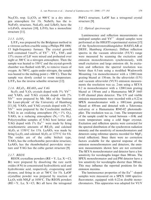
178L. van Pieterson et al. / Journal of Luminescence 91 (2000) 177–193Eu 3+ -ion in the same host lattices under chargetransfer excitation.2. Experimental2.1. Sample preparationFor all systems, undoped host lattices wereprepared, as well as the Yb 3+ (1% and 3%) andEu 3+ (1%) doped lattices. The crystalline powdersamples were analyzed with X-ray diffractionusing a Philips PW1710 Diffractometer Controlusing Cu K a radiation (1.542 A˚ ).Fig. 1. Configurational-coordinate diagrams of Eu 3+ (4f 6 ) andYb 3+ (4f 13 ). In this diagram the potential energy is plotted as afunction of a configurational coordinate, which can be relatedto the metal-ligand distance in the vibrating complex [1].This process is shown in Fig. 1(a). In Eu 3+ ,luminescence from the charge transfer state cannotbe observed, because there will always be fastrelaxation to the 5 D J states.Yb 3+ (4f 13 ) is an ion for which charge transferluminescence can be expected [3,4]. In this ion,the only excited 4f state, 2 F 5/2 , is located 10 000 cm 1above the ground state 2 F 7/2 . Fig. 1(b) shows theconfiguration-coordinate model of Yb 3+ [1].Because of the large energy difference betweenthe charge transfer state and the highest excited4f state, charge transfer luminescence can beobserved.Here we report a systematic study of chargetransfer luminescence from Yb 3+ . We studiedYb 3+ charge transfer luminescence in the phosphates,borates, aluminates, oxysulfides, oxyhalogenides,oxides and fluorides. The influence of thehost lattice and the size of the cation site for whichYb 3+ substitutes is investigated. Temperaturedependentmeasurements were performed to getan insight in the influence of the host lattice onthe quenching temperature of the Yb 3+ chargetransfer luminescence. The results are comparedwith the temperature quenching measured for the2.1.1. REPO 4Powder samples of REPO 4 (RE=Sc, Lu, Y, La)were prepared by firing RE 2 O 3 (4 N) and(NH 4 ) 2 HPO 4 in air at 13508C for 3 h. ScPO 4 ,LuPO 4 and YPO 4 all have a tetragonal crystalstructure (zircon-type), LaPO 4 has a monoclinicstructure (monazite type) [6].2.1.2. REBO 3Powder samples of REBO 3 (RE=Sc, Y, La)were prepared by firing stoichiometric amounts ofRE 2 O 3 (4 N) and boric acid in air at 5008C for 5 h.Then, after adding 20% excess boric acid, thesamples were heated for 8 h at 9508C. ScBO 3 hasthe rhombohedral calcite structure, YBO 3 has thehexagonal pseudovaterite structure, and LaBO 3had the orthorhombic aragonite structure [7].2.1.3. RE 2 O 2 SY 2 O 2 S and La 2 O 2 S were prepared by the sulfidefusionmethod [8] which involves a solid–meltreaction between RE 2 O 3 (4 N) and Na 2 S x .RE 2 O 3 ,Na 2 CO 3 , and S (ratio 1 : 1 : 2.97) were mixed andpreheated at 2008C, then fired at 11008C innitrogen atmosphere for 4 h. It is then washedseveral times with hot water and dilute HCl (2%).Y 2 O 2 S and La 2 O 2 S both have a hexagonal crystalstructure [9].2.1.4. Na/LiREO 2NaREO 2 and LiREO 2 powders (RE=Sc, Y,La) were prepared by firing RE 2 O 3 (4 N) and
L. van Pieterson et al. / Journal of Luminescence 91 (2000) 177–193 179Na 2 CO 3 resp. Li 2 CO 3 at 9008C in a dry nitrogenatmosphere for 3 h. NaScO 2 has the a-NaFeO 2 structure, NaLaO 2 and LiScO 2 have thea-LiFeO 2 structure [10], LiYO 2 has a monoclinicstructure [11].2.1.5. LiYF 4LiYF 4 was prepared by the Bridgman method ina vitreous carbon crucible using a Philips PH 1006/13 high-frequency furnace. The crystal growthmelt contained 5 mol% of Yb 3+ .YF 3 , YbF 3 and15% excess of LiF were mixed and preheated overnight at 3008C in a nitrogen atmosphere. Then thesample was heated to 5508C andthecrystal-growthchamber was flushed with SF 6 to remove traces ofoxygen. At 7008C, again under nitrogen, the samplewas heated to the melting point (9008C). Then thesample was slowly cooled to room temperature.LiYF 4 has the inverse scheelite structure [12].2.1.6. RE 2 O 3 , REAlO 3 and YAGSc 2 O 3 and Y 2 O 3 crystals doped with 3% Yb 3+and YAlO 3 and YAG crystals doped with 2%Yb 3+ were prepared by Mr. Mix of the ‘Institutfu¨ r Laser-physik’ of the University of Hamburg[13,14]. YAlO 3 and YAG crystals doped with 2%Yb 3+ were prepared by the Czochralski method,YAG in an oxidizing atmosphere (N 2 +1% O 2 ),YAlO 3 in a reducing atmosphere (N 2 +1% H 2 ).Polycrystalline samples of YAG host lattice andYAG doped with 1% Eu 3+ were made by firingstoichiometric amounts of RE 2 O 3 and calcinedAl 2 O 3 at 13508C for 15 h. LaAlO 3 was made byfiring La 2 O 3 and calcined Al 2 O 3 at 12758C for 8 h.The oxides are of the cubic bixbyit type,YAlO 3 has the orthorhombic perovskite structure,LaAlO 3 has the rhombohedral perovskite structureand YAG has the cubic garnet structure [9].2.1.7. REOXREOX crystalline powders (RE=Y, La; X=Cl,Br) were prepared by dissolving the rare earthoxides (4 N) in concentrated hydrochloric acid andhydrobromic acid, respectively, evaporating untildryness, and firing in air at 7008C for 5 h. LaOFcrystalline powder was prepared by reaction ofLa 2 O 3 with NH 4 F at 10508C. The REOX powders(RE=Y, La; X=Cl, Br) all have the tetragonalPbFCl structure, LaOF has a tetragonal crystalstructure [9].2.2. Optical spectroscopyLuminescence and reflection measurements onundoped samples and Yb 3+ -doped samples wereperformed at the HIGITI experimental station [15]of the Synchrotronstrahlungslabor HASYLAB atDESY, Hamburg (Germany). Diffuse reflectionspectra in the vacuum ultraviolet (VUV) rangewere measured by scanning the excitation andemission monochromators synchronously, withsmall excitation and large emission slit. In excitationand reflection the spectral resolution wasbetter than 0.5 nm using a modified WadsworthMounting 1 m monochromator with a 1200 l/mmgrating blazed at 150 nm. In the ultraviolet (UV)and vacuum ultraviolet (VUV) emission measurementsthe resolution was ca. 2 nm using a SEYA0.2 m monochromator with a 1200 l/mm gratingblazed at 150 nm and a Hamamatsu MCP 1645U-09 channel plate detector. The UV and visibleemission measurements were recorded with a 0.22 mSPEX monochromator with a 100 l/mm gratingblazed at 450 nm and detected with a Tektronicsccd-array or a Hamamatsu R943-02 photomultiplier.The resolution was ca. 1 nm. The temperatureof the sample could be varied between 10 K androom temperature using a cold finger cryostat.Excitation and reflection spectra were corrected forthe spectral distribution of the synchrotron radiationintensity and the sensitivity of monochromators anddetectors using reference spectra recorded for MgF 2(total reflection). Since there were no correctionfactors available for the spectral response of theemission monochromators and detectors, the emissionmeasurements shown here are not corrected.The SEYA monochromator and detector have a lowsensitivity for wavelengths longer than 300 nm. TheSPEX monochromator and ccd/PM detector have alow sensitivity for wavelengths shorter than 300 nm.This may influence the reported positions for theemission maxima.The luminescence properties of the Eu 3+ -dopedsamples were measured on a SPEX 1680 spectrofluorometer,equipped with 0.22 m double monochromators.This apparatus was adapted for VUV
- Page 1: Journal of Luminescence 91 (2000) 1
- Page 5 and 6: L. van Pieterson et al. / Journal o
- Page 7 and 8: L. van Pieterson et al. / Journal o
- Page 9 and 10: L. van Pieterson et al. / Journal o
- Page 11 and 12: L. van Pieterson et al. / Journal o
- Page 13 and 14: L. van Pieterson et al. / Journal o
- Page 15 and 16: L. van Pieterson et al. / Journal o
- Page 17: L. van Pieterson et al. / Journal o
L. van Pieterson et al. / Journal <strong>of</strong> <strong>Luminescence</strong> 91 (2000) 177–193 179Na 2 CO 3 resp. Li 2 CO 3 at 9008C in a dry nitrogenatmosphere for 3 h. NaScO 2 has the a-NaFeO 2 structure, NaLaO 2 and LiScO 2 have thea-LiFeO 2 structure [10], LiYO 2 has a monoclinicstructure [11].2.1.5. LiYF 4LiYF 4 was prepared by the Bridgman method ina vitreous carbon crucible using a Philips PH 1006/13 high-frequency furnace. The crystal growthmelt contained 5 mol% <strong>of</strong> Yb 3+ .YF 3 , YbF 3 and15% excess <strong>of</strong> LiF were mixed and preheated overnight at 3008C in a nitrogen atmosphere. Then thesample was heated to 5508C andthecrystal-growthchamber was flushed with SF 6 to remove traces <strong>of</strong>oxygen. At 7008C, again under nitrogen, the samplewas heated to the melting point (9008C). Then thesample was slowly cooled to room temperature.LiYF 4 has the inverse scheelite structure [12].2.1.6. RE 2 O 3 , REAlO 3 and YAGSc 2 O 3 and Y 2 O 3 crystals doped with 3% Yb 3+and YAlO 3 and YAG crystals doped with 2%Yb 3+ were prepared by Mr. Mix <strong>of</strong> the ‘Institutfu¨ r Laser-physik’ <strong>of</strong> the University <strong>of</strong> Hamburg[13,14]. YAlO 3 and YAG crystals doped with 2%Yb 3+ were prepared by the Czochralski method,YAG in an oxidizing atmosphere (N 2 +1% O 2 ),YAlO 3 in a reducing atmosphere (N 2 +1% H 2 ).Polycrystalline samples <strong>of</strong> YAG host lattice andYAG doped with 1% Eu 3+ were made by firingstoichiometric amounts <strong>of</strong> RE 2 O 3 and calcinedAl 2 O 3 at 13508C for 15 h. LaAlO 3 was made byfiring La 2 O 3 and calcined Al 2 O 3 at 12758C for 8 h.The oxides are <strong>of</strong> the cubic bixbyit type,YAlO 3 has the orthorhombic perovskite structure,LaAlO 3 has the rhombohedral perovskite structureand YAG has the cubic garnet structure [9].2.1.7. REOXREOX crystalline powders (RE=Y, La; X=Cl,Br) were prepared by dissolving the rare earthoxides (4 N) in concentrated hydrochloric acid andhydrobromic acid, respectively, evaporating untildryness, and firing in air at 7008C for 5 h. LaOFcrystalline powder was prepared by reaction <strong>of</strong>La 2 O 3 with NH 4 F at 10508C. The REOX powders(RE=Y, La; X=Cl, Br) all have the tetragonalPbFCl structure, LaOF has a tetragonal crystalstructure [9].2.2. Optical spectroscopy<strong>Luminescence</strong> and reflection measurements onundoped samples and Yb 3+ -doped samples wereperformed at the HIGITI experimental station [15]<strong>of</strong> the Synchrotronstrahlungslabor HASYLAB atDESY, Hamburg (Germany). Diffuse reflectionspectra in the vacuum ultraviolet (VUV) rangewere measured by scanning the excitation andemission monochromators synchronously, withsmall excitation and large emission slit. In excitationand reflection the spectral resolution wasbetter than 0.5 nm using a modified WadsworthMounting 1 m monochromator with a 1200 l/mmgrating blazed at 150 nm. In the ultraviolet (UV)and vacuum ultraviolet (VUV) emission measurementsthe resolution was ca. 2 nm using a SEYA0.2 m monochromator with a 1200 l/mm gratingblazed at 150 nm and a Hamamatsu MCP 1645U-09 channel plate detector. The UV and visibleemission measurements were recorded with a 0.22 mSPEX monochromator with a 100 l/mm gratingblazed at 450 nm and detected with a Tektronicsccd-array or a Hamamatsu R943-02 photomultiplier.The resolution was ca. 1 nm. The temperature<strong>of</strong> the sample could be varied between 10 K androom temperature using a cold finger cryostat.Excitation and reflection spectra were corrected forthe spectral distribution <strong>of</strong> the synchrotron radiationintensity and the sensitivity <strong>of</strong> monochromators anddetectors using reference spectra recorded for MgF 2(total reflection). Since there were no correctionfactors available for the spectral response <strong>of</strong> theemission monochromators and detectors, the emissionmeasurements shown here are not corrected.The SEYA monochromator and detector have a lowsensitivity for wavelengths longer than 300 nm. TheSPEX monochromator and ccd/PM detector have alow sensitivity for wavelengths shorter than 300 nm.This may influence the reported positions for theemission maxima.The luminescence properties <strong>of</strong> the Eu 3+ -dopedsamples were measured on a SPEX 1680 spectr<strong>of</strong>luorometer,equipped with 0.22 m double monochromators.This apparatus was adapted for VUV


