Charge Transfer Luminescence of Yb3+
Charge Transfer Luminescence of Yb3+
Charge Transfer Luminescence of Yb3+
- No tags were found...

You also want an ePaper? Increase the reach of your titles
YUMPU automatically turns print PDFs into web optimized ePapers that Google loves.
Journal <strong>of</strong> <strong>Luminescence</strong> 91 (2000) 177–193<strong>Charge</strong> transfer luminescence <strong>of</strong> Yb 3+L. van Pieterson*, M. Heeroma, E. de Heer, A. MeijerinkDebye Institute, Utrecht University, P. O. Box 80 000, 3508 TA Utrecht, The NetherlandsReceived 13 December 1999; received in revised form 21 February 2000; accepted 28 February 2000AbstractA systematic study <strong>of</strong> charge transfer (CT) luminescence from Yb 3+ is presented. CT luminescence was observed inLiYF 4 (7 eV), aluminates, phosphates, oxides (4 eV) and oxysulfides (3 eV). In all cases the CT emission bands arebroad and the Stokes shifts are large, ranging from 7000 cm 1 in oxysulfides and LiYF 4 and up to 17 000 cm 1 inaluminates. The influence <strong>of</strong> the size <strong>of</strong> the host lattice cation site on the charge transfer luminescence was investigated.A shift to longer wavelengths <strong>of</strong> the CT emission bands with increasing size <strong>of</strong> the cation site was observed. Typical(radiative) decay times for the CT luminescence are between 100 and 200 ns. The quenching <strong>of</strong> the Yb 3+ CTluminescence in the various host lattices was investigated and compared with the quenching <strong>of</strong> the Eu 3+ emission underCT excitation in the same lattices. A good correlation was found. The quenching temperatures <strong>of</strong> the Eu 3+ emission aremuch higher than that <strong>of</strong> the Yb 3+ CT luminescence. In lattices where Eu 3+ emission quenches at very high temperatures(>500 K) Yb 3+ CT luminescence could be observed. # 2000 Elsevier Science B.V. All rights reserved.PACS: 87.20e; 78.55m; 78.90+tKeywords: Yb 3+ ; <strong>Charge</strong> transfer1. IntroductionIn this century the luminescence <strong>of</strong> rare earthions has been well studied [1]. Especially since theapplication <strong>of</strong> luminescence from rare earth ionsin color television, fluorescent tubes and X-rayphosphors, numerous papers have appeared on4f n -4f n and 4f n1 5d–4f n emission. One kind <strong>of</strong> rareearth luminescence is still relatively unknown:charge transfer luminescence. This transition isthe reverse <strong>of</strong> the well-known charge transfer*Corresponding author. Tel.: +31-30-2533545; fax:+31-30-2532403.E-mail address: l.vanpieterson@phys.uu.nl (L. van Pieterson).absorption [2]. Up till now only three papers havereported charge transfer luminescence <strong>of</strong> a rareearth ion. In 1977, the first observation <strong>of</strong> thisphenomenom was made by Nakazawa. He reportedon charge transfer luminescence <strong>of</strong> theYb 3+ ion in phosphate and oxysulfide lattices[3,4]. Recently, Danielson et al., published oncharge transfer luminescence <strong>of</strong> the Ce 4+ ion inSr 2 CeO 4 [5].The charge transfer state is important inapplications. For example, in the red phosphorused in fluorescent tubes (Y 2 O 3 :Eu 3+ ), UVradiation is efficiently absorbed by a transition tothe charge transfer state (CTS) <strong>of</strong> the Eu 3+ -ion.After non-radiative decay to the lower 4f levels,luminescence occurs from the 5 D J states <strong>of</strong> Eu 3+ .0022-2313/00/$ - see front matter # 2000 Elsevier Science B.V. All rights reserved.PII: S 0022-2313(00)00214-3
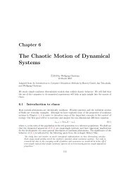
178L. van Pieterson et al. / Journal <strong>of</strong> <strong>Luminescence</strong> 91 (2000) 177–193Eu 3+ -ion in the same host lattices under chargetransfer excitation.2. Experimental2.1. Sample preparationFor all systems, undoped host lattices wereprepared, as well as the Yb 3+ (1% and 3%) andEu 3+ (1%) doped lattices. The crystalline powdersamples were analyzed with X-ray diffractionusing a Philips PW1710 Diffractometer Controlusing Cu K a radiation (1.542 A˚ ).Fig. 1. Configurational-coordinate diagrams <strong>of</strong> Eu 3+ (4f 6 ) andYb 3+ (4f 13 ). In this diagram the potential energy is plotted as afunction <strong>of</strong> a configurational coordinate, which can be relatedto the metal-ligand distance in the vibrating complex [1].This process is shown in Fig. 1(a). In Eu 3+ ,luminescence from the charge transfer state cannotbe observed, because there will always be fastrelaxation to the 5 D J states.Yb 3+ (4f 13 ) is an ion for which charge transferluminescence can be expected [3,4]. In this ion,the only excited 4f state, 2 F 5/2 , is located 10 000 cm 1above the ground state 2 F 7/2 . Fig. 1(b) shows theconfiguration-coordinate model <strong>of</strong> Yb 3+ [1].Because <strong>of</strong> the large energy difference betweenthe charge transfer state and the highest excited4f state, charge transfer luminescence can beobserved.Here we report a systematic study <strong>of</strong> chargetransfer luminescence from Yb 3+ . We studiedYb 3+ charge transfer luminescence in the phosphates,borates, aluminates, oxysulfides, oxyhalogenides,oxides and fluorides. The influence <strong>of</strong> thehost lattice and the size <strong>of</strong> the cation site for whichYb 3+ substitutes is investigated. Temperaturedependentmeasurements were performed to getan insight in the influence <strong>of</strong> the host lattice onthe quenching temperature <strong>of</strong> the Yb 3+ chargetransfer luminescence. The results are comparedwith the temperature quenching measured for the2.1.1. REPO 4Powder samples <strong>of</strong> REPO 4 (RE=Sc, Lu, Y, La)were prepared by firing RE 2 O 3 (4 N) and(NH 4 ) 2 HPO 4 in air at 13508C for 3 h. ScPO 4 ,LuPO 4 and YPO 4 all have a tetragonal crystalstructure (zircon-type), LaPO 4 has a monoclinicstructure (monazite type) [6].2.1.2. REBO 3Powder samples <strong>of</strong> REBO 3 (RE=Sc, Y, La)were prepared by firing stoichiometric amounts <strong>of</strong>RE 2 O 3 (4 N) and boric acid in air at 5008C for 5 h.Then, after adding 20% excess boric acid, thesamples were heated for 8 h at 9508C. ScBO 3 hasthe rhombohedral calcite structure, YBO 3 has thehexagonal pseudovaterite structure, and LaBO 3had the orthorhombic aragonite structure [7].2.1.3. RE 2 O 2 SY 2 O 2 S and La 2 O 2 S were prepared by the sulfidefusionmethod [8] which involves a solid–meltreaction between RE 2 O 3 (4 N) and Na 2 S x .RE 2 O 3 ,Na 2 CO 3 , and S (ratio 1 : 1 : 2.97) were mixed andpreheated at 2008C, then fired at 11008C innitrogen atmosphere for 4 h. It is then washedseveral times with hot water and dilute HCl (2%).Y 2 O 2 S and La 2 O 2 S both have a hexagonal crystalstructure [9].2.1.4. Na/LiREO 2NaREO 2 and LiREO 2 powders (RE=Sc, Y,La) were prepared by firing RE 2 O 3 (4 N) and
L. van Pieterson et al. / Journal <strong>of</strong> <strong>Luminescence</strong> 91 (2000) 177–193 179Na 2 CO 3 resp. Li 2 CO 3 at 9008C in a dry nitrogenatmosphere for 3 h. NaScO 2 has the a-NaFeO 2 structure, NaLaO 2 and LiScO 2 have thea-LiFeO 2 structure [10], LiYO 2 has a monoclinicstructure [11].2.1.5. LiYF 4LiYF 4 was prepared by the Bridgman method ina vitreous carbon crucible using a Philips PH 1006/13 high-frequency furnace. The crystal growthmelt contained 5 mol% <strong>of</strong> Yb 3+ .YF 3 , YbF 3 and15% excess <strong>of</strong> LiF were mixed and preheated overnight at 3008C in a nitrogen atmosphere. Then thesample was heated to 5508C andthecrystal-growthchamber was flushed with SF 6 to remove traces <strong>of</strong>oxygen. At 7008C, again under nitrogen, the samplewas heated to the melting point (9008C). Then thesample was slowly cooled to room temperature.LiYF 4 has the inverse scheelite structure [12].2.1.6. RE 2 O 3 , REAlO 3 and YAGSc 2 O 3 and Y 2 O 3 crystals doped with 3% Yb 3+and YAlO 3 and YAG crystals doped with 2%Yb 3+ were prepared by Mr. Mix <strong>of</strong> the ‘Institutfu¨ r Laser-physik’ <strong>of</strong> the University <strong>of</strong> Hamburg[13,14]. YAlO 3 and YAG crystals doped with 2%Yb 3+ were prepared by the Czochralski method,YAG in an oxidizing atmosphere (N 2 +1% O 2 ),YAlO 3 in a reducing atmosphere (N 2 +1% H 2 ).Polycrystalline samples <strong>of</strong> YAG host lattice andYAG doped with 1% Eu 3+ were made by firingstoichiometric amounts <strong>of</strong> RE 2 O 3 and calcinedAl 2 O 3 at 13508C for 15 h. LaAlO 3 was made byfiring La 2 O 3 and calcined Al 2 O 3 at 12758C for 8 h.The oxides are <strong>of</strong> the cubic bixbyit type,YAlO 3 has the orthorhombic perovskite structure,LaAlO 3 has the rhombohedral perovskite structureand YAG has the cubic garnet structure [9].2.1.7. REOXREOX crystalline powders (RE=Y, La; X=Cl,Br) were prepared by dissolving the rare earthoxides (4 N) in concentrated hydrochloric acid andhydrobromic acid, respectively, evaporating untildryness, and firing in air at 7008C for 5 h. LaOFcrystalline powder was prepared by reaction <strong>of</strong>La 2 O 3 with NH 4 F at 10508C. The REOX powders(RE=Y, La; X=Cl, Br) all have the tetragonalPbFCl structure, LaOF has a tetragonal crystalstructure [9].2.2. Optical spectroscopy<strong>Luminescence</strong> and reflection measurements onundoped samples and Yb 3+ -doped samples wereperformed at the HIGITI experimental station [15]<strong>of</strong> the Synchrotronstrahlungslabor HASYLAB atDESY, Hamburg (Germany). Diffuse reflectionspectra in the vacuum ultraviolet (VUV) rangewere measured by scanning the excitation andemission monochromators synchronously, withsmall excitation and large emission slit. In excitationand reflection the spectral resolution wasbetter than 0.5 nm using a modified WadsworthMounting 1 m monochromator with a 1200 l/mmgrating blazed at 150 nm. In the ultraviolet (UV)and vacuum ultraviolet (VUV) emission measurementsthe resolution was ca. 2 nm using a SEYA0.2 m monochromator with a 1200 l/mm gratingblazed at 150 nm and a Hamamatsu MCP 1645U-09 channel plate detector. The UV and visibleemission measurements were recorded with a 0.22 mSPEX monochromator with a 100 l/mm gratingblazed at 450 nm and detected with a Tektronicsccd-array or a Hamamatsu R943-02 photomultiplier.The resolution was ca. 1 nm. The temperature<strong>of</strong> the sample could be varied between 10 K androom temperature using a cold finger cryostat.Excitation and reflection spectra were corrected forthe spectral distribution <strong>of</strong> the synchrotron radiationintensity and the sensitivity <strong>of</strong> monochromators anddetectors using reference spectra recorded for MgF 2(total reflection). Since there were no correctionfactors available for the spectral response <strong>of</strong> theemission monochromators and detectors, the emissionmeasurements shown here are not corrected.The SEYA monochromator and detector have a lowsensitivity for wavelengths longer than 300 nm. TheSPEX monochromator and ccd/PM detector have alow sensitivity for wavelengths shorter than 300 nm.This may influence the reported positions for theemission maxima.The luminescence properties <strong>of</strong> the Eu 3+ -dopedsamples were measured on a SPEX 1680 spectr<strong>of</strong>luorometer,equipped with 0.22 m double monochromators.This apparatus was adapted for VUV
180L. van Pieterson et al. / Journal <strong>of</strong> <strong>Luminescence</strong> 91 (2000) 177–193excitation measurements [16]. The spectral resolution<strong>of</strong> this setup is about 0.5 nm. Excitationspectra measured on this apparatus were correctedfor the lamp intensity using excitation spectra <strong>of</strong>sodium salysilate emission as a reference. Emissionspectra were corrected for the spectral response<strong>of</strong> the monochromator and the detector usingcorrection spectra provided by the manufacturer.Temperature-dependent measurements could beperformed between 5 and 850 K. In the lowtemperatureregion a cold finger cryostat was used.For measurements at temperatures higher thanroom temperature a home-made high-temperaturesample holder was used.3. ResultsTo distinguish between Yb 3+ charge transferluminescence and luminescence <strong>of</strong> the host lattice,it is necessary to have a good knowledge abouthost lattice luminescence. <strong>Luminescence</strong> bandsobserved in the spectra <strong>of</strong> Yb 3+ -doped lattices,which are not observed in the spectra <strong>of</strong> theundoped samples, can be assigned to Yb 3+ . Forthis reason, both undoped samples and Yb 3+ -doped samples were prepared and measured. Acareful analysis <strong>of</strong> the reflection and luminescencespectra <strong>of</strong> the doped and undoped samplesprovides information on the CT luminescence <strong>of</strong>Yb 3+ in the different host lattices.3.1. PhosphatesFig. 2 shows the reflection spectra <strong>of</strong> ScPO 4 andScPO 4 doped with 3% Yb 3+ . The host latticeabsorption edge is observed at 168 nm. In thispaper the absorption edge is defined as the lowestenergy at which the absorption reaches its maximumvalue (see arrow in Fig. 2). The reflectionspectrum <strong>of</strong> ScPO 4 doped with 3 mol% <strong>of</strong> Yb 3+shows the same absorption bands as the undopedsample and in addition a strong absorption bandwith a maximum at 195 nm. This extra absorptioncan be assigned to the 2 F 7/2 ! CT transition <strong>of</strong> theYb 3+ ion in ScPO 4 . At wavelengths longer than200 nm the signal decreases slowly. The slowFig. 2. Reflection spectrum <strong>of</strong> the ScPO 4 host lattice (brokenline) and ScPO 4 doped with 3% Yb 3+ (solid line), measuredat 10 K.decrease may be related to the very low intensity<strong>of</strong> the synchrotron in this wavelength region whichintroduces relatively large errors in the correctedspectrum.Fig. 3 shows the emission and excitation spectra<strong>of</strong> ScPO 4 and ScPO 4 doped with 1% Yb 3+ . Theemission spectrum <strong>of</strong> the ScPO 4 host lattice showsseveral broad bands with maxima at 211, 350 and470 nm. The emission at 211 nm can only beexcited with radiation <strong>of</strong> energies higher than theband gap (l5168 nm). The other emission bandscan also be excited at wavelengths longer than168 nm and are attributed to defect centers in theScPO 4 lattice.If we compare the emission spectrum <strong>of</strong> ScPO 4doped with Yb 3+ with the spectrum <strong>of</strong> the hostlattice, two strong extra bands are observed withmaxima at 270 and 370 nm. These bands areassigned to transitions from the charge transferstate to the 2 F 7/2 and 2 F 5/2 state <strong>of</strong> Yb 3+ . Theenergy separation between the two emission bandsis 10 000 cm 1 , in agreement with the separationbetween the 2 F 5/2 and 2 F 7/2 states <strong>of</strong> Yb 3+ . Thefull-width at half-maximum (FWHM) <strong>of</strong> thebands is 6 000 cm 1 . The Stokes shift <strong>of</strong> the CT! 2 F 7/2 emission is 14 500 cm 1 . The Stokes shiftis the energy difference between the maximum <strong>of</strong>the CT absorption band and the CT ! 2 F 7/2emission band. The CT ! 2 F 7/2 emission band is<strong>of</strong>ten located in a spectral region (250–400 nm)where both SEYA monochromator with detectorand SPEX monochromator with ccd-array have alow sensitivity. For this reason, in this paper theStokes shift is calculated from the position <strong>of</strong> theCT ! 2 F 5/2 emission minus 10 000 cm 1 , this
L. van Pieterson et al. / Journal <strong>of</strong> <strong>Luminescence</strong> 91 (2000) 177–193 181emission is in most cases observed in a spectralregion where the SPEX/ccd sensitivity curve isrelatively flat. Emission due to the transitionbetween the two 4f states can be observed assharp peaks in the infrared at 979 nm.In the excitation spectrum <strong>of</strong> the Yb 3+ emissionat 270 nm, the host lattice absorption edge isobserved at 168 nm. At 195 nm the 2 F 7/2 ! CTexcitation band is observed. The position <strong>of</strong> thisexcitation band is in good agreement with thecharge transfer absorption band in the reflectionspectrum.At low temperature, a decay time <strong>of</strong> 163 25 nshas been measured for the charge transfer luminescence,similar to the decay times measured forYb 3+ charge transfer luminescence in LuPO 4 andYPO 4 [3,4]. To study the temperature quenching<strong>of</strong> the charge transfer luminescence, temperaturedependentlifetime measurements and intensitymeasurements were performed. In Fig. 4, theintensity <strong>of</strong> the 270 nm emission band and thedecay time <strong>of</strong> this emission are given as a function<strong>of</strong> temperature. There is a good agreementbetween the decrease in intensity and decay time.If we define the quenching temperature as thetemperature at which the luminescence intensity orthe decay time have decreased to half <strong>of</strong> its initialvalue, the quenching temperature <strong>of</strong> the Yb 3+charge transfer luminescence is estimated to beabout 225 K.Also for Yb 3+ in other orthophosphates luminescence,reflection and lifetime measurementswere performed and compared with the luminescence<strong>of</strong> the undoped host lattices. In Table 1, theFig. 3. Emission and excitation spectra <strong>of</strong> the ScPO 4 hostlattice (a, b) and <strong>of</strong> ScPO 4 doped with 1% Yb 3+ (c, d) measuredat 10 K. The emission spectra for the ScPO 4 host lattice andYb 3+ -doped sample were both recorded for excitation at140 nm, for the excitation spectrum <strong>of</strong> the host lattice the emissionwavelength was at 210 nm, for the Yb 3+ -doped sample itwas at 270 nm. In the emission spectra the broken linerepresents measurements performed with a SEYA monochromator,the solid line represents measurements performed with aSPEX monochromator equipped with a Tektronics ccd-array.Fig. 4. Decay time (*), on the left axis, and luminescenceintensity (&), on the right axis, <strong>of</strong> the charge transferluminescence at 270 nm as a function <strong>of</strong> temperature forScPO 4 :Yb 3+ (l exc =140 nm).
182L. van Pieterson et al. / Journal <strong>of</strong> <strong>Luminescence</strong> 91 (2000) 177–193Table 1Positions <strong>of</strong> absorption edges and emission bands in the orthophosphatehost lattices REPO 4 (RE=Sc, Lu, Y, La)data obtained for the orthophosphate host latticesare summarized and Table 2 summarizes theresults <strong>of</strong> the orthophosphates doped with Yb 3+ .In the case <strong>of</strong> LaPO 4 :Yb 3+ no evidence for Yb 3+charge transfer luminescence could be obtained,indicating that the charge transfer luminescence isquenched already at the lowest temperatures inthis host lattice.3.2. OxidesAbsorption edge (nm)Emission (nm)ScPO 4 168 211, 350, 470LuPO 4 140 Not measuredYPO 4 144 233, 440LaPO 4 153 262, 328Fig. 5 shows the reflection spectrum <strong>of</strong> LiScO 2and LiScO 2 doped with 3% Yb 3+ . The host latticeabsorption edge is observed at 187 nm. In theYb 3+ -doped sample, an extra absorption isobserved with a maximum at 209 nm. Thisabsorption band is assigned to the charge transferband <strong>of</strong> Yb 3+ .Fig. 6 shows the emission and excitation spectra<strong>of</strong> the LiScO 2 host lattice and LiScO 2 doped with3% Yb 3+ . The emission spectrum <strong>of</strong> the hostlattice shows one broad band at 250 nm whenexciting at 180 nm. In the excitation spectrum theabsorption edge is observed at 186 nm. Whenexciting at longer wavelengths, defect emission isobserved between 350 and 450 nm.In the emission spectrum <strong>of</strong> LiScO 2 :Yb 3+ twobroad bands are observed at 312 and 448 nmwhich are not present in the spectrum <strong>of</strong> theundoped lattice. The energy separation betweenthese bands is about 10 000 cm 1 . The FWHM <strong>of</strong>the bands is 6000 cm 1 and the Stokes shift is16 000 cm 1 . At 972 nm the transition between the4f states <strong>of</strong> Yb 3+ is observed. In the excitationspectrum <strong>of</strong> LiScO 2 :Yb 3+ the host lattice absorptionedge is observed at 188 nm. At longerwavelengths an extra band is observed with theonset at 225 nm and a maximum at 208 nm, whichcan be assigned to the charge transfer band <strong>of</strong>Yb 3+ .The decay time <strong>of</strong> the charge transfer luminescenceis 190 25 ns and the quenching temperatureis about 180 K in LiScO 2 . Table 3 summarizesthe results for the NaREO 2 and LiREO 2 hostlattices. Data on NaYO 2 and LiYO 2 host latticesare lacking due to problems with synthesis. Table 4shows charge transfer luminescence data obtainedfor NaREO 2 and LiREO 2 lattices doped withYb 3+ . In NaLaO 2 and LiLaO 2 no Yb 3+ chargetransfer luminescence was observed, indicating thatFig. 5. Reflection spectrum <strong>of</strong> LiScO 2 (broken line) and LiScO 2doped with 3% Yb 3+ (solid line) at 10 K.Table 2Positions <strong>of</strong> the charge transfer emission and absorption bands for Yb 3+ in orthophosphate host lattices. For decay time ðtÞ measurements(at 10 K) and determination <strong>of</strong> the quenching temperature ðT q Þ, the CT ! 2 F 7/2 emission is monitoredAbsorption max. (nm) Emission (nm) Stokes shift (cm 1 ) tðnsÞ T q ðKÞScPO 4 195 270, 370 14 500 163 225LuPO 4 210 290, 440 15 000 175 250YPO 4 210 304, 445 15 000 120 290LaPO 4 228 } } } }
L. van Pieterson et al. / Journal <strong>of</strong> <strong>Luminescence</strong> 91 (2000) 177–193 183Table 3Positions <strong>of</strong> absorption edges and emission bands in NaREO 2(RE=Sc, La) and LiREO 2 (RE=Sc, La) host latticesAbsorption edge (nm)Emission (nm)NaScO 2 189 400 (very broad)NaLaO 2 242 Not measuredLiScO 2 188 250, 350–450LiLaO 2 229 400Fig. 6. Emission and excitation spectra <strong>of</strong> the LiScO 2 host lattice(a, b) and LiScO 2 doped with 3% Yb 3+ (c, d), T ¼ 10 K. Thehost lattice emission spectrum was recorded for excitation at180 nm, the excitation spectrum was measured for the 272 nmemission. The emission spectrum <strong>of</strong> the Yb 3+ -doped sample wasrecorded for excitation at 200 nm, while the excitation spectrumwas measured for the 300 nm emission. In Fig. 6 (c), the brokenline represents measurements performed with a SEYA monochromator,the solid line represents measurements performed witha SPEX monochromator equipped with a Tektronics ccd-array.the CT luminescence is quenched in these hostlattices.The reflection spectra <strong>of</strong> Y 2 O 3 and Y 2 O 3 dopedwith 3% Yb 3+ are shown in Fig. 7. The hostlattice absorption edge is observed at 208 nm, inagreement with results by Tomiki et al., whoobserved the exciton creation at 5.88 eV (210 nm)[17]. In the Yb 3+ -doped Y 2 O 3 the charge transferabsorption band is observed at 227 nm.Fig. 8 shows the emission and excitation spectra<strong>of</strong> the Y 2 O 3 host lattice and Y 2 O 3 :Yb 3+ . Hostlattice emission is observed at 370 nm. In theexcitation spectrum, the host lattice excitationedge is observed at 206 nm.The emission spectrum <strong>of</strong> Y 2 O 3 doped with 3%Yb 3+ shows two broad bands with maxima at 368and 533 nm. The energy separation between thesebands is 8500 cm 1 , which is less than the energyseparation <strong>of</strong> 10 000 cm 1 between the 2 F 5/2 and2 F 7/2 states <strong>of</strong> Yb 3+ . This may be caused byunderlying host lattice defect emission and by thefact that the emission spectra are not corrected.The FWHM <strong>of</strong> the bands is 7000 cm 1 . At 995 nm,the transition between the 2 F 5/2 and 2 F 7/2 state canbe observed. In the excitation spectrum, an extraband is observed with a maximum at 228 nm, ingood agreement with the position <strong>of</strong> the chargetransfer band in the reflection spectrum. TheStokes shift is 15 000 cm 1 . In Table 5, data onluminescence properties <strong>of</strong> Yb 3+ -doped and undopedoxide lattices are summarized.In addition to the oxides discussed above, welooked for Yb 3+ CT luminescence in LaYO 3 .Inthis lattice the host lattice absorption edge is observedat 206 nm. No indication for CT luminescencefrom Yb 3+ was found, not even at thelowest temperatures.
184L. van Pieterson et al. / Journal <strong>of</strong> <strong>Luminescence</strong> 91 (2000) 177–193Table 4Positions <strong>of</strong> the charge transfer emission and absorption bands for Yb 3+ in NaREO 2 and LiREO 2 host lattices. For decay time (t)measurements (at 10 K) and determination <strong>of</strong> the quenching temperature ðT q Þ, the CT ! 2 F 7/2 emission is monitoredAbsorption (nm) Emission (nm) Stokes shift (cm 1 ) tðnsÞ T q ðKÞNaScO 2 208 322, 431 15 000 130 225NaLaO 2 262 } } } }LiScO 2 206 312, 448 16 000 190 180LiYO 2 214 355, 495 16 500 55 125LiLaO 2 252 } } } }Fig. 7. Reflection spectra <strong>of</strong> Y 2 O 3 (broken line) and Y 2 O 3doped with 3% Yb 3+ (solid line), measured at 10 K.3.3. AluminatesThe reflection spectra <strong>of</strong> the Y 3 Al 5 O 12 (YAG)host lattice and YAG doped with 2% Yb 3+ areshown in Fig. 9. At 178 nm the host latticeabsorption edge is observed. When Yb 3+ is incorporatedin the lattice, an extra absorption band isobserved at 209 nm, which is assigned to thetransition from the 2 F 7/2 ground state <strong>of</strong> Yb 3+ tothe charge transfer state.Fig. 10 shows the emission and excitationspectra <strong>of</strong> YAG and YAG : Yb 3+ . In the emissionspectrum <strong>of</strong> undoped YAG, host lattice luminescenceis observed at 256 nm. When exciting atlonger wavelengths defect emission around 450 nmis observed. In the Yb 3+ -doped samples strongemission bands are observed at 334 and 493 nmwhich are not present in the emission spectrum <strong>of</strong>the undoped sample. The energy separationbetween the bands is close to 10 000 cm 1 . Basedon these observations, the bands are assigned toCT luminescence from Yb 3+ . In the excitationspectrum <strong>of</strong> the 343 nm emission, the CT excitationband is observed at 210 nm, in agreement withthe position in the reflection spectrum. TheStokes shift <strong>of</strong> the charge transfer transition issome 17 500 cm 1 and the FWHM <strong>of</strong> both bandsis 6000 cm 1 . The decay time measured for theYb 3+ charge transfer luminescence is 30 5ns at10 K, much shorter than the decay times which aretypically observed for the Yb 3+ CT emission100–200 ns) at low temperatures. Probably, thecharge transfer luminescence is already partlyquenched at very low temperatures. The relativelyhigh intensity <strong>of</strong> the 2 F 5/2 ! 2 F 7/2 transitioncompared to the charge transfer luminescence alsosuggests that the CT emission is already partlyquenched at 10 K.In Fig. 11, the CT luminescence intensity and itsdecay time are given as a function <strong>of</strong> temperature.The immediate decrease in intensity in the lowtemperatureregion confirms that the luminescenceTable 5Positions <strong>of</strong> emission and absorption bands for undoped andYb 3+ -doped oxides RE 2 O 3 (RE=Sc, Y). For the undopedoxides, the absorption edge is given, for the Yb 3+ -doped oxidesthe CT absorption maximum is given. For decay time ðtÞmeasurements and determination <strong>of</strong> the quenching temperatureðT q Þ, the CT ! 2 F 7/2 emission is monitoredAbsorption(nm)Emission(nm)Stokesshift(cm 1 )tðnsÞ T q (K)Sc 2 O 3 196 Not measuredY 2 O 3 208 370Sc 2 O 3 :Yb 3+ 225 367, 485 14 000 13 530(very weak)Y 2 O 3 :Yb 3+ 227 368, 533 15 500 72 130
L. van Pieterson et al. / Journal <strong>of</strong> <strong>Luminescence</strong> 91 (2000) 177–193 185Table 6Positions <strong>of</strong> the emission and absorption bands for aluminatehost lattices and Yb 3+ -doped aluminates. For the undopedaluminates, the absorption edge is given, for the Yb 3+ -dopedaluminates the CT absorption maximum is givenAbsorption(nm)Emission(nm)Stokesshift(cm 1 )tðnsÞ T q ðKÞYAG 178 260, 460YAlO 3 158 Not measuredLaAlO 3 211 280, 600YAG : Yb 3+ 210 334, 493 17 500 28 580YAlO 3 :Yb 3+ 235 360, 533 14 000 100 100LaAlO 3 :Yb 3+ 244 } } } }Fig. 9. Reflection spectra <strong>of</strong> YAG (broken line) and YAGdoped with 2% Yb 3+ (solid line), measured at 10 K.Fig. 8. Emission (for l exc ¼ 170 nm) and excitation (forl em ¼ 370 nm) spectra <strong>of</strong> the Y 2 O 3 host lattice (a, b) andemission (for l exc ¼ 215 nm) and excitation (for l em ¼ 372 nm)spectra <strong>of</strong> Y 2 O 3 doped with 3% Yb 3+ (c, d), T ¼ 10 K.is already quenched at very low temperature.From this figure we can estimate that the quenchingtemperature is lower than 80 K.Fig. 12 shows the emission and excitationspectra <strong>of</strong> YAlO 3 doped with 2% Yb 3+ . Thebroad emission bands with maxima at 360 and533 nm are assigned to CT transitions <strong>of</strong> Yb 3+ .InFig. 13, the decay curves <strong>of</strong> the CT emissionsmeasured at 350 and 516 nm are shown. The decaytime <strong>of</strong> these emissions is 100 10 ns. In theexcitation spectrum <strong>of</strong> the 350 nm emission theCT band can be observed at 235 nm.In LaAlO 3 no evidence for CT luminescencecould be obtained. Table 6 summarizes theluminescence properties <strong>of</strong> the undoped andYb 3+ -doped aluminates.3.4. OxysulfidesThe charge transfer absorption for Yb 3+ in theoxysulfides is at relatively low energies andluminescence spectra could be recorded with the
186L. van Pieterson et al. / Journal <strong>of</strong> <strong>Luminescence</strong> 91 (2000) 177–193Fig. 11. Decay time (*), on the left axis, and CT luminescenceintensity (&), on the right axis, <strong>of</strong> YAG doped with 2% Yb 3+as a function <strong>of</strong> temperature.Fig. 12. Emission (a, l exc ¼ 220 nm) and excitation (b,l em ¼ 350 nm) spectra <strong>of</strong> YAlO 3 doped with 2% Yb 3+ atT ¼ 10 K.Fig. 10. Emission (for l exc ¼ 160 nm) and excitation (forl em ¼ 255 nm) spectra <strong>of</strong> the YAG host lattice (a, b) andemission (for l exc ¼ 170 nm) and excitation (for l em ¼ 345 nm)spectra <strong>of</strong> YAG doped with 2% Yb 3+ (c, d). In Fig. 10 (c), thebroken line represents measurements performed with a SEYAmonochromator, the solid line represents measurements performedwith a SPEX monochromator equipped with aTektronics ccd-array.Fig. 13. Decay curves (at 10 K) <strong>of</strong> the Yb 3+ CT emissions at350 (dotted) and 516 nm (broken line) in YAlO 3 .
L. van Pieterson et al. / Journal <strong>of</strong> <strong>Luminescence</strong> 91 (2000) 177–193 187Fig. 15. Decay time (*), on the left axis, and CT luminescenceintensity at 420 nm (&) and at 822 nm (n), on the right axis, <strong>of</strong>La 2 O 2 S doped with Yb 3+ as a function <strong>of</strong> temperature.SPEX 1680 spectr<strong>of</strong>luorometer. Fig. 14 shows theemission and excitation spectra <strong>of</strong> Y 2 O 2 S:Yb 3+and La 2 O 2 S:Yb 3+ at 10 K. In Y 2 O 2 S, Yb 3+charge transfer luminescence is observed at 390and 612 nm. The energy difference between theCT bands is 9500 cm 1 , close to the expected10 000 cm 1 . The FWHM <strong>of</strong> the bands is about3500 cm 1 . In the excitation spectrum the chargetransfer band is observed at 309 nm so the Stokesshift is relatively small, about 6000 cm 1 . Thedecay time measured at 390 nm is about 250 ns.The Y 2 O 2 S host lattices luminesce at 318 nm whenexciting into the conduction band (l5260 nm).This emission quenches already below 50 K. Also abroad defect-related emission around 590 nm isobserved which quenches at about 275 K.In La 2 O 2 SYb 3+ CT luminescence is observed at439 and 759 nm, the energy difference betweenthese bands is 9500 cm 1 . The CT excitation bandis observed at 317 nm, giving a Stokes shift <strong>of</strong>approximately 8500 cm 1 . In Fig. 15, the intensity<strong>of</strong> the Yb 3+ CT luminescence in La 2 O 2 S and itsdecay time are given as a function <strong>of</strong> temperature.From this figure we can estimate the quenchingtemperature at 150 K. Table 7 summarizes theluminescence properties <strong>of</strong> the oxysulfide hostlattices and Yb 3+ -doped samples.Fig. 14. Emission (for l exc ¼ 240 nm) and excitation (forl em ¼ 393 nm) spectra <strong>of</strong> Y 2 O 2 S doped with 1% Yb 3+ (a, b)and emission (for l exc ¼ 270 nm) and excitation (for l em ¼460 nm) spectra <strong>of</strong> La 2 O 2 S (c, d) doped with 1% Yb 3+ ,T ¼ 10 K.3.5. FluoridesFig. 16 shows the emission and excitationspectrum <strong>of</strong> LiYF 4 doped with 3% Yb 3+ . Theemission spectrum has been recorded using gateddetection (delay 5 ns, gate 10 ns) and shows two
188L. van Pieterson et al. / Journal <strong>of</strong> <strong>Luminescence</strong> 91 (2000) 177–193Table 7Positions <strong>of</strong> emission and absorption bands for oxysulfide host lattices and for Yb 3+ -doped RE 2 O 2 S (RE=Y, La) host lattices. Forthe undoped lattices, the absorption edge is given, for the Yb 3+ -doped lattices the CT absorption maximum is given. For decay timeðtÞ measurements (at 10 K) and determination <strong>of</strong> the quenching temperature ðT q Þ, the CT ! 2 F 7/2 emission is monitoredAbsorption edge (nm) Emission (nm) Stokes shift (cm 1 ) t (ns) T q (K)Y 2 O 2 S 263 318, 590 550 resp 275La 2 O 2 S 276 377, 560 60 resp 250Y 2 O 2 S:Yb 3+ 309 390, 612 6000 250 140La 2 O 2 S:Yb 3+ 317 439, 759 8500 223 150band between 148 and 163 nm observed bySzcezurek and Schlesinger for Yb 3+ in CaF 2 [18],which could be assigned to the CT transition <strong>of</strong>Yb 3+ .3.6. Other latticesFig. 16. Emission (a, l exc ¼ 155 nm) and excitation (b, l em ¼181 nm) spectra <strong>of</strong> LiYF 4 doped with 3% Yb 3+ at 10 K.very weak bands with maxima at 182 and 228 nm.The energy separation between both bands is11 000 cm 1 . The two emission bands have thesame excitation spectrum and also the decay timeis approximately the same: 13 2 ns. Therefore,these bands are assigned to transitions from thecharge transfer state to the 2 F 7/2 resp. 2 F 5/2 state <strong>of</strong>Yb 3+ . The fact that the emission is very weak andthe decay time is short suggests that the luminescenceis already quenched at very low temperatures.At 250 nm, the onset <strong>of</strong> the host latticeemission band can be observed.In the excitation spectrum, the charge transferband is observed at 159 nm, so the Stokes shift isca. 8000 cm 1 . The position <strong>of</strong> the CT excitationband is in agreement with the broad absorptionIn the previous sections the charge transferluminescence <strong>of</strong> Yb 3+ in many compounds hasbeen described. In view <strong>of</strong> the very limited number<strong>of</strong> compounds in which the CT luminescence <strong>of</strong>Yb 3+ has been reported before, a large number<strong>of</strong> compounds has been added. Yet, it is notsurprising that until now the number <strong>of</strong> compoundsin which CT luminescence from Yb 3+ hasbeen reported, is so small. In many compounds inwhich CT luminescence is observed, the Yb 3+ CTluminescence has a quenching temperature belowroom temperature. In many other compounds noCT luminescence is observed at all, not even at4 K. In addition to the compounds discussed inthe previous sections, we looked for Yb 3+ CTluminescence in the orthoborates REBO 3(RE=Sc, Y, La) and the oxyhalogenides REOX(RE=Y, La; X=F, Cl, Br) by comparing theluminescence spectra <strong>of</strong> undoped host lattices andYb 3+ -doped samples. For none <strong>of</strong> the borates oroxyhalogenides evidence for Yb 3+ CT luminescencewas obtained, indicating that the luminescence<strong>of</strong> Yb 3+ is already quenched at 10 K in thesehost lattices.3.7. Comparison with Eu 3+For comparison, the luminescence <strong>of</strong> Eu 3+under charge transfer excitation was measured
L. van Pieterson et al. / Journal <strong>of</strong> <strong>Luminescence</strong> 91 (2000) 177–193 189Table 8Position <strong>of</strong> the Eu 3+ and Yb 3+ charge transfer band and quenching temperature <strong>of</strong> the 5 D 0 ! 7 F J luminescence after excitation in thecharge transfer band resp. the quenching temperature <strong>of</strong> the Yb 3+ CT luminescenceEu 3+ CT excitation band (nm) Eu 3+ T q (K) Yb 3+ CT absorption band (nm) Yb 3+ T q (K)ScPO 4 205 760 a 195 225LuPO 4 215 850 a 210 250YPO 4 218 >850 210 290LaPO 4 251 400 228 510NaScO 2 225 650 208 225NaLaO 2 264 320 262 510LiScO 2 223 >850 206 180LiYO 2 247 600 214 125LiLaO 2 274 475 252 510Y 2 O 3 250 >850 227 130Y 2 O 2 S 334 725 310 140La 2 O 2 S 347 480 317 150YAG 237 >850 212 580LaAlO 3 306 500 244 510ScBO 3 218 420 } 510YBO 3 228 >850 216 510LaBO 3 275 350 } 510a The luminescence did not quench completely at high temperatures, but stayed at more than a quarter <strong>of</strong> the maximum intensity.for all host lattices described above. In Table 8, theposition <strong>of</strong> the Eu 3+ charge transfer band and thequenching temperature <strong>of</strong> the 5 D 0 ! 7 F J luminescencewhen excited in the Eu 3+ charge transferband, are given for all lattices. For some lattices, inTable 8 marked with a , the luminescence did notquench completely at high temperatures but stayedat more than a quarter <strong>of</strong> the maximum intensity,although the quenching curve <strong>of</strong> the Eu 3+luminescence had the usual S-shape.4. Discussion4.1. <strong>Charge</strong> transfer absorptionIf we compare the position <strong>of</strong> the Yb 3+ chargetransfer absorption bands in the lattices weinvestigated, we can identify three classes <strong>of</strong>compounds. In LiYF 4 :Yb 3+ charge transferabsorption takes place at very high energy(7.5 eV), in the oxidic lattices (phosphates,oxides and aluminates) at lower energies(5.5 eV) and in the oxysulfides, charge transferabsorption takes place at lowest energies (3 eV).Jrgensen [2] has given an expression to estimatethe position <strong>of</strong> charge transfer transitions, viz.s ¼½wðXÞwðMÞŠ 30 kK:Here s gives the position <strong>of</strong> the charge transferband in kK (1000 cm 1 ), wðXÞ the optical electronegativity<strong>of</strong> the anion and wðMÞ that <strong>of</strong> thecentral metal ion. Using wðFÞ ¼3:9 [19] andwðOÞ ¼3:2 [20], we can estimate the value <strong>of</strong> w(Yb)at 1.68, somewhat higher than the value given byJrgensen (1.6) [21]. For Eu 3+ w has been determinedto be about 1.75 [21]. The slightly lowervalue for Yb 3+ is in agreement with the observation<strong>of</strong> Jrgensen that w uncorr varies in the orderEu(III)>Yb (III)>Sm (III). When for S we usewðSÞ ¼2:8 [19], the calculated positions <strong>of</strong> thecharge transfer bands <strong>of</strong> the S 2 –RE 3+ , O 2 –RE 3+ and F –RE 3+ transitions are: s(Yb 3+ –S 2CT) ffi 33 000 cm 1 , s(Eu 3+ –S 2 CT) ffi30 000 cm 1 ,s(Yb 3+ –O 2 CT) ffi 45 000 cm 1 , s(Eu 3+ –O 2CT) ffi 42 000 cm 1 , s(Yb 3+ –F CT) ffi 66 000 cm 1and s(Eu 3+ –F CT) ffi 63 000 cm 1 . These calculatedvalues are in good agreement with theexperimental values. The shift <strong>of</strong> the CT band<strong>of</strong> about 3000 cm 1 to higher energies for Yb 3+in comparison to Eu 3+ is in agreement withthe average value in our observations, although
190L. van Pieterson et al. / Journal <strong>of</strong> <strong>Luminescence</strong> 91 (2000) 177–193there is a spread in the experimentally observeddifferences.The position <strong>of</strong> the CT band is not onlydetermined by the nature <strong>of</strong> the ligand and themetal ion. Also the coordination and the size <strong>of</strong>the cation site influence the energy <strong>of</strong> the CTtransition. In isostructural host lattices the Yb 3+and Eu 3+ charge transfer absorption band shiftsto longer wavelengths when the rare earth ion isincorporated on a larger cationic site. Forexample, in ScPO 4 (cation radius is 0.87 A˚ forVIII-coordination), the Yb 3+ charge transfer bandis located at 195 nm, whereas in LaPO 4 (cationradius is 1.18 A˚ for VIII-coordination) the Yb 3+charge transfer band is observed at 228 nm. Theshift to lower energies is in agreement with thework <strong>of</strong> Hoefdraad who showed that for VIII andXII coordination the position <strong>of</strong> the Eu 3+ chargetransfer band in oxides shifts to longer wavelengthswhen the RE–O distance increases [22].4.2. <strong>Charge</strong> transfer luminescenceIn a number <strong>of</strong> host lattices Yb 3+ chargetransfer luminescence can be observed, i.e., inorthophosphates, in oxides, in oxysulfides, inaluminates and in LiYF 4 . The Yb 3+ chargetransfer luminescence observed in LiYF 4 is thehighest energetic charge transfer luminescenceobserved till now as far as we are aware. In allcases the CT emission bands are broad and theStokes shift is large, between 6000 and 8000 cm 1(in oxysulfides and LiYF 4 ) and up to 17 000 cm 1in the aluminates. The observation <strong>of</strong> large Stokesshifts and broad bands is expected for chargetransfer transitions. Often, the charge transfertransition is described as the transfer <strong>of</strong> an electronfrom the ligands to the central metal ion. Inreality, the transition will not involve the transfer<strong>of</strong> one electron, but it certainly involves aconsiderable reorganization <strong>of</strong> the charge densitydistribution around the metal ion. This reorganizationis accompanied by an expansion <strong>of</strong> themetal-ligand bonds in the excited state, whichgives rise to the observation <strong>of</strong> large Stokes shiftsand broad bands.Also the size <strong>of</strong> the cationic site for which Yb 3+substitutes is expected to influence the Stokes shift.Smaller Stokes shifts are expected for Yb 3+ onsmaller sites. This is indeed observed. When welook at the orthophosphates, the lithium rare earthoxides and the oxysulfides, for which Yb 3+ chargetransfer luminescence is measured as a function <strong>of</strong>the size <strong>of</strong> the cation site, we see that also thecharge transfer emission shifts to longer wavelengthswith increasing cation size. From Sc to La,the radius increases: 0.87 A˚ for Sc 3+ , 0.97 A˚ forLu 3+ , 1.01 A˚ for Y 3+ and 1.18 A˚ for La 3+ ineightfold coordination [23]. The size <strong>of</strong> the siteavailable to the Yb 3+ ion influences the relaxationin the charge transfer state. When Yb 3+ isincorporated in a lattice on a larger cationic site,the relaxation in the excited charge transfer state islarger and therefore the Stokes shift is larger.Estimation <strong>of</strong> the Stokes shift <strong>of</strong> the chargetransfer luminescence (ScPO 4 , LuPO 4 and YPO 4 ,resp. 14 300, 14 900 and 15 100 cm 1 , LiScO 2 andLiYO 2 , resp. 16 200 and 16 500 cm 1 ,Y 2 O 2 S andLa 2 O 2 S, resp. 6000 cm 1 and 8500 cm 1 ) seems toconfirm that relaxation in the excited state is largerwhen Yb 3+ has been incorporated on a largercationic site. However, the Stokes shifts have beenestimated from uncorrected (emission) spectra,and therefore the error may be large. In Fig. 17,the influence <strong>of</strong> the size <strong>of</strong> the cation site for Yb 3+on the position <strong>of</strong> the charge transfer state and therelaxation (Stokes shift) is represented in aconfiguration-coordinate diagram.The radiative lifetime <strong>of</strong> the Yb 3+ CT emissionis typically between 100 and 200 ns. This is longerthan what one would expect for a fully allowedtransition. In view <strong>of</strong> the large change in theelectronic configuration, a large transition dipolemoment is expected. Typically, lifetimes <strong>of</strong> theorder <strong>of</strong> 10–20 ns are measured for such fullyallowed transitions. To understand why the presentlyobserved lifetimes are typically 10 timeslonger, one would need to calculate the electronicconfiguration <strong>of</strong> the ground state and the excitedcharge transfer state and determine the transitiondipole moment. Without these calculations, onecan only speculate about the reason for the relativelong lifetime. For example, the wave functionoverlap between the ‘hole’ on the ligands and theelectron on the metal ion may be small due todelocalization <strong>of</strong> the hole over the ligands. This
L. van Pieterson et al. / Journal <strong>of</strong> <strong>Luminescence</strong> 91 (2000) 177–193 191Fig. 17. Configurational-coordinate diagrams for Yb 3+ indifferent types <strong>of</strong> host lattices. For Yb 3+ on a larger cationsite (e.g. La 3+ ) the CT state shifts to lower energies and therelaxation in the excited state is larger, yielding a larger Stokesshift and lower quenching temperatures, or even quenching atthe lowest temperatures by fast cross-over (curved arrow).would result in a smaller transition probability forthe emission from the CT state.4.3. Quenching mechanismBesides the position <strong>of</strong> the charge transferabsorption and emission bands, it is interestingto discuss the quenching <strong>of</strong> the charge transferluminescence. Based on a simple configurationcoordinatemodel, one would expect the lowestquenching temperature for Yb 3+ incorporated onthe largest site. Due to the large relaxation in theexcited state, quenching by fast cross-over to theground state can occur (see also Fig. 17). Indeed,in most lattices in which Yb 3+ occupies the site <strong>of</strong>the La 3+ -ion, charge transfer luminescence isquenched at the lowest temperatures. An exceptionis La 2 O 2 S, which also has a relative small Stokesshift (8500 cm 1 ). However, not in all lattices thebehavior for the quenching temperature <strong>of</strong> thecharge transfer luminescence is in agreement withwhat is expected on the basis <strong>of</strong> the simpleconfiguration-coordinate diagram. For example,in the orthophosphates, the Yb 3+ charge transferluminescence in ScPO 4 (smallest site) is quenchedat lower temperatures (225 K) than in the LuPO 4(250 K) and YPO 4 (290 K) host lattices. The sametrend is observed for Eu 3+ in the orthophosphates.The reason for the increase <strong>of</strong> T q for Yb 3+ orEu 3+ on larger sites is not clear. It may be due to astrong distortion <strong>of</strong> the local environment <strong>of</strong> Yb 3+on the much smaller Sc 3+ site. Another explanationmay be that the quenching mechanism is notthermally activated cross-over from the excitedstate to the 4f 13 ground state, but photoionization.If this is the case, the position <strong>of</strong> the chargetransfer state in the band gap determines thequenching temperature and this is hard to predict.The CT excited state may very well be more stablefor Yb 3+ on a larger site, which would give rise toa higher quenching temperature. Thermally activatedphotoionization is well known for4f n ! 4f n1 5d (fd) transitions on lanthanides. Inthe 4f n1 5d state the 5d electron is delocalized overthe ligands and if the fd state is close to the edge <strong>of</strong>the conduction band (CB), the electron can bethermally excited from the fd state to the conductionband and the fd emission is quenched. Insome cases (e.g. BaF 2 :Eu 2+ and SrF 2 :Yb 2+ ) thelowest fd excited state is situated above the CBedge and no fd emission is observed at all [24,25].In Fig. 18(a), this well-known situation <strong>of</strong> thermallyactivated photoionization is depicted schematically.For Yb 3+ the photoionization processis expected to be different. The (excited) chargetransfer state corresponds to a situation in which ahole is delocalized over the ligands [26]. Theenergy <strong>of</strong> the hole will be close to the energy fora hole in the valence band (which usually has alarge contribution <strong>of</strong> the 2p wave function <strong>of</strong>ligands like O 2 and F ). Depending on the energy<strong>of</strong> the excited CT state, the hole can escape atelevated temperatures to the valence band (VB)and the CT luminescence is quenched [27]. InFig. 18(b), the suggested situation for thermallyactivated photoionization from a CT state <strong>of</strong> alanthanide is depicted. In host lattices in which noCT luminescence from Yb 3+ is observed at all,photoionization may occur without thermal activation(CTS in VB) and quenching already occursat the lowest temperatures. In Fig. 18, the energy
192L. van Pieterson et al. / Journal <strong>of</strong> <strong>Luminescence</strong> 91 (2000) 177–193Fig. 18. Thermal quenching due to photoionization for a4f n ! 4f n1 5d transition (a) and for a CT transition (b). VB andCB are the valence band and conduction band, respectively,E e and E h represent the energy <strong>of</strong> an electron and a hole,respectively, RE=rare earth ion and L=ligand. The position <strong>of</strong>the excited CTS for Eu 3+ (broken line in (b)) is at slightly lowerenergies than for Yb 3+ (drawn line in (b)).levels are shown in an energy diagram as iscommonly used for semiconductors. In this pictureinformation on lattice relaxation in the excitedstate is lost. An alternative picture for photoionizationfrom a charge transfer state has beenused in Refs. [24,25], where a configurationalcoordinate diagram like Fig. 17 is shown. Thecharge transfer state, the ionized state and the freeelectron–hole pair are represented by parabolaewith a large (CT) or small (electron–hole) <strong>of</strong>f-setwith respect to the ground state. In this representationthe information on relaxation is evident, butthe mobility <strong>of</strong> the charge carriers (holes in the VBor electrons in the CB) is less evident. Regardless<strong>of</strong> which picture is used, one should always beaware <strong>of</strong> the limitations in each representation,especially when both localized and delocalizedprocesses are shown in one picture.Evidence supporting the explanation <strong>of</strong> quenchingby thermally activated photoionization comesfrom the results on the quenching <strong>of</strong> the Eu 3+emission under CT excitation. The Eu 3+ emission( 5 D 0 ! 7 F J ) under charge transfer excitationquenches at much higher temperatures. The lowerquenching temperature for the CT luminescencefrom Yb 3+ can be related to the lower stability <strong>of</strong>Yb 2+ compared to Eu 2+ (the most stable divalentlanthanide). In the excited CT state the lanthanideis (formally) 2+ and due to the lower stability <strong>of</strong>Yb 2+ the CTS will be closer to the valence bandand quenching due to photoionization can beexpected at lower temperatures.The order in quenching temperatures is similarfor Yb 3+ and Eu 3+ . In the host lattices whereEu 3+ is incorporated on a La 3+ -site, the quenchingtemperature is much lower than in latticeswhere Eu 3+ occupies a Sc 3+ or Y 3+ site. This is inagreement with the low-quenching temperature forYb 3+ in these lattices. In the host lattices with veryhigh-quenching temperatures for the Eu 3+ emissionunder charge transfer excitation, Yb 3+ CTluminescence is observed, but has a quenchingtemperature below room temperature. An exceptionis YBO 3 . In this lattice, the Eu 3+ luminescenceafter CT excitation quenches at very hightemperature (>850 K), but no Yb 3+ CT luminescenceis observed at all. In general, CT luminescencefrom Yb 3+ is only observed in host latticesin which the quenching temperature for Eu 3+ ishigher than about 500 K.An alternative explanation for the higherquenching temperatures <strong>of</strong> Eu 3+ compared toYb 3+ is that for Yb 3+ radiative decay from thecharge transfer state (t 150 ns) has to competewith non-radiative decay to the 2 F 5/2 and 2 F 7/2states. In Eu 3+ , fast non-radiative decay (t ps)to the 5 D states has to compete with non-radiativedecay to the ground state 7 F levels, and once the5 D 0 state is populated, radiative decay occurs. Atpresent, new experiments are planned to obtainbetter insight in the quenching mechanism.5. ConclusionsThe charge transfer luminescence <strong>of</strong> Yb 3+ hasbeen investigated in a large variety <strong>of</strong> host lattices.In some host lattices (LiYF 4 , orthophosphates,aluminates, oxides and oxysulfides) charge transferluminescence has been observed, while in otherhost lattices (orthoborates, oxyhalogenides) thecharge transfer luminescence is quenched even at10 K. Besides quenching due to cross-over fromthe CT excited state to the ground state, alsoindications for quenching by (thermally activated)photoionization have been obtained. Most likely,
L. van Pieterson et al. / Journal <strong>of</strong> <strong>Luminescence</strong> 91 (2000) 177–193 193the photoionization involves the escape <strong>of</strong> a holefrom the charge transfer state to the valence band.The position <strong>of</strong> the charge transfer absorptionand emission bands shifts to longer wavelengthswith increasing covalency <strong>of</strong> the host lattice andwith increasing size <strong>of</strong> the cation site. The radiativelifetime <strong>of</strong> the charge transfer emission is typicallybetween 100 and 200 ns, which is relatively long fora fully allowed transition. A comparison has beenmade with the luminescence <strong>of</strong> Eu 3+ . The chargetransfer absorption band is higher in energy forYb 3+ than for Eu 3+ in the same host lattice. Thereis a clear relation between the quenching temperature<strong>of</strong> the Eu 3+ emission under CT excitation andthe CT emission from Yb 3+ . Only for host latticesin which the quenching temperature <strong>of</strong> the Eu 3+emission is higher than about 500 K charge transferluminescence from Yb 3+ could be observed,but this emission is already quenched below roomtemperature.AcknowledgementsThe authors are grateful to Dr. P. Gu¨ rtlerand Mr. S. Petersen from HASYLAB for theopportunity to use the excellent facilities for VUVspectroscopy at the DESY synchrotron, Hamburg(Germany) and their support whenever needed.The authors are also grateful to Mr. E. Mix andDr. S. Ku¨ ck <strong>of</strong> the ‘Institut fu¨ r Laser-physik’ <strong>of</strong> theUniversity <strong>of</strong> Hamburg for supplying the RE 2 O 3 ,YAlO 3 and YAG crystals. The financial support <strong>of</strong>Philips Lighting is gratefully acknowledged.References[1] G. Blasse, B.C. Grabmaier, Luminescent Materials,Springer, Berlin, 1994.[2] C.K. Jrgensen, Progr. Inorg. Chem. 12 (1970) 101.[3] E. Nakazawa, Chem. Phys. Lett. 56 (1978) 161.[4] E. Nakazawa, J. Lumin. 18/19 (1979) 272.[5] E. Danielson, M. Devenney, D.M. Giaquinta, J.H. Golden,R.C. Haushalter, E.W. McFarland, D.M. Poojary,C.M. Reaves, W.H. Weinberg, X. Di Wu, Science 279(1998) 837.[6] A.T. Aldred, Acta Crystallogr. B 40 (1984) 569.[7] G. Chadeyron, M. El-Ghozzi, R. Mahiou, A. Arbus,J.C. Cousseins, J. Solid State Chem. 128 (1997) 261.[8] M. Kottaisamy, R. Jagannathan, Ravi P. Rao,M. Avudaithai, L.K. Srinivasan, V. Sundaram, J. Electrochem.Soc. 142 (1995) 3205.[9] R.W.G. Wyck<strong>of</strong>f, Crystal Structures, 2nd Ed., vol. 1–3,Wiley, New York, 1960.[10] G. Blasse, J. Inorg. Nucl. Chem. 28 (1966) 2444.[11] F. Stewner, R. Hoppe, Z. Anorg. Allgem. Chem. 380(1971) 250.[12] R.E. Thoma, C.F. Weaver, H.A. Friedman, H. Insley,L.A. Harris, H.A. Yakel Jr., J. Phys. Chem. 65 (1961)1096.[13] L. Fornaseiro, E. Mix, V. Peters, K. Petermann, G. Huber,Cryst. Res. Technol. 34 (1999) 255.[14] E. Mix, L. Fornasiero, V. Peters, K. Petermann, G. Huber,Proceedings <strong>of</strong> the Twelfth International Conference onCrystal Growth, July 26–31, Jerusalem, 1998.[15] U. Hahn, N. Schwentner, G. Zimmerer, Nucl. Instrum.and Meth. 152 (1978) 261.[16] R.T. Wegh, H. Donker, A. Meijerink, Phys. Rev. B 56(1997) 13 841.[17] T. Tomiki, T. Shikenbaru, Y. Ganaha, T. Futemma,H. Kato, M. Yuri, H. Fukutani, T. Miyahara, S. Shin,M. Ishigame, J. Tamashiro, J. Phys. Soc. Japan 61 (1992)2951.[18] T. Szcezurek, M. Schlesinger, in: B. Jezowska-Trzebiatowska, J. Legendziewic, W. Strek (Eds.), RareEarths Spectroscopy, World Scientific, Singapore, 1985,p. 309.[19] C.K. Jrgensen, Modern Aspects <strong>of</strong> Ligand-Field Theory,North-Holland, Amsterdam, 1971.[20] N. Vugt, T. Wigmans, G. Blasse, J. Inorg. Nucl. Chem. 35(1973) 2602.[21] C.K. Jrgensen, Molec. Phys. 5 (1962) 271.[22] H.E. Hoefdraad, J. Solid State Chem. 15 (1975) 175.[23] R.D. Shannon, C.T. Prewitt, Acta Crystallogr. B 25 (1969)925.[24] C. Dujardin, B. Moine, C. Pedrini, J. Lumin. 54 (1993)259.[25] B. Moine, B. Courtois, C. Pedrini, J. Phys. (France) 50(1989) 2105.[26] T. Hoshina, S. Imanaga, S. Yokono, J. Lumin. 15 (1977)455.[27] C.W. Struck, W.H. Fonger, Phys. Rev. B 4 (1971) 22.