Aortic Dissection
Aortic Dissection
Aortic Dissection
- No tags were found...

Create successful ePaper yourself
Turn your PDF publications into a flip-book with our unique Google optimized e-Paper software.
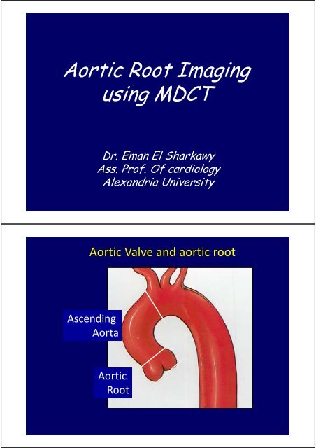
1.Annulus2. Bisinal diameterNormal aortic root3. Sino‐tubular junction4. Ascending aortaNormal Size of aortic root
<strong>Aortic</strong> valve anatomy
<strong>Aortic</strong> Root aneurysmDiagnose site, localized or diffuseNumber (single or multiple)shape (succular , fusiform )involvement of great vessels, coronary arteriesInvolvement of aortic valve , sinotubular junctioncomplicationsAneurysm of the aorta is defined as dilation ofthe aorta involving all three wall layersequaling or exceeding twice the normaldiameter.Pseudoaneurysms (false aneurysms) representsaccular dilations that do not contain aorticintima
<strong>Aortic</strong> aneurysm62Y old female had Bicuspid aortic valve and severe AIreferred for coronary angiography before surgery Failed
62Y old female had Bicuspid aortic valve and severe AIreferred for coronary angiography before surgery Failed62Y old female had Bicuspid aortic valve and severe AIreferred for coronary angiography before surgery Failed
<strong>Aortic</strong> <strong>Dissection</strong>The goals of MDCT imaging are to:1. identify the intimal flap and entry site2. identify any branch vessel involvement3. identify the presence of pericardial fluid(intrapericardial rupture) or periaortic hematoma4. identify extent of dissection5. evaluate size of the aorta6. evaluate the patency of the false lumen and degree oftrue lumen compression
<strong>Aortic</strong> <strong>Dissection</strong>
<strong>Aortic</strong> <strong>Dissection</strong><strong>Aortic</strong> <strong>Dissection</strong>Stanford type A
<strong>Aortic</strong> <strong>Dissection</strong><strong>Aortic</strong> <strong>Dissection</strong>
<strong>Aortic</strong> <strong>Dissection</strong><strong>Aortic</strong> <strong>Dissection</strong>
<strong>Aortic</strong> <strong>Dissection</strong><strong>Aortic</strong> valvenot an indication of MDCT but obtained during CT aortography* Number of cusps* annular diameter* Calcification* Vegetations* Valvular, Supra valvular or subvalvularstenosis (AVA by planimetry)
<strong>Aortic</strong> valveBicuspid aortic valve
Degenerative aortic stenosisSupravalvular aortic stenosis
Supravalvular aortic stenosis20Y male, Dyslipidemia, chest pain
Prosthetic aortic valve
Prosthetic aortic valveCoronary arteries
Marfan’s Syndrome‐<strong>Aortic</strong> root dilatation‐Sinotubular junctioneffacement‐Loss of leaflet cooptation‐Dilated LV
Annuloaortic ectasia without valvular insufficiency in a 20-year-old man.Sinus of Valsalva aneurysm
Sinus of valsalva aneurysmRuptured sinus of valsalva
<strong>Aortic</strong> root abscess* diagnose site, size,* Opening into the aortic root* Relation to aortic valve and coronary arteries* Relation to surrounding structures* Complications 9leak , rupture, effusion….52 Y old M, presented with IE onBicuspid aortic valve
52 Y old M, presented with IE onBicuspid aortic valve
52 Y old M, presented with IE onBicuspid aortic valve42y old female 2 months after open Mitralcommissurotomy
42y old female 2 months after openMitral commissurotomy
Diagnosis of rare congenital abnormalities30 Y old F with Turner’s S. Bicuspid aortic valve and turbulencein LVOT and aortic root by Echocardiography30 Y old F with Turner’s S. Bicuspid aortic valve and turbulencein LVOT and aortic root by Echocardiography
Virtual endoscopic views* can enable a more accurate presurgicalevaluation of the aortic valve* <strong>Aortic</strong> wall composition, and calcification* Prosthetic valve mobility* Diagnose coronary ostia esp. with ostial lesionor calcificationVirtual endoscopic views
Virtual endoscopic viewsCalcific aortic valveVirtual endoscopic viewsaortic root dissection flap
Virtual endoscopic viewsprosthetic aortic valve
Virtual endoscopic views<strong>Aortic</strong> valve vegetationFly Through Technique
Take Home MessageMDCT is an accurate , rapid and non‐invasiveidiagnostic modality for diseases of the aortic root.Multi‐modality imaging helps to delineate bothanatomical and functional abnormalities in aorticroot diseases.MDCT is Mandatory for Pre‐operative evaluationbefore acute aortic syndromes, aortic rootabscess, undiagnosed fistulas tracts, or suspectedcomplicationsThank YouDr. Eman El Sharkawy