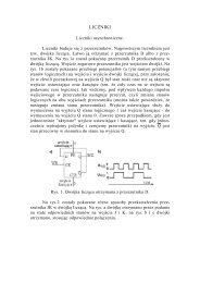
Lecture 7
Lecture 7
Lecture 7

Create successful ePaper yourself
Turn your PDF publications into a flip-book with our unique Google optimized e-Paper software.
Cell Membrane and Transport
Functions of membranes
All of the lipid molecules in cell membranes areAMPHIPHILIC (AMPHIPATHIC)-that is, they have HYDROPHILIC (”water-loving”) orPOLAR end and a HYDROPHOBIC (”water-fearing”) orNONPOLAR end.There are three major classes of membrane lipids –PHOSPHOLIPIDS, CHOLESTEROL, and GLYCOLIPIDSThe lipid compositions of the inner and outer monolayersare different, reflecting the different functions of the twofaces of a cell membrane.
Membrane lipids
Phospholipids in waterPhospholipids associate as bilayers in water because they are roughly cylindricalamphipathic molecules. The hydrophobic fatty acid chains are not exposed towater, whereas the hydrophilic phospholipid heads are in contact with water.
Eukaryotic plasma membranes can contain large amounts of cholesterol— as many as one molecule for every phospholipid molecule.
Asymmetric distribution of phospholipids between the inner and outer monolayersof the erythrocyte plasma membrane.
Diffusion of lipids within a bilayer. (a) Lateral diffusion of lipids is relativelyrapid. (b) Transverse diffusion, or flip-flop, of lipids is very slow.
Membrane fluidityThe ordered arrangement of phospholipid molecules makes the cellmembrane a liquid crystal. The hydrocarbon chains are in constantmotion, allowing each molecule to move laterally on the same side ofthe bilayer.
FLUID MOSAIC MODEL OF PLASMA MEMBRANEThe plasma membrane is described to be fluid because of its hydrophobic integralcomponents such as lipids and membrane proteins that move laterally or sidewaysthroughout the membrane. That means the membrane is not solid, but more like a'fluid'.The membrane is depicted as mosaic because like a mosaic that is made up ofmany different parts the plasma membrane is composed of different kinds ofmacromolecules, such as integral proteins, peripheral proteins, glycoproteins,phospholipids, glycolipids, and in some cases cholesterol, lipoproteins.-Lipids have rapid lateral movement-Lipids flip-flop extremely slowly-Lipids are asymmetrically distributed in membrane (different lipids in each side ofbilayer)-Fluidity depends on lipid compositionSaturated fatty acids - Make membrane less fluid; Solid at room temperatureUnsaturated fatty acids - Make membrane more fluid; Liquid at room temperatureCholesterol - Reduces membrane fluidity by reducing phospholipid movementLipids differ in the tail length - the longer the tail, the less fluid the membrane
Diagram of how various classes of proteins associate with the lipid bilayer.
Membrane proteinsThree transmembrane proteins are shown in (a), (b), and (c). The rolled-up β-pleated sheetshown in (c) forms a pore through the membrane. Water molecules and ions pass throughspecific types of pores. (d, e) Peripheral proteins are bound to integral proteins bynoncovalent interactions.
A selectively permeablemembrane. An artificialmembrane composedpurely of phospholipidswould be permeable tosome substances butwould repel others.
Movement of solutes across a permeable membrane
Process of diffusion.Diffusion is spontaneous, and no chemical energy is required to bring it about. a.When dye crystals are placed in water, they are concentrated in one area.b. The dye dissolves in the water, and there is a net movement of dye moleculesfrom higher to lower concentration. There is also a net movement of watermolecules from a higher to a lower concentration. c. Eventually, the water and thedye molecules are equally distributed throughout the container.
During osmosis, water diffuses across a selectively permeable membrane.Notice that the number of sugar molecules did not change on each sideof the membrane, but the number of water molecules on either side ofthe membrane did change.
In an isotonic solution,water molecules moveinto and out of the cell atthe same rate, and cellsretain their normal shape
In a hypotonic solution,water enters a cell byosmosis, causing the cellto swell
In a hypertonic solution,water leaves a cell byosmosis, causing the cellto shrink
macromolecules small organic molecules small inorganic ionsA cell that does nothing to control itsosmolarity will have a higher concentrationof solutes inside than outside. As a result,water will be higher in concentrationoutside the cell than inside. This differencein water concentration across the plasmamembrane will cause water to movecontinuously into the cell by osmosis.
Animal cells and bacteria control their intracellular osmolarity by activelypumping out inorganic ions, such as Na+, so that their cytoplasm containsa lower total concentration of inorganic ions than the extracellular fluid,thereby compensating for their excess of organic solutes.
Generation of a transmembrane electric potential (voltage) depends on theselective movement of ions across a semipermeable membrane
The K+ Channel
The gating of ion channels
A Gated Channel Protein Opens in Response to a Stimulus The membraneprotein changes its three-dimensional shape when a stimulus molecule binds to it.
Voltage-gated Na + channel
Active transport
Transport processes
Three Types of Proteins for Active TransportNote that in each of the three cases transport is directional.Transporters can be classified with regard to the direction of movement andwhether one or more unique molecules are moved.
The carrier protein allows glucose to enter the cell at a faster rate than would bepossible by simple diffusion across the membrane barrier.
The four classes of ATP-powered transport proteins.
ATP is a carrier of energy in cells. It is the common energy currencybecause it supplies energy for many different types of reactions.
Primary and secondary active transport rely ondifferent energy sourcesThere are two basic types of active transport processes:Primary active transport requires the direct participationof the energy-rich molecule ATP.Secondary active transport does not use ATP directly;rather, its energy is supplied by an ion concentrationgradientestablished by primary active transport.
The sodium–potassium pump is a carrier protein that maintains an electrochemicalgradient across the plasma membrane.
Primary Active Transport:The Sodium– Potassium PumpIn active transport, energy is used to move a solute against its concentrationgradient. Even though the Na+ concentration is higher outside the cell and the K+concentration is higher inside the cell, for each molecule of ATP used, two K+ arepumped into the cell and three Na+ are pumped out of the cell.
Sarcoplasmic Ca 2+ pump
Eukaryotic cells mantain very low concentration offree Ca +2 in their cytosol in the face of a very muchhigher extracellular Ca +2 concentration.The best understood P-type transport Atp-ase is theCa +2 pump in the sarcoplasmic reticulum (SR)membrane of skeletal muscle cells.The SR is a specialized type of endoplasmicreticulum found in the muscle cells and serves as anintracellular store of Ca +2 .
Secondary Active Transport The Na+ concentration gradient established byprimary active transport (right) powers the secondary active transport of glucose(left).The movement of glucose across the membrane against its concentrationgradient is coupled by a symport protein to the movement of Na+ into the cell.
A carrier protein transports sodium ions down their concentration gradient anduses that energy to cotransport glucose molecules against their concentrationgradient.Note that this carrier protein is a symporter.
PhagocytosisIn this type of endocytosis, a cellingests relatively large solidparticles.The white blood cell (a neutrophil)shown is phagocytizingbacteria. The vacuoles containbacteria that have already beeningested. Lysosomes containdigestive enzymes that breakdown the ingested material. Otherbacteria are visible outside thecell.
Receptor-mediated endocytosis. Cells that undergo receptor-mediated endocytosishave pits coated with the protein clathrin that initiate endocytosis when target moleculesbind to receptor proteins in the plasma membrane. A coated pit appears in the plasmamembrane of a developing egg cell, covered with a layer of proteins. When anappropriate collection of molecules gathers in the coated pit, the pit deepens and sealsoff to form a coated vesicle, which carries the molecules into the cell.
Exocytosis (a) Proteins and other molecules are secreted from cells in smallpackets called vesicles, whose membranes fuse with the plasma membrane,releasing their contents to the cell surface. (b) A transmission electron micrographshowing exocytosis.
ExocytosisIn exocytosis, a cell ejects waste products,or specific products of secretion such ashormones, by the fusion of a vesicle withthe plasma membrane
Chapter 7Membrane Structure and FunctionChapter 48Neurons, Synapses, and SignallingCONCEPT 48.2Ion pumps and ion channels maintain the restingpotential of a neuronCONCEPT 48.3Action potentials are the signals conducted by axons