74Michał Szpinda, Marcin DaroszewskiINTRODUCTIONQuantitative studies on the fetal aorta have been focusedpreviously on its diameter only, using echocardiographic[1-3] <strong>and</strong> autopsy [2, 4-7] techniques.Until now, there has been no information concerningboth the volume of the different aortic segments inhuman fetuses <strong>and</strong> their proportions. Gielecki et al. [8]suggested that volume of the aortic arch increasedaccording to the square root function as fetal age advanced.The present study was undertaken to clarify the increasein volume of the various aortic segments inhuman fetuses. Our objectives were to investigate:- the reference ranges of the volume for variousaortic segments at varying gestational ages,- the growth curves for the volume of various aorticsegments vs. fetal age,- the relationships between the volumes of the ascendingaorta, aortic arch <strong>and</strong> thoracic aorta,- the influence of sex on the value of the volumesstudied.MATERIAL AND METHODSThe examinations were carried out on 128 spontaneouslyaborted human fetuses of both genders (63males, 65 females) whose age varied from 15 to 34weeks (Table I).Table I. Age <strong>and</strong> number of fetuses studiedFetal age Crown-rump length (CRL) Number Sexmonths weeks(mm)male female(Hbdlife)mean SD min. max.4 15 89.4 6.1 85.0 92.0 10 5 516 103.7 6.1 95.0 106.0 7 3 417 114.9 8.2 111.0 121.0 6 4 218 129.3 6.6 124.0 134.0 8 3 55 19 142.7 7.7 139.0 148.0 6 3 320 155.3 5.8 153.0 161.0 4 1 321 167.1 4.7 165.0 173.0 3 2 122 178.1 6.9 176.0 186.0 7 4 36 23 192.3 6.3 187.0 196.0 9 4 524 202.9 5.7 199.0 207.0 11 6 525 215.2 4.8 211.0 218.0 7 5 226 224.7 5.2 220.0 227.0 7 4 37 27 234.1 4.3 231.0 237.0 4 0 428 244.2 5.1 240.0 246.0 5 2 329 253.8 4.5 249.0 255.0 6 1 530 262.7 3.1 260.0 264.0 6 5 18 31 270.7 5.2 268.0 275.0 4 1 332 281.4 3.7 279.0 284.0 5 4 19 33 290.3 6.1 286.0 293.0 9 4 534 301.4 3.2 296.0 302.0 4 2 2Total 128 63 65The crown-rump length (CRL) measurements weretaken as the basis for determining gestational age,according to Iffy et al. [9]. The study was approved bythe research ethics committee of the Nicolaus CopernicusUniversity (KB/217/2006). Specimens that haddetectable visible malformations were excluded fromthe study. Fetuses were divided into 6 monthly cohorts,from 4 th to 9 th month of gestation.The fetal arteries were injected with white latexLBS 3060 through the abdominal aorta, under controlledpressure of 50-60 mm Hg, using a syringe infusionpump SEP 11S (Ascor S.A. Medical Equipment,Warsaw 2001). The fetuses were immersed in a 10%neutral formalin solution for 4-24 months, <strong>and</strong> thendissected under a stereoscope with Huygens ocular ata magnification of 10. In each specimen, the dissectedascending aorta, aortic arch <strong>and</strong> thoracic aorta with themillimeter scale were placed perpendicular to the opticallens axis, recorded <strong>and</strong> digitalized to JPEG images.Next, digital pictures of the fetal aorta were analyzedby the digital-image analysis system of Leica Q WinPro 16 (Cambridge). Automatic volume measurementsof the various aortic segments were derived by assumingthat the filled aorta can be divided into small irregularcylinders of varying diameter <strong>and</strong> varyingheight. The sum of volume of such cylinders approximatingthe vessel was given in mm 3 as the volume ofthe different parts of the aorta was.For each fetus, the three following volume measurementswere performed:1. the volume of the ascending aorta - from its origin(at the level of the aortic valve annulus) to its ending(just proximal to the brachiocephalic trunk origin),2. the volume of the aortic arch - from its origin (justproximal to the brachiocephalic trunk origin) to itsending (just proximal to the entry of the ductus arteriosus),3. the volume of the thoracic aorta - from its origin(just proximal to the entry of the ductus arteriosus)to its ending (at the level of the diaphragm).The results obtained were analyzed by one wayANOVA test for unpaired data <strong>and</strong> then post-hoc intergroupcomparisons were performed using RIRTukey test.Regression analysis was used:- to derive the line of best fit for the plot for eachvolume examined against gestational age,- to calculate the relationships between volumes ofthe various aortic segments.
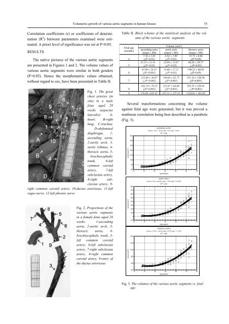
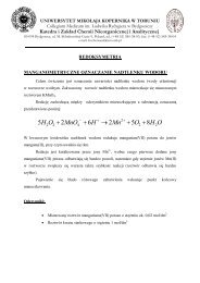
Volumetric growth of various aortic segments in human fetuses 75Correlation coefficients (r) or coefficients of determination(R 2 ) between parameters examined were estimated.A priori level of significance was set at P0.05). Hence the morphometric values obtained,without regard to sex, have been presented in Table II.Fig. 1. The greatchest arteries (insitu) in a malefetus aged 28weeks (aspectuslateralis): A-heart, B-rightlung, C-trachea,D-abdominaldiaphragm, 1-ascending aorta,2-aortic arch, 3-aortic isthmus, 4-thoracic aorta, 5-brachiocephalictrunk, 6-leftcommon carotidartery, 7-leftsubclavian artery,8-right subclavianartery, 9-right common carotid artery, 10-ductus arteriosus, 11-leftvagus nerve, 12-left phrenic nerveTable II. Block scheme of the statistical analysis of the volumeof the various aortic segmentsFetal age(months)45678ascending aorta(mean ± SD)7.56 ± 2.65↓(P>0.01)23.13 ± 11.41↓(P
- Page 8:
8Wojciech J. Baranowskirating) move
- Page 13 and 14:
The benefits resulting from introdu
- Page 15 and 16:
The benefits resulting from introdu
- Page 17 and 18:
Medical and Biological Sciences, 20
- Page 19 and 20:
The history and the present of hern
- Page 21 and 22:
The history and the present of hern
- Page 23: The history and the present of hern
- Page 26: 26Anna Budzyńska et al.Gram-dodatn
- Page 32 and 33: 32Piotr Kamiński et al.Streszczeni
- Page 34 and 35: 34Piotr Kamiński et al.both in Pom
- Page 36 and 37: 36Piotr Kamiński et al.hemoglobin
- Page 39 and 40: Medical and Biological Sciences, 20
- Page 41 and 42: Impact of mandatory vaccination pro
- Page 43 and 44: Medical and Biological Sciences, 20
- Page 45 and 46: Bone turnover during pregnancy 45ra
- Page 47: Bone turnover during pregnancy 47pl
- Page 50 and 51: 50Jan Styczyński, Anna Jaworska(p
- Page 52 and 53: 52Jan Styczyński, Anna JaworskaRES
- Page 55 and 56: Medical and Biological Sciences, 20
- Page 57 and 58: Analysis of immunophenotype at seco
- Page 59: Analysis of immunophenotype at seco
- Page 62 and 63: 62Ana-Maria ŠimundićINTRODUCTIOND
- Page 64 and 65: 64Ana-Maria ŠimundićThe shape of
- Page 67 and 68: Medical and Biological Sciences, 20
- Page 69 and 70: Quantitative anatomy of the aortic
- Page 71 and 72: Quantitative anatomy of the aortic
- Page 73: Medical and Biological Sciences, 20
- Page 77 and 78: Volumetric growth of various aortic
- Page 79 and 80: Medical and Biological Sciences, 20
- Page 81 and 82: Effect of Low Level Laser Therapy a
- Page 83 and 84: Effect of Low Level Laser Therapy a
- Page 85 and 86: Medical and Biological Sciences, 20
- Page 87 and 88: Body weight support during treadmil
- Page 89 and 90: Body weight support during treadmil
- Page 91 and 92: Medical and Biological Sciences, 20