NanoDrop 1000 Spectrophotometer V3.7 User's Manual
NanoDrop 1000 Spectrophotometer V3.7 User's Manual
NanoDrop 1000 Spectrophotometer V3.7 User's Manual

Create successful ePaper yourself
Turn your PDF publications into a flip-book with our unique Google optimized e-Paper software.
The information in this publication is provided for reference only. All information contained in this publication isbelieved to be correct and complete. Thermo Fisher Scientific shall not be liable for errors contained herein norfor incidental or consequential damages in connection with the furnishing, performance or use of this material.All product specifications, as well as the information contained in this publication, are subject to change withoutnotice.This publication may contain or reference information and products protected by copyrights or patents anddoes not convey any license under our patent rights, nor the rights of others. We do not assume any liabilityarising out of any infringements of patents or other rights of third parties.We make no warranty of any kind with regard to this material, including but not limited to the implied warrantiesof merchantability and fitness for a particular purpose. Customers are ultimately responsible for validation oftheir systems.© 2008 Thermo Fisher Scientific Inc. All rights reserved. No part of this publication may be stored in a retrievalsystem, transmitted, or reproduced in any way, including but not limited to photocopy, photograph, magnetic orother record, without our prior written permission.For Technical Support, please contact:Thermo Fisher Scientific3411 Silverside RoadBancroft Building, Suite 100Wilmington, DE 19810 U.S.A.Telephone: 302-479-7707Fax: 302-792-7155E-mail: info@nanodrop.comwww.nanodrop.comFor International Support, please contact your local distributor.Microsoft, Windows, Windows NT and Excel are either trademarks or registered trademarks of MicrosoftCorporation in the United States and/or other countries.Adobe and Acrobat are trademarks of Adobe Systems, Incorporated. All other trademarks are the property ofThermo Fisher Scientific Inc. and its subsidiaries.<strong>NanoDrop</strong> is a trademark of Thermo Fisher Scientific.Revised 7/08
Table of Contents1. Overview....................................................................................... 1-1Instrument Description .................................................................. 1-1Operation....................................................................................... 1-1Applications ................................................................................... 1-1Patents .......................................................................................... 1-22. Initial Set Up................................................................................. 2-1Computer Requirements ............................................................... 2-1Software Installation ...................................................................... 2-1Software Upgrades........................................................................ 2-2Registering Your Instrument.......................................................... 2-33. General Operation ....................................................................... 3-1The Sample Retention System...................................................... 3-1Cleaning the Sample Retention System........................................ 3-2Software Architecture and Features.............................................. 3-4User Preferences........................................................................... 3-6Utilities and Diagnostics ................................................................ 3-7Account Management ................................................................... 3-8Dye/Chromophore Editor............................................................. 3-104. Common Module Functions ....................................................... 4-1Module Startup .............................................................................. 4-1Common Functions ....................................................................... 4-1Measure (F1) ................................................................................. 4-1Blank (F3) ...................................................................................... 4-1Re-blank (F2)................................................................................. 4-2Print Screen (F4) ........................................................................... 4-3Start Report / Recording................................................................ 4-3Print Report (F5)............................................................................ 4-4Show Report (F7) .......................................................................... 4-4Sample ID...................................................................................... 4-4Sample # ....................................................................................... 4-5Exit................................................................................................. 4-5Escape Key (ESC)......................................................................... 4-5Show Context Help (Ctrl+H).......................................................... 4-5User’s <strong>Manual</strong>................................................................................ 4-55. Nucleic Acids ............................................................................... 5-1Sample Volume Requirements...................................................... 5-1Measurement Concentration Range ............................................. 5-1Spectrum Normalization ................................................................ 5-3Spectrum Overlay Control ............................................................. 5-36. MicroArray.................................................................................... 6-1Fluorescent Dye Selection ............................................................ 6-1Sample Volume Requirements...................................................... 6-1Measurement Concentration Range ............................................. 6-2Baseline Calculation & Normalization ........................................... 6-27. UV-VIS........................................................................................... 7-1Sample Volume Requirements...................................................... 7-1Measurement Concentration Range ............................................. 7-1Unique Screen Features ............................................................... 7-18. Protein A280................................................................................. 8-1Sample Volume Requirements...................................................... 8-1Pedestal Reconditioning................................................................ 8-1Measurement Concentration Range ............................................. 8-1Unique Screen Features ............................................................... 8-2Spectrum Normalization ................................................................ 8-3
Spectrum Overlay Control ............................................................. 8-49. Proteins & Labels ........................................................................ 9-1Fluorescent Dye Selection ............................................................ 9-1Sample Volume Requirements...................................................... 9-1Pedestal Reconditioning................................................................ 9-2Measurement Concentration Range ............................................. 9-2Unique Screen Features ............................................................... 9-2Baseline Type................................................................................ 9-410. Protein BCA ............................................................................... 10-1Sample Volume Requirements.................................................... 10-1Pedestal Reconditioning.............................................................. 10-1Measurement Concentration Range ........................................... 10-1BCA Kits, Protocols, and Sample Preparation ............................ 10-2Unique Screen Features ............................................................. 10-2Making BCA Measurements........................................................ 10-3Standard Curve Features ............................................................ 10-5Delete Standard Points ............................................................... 10-5Exiting the BCA Module .............................................................. 10-611. Protein Lowry............................................................................. 11-1Sample Volume Requirements.................................................... 11-1Pedestal Reconditioning.............................................................. 11-1Measurement Concentration Range ........................................... 11-1Modified Lowry Kits, Protocols, and Sample Preparation ........... 11-2Unique Screen Features ............................................................. 11-2Making Lowry Measurements ..................................................... 11-3Standard Curve Features ............................................................ 11-5Delete Standard Points ............................................................... 11-5Exiting the Lowry Module ............................................................ 11-612. Protein Bradford........................................................................ 12-1Sample Volume Requirement ..................................................... 12-1Pedestal Reconditioning.............................................................. 12-1Measurement Concentration Range ........................................... 12-2Bradford Kits, Protocols, and Sample Preparation...................... 12-2Unique Screen Features ............................................................. 12-3Making Bradford Protein Measurements..................................... 12-3Standard Curve Features ............................................................ 12-5Delete Standard Points ............................................................... 12-6Exiting the Bradford Module ........................................................ 12-713. Protein Pierce 660 nm............................................................... 13-1Unique Screen Features ............................................................. 13-1Making Pierce 660 nm Protein Measurements ........................... 13-2Standard Curve Features ............................................................ 13-4Delete Standard Points ............................................................... 13-514. Cell Cultures .............................................................................. 14-1Sample Size Requirements......................................................... 14-2Cell Suspension Concentrations ................................................. 14-2Sample Homogeneity .................................................................. 14-2Decontamination of Measurement Pedestals ............................. 14-215. Archived Data and Data Viewer ............................................... 15-1Archive File Creation ................................................................... 15-1Data Storage Hierarchy............................................................... 15-2Data Viewer ................................................................................. 15-2Archive File Converter................................................................. 15-9Opening Archived Data with Spreadsheet Programs................ 15-1016. Calibration Check...................................................................... 16-1Procedure .................................................................................... 16-117. Troubleshooting ........................................................................ 17-1
Error USB2000 ............................................................................ 17-1Connection Error ......................................................................... 17-2Signal Error.................................................................................. 17-4Saturated Detector ...................................................................... 17-5Liquid Column Breakage ............................................................. 17-6Other Software Error Messages.................................................. 17-8Sample Accuracy and Reproducibility....................................... 17-10260/280 Ratio ............................................................................ 17-11Unusual Spectrum..................................................................... 17-13Technical Support...................................................................... 17-1318. Maintenance and Warranty....................................................... 18-1Cleaning ...................................................................................... 18-1Calibration ................................................................................... 18-1Warranty ...................................................................................... 18-219. Appendices ................................................................................ 19-1Instrument Specifications ............................................................ 19-1Blanking and Absorbance Calculations....................................... 19-1Concentration Calculation (Beer’s Law)...................................... 19-2Solvent Compatibility................................................................... 19-3Decontamination of Measurement & Optical Surfaces ............... 19-3Setting Up a Dymo 400 Label Writer Printer ............................... 19-3
Section 1- Overview1. OverviewInstrument DescriptionThe Thermo Scientific <strong>NanoDrop</strong> <strong>1000</strong> <strong>Spectrophotometer</strong>measures 1 ul samples with high accuracy and reproducibility. The fullspectrum (220nm-750nm) spectrophotometer utilizes a patentedsample retention technology that employs surface tension alone tohold the sample in place. This eliminates the need for cumbersomecuvettes and other sample containment devices and allows for cleanup in seconds. In addition, the <strong>NanoDrop</strong> <strong>1000</strong> <strong>Spectrophotometer</strong>has the capability to measure highly concentrated samples withoutdilution (50X higher concentration than the samples measured by astandard cuvette spectrophotometer).OperationA 1 ul sample is pipetted onto the end of a fiber optic cable (thereceiving fiber). A second fiber optic cable (the source fiber) is thenbrought into contact with the liquid sample causing the liquid to bridgethe gap between the fiber optic ends. The gap is controlled to both1mm and 0.2 mm paths. A pulsed xenon flash lamp provides the lightsource and a spectrometer utilizing a linear CCD array is used toanalyze the light after passing through the sample. The instrument iscontrolled by PC based software, and the data is logged in an archivefile on the PC.ApplicationsUV/VIS spectrophotometry is simple for samples as small as 1 ulusing the <strong>NanoDrop</strong> <strong>1000</strong> <strong>Spectrophotometer</strong>. The small samplerequirement and ease of use make the <strong>NanoDrop</strong> <strong>1000</strong><strong>Spectrophotometer</strong> ideally suited for measuring:• Nucleic acid concentration and purity of nucleic acid samples upto 3700 ng/ul (dsDNA) without dilution• Fluorescent dye labeling density of nucleic acid microarraysamples• Purified protein analysis (A280) up to 100 mg/ml (BSA)• Expanded spectrum measurement and quantitation of fluorescentdye labeled proteins, conjugates, and metalloproteins• Bradford Assay analysis of protein• BCA Assay analysis of protein• Lowry Assay analysis of protein• Pierce Protein 660 nm Protein Assay• Cell density measurements1-1
Section 1- Overview• General UV-Vis spectrophotometryPatentsThe sample retention technology used in the <strong>NanoDrop</strong> <strong>1000</strong><strong>Spectrophotometer</strong> is covered under US patents 6,628,382 and6,809,826. Other patents are pending.1-2
Section 2-Initial Set-upswitch or give a visual indication of the operability of the 12V powersupply.Registering Your InstrumentPlease register your product! We periodically update our softwareand add new features free of charge. We would like to keep our userlist updated so that we may alert you to these updates and allinformation supplied is completely confidential. You can register yourinstrument on our website.2-3
Section 3-General Operation3. General OperationThe Sample Retention SystemBasic UseThe main steps for using the sample retention system are listedbelow:1. With the sampling arm open, pipette the sample onto the lowermeasurement pedestal.2. Close the sampling arm andinitiate a spectral measurementusing the operating software on thePC. The sample column isautomatically drawn between theupper and lower measurementpedestals and the spectralmeasurement made.3-1
Section 3-General Operation3. When the measurement iscomplete, open the samplingarm and wipe the sample fromboth the upper and lowerpedestals using a softlaboratory wipe. Simple wipingprevents sample carryover insuccessive measurements forsamples varying by more than<strong>1000</strong> fold in concentration. Seeour website for performancedata on sample carryover.Cleaning the Sample Retention SystemWiping the sample from both the upper and lower pedestals (asshown above) upon completion of each sample measurement isusually sufficient to prevent sample carryover and avoid residuebuildup. Although generally not necessary, 2 ul water aliquots can beused to clean the measurement surfaces after particularly highconcentration samples to ensure no residual sample is retained oneither pedestal. After measuring a large number of samples, however,it is recommended that the areas around the upper and lowerpedestals be cleaned thoroughly. This will prevent the wiping aftereach measurement from carrying previous samples onto themeasurement pedestals and affecting low-level measurements. Afinal cleaning of all surfaces with de-ionized water is alsorecommended after the user’s last measurement. Note: Please do notuse a squirt bottle to apply de-ionized water.Decontamination of Measurement PedestalsIf decontamination is necessary, a sanitizing solution, such as a 0.5%solution of sodium hypochlorite (1:10 dilution of common commercialbleach solutions – freshly prepared), can be used to ensure that nobiologically active material is present on the measurement pedestals.The metal fiber optic fittings are made from 303 stainless steel andare resistant to most common laboratory solvents (see “SolventCompatibility” appendix). Note: Please do not use a squirt bottle toapply diluted bleach.Pedestal ReconditioningThe Bradford reagent as well as other buffers containing surfactantscan “un-condition” the measurement pedestal surfaces so that theliquid column does not form well with 1ul samples. If this occurs,3-2
Section 3-General Operationspectrophotometry. Genomic DNA, lambda DNA and viscoussolutions of highly concentrated nucleic acids are common examplesknown to the molecular biologist. Proteins are subject to denaturation,precipitation, and aggregation and therefore may require specialhandling to ensure sample homogeneity.Effect of Evaporation and SolventsEvaporation of the sample during the measurement cycle usually hasjust a minimal effect on absorbance readings and may result in a 1-2% increase in sample concentration. This can be observed in thefield by measuring the same sample successively over time. Highlyvolatile solvents, such as hexane, will likely evaporate before themeasurement can be completed. Less volatile solvents such asDMSO can be used successfully.Sample RecoveryOne of the advantages of the sample retention system is that samplescan be recovered from the upper and lower measurement pedestalsby extraction with a pipette.Software Architecture and FeaturesMain MenuWith the sampling arm in the down position, start the operatingsoftware by selecting the following path:Start Programs <strong>NanoDrop</strong> ND-<strong>1000</strong> (version)3-4
Section 3-General OperationApplication ModulesThe operating software has been tailored to meet the life scientist’sneeds. It includes the following application modules:• Nucleic Acid – concentration and purity of nucleic acid• MicroArray – dye incorporation concentration and purity ofnucleic acid• UV-Vis – general UV-Vis measurements• Cell Cultures – “absorbance” (light scattering) measurement ofsuspended microbial cells• Protein A280 – concentration and purity of purified protein• Proteins & Labels – concentration of dye-labeled proteins,conjugates, and metalloproteins3-5
Section 3-General Operation• UV/VisThe default settings for the two cursors used to monitor specificwavelengths are 300 nm for λ1 and 700 nm for λ2. The user mayelect to have the HiAbs on (automatic utilization of the 0.2 mmpath). An additional option is to elect to normalize the data andspectra using the absorbance value of the wavelength between400 nm and 700 with the lowest andabsorbance value.• MicroarrayThe default setting is ssDNA-33 for the nucleic acid. The defaultsettings remain Dye 1 set to Cy3, Dye 2 set to Cy5, withabsorbance normalized to the absorbance value at 750nm.Other options include RNA-40, ssDNA-33 and Other. There areseveral hard-coded dye choices including common Alexa fluordyes. See the explanation of the Dye/Chromophore Editor later inthis section for information about adding custom dyes andchromophores.• A280There are six sample type options available for purified proteinanalysis and concentration measurement. The default setting isOther protein (E1%). See Section 8 for additional information abouteach sample type option. Note: Software versions 3.5.1 and higherinclude the option to select whether or not to have the data andspectrum normalized to the absorbance value at 340 nm.• Proteins and LabelsThere are six sample type options available for purified proteinanalysis and concentration measurement. The default setting isOther protein (E1%).The user may also elect whether or not to use a bichromaticnormalization of the 280 nm absorbance value to the absorbancevalue at 340 nm.The default dye setting remains Dye 1 set to Cy3 with absorbancenormalized at 750nm.Utilities and DiagnosticsThis module is used to both confirm that the instrument is performingwithin the pathlength calibration specifications and help troubleshootoperational problems with the instrument. Note: The calibration checkutility is available in software versions 3.5.1 and higher.3-7
Section 3-General OperationFor more information on using this module, refer to both Section 15(Calibration Check) and Section 16 (Troubleshooting) of this manual.Account ManagementThe Account Management module provides options for directingwhere specific data files are archived by allowing users to segregatetheir data into personal folders. The Account Management module isaccessible to the administrator only.Account TypesThere are three types of user accounts:• Level 10- this is the highest security setting and all level 10 userscan add new users, modify a user, delete a user and set passwordoptions. At the time of software installation, the only level 10account is Administrator whose initial password is “nanodrop”. It isstrongly recommended that the password be changed after initialaccount set up. Any user can be set to a level 10 access, althoughthis is not recommended (see Level 5). Note: The administrator (orthe last level 10 user) account may not be deleted.• Level 5- this is the security setting recommended for an ordinaryuser account. An account with this access will be passwordprotected and will be able to select specific user preferences. Also,all data generated will be automatically archived to the user’saccount in C:\Nanodrop data (and the user specified location if thatpreference is selected).• Default (level 0 security) - this access level is reserved for theDefault account only. This account enables any user without anaccount to access all the active software measurement modules.Although it is not password protected, user preferences can be set3-8
Section 3-General Operationfor this account. All data generated will be automatically archived tothe Default folder within the c:\Nanodrop Data folder. Note: Forlaboratories requiring that every user have a unique user-account,the administrator may disable the default user account.Account Log-in/Log-out and Time OutThe user’s account will remain active until 1) a user logs out of his/heraccount by using the pull down menu to select either Default oranother user name or 2) the user closes the software.A user account may also be logged out automatically if the software“System Idle Timeout” is exceeded. After 4 hours of inactivity thesoftware account will automatically revert back to the Default user. Ascreen will appear indicating that the time is about to expire, with a30-second countdown. If the user elects ‘CANCEL’, the clock withreset and the user account and application module will remain activefor another 4 hours. If the time expires, the open application modulewill close, returning to the Main Menu and the Default user.Account LockoutUser-specific accounts can become locked out in several ways asnoted below:• Failure to change password within the allotted time• Incorrectly entering the password 99 consecutive times• The administrator locks a specific accountOnly the administrator (level 10) can unlock a locked account. This isdone by using the ‘Modify User’ entry in the Account Managementmodule. Note: All accounts (even the administrator) can be locked ifthe incorrect password entry occurs as previously described.Change PasswordThis module enables each user having an authorized account ID tochange their respective password.Note: The administrator, using the ‘Options’ or the ‘Modify User’entries in the ‘Account Management’ module, establishes whetherindividual user passwords will expire and, if so, after how many days.3-9
Section 3-General OperationPasswords.log fileThis file contains the User ID & password for all accounts and isreadable only by the software. It can be found in the c:\nanodropdata\log files folder. It is strongly recommended that the administratormake a copy of that file and store it in the same log files folder asabove each time a new user account is added or a password ischanged. If the administrator’s account becomes locked, the up-todatecopy can be renamed and used as the password.log file.Note: If upgrading from a previous version, the “passwords.log” and“user preferences.log” files should be automatically copied to thec:\<strong>NanoDrop</strong> Data\Log Files directory. If for some reason these filesare not copied automatically, they must be manually copied from theC:\Program files\<strong>NanoDrop</strong> (version) to the C:\Programfiles\<strong>NanoDrop</strong> (version) directory.Dye/Chromophore EditorThe Dye/Chromophore Editor gives the user the ability to add theirown dyes or chromophores in addition to the predefined fluorescentdyes available for use with the MicroArray and Proteins and Labelsmodules. Note 1: Predefined dye methods are indicated by adiamond and can’t be modified. Note 2: Absorbance contribution at260 nm and 280 nm from the respective dye can be corrected byentering the appropriate decimal correction % field. Refer to the dyemanufacturer to find the 260 nm and 280 nm % factor for dyes notpre-defined in the Dye/Chromophore List.3-10
Section 3-General OperationNote: If upgrading from a version prior to 3.3, zero values (0) for 260nm % and 280 nm % correction factors will be entered for all userdefined dyes.3-11
Section 4-Common Module Functions4. Common Module FunctionsModule StartupWhen the software starts, you should see this message:For best results, ensure measurement pedestal surfaces areclean, load a water sample onto the lower measurement pedestal andthen click ‘OK’. The message “Initializing Spectrometer- pleasewait” will appear. When this message disappears, the instrument willbe ready for use. All data taken will automatically be logged in theappropriate archive file.Common FunctionsMeasure (F1)Each time a software module is opened (initiated), the Measurebutton is inactive as noted by its “grayed-out” appearance. A blankmust first be measured before the Measure button will become active.The Measure button is used to initiate the measurement sequence forall samples (non-blanks). It is actuated by depressing the F1 key orclicking the Measure button. The entire measurement cycle takesapproximately 10 seconds.Blank (F3)Before making a sample measurement, a blank must be measuredand stored (see “Blanking and Absorbance Calculations” in theappendix for more details on absorbance calculations). After makingan initial blank measurement, a straight line will appear on the screen;subsequent blanks will clear any sample spectrum and display astraight line as shown in the following image:4-1
Section 4-Common Module FunctionsFor the most consistent results, it is best to begin any measurementsession with a blanking cycle. This will assure the user that theinstrument is working properly and that the pedestal is clean. Followthe steps below to perform a blanking cycle:1. Load a blank sample (the buffer, solvent, or carrier liquid usedwith your samples) onto the lower measurement pedestal andlower the sampling arm into the ‘down’ position.2. Click on the ‘Blank’ (F3) button.3. When the measurement is complete, wipe the blanking bufferfrom both pedestals using a laboratory wipe.4. Analyze an aliquot of the blanking solution as though it were asample. This is done using the ‘Measure’ button (F1). The resultshould be a spectrum with a relatively flat baseline. Wipe theblank from both measurement pedestal surfaces and repeat theprocess until the spectrum is flat.See “Blanking and Absorbance Calculations” in the appendix for moreinformation on blanking and absorbance calculations.Re-blank (F2)The Re-blanking option (F2) establishes a new reference (blank) thatis used for the absorbance calculations of subsequent samples.However, unlike the Blank (F3) function, the Re-blank featurerecalculates the absorbance spectrum for the most recent sample anddisplays this on the screen. When the Re-blank function is used, thefollowing message appears:4-2
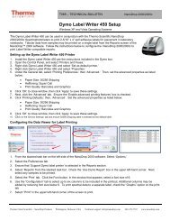
Section 4-Common Module FunctionsPrint Screen (F4)The ‘Print Screen’ button will print a copy of the current operatingscreen to the default printer attached to the operating PC.Note: The system is configured to work with the Dymo Label Writer400 printing on #30256 [2-5/16” X 4”] shipping labels, but can print onany printer connected to the PC.Print WindowA Print dialogue can be initiated from the ‘File’ pull-down menu or bytyping ‘Ctrl+P’. The user can specify any connected printer from thePrint dialogue.Saving Current Screen as .JPG ImageThe current screen can be saved as a .jpg image file by selecting‘Save Window’ from the ‘File’ pull down menu.Start Report / RecordingThe user can log measurement results in a report table and printthem to the desired printer. To initiate this feature, select the ‘StartReport’ button. The default setting has the Recording featureactivated. Refer to Data Viewer in Section 14 for additional details.Note: To override this feature, click on the ‘Recording’ button. Oncede-selected, the button will read Start Report.When the specified maximum number of entries for that specificreport has been reached, there are 4 options: ‘Ignore’, ‘Save’, ‘Print’,‘Save and Print’.4-3
Section 4-Common Module FunctionsAll data is stored in the archive file at C:\<strong>NanoDrop</strong> Data (and in aduplicate location if selected in User Preferences).Note: This feature can be set so that ‘Recording’ is the default mode.See User Preferences in Section 3 for more information.Print Report (F5)Selecting the ‘Print Report’ (F5) button will print the existing samplereport to the default printer. It can be configured to clear the samplereport contents. The user also has options as to how the buffer ishandled. Refer to Data Viewer in Section 14 for additionalinformation. All data is stored in the archive file at C:\<strong>NanoDrop</strong> Dataand in a duplicate location if selected in User Preferences.Note: The system is configured to work with the Dymo Label Writer400 printing on #30256 [2-5/16” X 4”] shipping labels, but can print toany printer connected to the PC.Show Report (F7)The user can display the entries comprising the current SampleReport at any time by selecting the ‘Show Report’ button. Thisfunction will enable the Data Viewer software described in Section 14.Parameters specific for the individual application modules arepopulated for each individual Sample ID.Sample IDThe ‘Sample ID’ is highlighted for overtyping or barcode scanning.The user may input a sample ID that will be used to identify themeasurement in a report print out and in the archived data file. Thesample ID entry is “key focused”, meaning it is the default selection on4-4
Section 4-Common Module Functionsthe screen and should have a flashing text cursor when theinstrument is waiting to make a new measurement.Sample #The ‘Sample #’ indicator is activated when a sample report is beingrecorded. It indicates the sample number of the last sampleprocessed in the current report and increments with each successivemeasurement until the sample report is fully populated. The samplebuffer limit can be modified on the report page.ExitThis command closes all application modules and supporting options.After clicking the ‘Exit’ button, the user has 10 seconds to cancel theexit command. If no action is taken within 10 seconds, the exitcommand is carried out. Note: All measurement data is automaticallysaved to an archive file and requires no user action.Escape Key (ESC)The escape key is set to exit out of all screens. Hitting the escapekey twice will log the user out of an application module.Show Context Help (Ctrl+H)Context Help is enabled in the Main Menu, all function modules, andthe application modules. The help feature is enabled by choosing‘Show Context Help’ from the ‘Help’ menu pull down or by selecting‘Ctrl+H’. Once enabled, placing the cursor on elements of the screenwill automatically generate an explanation of that element. ContextHelp remains active until deselected.User’s <strong>Manual</strong>A .PDF version of this User’s <strong>Manual</strong> is accessible from the MainMenu and from the Help menu in all of the application modules. It canalso be accessed by selecting from the Help pull down menu in anyapplication module or from Start Programs <strong>NanoDrop</strong> ND-<strong>1000</strong> (version).4-5
Section 5- Nucleic Acids5. Nucleic AcidsNucleic acid samples can be readily checked for concentration andquality using the <strong>NanoDrop</strong> <strong>1000</strong> <strong>Spectrophotometer</strong>. To measurenucleic acid samples select the ‘Nucleic Acid’ application module.Sample Volume RequirementsField experience has indicated that 1ul samples are sufficient toensure accurate and reproducible results when measuring aqueousnucleic acid samples. However, if you are unsure about your sampleor your pipettor accuracy, a 1.5-2ul sample is recommended toensure that the liquid sample column is formed and the light path iscompletely covered by sample.Measurement Concentration RangeThe <strong>NanoDrop</strong> <strong>1000</strong> <strong>Spectrophotometer</strong> will accurately measuredsDNA samples up to 3700 ng/ul without dilution. To do this, theinstrument automatically detects the high concentration and utilizesthe 0.2mm pathlength to calculate the absorbance.DetectionLimit(ng/ul)2Approx.Upper Limit(ng/ul)3700 ng/ul(dsDNA)3000 (RNA)2400 (ssDNA)Typical Reproducibility(minimum 5 replicates)(SD= ng/ul; CV= %)sample range 2-100 ng/ul: ± 2ng/ulsample range >100 ng/ul: ± 2%Unique Screen Features5-1
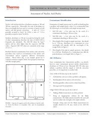
Section 5- Nucleic AcidsSample Type: used to select the (color-keyed) type of nucleic acidbeing measured. The user can select ‘DNA-50’ for dsDNA, ‘RNA-40’for RNA, ‘ssDNA-33’ for single-stranded DNA, or ‘Other’ for othernucleic acids. The default is DNA-50. If ‘Other’ is selected, the usercan select an analysis constant between15-150. When navigatingamongst the three general sample types within the Nucleic Acidsmodule, the last constant value entered within the ‘Constant’ sampletype will be retained. See the “Concentration Calculation (Beer’sLaw)” Appendix for more details on this calculation.λ and Abs: the user selected wavelength and correspondingabsorbance. The wavelength can be selected by moving the cursoror using the up/down arrows to the right of the wavelength box. Note:The user-selected wavelength and absorbance are not utilized in anycalculations.A260 10 mm path: absorbance of the sample at 260 nm representedas if measured with a conventional 10 mm path. Note: This is 10X theabsorbance actually measured using the 1 mm path length and 50Xthe absorbance actually measured using the 0.2 mm path length.A280 10 mm path: sample absorbance at 280 nm represented as ifmeasured with a conventional 10 mm path. Note: This is 10X theabsorbance actually measured using the 1 mm path length and 50Xthe absorbance actually measured using the 0.2 mm path length.260/280: ratio of sample absorbance at 260 and 280 nm. The ratio ofabsorbance at 260 and 280 nm is used to assess the purity of DNAand RNA. A ratio of ~1.8 is generally accepted as “pure” for DNA; aratio of ~2.0 is generally accepted as “pure” for RNA. If the ratio is5-2
Section 5- Nucleic Acidsappreciably lower in either case, it may indicate the presence ofprotein, phenol or other contaminants that absorb strongly at or near280 nm. See “260/280 Ratio” in the Troubleshooting section for moredetails on factors that can affect this ratio.260/230: ratio of sample absorbance at 260 and 230 nm. This is asecondary measure of nucleic acid purity. The 260/230 values for“pure” nucleic acid are often higher than the respective 260/280values. They are commonly in the range of 1.8-2.2. If the ratio isappreciably lower, this may indicate the presence of co-purifiedcontaminants.ng/ul: sample concentration in ng/ul based on absorbance at 260 nmand the selected analysis constant. See the “ConcentrationCalculation (Beer’s Law)” in the appendix for more details on thiscalculation.Spectrum NormalizationThe baseline is automatically set to the absorbance value of thesample at 340 nm, which should be very nearly zero absorbance. Allspectra are referenced off of this zero.Spectrum Overlay ControlThe user can display more than one spectrum in the same displayusing this feature. The current sample plot will be displayed in boldand previous plots will be distinguished by different colors as seen inthe following example:5-3
Section 5- Nucleic AcidsThe default option is set to clear the display for the next reading. Theuser may set the overlay control to clear after each sample plot,(default setting), after each new report, or accumulate plots untilprompted to clear. The ‘Clear Now’ setting will clear all current andprevious plots. When the overlay function is active, the software willauto scale the y-axis based on the sample with the highestabsorbance at 260 nm. Note: When the overlay function is active, the‘Blank’ function does not clear the existing overlaid sample spectra.5-4
Section 6- MicroArray6. MicroArrayThe capability to pre-select viable fluorescent-tagged hybridizationprobes for gene expression in micro arrays can eliminate potentiallyflawed samples and improve research effectiveness. The <strong>NanoDrop</strong><strong>1000</strong> <strong>Spectrophotometer</strong> measures the absorbance of the fluorescentdye, allowing detection at dye concentrations as low as 0.2 picomoleper microliter.Fluorescent Dye SelectionThere are currently nine fluorescent dyes that are hard-coded for usewith the MicroArray module (see table below). Users can also enter &save fluorescent dyes not coded within the <strong>NanoDrop</strong> <strong>1000</strong><strong>Spectrophotometer</strong> software using the ‘Dye/Chromophore Editor’button found in the main menu. Dyes can be selected using the scrollarrows or by highlighting the Dye 1 or Dye 2 box. The respectiveabsorbance wavelength, extinction coefficient, and 260nm and 280nm% corrections will be automatically utilized for measurement andconcentration calculation. The default settings remain Dye 1 set toCy3 and Dye 2 set to Cy5. In addition to the fluorescent dyesavailable from the drop-down menu, an option entitled ‘None’ is alsoavailable. Selecting ‘None’ disables the respective calculations &numeric displays corresponding to that dye.Note: Please refer to the dye manufacturer for the appropriatecorrection factors for user entered dyes.Sample Volume RequirementsField experience has indicated that 1 ul samples are sufficient toensure accurate and reproducible results when measuring aqueous6-1
Section 6- MicroArraynucleic acid samples containing incorporated fluorescent dyes.However, if you are unsure about the surface tension properties ofyour sample or your pipettor accuracy, a 1.5-2 ul sample isrecommended to ensure that the liquid sample column is formed andthe light path is completely covered by sample.Measurement Concentration RangeThe <strong>NanoDrop</strong> <strong>1000</strong> <strong>Spectrophotometer</strong> will accurately measurefluorescent-dye and nucleic acid concentrations up to 100 pmols/ul(Cy3) and 750 ng/ul (DNA) respectively without dilution. A table ofsample concentration ranges is listed below.SampleTypeCy3, Cy3.5,Alexa Fluor 555and Alexa Fluor660Cy5, Cy5.5 andAlexa Fluor 647DetectionLimit(pmol/ul)Approx. UpperLimit (pmol/ul)0.20 <strong>1000</strong>.12 60Alexa Fluor 488and Alexa Fluor594 0.40 215Alexa Fluor 546 0.30 145Typical Reproducibility(minimum 5 replicates)(SD= pmol/ul; CV= %)sample range 0.20-4.0pmol/ul:± 0.20 pmol/ulsample range >4.0 pmol/ul:± 2%sample range 0.12-2.4pmol/ul:± 0.12 pmol/ulsample range >2.4 pmol/ul:± 2%sample range 0.40-8.0pmol/ul:± 0.40 pmol/ulsample range >8.0 pmol/ul:± 2%sample range 0.30-6.0pmol/ul:± 0.30 pmol/ulsample range >6.0 pmol/ul:± 2%Baseline Calculation & NormalizationThe software normalizes the visual spectrum display for all readingsat 750nm and will automatically calculate a baseline between 400 and750 nm for dye concentration calculations. The green vertical line onthe screen represents the peak wavelength position for Dye 1, andthe red vertical line represents the peak wavelength position for Dye2.6-2
Section 6- MicroArrayUnique Screen FeaturesMax Absorbance: used to rescale the upper limit of the vertical axis.Sample Type: used to select the (color-keyed) type of nucleic acidbeing measured. The user can select ‘DNA-50’ for dsDNA, ‘RNA-40’for RNA, ssDNA-33’ for single-stranded DNA, or ‘Other’ for othernucleic acids. The default is ssDNA-33’. If ‘other’ is selected, theuser can select an analysis constant between 15-150. Whennavigating amongst the three (3) general sample types within theMicro Array module, the last value of the constant entered within the‘Constant’ Sample Type will be retained. See “ConcentrationCalculation (Beer’s Law)” in the appendix for more details on thiscalculation.λ and Abs Norm: the user selected wavelength (black cursor) andcorresponding absorbance at the 1mm pathlength. The wavelengthcan be selected by dragging the black cursor or using the up/downarrows in the wavelength box. Note: The user-selected wavelengthand absorbance at the 1 mm pathlength are not utilized in anycalculations.Dye 1 (or 2): user selected dyeAbs. Norm: normalized absorbance of selected Dye at the 1 mmpathlength.pmol/ul: concentration based upon selected Dye’s extinctioncoefficient. See “Concentration Calculation (Beer’s Law)” in theappendix for more details on this calculation.ng/ul: concentration of nucleic acids in the sample calculated usingthe absorbance at 260 nm minus the absorbance at 340 nm (i.e.6-3
Section 6- MicroArraynormalized at 340 nm) and the nucleic acid analysis constant. See“Concentration Calculation (Beer’s Law)” in the appendix for moredetails on this calculation.260/280: ratio of sample absorbance at 260 and 280 nm. The ratio ofabsorbance at 260 and 280 nm is used to assess the purity of DNAand RNA. A ratio of ~1.8 is generally accepted as “pure” for DNA; aratio of ~2.0 is generally accepted as “pure” for RNA. If the ratio isappreciably lower in either case, it may indicate the presence ofprotein, phenol or other contaminants that absorb strongly at or near280 nm. See “260/280 Ratio” in the Troubleshooting section for moredetails on factors that can affect this ratio.6-4
Section 7- UV/Vis7. UV-VISThe ‘UV/VIS Absorbance’ module allows the <strong>NanoDrop</strong> <strong>1000</strong><strong>Spectrophotometer</strong> to function as a conventional spectrophotometer.Sample absorbance is displayed on the screen from 220 nm to 750nm and cursors permit the measurement of individual peaks.Sample Volume RequirementsField experience has indicated that 1 ul samples are sufficient toensure accurate and reproducible results when measuring mostaqueous samples. If you are unsure about your sample compositionor your pipettor accuracy, a 1.5-2 ul sample is recommended toensure that the liquid sample column is formed and the light pathcompletely is covered by sample.Measurement Concentration RangeAll <strong>NanoDrop</strong> <strong>1000</strong> spectrophotometers will measure absorbance upto the 10mm pathlength equivalent of 15 A. <strong>NanoDrop</strong> <strong>1000</strong><strong>Spectrophotometer</strong> instruments with serial numbers >500 or thosethat have been field retrofitted, can utilize the short path length(0.2mm) which enables the 10mm path length equivalent of 75 A.Unique Screen Featuresλ1/Abs1 and λ2/Abs2: current values of the user-selectablewavelength cursors and corresponding absorbencies for a 1 mm7-1
Section 7- UV/Vispathlength. The wavelengths can be set by dragging the cursor,using the up/down arrows or typing in the desired wavelength.Baseline: the absorbance of the user selectable-baseline (horizontal)cursor. The user may drag this cursor to a new vertical position tocreate a new baseline. The absorbance value of the baseline issubtracted from the absorbance of the spectrum.Max Absorbance: used to rescale the upper limit of the vertical axis.Hi Abs: samples with high absorbance (up to 75 A equivalent @ 10mm path) can be measured directly (applicable for instruments withserial numbers >500 only or that have been field-retrofitted). Thiscapability is selected by choosing the ‘Hi Abs’ button on the headerbar. When this is selected, the absorbance is measured using theshort path (0.2mm) and plotted as a red line normalized to a 0.1mmpath for easier visual comparison. Sample ID label will be stored withsample data in the file folder “C:\<strong>NanoDrop</strong> Data\User name\ HiAbs”.This data cannot be imported into the Data Viewer but can be openedwith Excel type spreadsheets. This feature may be selected as thedefault option using the User Preferences module.Normalize: This is a user-selectable feature in this module. Ifselected, the software will automatically normalize the spectrumbased on the lowest absorbance value in the range 400-750 nm. Thisfeature may be selected as the default option using the UserPreferences module.7-2
Section 8- Protein A2808. Protein A280Proteins, unlike nucleic acids, can exhibit considerable diversity. TheA280 method is applicable to purified proteins exhibiting absorbanceat 280nm. It does not require generation of a standard curve and isready for quantitation of protein samples at startup. This moduledisplays the UV spectrum, measures the protein’s absorbance at 280nm (A280) and calculates the concentration (mg/ml). Like the NucleicAcid module, it automatically switches to the 0.2 mm pathlength atvery high concentrations of protein. Also analogous to the NucleicAcid module, the Protein A280 module displays and records 10 mm (1cm) equivalent data on the screen and in the archived data file.Sample Volume RequirementsSome proteins are hydrophobic and others hydrophilic giving rise tovariable surface tension properties in the sample to be measured.Additionally the presence of surfactants or detergents in reagents,such as the Bradford reagent, can significantly alter the surfacetension resulting is difficulty in forming adequate columns formeasurement. The column formation issue can be overcome withoutaffecting the sample’s absorbance by using a larger sample volume.A 2 ul sample size is recommended for protein measurements.Pedestal ReconditioningSolutions and reagents containing surfactants may “un-condition” themeasurement pedestal surfaces so that the liquid column does notform. If this occurs, “buff” the measurement pedestal surfaces byrubbing each measurement surface aggressively with a dry laboratorywipe 30-40 times. This will “re-condition” the surface allowing theliquid sample column to form. Alternatively, use the <strong>NanoDrop</strong>Pedestal Reconditioning Compound (PR-1) as a rapid means ofreconditioning the pedestals when the surface properties have beencompromised and liquid columns break during measurement.Additional information about the PR-1 kit may be found on ourwebsite.Measurement Concentration RangeThe <strong>NanoDrop</strong> <strong>1000</strong> <strong>Spectrophotometer</strong> will accurately measureprotein samples up to 100 mg/ml (BSA) without dilution. To do this,the instrument automatically detects the high concentration andutilizes the 0.2mm pathlength to calculate the absorbance. A table ofconcentration range and typical reproducibility is listed below.8-1
Section 8- Protein A280SampleTypePurifiedBSADetectionLimit0.10mg/mlApprox.UpperLimit100 mg/mlTypical Reproducibility(minimum 5 replicates)(SD= mg/ml; CV= %)sample range 0.10-10 mg/ml: ±0.10 mg/mlsample range >10mg/ml: ± 2%Unique Screen FeaturesSample Type: There are six sample types (options) available forpurified protein analysis and concentration measurement. All of theoptions can be viewed by clicking the mouse while it is positionedwithin the ‘Sample Type’ box. The sample type (color-keyed) can beselected by clicking on the preferred option or by scrolling through theselections using the up or down arrow keys located to the left of thesample type box. A description of each sample type is given below.A general reference setting based on a 0.1%(1 mg/ml) protein solution producing anAbsorbance at 280 nm of 1.0 A (where thepathlength is 10 mm or 1 cm).Bovine Serum Albumin reference. Unknown(sample) protein concentrations are calculatedusing the mass extinction coefficient of 6.7 at280 nm for a 1% (10 mg/ml) BSA solution.IgG reference. Unknown (sample) proteinconcentrations are calculated using the massextinction coefficient of 13.7 at 280 nm for a1% (10 mg/ml) IgG solution.8-2
Section 8- Protein A280Lysozyme reference. Unknown (sample)protein concentrations are calculated using themass extinction coefficient of 26.4 at 280 nmfor a 1% (10 mg/ml) Lysozyme solution.User-entered values for molar extinctioncoefficient (M -1 cm -1 ) and molecular weight(MW) in kilo Daltons for their respectiveprotein reference. Maximum value for e is9999 X <strong>1000</strong> and maximum value for M.W. is99999 X <strong>1000</strong>.User-entered mass extinction coefficient (Lgm -1 cm -1 ) for a 10 mg/ml (1%) solution of therespective reference protein.λ and Abs: current value of the user-selectable wavelength cursorand corresponding absorbance. The wavelength can be set bydragging the cursor, using the up/down arrows or typing in the desiredwavelength. Note: The user-selected wavelength and absorbanceare not utilized in any calculations.A280 10-mm Path: 10 mm-equivalent absorbance at 280 nm for theprotein sample measuredA260/280: ratio of sample absorbance at 260 and 280 nmSpectrum NormalizationThe baseline is automatically set to the absorbance value of thesample at 340 nm, which should be very nearly zero absorbance. Allspectra are referenced off of this zero. All data are now normalizedand archived in the same format. The user may elect to turn off thebaseline normalization, which may result in the spectra being offsetfrom the baseline.8-3
Section 8- Protein A280Note: If the spectra baseline offset is significant, the calculatedprotein concentration may be higher than the true value. Somesamples may exhibit a greater baseline offset than the exampleabove.Spectrum Overlay ControlThe user can display more than one spectrum in the same displayusing this feature. The current sample plot will be displayed in boldand previous plots will be distinguished by different colors as seen inthe following example:The default option is set to clear the display for the next reading. Theuser may set the overlay control to clear after each sample plot8-4
Section 8- Protein A280(default setting), after each new report, or accumulate plots untilprompted to clear. The ‘Clear Now’ setting will clear all current andprevious plots. When the overlay function is active, the softwaremodule will auto scale the y-axis based on the sample with thehighest absorbance at 280nm. Note: When the overlay function isactive, the ‘Blank’ function does not clear the existing overlaid samplespectra.8-5
Section 9- Proteins & Labels9. Proteins & LabelsThis software module can be used to determine protein concentration(A280nm) as well as fluorescent dye concentration (protein arrayconjugates), or to measure the purity of metalloproteins (such ashemoglobin) using wavelength ratios.Fluorescent Dye SelectionThere are currently nine fluorescent dyes that are hard-coded for usewith the Proteins and Labels module (see table below). Users canalso enter & save fluorescent dyes not coded within the <strong>NanoDrop</strong><strong>1000</strong> <strong>Spectrophotometer</strong> software using the ‘Dye/ChromophoreEditor’ button found in the main menu.Dyes can be selected using the scroll arrows or by highlighting theDye 1 or Dye 2 box. The respective absorbance wavelength,extinction coefficient, and 280nm % corrections will be automaticallyutilized for measurement and concentration calculation. The defaultsettings remain Dye 1 set to Cy3. In addition to the fluorescent dyesavailable from the drop-down menu, an option entitled ‘None’ is alsoavailable. Selecting ‘None’ disables the respective calculations &numeric displays corresponding to that dye.Note: Please refer to the dye manufacturer for the appropriatecorrection factors for user entered dyes.Sample Volume RequirementsSome proteins are hydrophobic and others hydrophilic giving rise tovariable surface tension in the samples to be measured. Additionally9-1
Section 9- Proteins & Labelsthe presence of surfactants or detergents in reagents, such as theBradford reagent, can significantly alter surface tension. Thisoccurrence can be overcome without affecting the sample’sabsorbance by using a larger sample volume. A 2 ul sample size isrecommended for protein measurements.Pedestal ReconditioningSolutions and reagents containing surfactants may “un-condition” themeasurement pedestal surfaces so that the liquid column does notform. If this occurs, “buff” the measurement pedestal surfaces byrubbing each measurement surface aggressively with a dry laboratorywipe 30-40 times. This will “re-condition” the surface allowing theliquid sample column to form. Alternatively, use the <strong>NanoDrop</strong>Pedestal Reconditioning Compound (PR-1) as a rapid means ofreconditioning the pedestals when the surface properties have beencompromised and liquid columns break during measurement.Additional information about the PR-1 kit may be found on ourwebsite.Measurement Concentration RangeThe <strong>NanoDrop</strong> <strong>1000</strong> <strong>Spectrophotometer</strong> will accurately measureprotein samples up to 20 mg/ml (BSA) without dilution. A table ofconcentration range and typical reproducibility is listed below.SampleTypePurifiedBSACy3DetectionLimit0.10mg/ml0.2uMApprox.UpperLimit20mg/ml100uMTypical Reproducibility(minimum 5 replicates)(SD= mg/ml; CV= %)sample range 0.10-10 mg/ml: ± 0.10mg/mlsample range >10mg/ml: ± 2%sample range 0.20-4.0 pmol/ul: ±0.20 pmol/ulsample range >4.0 pmol/ul: ± 2%Unique Screen Features9-2
Section 9- Proteins & LabelsMax Absorbance: used to rescale the upper limit of the vertical axis.Sample Type: The same six sample types (options) listed underProtein A280 (Section 8) are available for purified Proteins & Labelsanalysis and concentration measurement. All of the options can beviewed by clicking the mouse while it is positioned within the sampletype box. The sample type (color-keyed) can be selected by clickingon the preferred option or by scrolling through the selections using theup or down arrow keys located to the left of the sample type box. SeeProtein A280 (Section 8) for a detailed description of each sampletype.λ3: user-selected wavelengthNorm Abs @ λ3: 10mm equivalent normalized absorbance at therespective wavelength.Dye 1 (or 2): user-selected dyeNorm Abs @ λ1: normalized 10 mm equivalent absorbance ofselected DyeuM: concentration based upon selected Dye’s extinctioncoefficient. See the “Concentration Calculation (Beer’s Law)” inthe Appendices for more details on this calculation.Abs ratio λ1/λ3: ratio of the absorbance of Dye 1 to the absorbanceat the user-selected wavelength (λ3).mg/ml: concentration of proteins in the sample calculated using theabsorbance at 280 nm minus the absorbance at 340 nm (i.e.9-3
Section 9- Proteins & Labelsnormalized at 340 nm). See the “Concentration Calculation (Beer’sLaw)” Appendices for more details on this calculation.Baseline TypeThis application module has two user-selectable “Baseline Type”options. The default setting is set to normalize the display spectrumat 750nm. Alternatively, the 400-750 Slope Baseline Type willnormalize the display at 750 nm and accommodate any linearbaseline offset across the 400 to 750 nm range.The user may also elect whether or not to use a bichromaticnormalization of the 280 nm absorbance value to the absorbancevalue at 340 nm.9-4
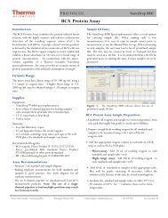
Section 10- Protein BCA10. Protein BCAThe BCA (Bicinchoninic Acid) Protein Assay is an alternative methodfor determining protein concentration. It is often used for more diluteprotein solutions and/or in the presence of components that also havesignificant UV (280 nm) absorbance. Unlike the Protein A280 method,the BCA Assay requires a standard curve to be generated each time itis run, before unknown proteins can be measured. The resulting Cu-BCA chelate formed in the presence of protein is measured at itswavelength maximum of 562 nm and normalized at 750 nm. Preformulatedreagents of BCA and CuSO 4 , utilized in the assay, areavailable in kit form from numerous manufacturers. Follow theirrecommendations when mixing the respective reagents at the timethe assay is to be performed.Sample Volume RequirementsSome proteins are hydrophobic and others hydrophilic giving rise tovariable surface tension properties in the sample to be measured.Additionally the presence of surfactants or detergents in reagents,such as the Bradford reagent, can significantly alter surface tension.This occurrence can be overcome without affecting the sample’sabsorbance by using a larger sample volume. A 2 ul sample size isrecommended for protein measurements.Pedestal ReconditioningSolutions and reagents containing surfactants may “un-condition” themeasurement pedestal surfaces so that the liquid column does notform. If this occurs, “buff” the measurement pedestal surfaces byrubbing each measurement surface aggressively with a dry laboratorywipe 30-40 times. This will “re-condition” the surface allowing theliquid sample column to form. Alternatively, use the <strong>NanoDrop</strong>Pedestal Reconditioning Compound (PR-1) as a rapid means ofreconditioning the pedestals when the surface properties have beencompromised and liquid columns break during measurement.Additional information about the PR-1 kit may be found on ourwebsite.Measurement Concentration RangeWhen using a 20:1 reagent to sample volume dilution, the BCA assayconcentration range of detection is ~0.20 mg/ml to 8.0 mg/ml on the<strong>NanoDrop</strong> <strong>1000</strong>. Using a 1:1 reagent to sample volume dilution theconcentration range of detection is 0.01 mg/ml – 0.20 mg/ml.10-1
AssayTypeApprox.LowerLimitApprox.UpperLimitSection 10- Protein BCATypical Reproducibility(minimum 5 replicates)(SD= mg/ml; CV= %)Regular BCA 0.2 mg/ml 8.0 mg/ml ± 2% (over entire range)Mini BCA 0.01 mg/ml 0.20 mg/ml ± 0.01 mg/ml (over entire range)BCA Kits, Protocols, and Sample PreparationCommercial BCA Protein kit manufacturers typically outlineprocedures for two different protein concentration ranges:• A regular assay – using a 20:1 reagent / sample volume ratio. Toaccurately prepare standards, we suggest using a minimumsample volume of 4 ul in 80 ul of BCA reagent (larger samplevolume is preferable).• A mini assay – using a 1:1 reagent / sample volume ratio. Toprepare sufficient volume of these 1:1 mixtures, we suggest usinga minimum of 10 ul of sample and 10 ul of BCA reagent in a PCRtube. Using the same pipettor for both volumes will eliminate anypipette-to-pipette accuracy differences.Note : If you run the assay at 60°C, doubling the volumes mayafford greater insurance against skewed results from evaporation/ condensation within the sealed reaction tube.Protein standards (BSA) for generating a standard curve may also beprovided by the manufacturer for the BCA assay kit. Follow themanufacturer’s protocol for the assay including recommendedincubation times and temperature. Additionally, use the respectivestandard (e.g. BSA) and dilutions that cover the analytical range(mg/ml) of interest. Note: Since the <strong>NanoDrop</strong> <strong>1000</strong><strong>Spectrophotometer</strong> measures higher protein concentrations thantraditional cuvette-based spectrophotometers, you may need tosupply your own protein standards at higher concentrations thanprovided by the manufacturer.Unique Screen FeaturesView Standard Curve (F8): selecting this button allows the user toview the standard curve at any time.Sample Type: choices are Reference, Standards 1-7, and Sample.The software will guide you to measure your reference, then at leastone standard before allowing measurement of samples. Reference10-2
Section 10- Protein BCAsamples refer to the dye reagent without any protein added (ie the “0”sample).Replicate #: counter for tracking replicate number during referenceand standard measurement.Reset all Standards (F11): clears all replicates of all standards.Reset 1 Standard (F12): clears all replicates of the selectedstandard.Absorbance at 562nm: the Cu-BCA complex’s absorbance at 562nm for the 1mm pathlength.Cursor λ and Absorbance: this feature allows the user to adjust thecursor wavelength and view the corresponding absorbance. Thecursor wavelength can be set by the user. Note: The user-selectedwavelength and absorbance are not utilized in any calculations.mg/ml: the concentration of the sample (unknown).Making BCA MeasurementsA standard curve is required every time the BCA assay is run.Although curves can be saved and reloaded in the <strong>NanoDrop</strong> <strong>1000</strong><strong>Spectrophotometer</strong> software, it is recommended that the user followmanufacturers’ guidelines and generate fresh standard curves foreach assay. Additionally, a standard curve ‘set-up’ may be reloaded.This feature will recall the respective standard series used in apreviously saved standard curve. Both single and multi-pointstandard curve generation is incorporated into the software. Astandard curve can be developed using a reference (BCA reagentonly – no protein) and a single replicate of one standard. The multipointstandard curve generator allows a maximum of five replicatesfor up to seven different standards. There is no set order in whichstandards must be run.A blank must be measured before the standard curve may begenerated. It is advisable to use water as the blank and use the dyereagent without any protein added as the “0” or reference sample.There are three general procedural steps to unknown proteinconcentration measurements. The requisite order, includinggenerating the standard curve, is as follows:10-3
Section 10- Protein BCAStep 1: Measure the‘Reference’ (BCAreagent – a ‘zero’Standard)Note: The softwarewill guide the userwith instructions in thelarge text box on theright side of thescreen.Step 2: MeasureStandardsUp to 5 replicates ofeach of up to 7standards can bemeasured.The software will notallow measurement ofsamples until aminimum of 1standard andreferences – or 2standards – aremeasured.Polynomial curvefitting requires morestandard points.10-4
Section 10- Protein BCAStep 3: MeasureSamplesSampleconcentrations can becalculated by usinglinear interpolation(point-to-point)between the twostandards flanking theunknown sample orby using polynomialfitting. [Note: In orderto obtain aconcentration value(mg/ml) the sample(unknown) must fallwithin the limits of thestandard curve].Standard Curve FeaturesStandard curves can be saved and reloaded for reference use byusing the ‘Standard Curve’ pull down menu and choosing the’ Saveas’ or ‘Load Standard Curve’ functions. Selecting the ‘View StandardCurve’ button allows the user to review the standard curve at anytime. Software versions 3.5.1 and higher include the ability to reloada standard curve concentration series using the ‘Load Standard CurveSet-up’.Delete Standard PointsThe Delete Point button allows the user to delete points by firstselecting the data point to be deleted in the table and then choosingthe ‘Delete Point’ button. If erroneous or non-representative readingsare encountered for a specific standard, all replicates of that standardare cleared by selecting ‘Delete 1 Standard’. Additionally, allstandards can be deleted at once using the ‘Reset all Standards’button.10-5
Section 10- Protein BCARegular BCA StandardCurve:0.2 – 8.0 mg/mlmini-BCA Standard Curve:0.01 – 0.20 mg/mlExiting the BCA ModuleIt is recommended that you process all of the unknowns to beassayed before exiting the BCA software module.10-6
Section 11-Protein Lowry11. Protein LowryThe Modified Lowry Protein Assay is an alternative method fordetermining protein concentration based on the widely used and citedLowry procedure for protein quantitation. Like the BCA and BradfordAssays, the Modified Lowry Assay requires standard curve generationeach time it is run, before unknown proteins can be measured. TheModified Lowry procedure involves reaction of protein with cupricsulfate in alkaline solution, resulting in formation of tetradentatecopper-protein complexes. The Folin-Ciocalteu Reagent is effectivelyreduced in proportion to the chelated copper-complexes resulting in awater-soluble blue product that is measured at 650 nm andnormalized at 405 nm. Pre-formulated reagents, utilized in the assay,are available in kit form from numerous manufacturers. Follow theirrecommendations when mixing the respective reagents at the timethe assay is to be performed.Sample Volume RequirementsSome proteins are hydrophobic and others hydrophilic giving rise tovariable surface tension in the sample to be measured. Additionallythe presence of surfactants or detergents in reagents, such as theBradford reagent, can significantly alter surface tension. Thisoccurrence can be overcome without affecting the sample’sabsorbance by using a larger sample volume. A 2 ul sample size isrecommended for protein measurements.Pedestal ReconditioningSolutions and reagents containing surfactants may “un-condition” themeasurement pedestal surfaces so that the liquid column does notform. If this occurs, “buff” the measurement pedestal surfaces byrubbing each measurement surface aggressively with a dry laboratorywipe 30-40 times. This will “re-condition” the surface allowing theliquid sample column to form. Alternatively, use the <strong>NanoDrop</strong>Pedestal Reconditioning Compound (PR-1) as a rapid means ofreconditioning the pedestals when the surface properties have beencompromised and liquid columns break during measurement.Additional information about the PR-1 kit may be found on ourwebsite.Measurement Concentration RangeThe Modified Lowry assay concentration range of detection is ~0.20mg/ml to 8.0 mg/ml (BSA) on the <strong>NanoDrop</strong> 8000.11-1
Section 11-Protein LowryApprox. Approx. Typical ReproducibilityAssayLower Upper (minimum 5 replicates)TypeLimit Limit(CV= %)Modified Lowry 0.2 mg/ml 4.0 mg/ml ± 2% (over entire range)Modified Lowry Kits, Protocols, and Sample PreparationCommercial Modified Lowry Protein kit manufacturers typically outlineprocedures for their assays.Modified Lowry assay – using a 5:1 reagent / sample volume ratio.• To accurately prepare Standards, we suggest using a minimumsample volume of 20 ul and 100ul of Modified Lowry reagent.Using the same pipettor for both volumes will eliminate anypipette-to-pipette accuracy differences.• After the initial 10 minute room temperature (RT) incubationperiod, add 10 ul of the Folin –Ciocalteu reagent and incubate for30 minutes at RT.Protein standards (BSA) for generating a standard curve may also beprovided by the manufacturer for the Modified Lowry assay kit. Followthe manufacturer’s protocol for the assay including recommendedincubation times and temperature. Additionally, use the respectivestandard (e.g. BSA) and dilutions that cover the analytical range(mg/ml) of interest. Note: Since the <strong>NanoDrop</strong> <strong>1000</strong> can measurehigher protein concentrations than traditional cuvette-basedspectrophotometers, you may need to supply your own proteinstandards at higher concentrations than provided by themanufacturer.Unique Screen FeaturesView Standard Curve (F8): selecting this button allows the user toview the standard curve at any time.Sample Type: choices are Reference, Standards 1-7, and Sample.The software will guide you to measure your reference, then at leastone standard before allowing measurement of samples. Referencesamples refer to the dye reagent without any protein added (ie the “0”sample).Replicate #: counter for tracking replicate number during referenceand standard measurement.11-2
Section 11-Protein LowryReset all Standards (F11): clears all replicates of all standards.Reset 1 Standard (F12): clears all replicates of the selectedstandard.Absorbance at 650 nm: the Cu-complex’s absorbance at 650 nm forthe 1mm pathlength.Cursor λ and Absorbance: this feature allows the user to adjust thecursor wavelength and view the corresponding absorbance. Thecursor wavelength can be set by the user. Note: The user-selectedwavelength and absorbance are not utilized in any calculations.mg/ml: the concentration of the sample (unknown).Making Lowry MeasurementsA standard curve is required every time the Modified Lowry assay isrun. Although curves can be saved and reloaded in the <strong>NanoDrop</strong><strong>1000</strong> <strong>Spectrophotometer</strong> software, it is recommended that the userfollow manufacturers’ guidelines and generate fresh standard curvesfor each assay. Additionally, a standard curve ‘set-up’ may bereloaded. This feature will recall the respective standard series usedin a previously saved standard curve. Both single and multi-pointstandard curve generation is incorporated into the software. Astandard curve can be developed using a reference (Modified Lowryreagent only – no protein) and a single replicate of one standard. Themulti-point standard curve generator allows a maximum of fivereplicates for up to seven different standards. There is no set order inwhich standards must be run.A blank must be measured before the standard curve may begenerated. It is advisable to use water as the blank and use the dyereagent without any protein added as the “0” or reference sample.There are three general procedural steps to unknown proteinconcentration measurements. The requisite order, includinggenerating the standard curve, is as follows:11-3
Section 11-Protein LowryStep 1: Measure the‘Reference’ (Lowryreagent – a ‘zero’Standard)Note: The softwarewill guide the userwith instructions in thelarge text box on theright side of thescreen.Step 2: MeasureStandardsUp to 5 replicates ofeach of up to 7standards can bemeasured.The software will notallow measurement ofsamples until aminimum of 1standard andreferences – or 2standards – aremeasured.Polynomial curvefitting requires morestandard points.11-4
Section 11-Protein LowryStep 3: MeasureSamplesSampleconcentrations can becalculated by usinglinear interpolation(point-to-point)between the twostandards flanking theunknown sample orby using polynomialfitting. [Note: In orderto obtain aconcentration value(mg/ml) the sample(unknown) must fallwithin the limits of thestandard curve].Standard Curve FeaturesStandard curves can be saved and reloaded for reference use byusing the ‘Standard Curve’ pull down menu and choosing the’ Saveas’ or ‘Load Standard Curve’ functions. Selecting the ‘View StandardCurve’ button allows the user to review the standard curve at anytime. Software versions 3.5.1 and higher include the ability to reload astandard curve concentration series using the ‘Load Standard CurveSet-up’.Delete Standard PointsThe Delete Point button allows the user to delete points by firstselecting the data point to be deleted in the table and then choosingthe ‘Delete Point’ button. If erroneous or non-representative readingsare encountered for a specific standard, all replicates of that standardare cleared by selecting ‘Delete 1 Standard’. Additionally, allstandards can be deleted at once using the ‘Reset all Standards’button.11-5
Section 11-Protein LowryModified LowryStandardCurve: 0.2 –4.0 mg/mlExiting the Lowry ModuleIt is recommended that you process all of the unknowns before exitingthe Lowry software module.11-6
Section 12- Protein Bradford12. Protein BradfordThe Bradford Assay is a commonly used method for determiningprotein concentration. It is often used for more dilute protein solutionswhere lower detection sensitivity is needed and/or in the presence ofcomponents that also have significant UV (280 nm) absorbance. Likethe BCA and Lowry methods, the Bradford assay requires a standardcurve to be generated before unknown proteins can be measured.The Bradford uses the protein-induced absorbance shift ofCoomassie Blue dye to 595 nm as a measure of proteinconcentration. The bound protein-dye complex is measured at 595nm and normalized at 750 nm. A single stabilized reagent mixturecontaining Coomassie Blue dye, alcohol, and surfactant in kit form isavailable from numerous manufacturers. Follow the respectivemanufacturer’s recommendations for all standards and samples(unknowns), ensuring they are subjected to the identical conditionsand timing throughout the assay.Sample Volume RequirementSome proteins are hydrophobic and others hydrophilic giving rise tovariable surface tension in the sample to be measured. Additionallythe presence of surfactants or detergents in reagents, such as theBradford reagent, can significantly alter surface tension. Thisoccurrence can be overcome without affecting the sample’sabsorbance by using a larger sample volume. A 2 ul sample size isrecommended for protein measurements.Pedestal ReconditioningSolutions and reagents containing surfactants may “un-condition” themeasurement pedestal surfaces so that the liquid column does notform. If this occurs, “buff” the measurement pedestal surfaces byrubbing each measurement surface aggressively with a dry laboratorywipe 30-40 times. This will “re-condition” the surface allowing theliquid sample column to form. Alternatively, use the <strong>NanoDrop</strong>Pedestal Reconditioning Compound (PR-1) as a rapid means ofreconditioning the pedestals when the surface properties have beencompromised and liquid columns break during measurement.Additional information about the PR-1 kit may be found on ourwebsite.12-1
Section 12- Protein BradfordMeasurement Concentration RangeUnknown protein concentrations from ~100ug/ml up to severalthousand micrograms/ml (ug/ml) can be determined on the <strong>NanoDrop</strong><strong>1000</strong> <strong>Spectrophotometer</strong> using the regular Bradford assay. The bestlinearity is in the 100-<strong>1000</strong> ug/ml range. A mini-Bradford assaycovers the approximate range of 15-125 ug/ml.Coomassie dye-dye and Coomassie dye-protein aggregates arefrequently encountered in Coomassie dye-based protein assays.Particulates often form with increasing development time, which cancause significant fluctuations in Absorbance readings. It is alsoimportant to note the total analyte (protein-dye) signal at 595 nm islimited to ~ 0.150 A as a result of the 1.0mm pathlength of theinstrument, the Bradford (Coomassie dye) reagent concentration, andthe acidic pH. Making measurements in triplicate of standards andsamples (unknowns) is good practice, particularly with the limitedassay signal obtained with the Bradford Assay.AssayTypeRegularBradfordApprox.LowerLimit100ug/mlApprox.UpperLimit8000ug/mlMini Bradford 15 ug/ml 100 ug/mlTypical Reproducibility(minimum 5 replicates)(SD= ug/ml; CV= %)sample range 100-500 ug/ml: ±25 ug/mlsample range 500-8000 ug/ml: ±5%sample range 15-50 ug/ml: ± 4ug/mlsample range 50-125 ug/ml: ± 5%Bradford Kits, Protocols, and Sample PreparationCommercial Bradford Protein kit manufacturers typically outlineprocedures for two different concentration ranges:• A regular assay – using a 50:1 reagent / sample volume ratio. Toaccurately prepare standards, we suggest using a minimumsample volume of 4 ul in 200 ul of Bradford reagent (largersample volume is preferable).• A mini assay– using a 1:1 reagent / sample volume ratio. Toprepare sufficient volume of these 1:1 mixtures, we suggest usinga minimum of 10 ul of sample and 10 ul of Bradford reagent in aPCR tube. Use the same pipettor for both volumes to eliminateany pipette-to-pipette accuracy differences.12-2
Section 12- Protein BradfordProtein standards (BSA) for generating a standard curve may also beprovided by the manufacturer for the Bradford assay kit. Follow themanufacturer’s protocol for the assay including recommendedincubation times and temperature. Additionally, use the respectivestandard (e.g. BSA) and dilutions that cover the analytical range(mg/ml) of interest. Note: Since the <strong>NanoDrop</strong> <strong>1000</strong> can measurehigher protein concentrations than traditional cuvette-basedspectrophotometers, you may need to supply your own proteinstandards at higher concentrations than provided by themanufacturer.Unique Screen FeaturesView Standard Curve (F8): selecting this button allows the user toview the standard curve at any time.Sample Type: choices are Reference, Standards 1-7, and Sample.The software will guide you to measure your reference, then at leastone standard before allowing measurement of samples. Referencesamples refer to the dye reagent without any protein added (ie the “0”sample).Replicate #: counter for tracking replicate number during referenceand standard measurement.Reset all Standards (F11): clears all replicates of all standards.Reset 1 Standard (F12): clears all replicates of the selectedstandard.Absorbance at 595 nm: the protein-dye complex’s absorbance at595 nm for the 1mm pathlength.Cursor λ and Absorbance: this feature allows the user to adjust thecursor wavelength and view the corresponding absorbance. Thecursor wavelength can be set by the user. Note: The user-selectedwavelength and absorbance are not utilized in any calculations.ug/ml: concentration of the sample (unknown).Making Bradford Protein MeasurementsA standard curve is required every time the Bradford assay is run.Although curves can be saved and reloaded in the <strong>NanoDrop</strong> <strong>1000</strong><strong>Spectrophotometer</strong> software, it is recommended that the user followmanufacturers’ guidelines and generate fresh standard curves for12-3
Section 12- Protein Bradfordeach assay. Additionally, a standard curve ‘set-up’ may be reloaded.This feature will recall the respective standard series used in apreviously saved standard curve. Both single and multi-pointstandard curve generation is incorporated into the software. Astandard curve can be developed using a reference (ModifiedBradford reagent only – no protein) and a single replicate of onestandard. The multi-point standard curve generator allows amaximum of five replicates for up to seven different standards. Thereis no set order in which standards must be run.A blank must be measured before the standard curve may begenerated. It is advisable to use water as the blank and use the dyereagent without any protein added as the “0” or reference sample.There are three general procedural steps to unknown proteinconcentration measurements. The requisite order, includinggenerating the standard curve, is as follows:Step 1: Measure the‘Reference’(Bradford reagent –a ‘zero’ Standard)Note: The softwarewill guide the userwith instructions inthe large text box onthe right side of thescreen.12-4
Section 12- Protein BradfordStep 2: MeasureStandardsUp to 5 replicates ofeach of up to 7standards can bemeasured.The software will notallow measurementof samples until aminimum of 1standard andreferences – or 2standards – aremeasured.Polynomial curvefitting requires morestandard points.Step 3: MeasureSamplesSampleconcentrations canbe calculated byusing linearinterpolation (pointto-point)betweenthe two standardsflanking theunknown sample orby using polynomialfitting. [Note: In orderto obtain aconcentration value(mg/ml) the sample(unknown) must fallwithin the limits ofthe standard curve].Standard Curve FeaturesStandard curves can be saved and reloaded for reference use byusing the ‘Standard Curve’ pull down menu and choosing the’ Saveas’ or ‘Load Standard Curve’ functions. Selecting the ‘View StandardCurve’ button allows the user to review the standard curve at anytime. Software versions 3.5.1 and higher include the ability to reload a12-5
Section 12- Protein Bradfordstandard curve concentration series using the ‘Load Standard CurveSet-up’.Delete Standard PointsThe Delete Point button allows the user to delete points by firstselecting the data point to be deleted in the table and then choosingthe ‘Delete Point’ button. If erroneous or non-representative readingsare encountered for a specific standard, all replicates of that standardare cleared by selecting ‘Delete 1 Standard’. Additionally, allstandards can be deleted at once using the ‘Reset all Standards’button.Regular Bradford curvecovers 200-8000 ug/ml. Notethe linear range is 100-<strong>1000</strong>ug/mlA mini-Bradford assay coversan approximate range of 15-100 ug/ml.12-6
Section 12- Protein BradfordExiting the Bradford ModuleIt is recommended that you process all of the unknowns to beassayed before exiting the Bradford software module.12-7
Section 13- Protein Pierce 660 nm13. Protein Pierce 660 nmThe Thermo Scientific Pierce 660 nm Protein Assay reagent is aready-to-use formulation that offers rapid, accurate and reproduciblecolorimetric detection of minute amounts of protein in solution. Thereagent is ideal for measuring total protein concentration in samplescontaining both reducing agents and detergents.Dynamic RangeThe assay has a linear range for BSA of 50-2000 ug/ml using areagent to sample ratio of 15:1. The sensitivity of the assay may beincreased by using a 7.5 to 1 reagent to sample ratio yielding alinear range of 25-<strong>1000</strong> ug/ml.AssayTypeApprox.LowerLimitApprox.UpperLimitTypical Reproducibility(minimum 5 replicates)(SD= mg/ml; CV= %)15:1 50 ug/ml 2000 ug/ml ± 5% (over entire range)7.5:1 25 ug/ml <strong>1000</strong> ug/ml ± 5% (over entire range)• A regular assay – using a 15:1 reagent / sample volume ratio. Toaccurately prepare standards, we suggest using a minimumsample volume of 4 ul in 60 ul of the Pierce 660 nm reagent(larger sample volume is preferable).• A mini assay– using a 7.5:1 reagent / sample volume ratio. Toprepare sufficient volume of mixtures, we suggest using aminimum of 6 ul of sample and 45 ul of the Pierce 660 nm reagentin a PCR tube.Follow the manufacturer’s protocol for the assay includingrecommended incubation times and temperature. Additionally, usethe respective standard (e.g. BSA) and dilutions that cover theanalytical range (mg/ml) of interest.Note: Since the <strong>NanoDrop</strong> <strong>1000</strong> can measure higher proteinconcentrations than traditional cuvette-based spectrophotometers,you may need to supply your own protein standards at higherconcentrations than provided by the manufacturer.Unique Screen Features13-1
Section 13- Protein Pierce 660 nmView Standard Curve (F8): selecting this button allows the user toview the standard curve at any time.Sample Type: choices are Reference, Standards 1-7, and Sample.The software will guide you to measure your reference, then at leastone standard before allowing measurement of samples. Referencesamples refer to the dye reagent without any protein added (ie the “0”sample).Replicate #: counter for tracking replicate number during referenceand standard measurement.Reset all Standards (F11): clears all replicates of all standards.Reset 1 Standard (F12): clears all replicates of the selectedstandard.Absorbance at 660 nm: the protein-reagent complex’s absorbanceat 660 nm for the 1mm pathlength.Cursor λ and Absorbance: this feature allows the user to adjust thecursor wavelength and view the corresponding absorbance. Thecursor wavelength can be set by the user. Note: The user-selectedwavelength and absorbance are not utilized in any calculations.ug/ml: concentration of the sample (unknown).Making Pierce 660 nm Protein MeasurementsA standard curve is required by the software every time the PireceProtein 660 nm assay is run. Although curves can be saved andreloaded in the <strong>NanoDrop</strong> <strong>1000</strong> <strong>Spectrophotometer</strong> software, it isrecommended that the user follow manufacturers’ guidelinesregarding the use of saved curves when running this assay.Additionally, a standard curve ‘set-up’ may be reloaded. This featurewill recall the respective standard series used in a previously savedstandard curve. Both single and multi-point standard curvegeneration is incorporated into the software.A standard curve can be developed using a reference (reagent only –no protein) and a single replicate of one standard. The multi-pointstandard curve generator allows a maximum of five replicates for upto seven different standards. There is no set order in which standardsmust be run.13-2
Section 13- Protein Pierce 660 nmA blank must be measured before the standard curve may begenerated. It is advisable to use use the dye reagent without anyprotein added as both the Blank and the “0” reference sample.Note- This is unlike the other colorimetric assays on the <strong>NanoDrop</strong><strong>1000</strong> where it is recommended that water be used as the blank.There are three general procedural steps to unknown proteinconcentration measurements. The requisite order, includinggenerating the standard curve, is as follows:Step 1: Measure the‘Reference’ (660 nmreagent – a ‘zero’Standard)Note: The softwarewill guide the userwith instructions inthe large text box onthe right side of thescreen.13-3
Section 13- Protein Pierce 660 nmStep 2: MeasureStandardsUp to 5 replicates ofeach of up to 7standards can bemeasured.The software will notallow measurementof samples until aminimum of 1standard andreferences – or 2standards – aremeasured.Polynomial curvefitting requires morestandard points.Step 3: MeasureSamplesSampleconcentrations canbe calculated byusing linearinterpolation (pointto-point)betweenthe two standardsflanking theunknown sample orby using polynomialfitting. [Note: In orderto obtain aconcentration value(mg/ml) the sample(unknown) must fallwithin the limits ofthe standard curve].Standard Curve FeaturesStandard curves can be saved and reloaded for reference use byusing the ‘Standard Curve’ pull down menu and choosing the’ Saveas’ or ‘Load Standard Curve’ functions. Selecting the ‘View StandardCurve’ button allows the user to review the standard curve at anytime.13-4
Section 13- Protein Pierce 660 nmDelete Standard PointsThe Delete Point button allows the user to delete points by firstselecting the data point to be deleted in the table and then choosingthe ‘Delete Point’ button. If erroneous or non-representative readingsare encountered for a specific standard, all replicates of that standardare cleared by selecting ‘Delete 1 Standard’. Additionally, allstandards can be deleted at once using the ‘Reset all Standards’button.The 15:1 reagent to sampleratio assay covers 500-2000ug/ml.13-5
Section 14- Cell Cultures14. Cell CulturesUsing an absorbance spectrophotometer to monitor light scattered bynon-absorbing suspended cells is common practice in life sciencelaboratories. Such applications, more than any other, accentuate thedifferences amongst the optical systems of the numerousspectrophotometer designs.Note: The most distinct difference between the <strong>NanoDrop</strong> <strong>1000</strong><strong>Spectrophotometer</strong> “absorbance” values for microbial cell culturesand those observed using classical cuvette based systems will beattributable to the shorter pathlength (1 mm vs. 1 cm.) Values may notbe exactly 10 fold different as readings are dependent on both theoptics of a specific spectrophotometer as well as the cell type insuspension.The Cell Cultures module displays the sample spectrum from 250 nmto 700 nm. One cursor is fixed at the frequently used wavelength formonitoring cell suspensions (600nm) while the second cursor can beset to the wavelength of interest.Unique Screen Features600nm Absorbance: current value of the absorbance at 600 nm withthe Baseline absorbance subtracted. Note: The actual 1 mmabsorbance is displayed.14-1
Section 14- Cell Culturesλ and Abs: current value of the user-selectable wavelength cursorand corresponding absorbance. The wavelength can be set bydragging the cursor, using the up/down arrows or typing in the desiredwavelength.Baseline: the absorbance of the user selectable-baseline (horizontal)cursor. The user may drag this cursor to a new vertical position tocreate a new baseline. The absorbance value of the baseline issubtracted from the absorbance of the spectrum.Max Absorbance: used to rescale the upper limit of the vertical axis.Sample Size RequirementsField experience has indicated that 1ul samples are sufficient toensure accurate and reproducible results when measuring aqueoussamples. However, if you are unsure about your sample compositionor your pipettor accuracy, a 2 ul sample is recommended to ensurethat the liquid sample column is formed and the light path iscompletely covered by sample.Cell Suspension ConcentrationsDue to its shorter path length, the <strong>NanoDrop</strong> <strong>1000</strong> <strong>Spectrophotometer</strong>can measure absorbencies that are 10-fold higher than thosemeasurable on a standard cuvette spectrophotometer. This makes itpossible to directly monitor concentrated cell suspensions. Since theentire spectrum is displayed, diluted samples exhibiting very low‘Absorbance’ at 600 nm can be monitored at lower wavelengths, forexample 280 nm.Sample HomogeneityThe user must be sure to homogeneously suspend the cells whensampling for “absorbance” measurements and read the sampleimmediately to avoid significant cell settling. Vigorous mixing may berequired when measuring concentrated samples.Decontamination of Measurement PedestalsIf decontamination is necessary, a sanitizing solution, such as a 0.5%solution of sodium hypochlorite (1:10 dilution of common commercialbleach solutions – freshly prepared), can be used to ensure that nobiologically active material is present on the measurement pedestals.The metal fiber optic fittings are made from 303 stainless steel andare resistant to most common laboratory solvents (see “Solvent14-2
Section 14- Cell CulturesCompatibility” appendix.) Note: Please do not use a squirt bottle toapply either a cleaning solution or water to the pedestal.14-3
15. Archived Data and Data ViewerSection 15- Archived Data and Data ViewerSample data from all application modules are automatically stored inarchive files and can be opened by either the integrated Data Viewersoftware program or spreadsheet programs such as MS Excel.Archive File CreationEvery time an application module is started, an application-specificarchive file is created for the user that is logged in. All measurementsmade by the user (in that application module) for a given calendarday are stored in a single archive file. These files bear the name ofthe respective application module with the date appended. Forexample, an archived file entitled “Nucleic Acid 2007 06 04.ndj”corresponds to Nucleic Acid data from the software session thatbegan on June 4, 2007. A unique file extension (.ndj) has been givento these files to enable automatic startup with the Data Viewer (seedescription of Data Viewer below).The data may be edited and/or reformatted and stored under namesof the user’s choice. The spectrum can be re-plotted from thewavelength data if needed for further analysis.Note1: Absorbance data shown in archive files are represented asthey are displayed on the screen. For Nucleic Acids, Protein A280,and Protein and Labels application modules, data are stored basedon a 1.0 cm (10.0 mm) path. For MicroArray, UV-Vis, BCA Protein,Bradford, Lowry and Cell Culture application modules the data arenormalized to a 1.0 mm (0.1 cm) path. For high absorbance UV-Vissamples, data are stored based on a 0.1 mm path.Note 2: For data from all modules, a column entitled ‘MeasurementType’ is included. For each measurement, this column will contain‘Measure’, ‘Blank’, or ‘Reblank’. If the value is ‘Measure’, then thevalues in that row are from a normal measurement that has utilizedthe stored blank value. If the value is ‘Blank’, it indicates that themeasurement is the initial blank recorded. If the value is ‘Reblank’, itis the re-analysis of the previous measurement with a new blank.15-1
Section 15- Archived Data and Data ViewerData Storage HierarchyThe hierarchy for archive files is as follows:C:\<strong>NanoDrop</strong> Data User name Application Module (BCA Protein,Lowry, Bradford, Cell Culture, MicroArray, Nucleic Acid, Protein A-280, Proteins & Labels, UV-Vis or UV-Vis HiAbs)All archived data files are stored within an application module folderthat is within the User folder as shown below:User-Defined Archive File LocationIn addition to the primary data storage, users may elect to save theirdata to an additional location. This option can be chosen under theArchiving Tab in User Preferences on the Main Menu by setting the‘Duplicate data storage box’ to ‘On’ and then choosing the file path byclicking on the file folder icon under Duplicate Data Folder. Save thealternative path by clicking on the ‘Save Preferences’ button beforeexiting the User Preferences window.All data are written to the archive file immediately upon completion ofthe measurement. Inadvertent software or PC shutdowns should notaffect the archive file.Data ViewerData Viewer is a versatile data reporting software programincorporated into the operating software that offers the user the ability15-2
Section 15- Archived Data and Data Viewerto customize report structures, import stored data and re-plot datafrom previously generated data. Using the Data Viewer is the mostexpedient method to review data. This feature may be accessedduring measurement sessions from the Show Report function foundwithin each method module. It may also be accessed from the MainMenu page. A <strong>NanoDrop</strong> <strong>1000</strong> spectrophotometer does not need tobe connected to the PC to use the Data Viewer module.Data Viewer FeaturesThe Data Viewer is composed of two or three pages in a tabular formconsisting of Plots, Reports and Standard Curves (where utilized).The user may access any page by clicking on the tabs.The software opens to the Report page whether accessed throughthe Main Menu or Show Report. Note: Recording rather than StartReport must be selected in order to access the Data Viewer via ShowReport.Tool Bar Features common to all three pages include:• File: Options controlled by this tool bar function include ‘File PageSet-up’, ‘Print Window’ and ’Save Window’.• Configuration: Options controlled by this tool bar functioninclude ‘Auto Scale’, ‘Include graph in printout’ and ‘Includestandards in printout’.• Data: Includes options to import data, rename samples anddelete sample data. Note: After deleting all samples, it isimportant to exit out of the Data Viewer module and re-enter ifimporting data for a different application type.• Reports: This tool bar function allows the user to select columnsof interest to be included in a report. See Reports later in thissection for a more detailed explanation of the features of theReport Page.• Help/Context Help: This feature is enabled in the Main Menu, allfunction modules, and the application modules. The help featureis enabled by choosing Show Context Help from the Help menupull down or by selecting Ctrl H. Once enabled, placing thecursor on elements of the screen will automatically generate anexplanation of that element. Context Help remains active untildeselected.15-3
Section 15- Archived Data and Data ViewerImportTo import samples for viewing, select Import Samples from the Datadrop down of either the Plots or Report pages within the Data Viewersoftware. This will bring up a new window with an Import Folder boxand a Directory Tree as shown in the following image:Import Features include:• Import folder: Used to select folder from where data areimported. Folder selection must be at the level of user or highermay not select an application or method folder within a user inthis activity box.• Directory tree: Used to select specific data to be imported.Clicking on the square to the left of each file name will providefurther detail to each level. Users may choose to select eitherindividual samples within a file or the entire file. All importselections must be of the same application or method type. Note:Hi Abs data cannot be imported into the Data Viewer but can beopened with Excel type spreadsheets.• Archive File Converter: Data generated with earlier versions ofthe operating software and not stored in c:\nanodrop data willneed to be copied and converted to 3.2 version compatible .ndjfiles. See Archive File Converter later in this section for a moredetailed explanation of this feature. Note: The original archivefiles will not be altered during this process.• >>> or
Section 15- Archived Data and Data Viewer• Search: Function allows the user to locate specific data bysearching through sample ID names.• Sample Information and Spectrum: Populated with theinformation associated with the most recently highlighted sample.• Import and Return: Uses selected sample data to populate Plotsand Reports windows. Note: Holding down the shift or control PCfunction keys will allow the user to select multiple samples and/orfiles for importing. The keys can also be used to deselect multiplesamples as seen in the following example:PlotsThe Plots page displays selected sample spectra.Plot Features include:15-5
Section 15- Archived Data and Data Viewer• Test Type: Auto fills in module name.• Date: Auto fills in the date and time when a report is generated.• Selected Plot: There are two methods of selecting or highlightingindividual sample data. The user may simply move the cursorover the plot of interest and click or use the ‘Selected Plot’ dropdown box (which will also display the legend). The selectedsample will show up as a bold plot line.• Plots/Sets: Users may select the maximum number of individualplots (up to 20) graphed per page. Since a report can hold datafor many samples and a graph page is limited to 20 plots,additional sample spectra are displayed on new pages. Eachpage is then referred to as a set.• Legend: Positioning the cursor over the legend box will bring upa visual display matching the sample name to a plot color. Theuser is not able to select or highlight a sample from the legend.• Sample information: Automatically populates with dataassociated with selected sample. Data displayed is appropriatefor data type chosen. Note: Information is based on data collectedat the time the sample was measured and is not modified by achange in cursor position on the Data Viewer real time display.• Movable x and y axis: Available for all data types. If the cursor isout of view in either direction- rescale the axis by typing over oneof the outer limit numbers. The cursor absorbance informationdisplayed at the bottom of the page is determined by the positionof the movable cursors. The movable X determines the baselinefrom which the peak of the Y position is calculated. ResetBaseline will reposition the x axis back to zero.ReportsThe Reports page displays the data for selected samples in a tableformat. The user may modify column configurations for each methodtype and save multiple customized formats.15-6
Section 15- Archived Data and Data ViewerSome key options useful for the Reports page are accessible throughthe Report tool bar drop down.Choosing the ‘Configure Report’ option brings up the following box:Selecting ‘OK’ will return the user to the reports page displaying onlythe columns of interest.Report Features include:• Sort: Allows users to sort data by column (example-by date orsample name) and by either ascending or descending order.• Save Report Format: Saves the current report format as an .ndffile for retrieval and future use. To designate a saved reportformat as the default format, exit to the Main Menu, choose‘Users Preferences’ and click ’Reports’. Use the “Select Default15-7
Section 15- Archived Data and Data ViewerReport Format’ to see the list of saved formats available for thespecific method type.• Load Report Format: Allows saved report formats to be loadedeither before or after data is imported.• Print report: Will print out only the Report page by default. Usersmay choose whether or not to print out the standards or plotspages by selecting these options under the Configuration dropdown on the tool bar.• Save Report and Load Report: There are several options forthis feature as seen in the following window:Using the Full Report option will allow the user to use the Data Viewerto reload the report at a later date. The saved report may be recalledusing the pull down Load Report. If using the Load Report feature thereport will be displayed with the default column configuration. Note:Access the User Preferences module on the main menu to modifyand save preferred default configurations. Reports are saved in an.ndv format.The other two options are meant for reports that are expected to beopened in Excel type spreadsheets. To open these reports, go to theC:\<strong>NanoDrop</strong> Data\Reports folder and right click on the file of interest.Additional features of the Report page:• Test Type: Automatically populated with data method type.• Date and time: Auto fills in the date and time when a report isgenerated.• Report Name: User defined designation for the current report.15-8
Section 15- Archived Data and Data Viewer• Report Full Mode: Drop down box defining options for managingreports. The four options include ‘Ignore’, ‘Save’, ‘Print’, ‘Saveand Print’. The selected action will be executed when the reportreaches the designated ‘Max Report Size’.• Max Report Size: Default number is set at 200. Note: Thisfeature is not utilized if the Report Full Mode is set to “Ignore”.Standards PageThe Standards page will display the actual reference standardsapplied to each particular sample at the time of measurement. Note:This page is available for software modules utilizing a StandardCurve.Archive File ConverterAll archive files located in the c:\Nanodrop Data folder generated withearlier versions of the operating software will automatically be copiedand converted to version-compatible .ndj files upon first use of thesoftware. Archive files generated with earlier versions of software andnot stored in the folder C:\<strong>NanoDrop</strong> Data will need to be convertedmanually before reviewing with the Data Viewer. The File Convertercan be accessed from the Import Data page and the conversion canbe done one file at a time or by converting an entire file folder. Note:The original archive files are not altered during this process.15-9
Section 15- Archived Data and Data ViewerOpening Archived Data with Spreadsheet ProgramsThe archived files are in tab-delimited format and can be opened inMicrosoft Excel or an equivalent spreadsheet program. Whenopening with Excel, be sure to save the file as a new name beforemaking any changes to the data. If changes are made to the data inthe .ndj file, the Data Viewer module may not be able to recognizeand import the data.15-10
Section 16- Calibration Check16. Calibration CheckThe Calibration Check is found within the Utilities and Diagnosticsmodule and is accessed through the Main Menu. It is used to confirmthat both pathlengths are within calibration specifications.A vial of CF-1 is required to run the calibration check procedure. CF-1is an aqueous potassium dichromate (K2Cr2O 7) solution for use inconfirming calibration of <strong>NanoDrop</strong> <strong>1000</strong> <strong>Spectrophotometer</strong>s. It isroughly ten times more concentrated than other commerciallyavailable K2Cr2O 7 solutions used to confirm calibration of standardspectrophotometers and is available through Thermo Fisher Scientificor your local distributor.Procedure1. Ensure the measurement pedestals are clean and that a 1ulwater sample “beads” up on the lower pedestal.2. Remove any lint build-up from around the instrumentsolenoid. See the instructions at the end of this section forremoval of lint build-up3. Open the <strong>NanoDrop</strong> <strong>1000</strong> <strong>Spectrophotometer</strong> CalibrationCheck Software and follow the prompts in the CustomerGuidance text box of the software.4. Enter the Target Absorbance found on the CF-1 vial asdirected in the image below, Typically the target absorbanceis 0.734; the actual value will depend on the lot of CF-1.16-1
Section 16- Calibration Check5. Add 1ul of deionized water and select “Blank”.6. Before opening the ampoule of CF-1 Calibration Fluid, shakevigorously to ensure solution is thoroughly mixed. Ensure allsolution is collected in the bottom portion of the ampoule.7. Carefully break the neck of the ampoule to open the CF-1Calibration Fluid.8. Follow the on-screen prompts in the Customer Guidance textbox. Using individual 1ul aliquots of the CF-1 CalibrationFluid, measure 10 replicates. After the 10 th measurement,the calibration check results will be displayed on-screen inthe Customer Guidance text box.9. If the instrument does not pass the calibration check using1ul samples, immediately repeat the procedure again (step6) using 2ul samples.A table of the Calibration Check tolerances and specifications isavailable through the Help menu drop down.To print a copy of the results for your records, click the “PrintScreen” button. A .JPG of the final results is automaticallyarchived on the hard drive at: C:\<strong>NanoDrop</strong> Data\Calib checkIf recalibration is required, please contact your local distributor orTechnical Support.16-2
Section 16- Calibration CheckNOTE: The CF-1 Calibration Fluid is supplied in a single use vial.The CF-1 must be used within one hour of opening the vial.Exposure to the environment or transferring of the fluid to anothercontainer may cause a significant change in concentration.Procedure for Removal of Lint Build-up Under SolenoidOn occasion, the top and bottom pedestals may require“reconditioning”, which involves rubbing the pedestals aggressively30-40 times with a lab wipe. Some brands of lab wipes shred duringthe process and may result in a build-up of lint under the instrumentsolenoid. A significant build-up of lint may alter the absorbancepathlength, resulting in erroneous measurements. Refer to the imageand follow the instructions below to remove this lint build-up.1. Lay the instrument on its side with the source fiber (blackfiber optic cable facing up.2. Open the arm of the sampling mechanism.3. Using a paperclip or a small screwdriver, manually depressthe solenoid plunger and spray compressed air down thesolenoid plunger hole. Be sure to keep the can ofcompressed air upright so as not to spray the propellant intothe instrument.16-3
Section 17- Troubleshooting17. TroubleshootingError USB2000This error might appear upon software startup and usually indicatesthat the USB cable is not properly connected or the software is notloaded properly. To troubleshoot, do the following:1. Confirm that the USB cable is connected to both the PC and theinstrument.2. If the cable is connected properly but the error persists, run the‘USB Reset’ application located in Start Programs <strong>NanoDrop</strong> Utilities USB Reset. If USB Reset is not installedon your PC, you can download it from the ‘Downloads’ section ofour website.3. Follow the onscreen instructions from USB Reset. Afterunplugging and then reconnecting the USB cable, the Found NewHardware Wizard should start as shown below. (Windows XPSP2 operating system will ask to allow it to search the internet forthe proper software as shown- Select ‘No, not this time’). Followthe prompts for automatic installation of the software.Intro Page: Windows XP- SP2Other Windows Operating Systems4. Restart the operating software; if it works properly you arefinished. If it does not operate properly, go to step 5.5. Close the operating software and open the USBView utility toconfirm proper USB communication: Start Programs <strong>NanoDrop</strong> Utilities USBView. If USBView is not installed onyour PC, you can download it from the ‘Downloads’ section of ourwebsite.17-1
Section 17- Troubleshooting6. Click on the ‘Device Connected’ as shown below. If more thanone USB device is connected, view each of them. One of theconnected devices should give an “idVendor’”and “idProduct”as 0x2457 and 0x1002 respectively as shown below. If theseare present, the USB function of the instrument should be OK.If the “idVendor” and “idProduct” are different than below, or ifno USB device is present in the list, go to step 7.7. Install the instrument on another PC to rule out a faulty USBhub/port on the original PC. Run USBVIEW on the 2 nd PC. If theerror continues, contact your local distributor or TechnicalSupport.Connection ErrorThis error occurs whenever the USB connection is disrupted whileoperating a software module. In most cases, selecting ‘Retry’ willreconnect properly. Some possible causes and solutions are listedbelow:17-2
Section 17- TroubleshootingPower management scheme on the PC:If your PC is automatically going into standby or hibernate mode, theUSB communication will be lost whenever it occurs and ‘Retry’ willNOT reconnect the instrument. If this occurs, the USB cable will needto be disconnected/reconnected before selecting ‘Retry’.You can confirm that the power management settings are correct byopening the Power Options Properties page by choosing Start Control Panel Power Options. The ‘System Standby’ and ‘SystemHibernate’ should be set to ‘Never’ for the ‘Plugged In’ column asshown below.Static Electricity DischargeDischarge from the user to the instrument can be a problem in verydry environments. It may be necessary for the user to wear agrounding strap to prevent the discharges from occurring.Defective USB Port on PCIf your instrument operates properly most of the time, but theConnection Error appears intermittently, it could be caused by theUSB port on the PC. If this occurs, install the software and operateon another PC. If the error does not occur on the second PC, it maybe necessary to replace the USB card on the original PC.17-3
Section 17- TroubleshootingSignal ErrorThis error occurs because no light or not enough light is reaching thedetector. If the troubleshooting steps outlined in the message do notfix the problem, perform an Intensity Check:• Open the Utilities and Diagnostics module from the main menu.• With the sampling arm down, select OK to initialize thespectrometer and then select ‘Intensity Check’. You should see ared and black spectrum and a bias value greater than 65 asshown below. This indicates that the USB communication isnormal, the power supply is operational and the flash lamp isfunctioning.• If no spectra appear in the image, confirm that the power supply isfirmly connected to the instrument and the plug is connected to aworking outlet. Next, confirm that the power supply is operatingproperly. To do this, connect the leads of a volt-ohmmeter to theoutlet of the supply. The voltage should be 12-20 Vdc, centerpositive.17-4
Section 17- TroubleshootingIf none of the troubleshooting steps above solves the problem,contact your local distributor or Technical Support.UV Wavelength signal Intensity is lowThe above error message indicates that the detector signal in the UVregion is low. The most likely explanation for the signal is that asample with absorbance in the UV range (such as protein or nucleicacid) has dried down on the pedestal. Follow the cleaning stepsoutlined below:1. Apply 5 ul of dH 2 0 solution onto each bottom pedestal.2. Lower the upper pedestal arm to form a liquid column; let it sitfor approximately 2-3 minutes.3. Wipe away the water from both the upper and lower pedestalwith a clean lab wipe.Note: Typically dH 2 0 is sufficient for removal of samples that havedried on the optical pedestals. There are a few cases (i.e. driedproteins) that may require a more rigorous cleaning protocol. Forthese cases, we recommend that 0.5M HCl be substituted for thedH 2 0 in step 1 above. Follow with 5 ul of dH 2 0 to remove any residualHCl.. Note: Do not use a squirt bottle to apply de-ionized water orHCl.Saturated Detector17-5
Section 17- TroubleshootingThis error message can occur when the software calculates too highintegration times during initialization. This is most likely due to a driedsample left on the measurement surface after the last use of theinstrument. Cleaning both the top and bottom pedestals with deionizedwater and exiting out of the software module to the mainmenu should alleviate the problem. It is not necessary to close thesoftware completely as each module is re-initialized when it isopened. If the error persists, contact your local distributor orTechnical Support.Liquid Column BreakageThis warning occurs when a possible problem with the column isdetected. The software compares the long path and short pathabsorbances and issues a warning to the user if the short path is not20% of the long path absorbance, within a tolerance. The mostcommon explanation is that the column is not forming properly due tothe pedestal being “unconditioned”.When a pedestal becomes unconditioned, sample droplets applied tothe bottom pedestal will 'flatten-out' and cover the entire pedestalsurface rather than 'bead up'. Buffers containing detergents andvarious other reagents may cause the pedestal surfaces to becomeunconditioned. We have noted that routine use of the Bradfordreagent may result in difficulty forming columns with 1 ul samples.Reconditioning17-6
Section 17- TroubleshootingUse the <strong>NanoDrop</strong> Pedestal Reconditioning Compound (PR-1) as arapid means of reconditioning the pedestals when the surfaceproperties have been compromised and liquid columns break duringmeasurement.1. Open the vial containing PR-1 and use the applicator provided inthe kit to remove a pin-head sized amount of the compound.,2. Apply a very thin, even layer of PR-1 to the surface of the upperand lower pedestals.3. Wait 30 seconds for the PR-1 to dry.4. Fold a clean, dry laboratory wipe into quarters and remove thePR-1 by aggressively rubbing the surface of the upper and lowerpedestals until all compound residue is removed. Note: The blackappearance of the removed residue is normal.The reconditioning process is complete once the laboratory wipeshows is clean of black residue. To check the effectiveness of thereconditioning, load a 1 ul aliquot of dH 2 0 onto the lowermeasurement pedestals and visually verify that the water “beads” up.Additional information about the PR-1 kit may be found on ourwebsite.As an alternative to using the PR-1 Kit, the pedestals may bereconditioned as follows:1. Fold a clean dry lab wipe over several times to increase itsthickness.2. Press the lab wipe firmly down on the lower pedestal and"buff/rub" very aggressively at least 50 times - the lab wipe willrip during this procedure and will have to be refolded severaltimes throughout the procedure. The upper pedestal may also bebuffed but care should be taken not to put too much force on theupper arm.If the warning persists and the user visually confirms that the liquidcolumn is forming, contact your local distributor or Technical Support.Error: Code 817-7
Section 17- TroubleshootingThis error code is most likely to occur if the Windows account that iscurrently logged into Windows does not have read and write accessto the folder c:\nanodrop data or one of its subfolders. See your PCadministrator to make sure that all users of the operating softwarehave the appropriate Windows access level.Can’t Find LabView RunTime Engine…This error message likely means that one or more of the softwarecomponents have been removed or corrupted. If this occurs, reinstallthe Labview Runtime engine by selecting Start Programs<strong>NanoDrop</strong>Utilities Runtime Installer.Report Format Loading ErrorThis error occurs when the user does not have “Write” access to oneof the report format files located at C:\Nanodrop data\custom reportformats or the file has been moved from this folder. Contact your PCadministrator to give all users Read and Write access to this folder.Replace the files if they have been removed. If the file cannot belocated, reinstall the software.Other Software Error MessagesCan’t find file OOIDRV.INI…17-8
Section 17- TroubleshootingThis error occurs when trying to install the software withoutadministrator privileges. Contact your system administrator to installthe software.Source Error…This error indicates that there is insufficient light getting through tomake good absorbance measurements. Check that the sampling armis in the down position and the power is connected.Error 9000This error occurs when the passwords.log file is missing orcorrupt.Reinstall the operating software and "overwrite" the existing copywhen prompted." A new copy of the passwords.log file should appearin the C:\<strong>NanoDrop</strong> Data\Log Files folder.Error 9003This error indicates that the monitor resolution is below the 1024x768required. Check the computer settings. Be sure that the start menutool bar is set to the bottom and not along the side.Low Detector Bias…This occurs when the software has detected a problem with thedetector. Contact your local distributor or Technical Support if youencounter this error.OOIDRV\ TimeoutThis error occurs when a necessary file is deleted as a previousversion of the software is uninstalled. Reinstall the newest software tocorrect the problem.EZUSB.SYS Cannot Be Found…If this error message appears, do the following:• Windows 2000: Type C:\WINNT\INF in the file path text box.• Windows XP: Type C:\WINDOWS\INF in the file path text box.This should allow the installation to complete successfully.Driver X Configuration Failed- You Must <strong>Manual</strong>ly Edit theRegistryThis error message (or others with similar wording) occurs whenattempting to install the operating software on a computer runningWindows 2000 or XP. It occurs because the user does not have thenecessary authorization to install the software. Contact your systemadministrator if this occurs.17-9
Section 17- TroubleshootingInsufficient Memory…This error message (or others with similar wording) occurs whenattempting to install the operating software on a computer that doesnot have at least 40MB of free hard disk space.No Printer Connected…This error appears when attempting to print when a printer is notattached to the PC. It is non-fatal and will not cause the software toshut down.Sample Accuracy and ReproducibilityIf you are obtaining results that seem inaccurate or not reproducible, itcould be the result of sample or aliquot non-homogeneity or liquidcolumn breakage. It may be helpful to try the following to ensurerepresentative results:• Make sure the sample surfaces are clean before starting thesoftware moduleA dirty sample pedestal (i.e a pedestal with a sample dried on it)on startup can cause erroneous absorbance readings (evennegative values) and signal saturation. It is always a goodpractice to clean the sample surfaces with de-ionized waterbefore opening an acquisition module to remove any dried downsample that might be present. Note: Do not use a squirt bottle toapply de-ionized water.• Use a 1.5-2 ul sample sizeVery strange results can occur when the liquid sample column isnot completely formed during a measurement. While making ameasurement, visually confirm that the liquid column is formed. Ifnecessary, try 1.5-2 ul samples to ensure the column is formed.Also, proteins and solutions containing surfactants are known to“un-condition” the measurement pedestal surfaces so that theliquid column does not form. If this occurs, “buff” themeasurement pedestal surfaces by rubbing each with a drylaboratory wipe 15-20 times. This will “re-condition” the surfaceallowing for the liquid sample column to form.• Heat DNA samples to 55 °C and vortex before measurementDue to the small volumes required by the <strong>NanoDrop</strong> <strong>1000</strong><strong>Spectrophotometer</strong>, it is important to ensure that the samplebeing measured is homogeneous. Field experience has shownthat samples containing large molecules such as genomic or17-10
Section 17- Troubleshootinglambda DNA are particularly susceptible to this phenomenon.Note: Larger volumes used with cuvette-basedspectrophotometers will mask the effect of sample nonhomogeneity.• Perform a Blanking CycleThis will confirm that the instrument is working well and that anysample carryover from previous measurements is not a concern.To run a blanking cycle, perform the following:1. Open the application software module.2. Load an aliquot of the blank (the buffer, solvent, or carrier liquidused with your samples) onto the lower measurement pedestaland lower the sampling arm into the down position.3. Click on the ‘Blank’ (F3) button to store the blank reference.4. Analyze a fresh replicate of the blanking solution as though itwere a sample by selecting ‘Measure’ (F1). The result shouldbe a spectrum that varies no more than 0.050 A (10mmabsorbance equivalent).5. Wipe the blank from both measurement pedestal surfaces witha laboratory wipe and repeat the process until the spectrum iswithin 0.005 A (1mm path).• Confirm that reference (blank) solution and solvent are thesame materialSome buffer components absorb in the UV range and therefore itis critical to blank the instrument with exactly the same materialthat the sample is suspended in.• Confirm that your sample is not too diluteMeasuring samples at or near the detection limit will result inmeasurements that can vary a significant amount. Refer to the‘Measurement Concentration Range’ of the application modulethat you are using for the applicable measurement range.• Confirm instrument accuracy with CF-1CF-1 is a concentrated potassium dichromate calibration standardavailable from Thermo Fisher Scientific and its distributors. It is agood practice to check the instrument’s performance every sixmonths with a fresh vial of CF-1.260/280 RatioMany researchers encounter a consistent 260/280 ratio change whenswitching from a standard cuvette spectrophotometer to the17-11
Section 17- Troubleshooting<strong>NanoDrop</strong> <strong>Spectrophotometer</strong>. The three main causes for this arelisted below:Change in sample aciditySmall changes in solution pH will cause the 260:280 to vary ** . Acidicsolutions will under-represent the 260:280 ratio by 0.2-0.3, while abasic solution will over-represent the ratio by 0.2-0.3. If comparingthe <strong>NanoDrop</strong> <strong>1000</strong> <strong>Spectrophotometer</strong> <strong>Spectrophotometer</strong> to otherspectrophotometers, it is important to ensure that the pH of anundiluted sample measured on the <strong>NanoDrop</strong> <strong>1000</strong><strong>Spectrophotometer</strong> is at the same pH as the diluted sample measuredon the second spectrophotometer.** William W. Wilfinger, Karol Mackey, and Piotr Chomczynski, Effect of pHand Ionic Strength on the Spectrophotometric Assessment of Nucleic AcidPurity: BioTechniques 22:474-481 (March 1997)Wavelength accuracy of the spectrophotometersAlthough the absorbance of a nucleic acid at 260nm is generally on aplateau, the absorbance curve at 280nm is quite steeply sloped. Aslight shift in wavelength accuracy will have a large effect on 260:280ratios. For example, a +/- 1 nm shift in wavelength accuracy willresult in a +/- 0.2 change in the 260:280 ratio. Since manyspectrophotometers claim a 1 nm accuracy specification, it is possibleto see as much as a 0.4 difference in the 260:280 ratio whenmeasuring the same nucleic acid sample on two spectrophotometersthat are both within wavelength accuracy specification.Nucleotide mix in your sampleThe five nucleotides that comprise DNA and RNA exhibit widelyvarying 260:280 ratios ** . The following represent the 260:280 ratiosestimated for each nucleotide if measured independently:Guanine: 1.15Adenine: 4.50Cytosine: 1.51Uracil: 4.00Thymine: 1.47The resultant 260:280 ratio for the nucleic acid being studied will beapproximately equal to the weighted average of the 260:280 ratiosfor the four nucleotides present. It is important to note that thegenerally accepted ratios of 1.8 and 2.0 for DNA and RNA are “rules17-12
Section 17- Troubleshootingof thumb”. The actual ratio will depend on the composition of thenucleic acid. Note: RNA will typically have a higher 260:280 ratiodue to the higher ratio of Uracil compared to that of Thymine.** Leninger, A. L. Biochemistry, 2 nd ed., Worth Publishers, New York, 1975Unusual SpectrumA sample that exhibits jagged “cuts” out of the spectrum, but anotherwise normal shape, may be the result of detector saturation.This can be caused by the software selecting too high of anintegration time due to a dirty sample pedestal upon startup. Trycleaning both sample pedestals thoroughly and restarting thesoftware. For reference, examples of spectra generated with asaturated detector are shown below.Detector saturation- nucleic acid Detector saturation- BradfordmeasurementmeasurementA spectrum that is very “un-smooth” or “ragged” can be caused byinsufficient light intensity reaching the spectrometer. If you suspectthat this is occurring, contact your local distributor or TechnicalSupport for assistance.Technical SupportIf after referring to the above troubleshooting tips you are unable toresolve your problem, please contact your local distributor orTechnical Support for assistance. The following information will bevery helpful:• Serial Number of the instrumentThe number can be found on the bottom of the unit.• JPG image of Utilities and Diagnostics moduleTo get this, open this module and select ‘OK’ to initialize themodule. Select ‘Intensity Check’. Once the spectrum hasbeen created, choose File Save Window as shown below.17-13
Section 17- TroubleshootingSave to your hard drive and email as an attachment to yourlocal distributor or Technical Support.• Application Module Screen CapturesScreen captures of the actual spectrum as seen on your PC areof great use in diagnosing problems. Making a screen capture isquite easy. When in an application module, press ‘Alt+PrintScreen’. This copies the highlighted screen window to the PC’sclipboard. Next, paste this screen capture into MS Word, MSPaint (this program usually comes standard with the PC and canusually be found in the Start Accessories menu), or othergraphics programs. Save this as a .jpg or .doc file and send asemail attachment to your local distributor or Technical Support.Data Archive FilesIf you have questions about your data, please send the archive filecontaining the suspect data as an email attachment to your localdistributor or Technical Support. The archived file can be found atC:\<strong>NanoDrop</strong> Data User name Application Module (BCA Protein,Bradford, Cell Culture, Protein Lowry, Proteins and Labels,MicroArray, Nucleic Acid, Protein A-280, UV-Vis or UV-Vis HiAbs)..17-14
18. Maintenance and WarrantySection 18- Maintenance and WarrantyCleaningThe primary maintenance requirement of the <strong>NanoDrop</strong> <strong>1000</strong><strong>Spectrophotometer</strong> is keeping the measurement pedestal surfacesclean. Upon completion of each sample measurement, wipe thesample from the upper and lower pedestals to prevent samplecarryover and avoid residue buildup. It is also recommended thatboth measurement pedestals be cleaned with de-ionized water uponcompletion of a testing series.1. Apply 5 ul of dH 2 0 solution onto each bottom pedestal.2. Lower the upper pedestal arm to form a liquid column; let it sitfor approximately 2-3 minutes.3. Wipe away the water from both the upper and lower pedestalwith a clean lab wipe.Note: Typically dH 2 0 is sufficient for removal of samples that havedried on the optical pedestals. There are a few cases (i.e. driedproteins) that may require a more rigorous cleaning protocol. Forthese cases, we recommend that 0.5M HCl be substituted for the 5 ulof dH 2 0. Please follow an HCl application with 5 ul of dH20 to ensureany residual HCl is removed. Note: Do not use a squirt bottle to applyde-ionized water or HCl.CalibrationPathlength (Accuracy) Calibration Check)It is good practice to verify the calibration every six months using CF-1 calibration fluid.. This will verify the pathlength accuracy of theinstrument. The calibration check procedure is found within theUtilities and Diagnostics module.WavelengthEach time the software is started, the wavelength is auto-calibratedbased on known peaks in the xenon lamp spectra. No calibration isrequired by the user.Parts That Require ReplacementIn general, the xenon flash lamp is the only part that will need to bereplaced. The lamp has a lifespan of at least 30,000 measurements.When the flash lamp fails, the light output will become very erratic or18-1
Section 18- Maintenance and Warrantystop altogether. Contact Technical Support or your local distributor ifyou suspect your lamp may need replacing.WarrantyAll <strong>NanoDrop</strong> spectrophotometers and accessories manufactured byThermo Fisher Scientific are warranted against manufacturing defectsin parts and labor for a period of one year. Preventive Maintenance aswell as additional one, two, and three year warranty extensions areavailable. More information about the various plans may be found onour website.18-2
Section 19- Appendices19. AppendicesInstrument Specifications• Sample Size: 1 microliter• Path Length: 1 mm (with auto-ranging to 0.2 mm)• Light Source: Xenon flash lamp• Detector Type: 2048-element linear silicon CCD array• Wavelength Range: 220-750 nm• Wavelength Accuracy: 1 nm• Wavelength Resolution: 3 nm (FWHM at Hg 546 nm)• Absorbance Precision: 0.003 absorbance ( 1mm path)• Absorbance Accuracy: 2% (at 0.76 absorbance at 257 nm)• Absorbance Range: 0.02-75 (10 mm equivalent absorbance)• Detection Limit: 2 ng/microliter (dsDNA)• Maximum Concentration: 3700 ng/microliter (dsDNA)• Measurement Cycle Time: 10 seconds• Dimensions: 14 cm X 20 cm• Weight: 1.6 kg• Sample Pedestal Material of Construction: 303 stainless steel andquartz fiber• Operating Voltage: 12 Vdc• Operating Power Consumption: 6 W• Standby Power Consumption: 1.5 W• CE Approval: Units sold in Europe, Australia and New Zealand• UL/CSA Approval: Units sold in N. America, S. America, Asia andAfrica• Included in system: software, compatible with Windows 2000 orXPBlanking and Absorbance CalculationsWhen the <strong>NanoDrop</strong> <strong>1000</strong> <strong>Spectrophotometer</strong> is “blanked”, aspectrum is taken of a reference material (blank) and stored inmemory as an array of light intensities by wavelength. When ameasurement of a sample is taken, the intensity of light that hastransmitted through the sample is recorded. The sample intensitiesalong with the blank intensities are used to calculate the sampleabsorbance according to the following equation:Absorbance = -log (Intensity sample /Intensity blank )Thus, the measured light intensity of both the sample and of the blankare required to calculate the absorbance at a given wavelength.19-1
Section 19- AppendicesConcentration Calculation (Beer’s Law)GeneralThe Beer-Lambert equation is used to correlate the calculatedabsorbance with concentration:A = E * b * cWhere A is the absorbance represented in absorbance units (A), E isthe wavelength-dependent molar absorptivity coefficient (or extinctioncoefficient) with units of liter/mol-cm, b is the path length in cm, and cis the analyte concentration in moles/liter or molarity (M).Fluorescent Dyes (Microarray Measurement)The software uses the general form of the Beer-Lambert equation tocalculate fluorescent dye concentrations in the Microarray module.The table of extinction coefficients for each dye is below:Dye TypeExtinctionCoefficient(liter/mol-cm)MeasurementWavelength(nm)Cy3 150000 550Cy5 250000 650Alexa Fluor 488 7<strong>1000</strong> 495Alexa Fluor 546 104000 556Alexa Fluor 555 150000 555Alexa Fluor 594 73000 590Alexa Fluor 647 239000 650Alexa Fluor 660 132000 663Cy3.5 150000 581Cy5.5 250000 675Nucleic AcidsFor nucleic acid quantification, the Beer-Lambert equation is modifiedto use an extinction coefficient with units of ng-cm/ml. Using thisextinction coefficient gives a manipulated equation:c = (A * e)/bWhere c is the nucleic acid concentration in ng/microliter, A is theabsorbance in AU, e is the wavelength-dependent extinctioncoefficient in ng-cm/microliter and b is the path length in cm.The generally accepted extinction coefficients for nucleic acids are:• Double-stranded DNA: 50 ng-cm/ul• Single-stranded DNA: 33 ng-cm/ul19-2
Section 19- Appendices• RNA: 40 ng-cm/ulFor the <strong>NanoDrop</strong> <strong>1000</strong> <strong>Spectrophotometer</strong>, path lengths of 1.0 mmand 0.2 mm are used compared to a standard spectrophotometerusing a 10.0 mm path. Thus, the <strong>NanoDrop</strong> <strong>1000</strong> <strong>Spectrophotometer</strong>is capable of measuring samples that are 50 times more concentratedthan can be measured in a standard spectrophotometer.Note: Absorbance data shown in archive files are represented asdisplayed on the software screen. For Nucleic Acid, Protein A280 andProteins and Labels modules, data are normalized to a 1.0 cm (10.0mm) path. For MicroArray, UV-Vis, Protein BCA, Protein Bradford,Protein Lowry and Cell Culture modules the data are normalized to a0.1 cm (1.0 mm) path. For high absorbance UV-Vis samples, dataare normalized to a 0.1mm path.Solvent CompatibilityThe <strong>NanoDrop</strong> <strong>1000</strong> <strong>Spectrophotometer</strong> is compatible with mostsolvents typically used in life science laboratories. These include:methanol, ethanol, n-propanol, isopropanol, butanol, acetone, ether,chloroform, carbon tetrachloride, DMSO, DMF, Acetonitrile, THF,toluene, hexane, benzene, sodium hydroxide, sodium hypochlorite(bleach), dilute HCl, dilute HNO3, dilute acetic acid.All forms of Hydrofluoric Acid (HF) are incompatible, as the fluorideion will dissolve the quartz fiber optic cable.Decontamination of Measurement & Optical SurfacesIf decontamination is necessary, a sanitizing solution, such as a 0.5%solution of sodium hypochlorite (1:10 dilution of common commercialbleach solutions – freshly prepared), can be used to ensure that nobiologically active material is present on the measurement pedestals.The metal fiber optic fittings are made from 303 stainless steel andare resistant to most common laboratory solvents (see “SolventCompatibility” appendix).Note: Do not use a squirt bottle to apply thediluted bleach.Setting Up a Dymo 400 Label Writer PrinterTo set DYMO default label to print with the <strong>NanoDrop</strong> <strong>1000</strong><strong>Spectrophotometer</strong>:1. Open Control Panel, then Printers and Faxes2. Right click on Dymo printer then select Properties3. Under General tab, choose ‘Printing Preferences’ then‘Advanced’. Then set the following:• Paper Size: 3025619-3
Section 19- Appendices• Halftoning: Super Cell• Print Quality: Barcodes and Graphics4. Click ‘OK’ to close this window then click ‘Apply’5. Next, click on the ‘Advanced’ tab. Ensure that ‘Enable advancedprinting features’ box is checked.6. Click on ‘Printing Defaults’ then ‘Advanced’ and set the following:• Paper Size: 30256• Halftoning: Super Cell• Print Quality: Barcodes and Graphics7. Click ‘OK’ to close this window then click ‘Apply’8. Click on the ‘Device Settings’ tab and ensure 30256 ShippingLabel is selected as the ‘Default’ label.It is best to confirm that the shipping label selection has beenrecognized by checking the set-up from within the operating software.1. Open the ‘Data Viewer’ module and select ’Page Set-up’ underthe File drop down menu.2. Click on the ‘Printer Set-up’ button and then select the ‘DymoPrinter’ under the ‘Printer Name’ dropdown box.3. Next, click on ‘Properties’ and select ‘Advanced’ on the nextwindow.4. Select ‘Paper size: 30256’ and click ‘OK’ to save the selection.19-4