Thyroid Scan and Uptake - RadiologyInfo
Thyroid Scan and Uptake - RadiologyInfo
Thyroid Scan and Uptake - RadiologyInfo

You also want an ePaper? Increase the reach of your titles
YUMPU automatically turns print PDFs into web optimized ePapers that Google loves.
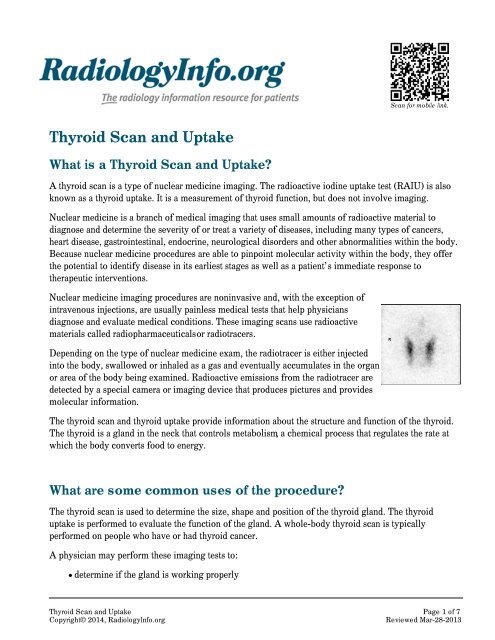
<strong>Scan</strong> for mobile link.<strong>Thyroid</strong> <strong>Scan</strong> <strong>and</strong> <strong>Uptake</strong>What is a <strong>Thyroid</strong> <strong>Scan</strong> <strong>and</strong> <strong>Uptake</strong>?A thyroid scan is a type of nuclear medicine imaging. The radioactive iodine uptake test (RAIU) is alsoknown as a thyroid uptake. It is a measurement of thyroid function, but does not involve imaging.Nuclear medicine is a branch of medical imaging that uses small amounts of radioactive material todiagnose <strong>and</strong> determine the severity of or treat a variety of diseases, including many types of cancers,heart disease, gastrointestinal, endocrine, neurological disorders <strong>and</strong> other abnormalities within the body.Because nuclear medicine procedures are able to pinpoint molecular activity within the body, they offerthe potential to identify disease in its earliest stages as well as a patient’s immediate response totherapeutic interventions.Nuclear medicine imaging procedures are noninvasive <strong>and</strong>, with the exception ofintravenous injections, are usually painless medical tests that help physiciansdiagnose <strong>and</strong> evaluate medical conditions. These imaging scans use radioactivematerials called radiopharmaceuticals or radiotracers.Depending on the type of nuclear medicine exam, the radiotracer is either injectedinto the body, swallowed or inhaled as a gas <strong>and</strong> eventually accumulates in the organor area of the body being examined. Radioactive emissions from the radiotracer aredetected by a special camera or imaging device that produces pictures <strong>and</strong> providesmolecular information.The thyroid scan <strong>and</strong> thyroid uptake provide information about the structure <strong>and</strong> function of the thyroid.The thyroid is a gl<strong>and</strong> in the neck that controls metabolism, a chemical process that regulates the rate atwhich the body converts food to energy.What are some common uses of the procedure?The thyroid scan is used to determine the size, shape <strong>and</strong> position of the thyroid gl<strong>and</strong>. The thyroiduptake is performed to evaluate the function of the gl<strong>and</strong>. A whole-body thyroid scan is typicallyperformed on people who have or had thyroid cancer.A physician may perform these imaging tests to:determine if the gl<strong>and</strong> is working properly<strong>Thyroid</strong> <strong>Scan</strong> <strong>and</strong> <strong>Uptake</strong> Page 1 of 7Copyright© 2014, <strong>RadiologyInfo</strong>.orgReviewed Mar-28-2013
help diagnose problems with the thyroid gl<strong>and</strong>, such as an overactive thyroid gl<strong>and</strong>, a conditioncalled hyperthyroidism, cancer or other growthsassess the nature of a nodule discovered in the gl<strong>and</strong>detect areas of abnormality, such as lumps (nodules) or inflammationdetermine whether thyroid cancer has spread beyond the thyroid gl<strong>and</strong>evaluate changes in the gl<strong>and</strong> following medication use, surgery, radiotherapy or chemotherapyHow should I prepare?You may be asked to wear a gown during the exam or you may be allowed to wear your own clothing.Women should always inform their physician or technologist if there is any possibility that they arepregnant or if they are breastfeeding. See the Safety page (www.<strong>RadiologyInfo</strong>.org/en/safety/) for moreinformation about pregnancy <strong>and</strong> breastfeeding related to nuclear medicine imaging.You should inform your physician <strong>and</strong> the technologist performing your exam of any medications youare taking, including vitamins <strong>and</strong> herbal supplements. You should also inform them if you have anyallergies <strong>and</strong> about recent illnesses or other medical conditions.You should tell your physician if you:have had any tests, such as an x-ray or CT scan, surgeries or treatments using iodinated contrastmaterial within the last two months.are taking medications or ingesting other substances that contain iodine, including kelp, seaweed,cough syrups, multivitamins or heart medications.have any allergies to iodine, medications <strong>and</strong> anesthetics.are breastfeeding.In the days prior to your examination, blood tests may be performed to measure the level of thyroidhormones in your blood. You may be told not to eat for several hours before your exam because eatingcan affect the accuracy of the uptake measurement.Jewelry <strong>and</strong> other metallic accessories should be left at home if possible, or removed prior to the exambecause they may interfere with the procedure.You will receive specific instructions based on the type of scan you are undergoing.What does the equipment look like?Special camera or imaging devices used in nuclear medicine include the gamma camera <strong>and</strong>single-photon emission-computed tomography (SPECT).The gamma camera, , also called a scintillation camera, detects radioactive energy that is emitted fromthe patient's body <strong>and</strong> converts it into an image. The gamma camera does not emit any radiation. Thegamma camera is composed of radiation detectors, called gamma camera heads, which are encased inmetal <strong>and</strong> plastic <strong>and</strong> most often shaped like a box, attached to a round circular donut shaped gantry. The<strong>Thyroid</strong> <strong>Scan</strong> <strong>and</strong> <strong>Uptake</strong> Page 2 of 7Copyright© 2014, <strong>RadiologyInfo</strong>.orgReviewed Mar-28-2013
metal <strong>and</strong> plastic <strong>and</strong> most often shaped like a box, attached to a round circular donut shaped gantry. Thepatient lies on the examination table which slides in between the parallel gamma camera heads which aresuspended over the examination table <strong>and</strong> located beneath the examination table. Sometimes, the gammacamera heads are oriented at a 90 degree angle <strong>and</strong> placed over the patient's body.SPECT involves the rotation of the gamma camera heads around the patient's body to produce moredetailed, three-dimensional images.A computer aids in creating the images from the data obtained by the gamma camera.A probe is a small h<strong>and</strong>-held device resembling a microphone that can detect <strong>and</strong> measure the amount ofthe radiotracer in a small area of your body.How does the procedure work?With ordinary x-ray examinations, an image is made by passing x-rays through the patient's body. Incontrast, nuclear medicine procedures use a radioactive material, called a radiopharmaceutical orradiotracer, which is injected into the bloodstream, swallowed or inhaled as a gas. This radioactivematerial accumulates in the organ or area of your body being examined, where it gives off a small amountof energy in the form of gamma rays. Special cameras detect this energy, <strong>and</strong> with the help of a computer,create pictures offering details on both the structure <strong>and</strong> function of organs <strong>and</strong> tissues in your body.How is the procedure performed?Nuclear medicine imaging is usually performed on an outpatient basis, but is often performed onhospitalized patients as well.<strong>Thyroid</strong> <strong>Scan</strong>You will be positioned on an examination table. If necessary, a nurse or technologist will insert anintravenous (IV) catheter into a vein in your h<strong>and</strong> or arm.Depending on the type of nuclear medicine exam you are undergoing, the dose of radiotracer is theninjected intravenously, swallowed or inhaled as a gas.When radiotracer is taken by mouth, in either liquid or capsule form, it is typically swallowed up to 24hours before the scan. The radiotracer given by intravenous injection is usually given up to 30 minutesprior to the test.When it is time for the imaging to begin, you will lie down on a moveable examination table with yourhead tipped backward <strong>and</strong> neck extended. The gamma camera will then take a series of images, capturingimages of the thyroid gl<strong>and</strong> from three different angles. You will need to remain still for brief periods oftime while the camera is taking pictures.When the examination is completed, you may be asked to wait until the technologist checks the images incase additional images are needed. Occasionally, more images are obtained for clarification or better<strong>Thyroid</strong> <strong>Scan</strong> <strong>and</strong> <strong>Uptake</strong> Page 3 of 7Copyright© 2014, <strong>RadiologyInfo</strong>.orgReviewed Mar-28-2013
visualization of certain areas or structures. The need for additional images does not necessarily meanthere was a problem with the exam or that something abnormal was found, <strong>and</strong> should not be a cause ofconcern for you.If you had an intravenous line inserted for the procedure, it will usually be removed unless you arescheduled for an additional procedure that same day that requires an intravenous line.Actual scanning time for a thyroid scan is 30 minutes or less.<strong>Thyroid</strong> <strong>Uptake</strong>You will be given radioactive iodine (I-123 or I-131) in liquid or capsule form to swallow. The thyroiduptake will begin several hours to 24 hours later. Often, two separate uptake measurements are obtainedat different times. For example, you may have uptake measurements at four to six hours <strong>and</strong> 24 hours.When it is time for the imaging to begin, you will sit in a chair facing a stationary probe positioned overthe thyroid gl<strong>and</strong> in the neck.When the examination is completed, you may be asked to wait until the technologist checks the images incase additional images are needed. Occasionally, more images are obtained for clarification or bettervisualization of certain areas or structures. The need for additional images does not necessarily meanthere was a problem with the exam or that something abnormal was found, <strong>and</strong> should not be a cause ofconcern for you.Actual scanning time for each thyroid uptake is five minutes or less.What will I experience during <strong>and</strong> after the procedure?Most thyroid scan <strong>and</strong> thyroid uptake procedures are painless. However, during the thyroid scan, youmay feel uncomfortable when lying completely still with your head extended backward while the gammacamera is taking images.When the radiotracer is given intravenously, you will feel a slight pin prick when the needle is insertedinto your vein for the intravenous line. When the radioactive material is injected into your arm, you mayfeel a cold sensation moving up your arm, but there are generally no other side effects.When swallowed, the radiotracer has little or no taste. When inhaled, you should feel no differently thanwhen breathing room air or holding your breath.It is important that you remain still while the images are being recorded. Though nuclear imaging itselfcauses no pain, there may be some discomfort from having to remain still or to stay in one particularposition during imaging.Unless your physician tells you otherwise, you may resume your normal activities after your nuclearmedicine scan. If any special instructions are necessary, you will be informed by a technologist, nurse orphysician before you leave the nuclear medicine department.Through the natural process of radioactive decay, the small amount of radiotracer in your body will loseits radioactivity over time. It may also pass out of your body through your urine or stool during the first<strong>Thyroid</strong> <strong>Scan</strong> <strong>and</strong> <strong>Uptake</strong> Page 4 of 7Copyright© 2014, <strong>RadiologyInfo</strong>.orgReviewed Mar-28-2013
its radioactivity over time. It may also pass out of your body through your urine or stool during the firstfew hours or days following the test. You should also drink plenty of water to help flush the radioactivematerial out of your body as instructed by the nuclear medicine personnel.Who interprets the results <strong>and</strong> how do I get them?A radiologist or other physician who has specialized training in nuclear medicine will interpret theimages <strong>and</strong> forward a report to your referring physician.What are the benefits vs. risks?BenefitsRisksNuclear medicine examinations provide unique information—including details on both function<strong>and</strong> anatomic structure of the body that is often unattainable using other imaging procedures.For many diseases, nuclear medicine scans yield the most useful information needed to make adiagnosis or to determine appropriate treatment, if any.Nuclear medicine is less expensive <strong>and</strong> may yield more precise information than exploratorysurgery.Because the doses of radiotracer administered are small, diagnostic nuclear medicine proceduresresult in relatively low radiation exposure to the patient, acceptable for diagnostic exams. Thus, theradiation risk is very low compared with the potential benefits.Nuclear medicine diagnostic procedures have been used for more than five decades, <strong>and</strong> there areno known long-term adverse effects from such low-dose exposure.The risks of the treatment are always weighed against the potential benefits for nuclear medicinetherapeutic procedures. You will be informed of all significant risks prior to the treatment <strong>and</strong> havean opportunity to ask questions.Allergic reactions to radiopharmaceuticals may occur but are extremely rare <strong>and</strong> are usually mild.Nevertheless, you should inform the nuclear medicine personnel of any allergies you may have orother problems that may have occurred during a previous nuclear medicine exam.Injection of the radiotracer may cause slight pain <strong>and</strong> redness which should rapidly resolve.Women should always inform their physician or radiology technologist if there is any possibilitythat they are pregnant or if they are breastfeeding. See the Safety page(www.<strong>RadiologyInfo</strong>.org/en/safety/) for more information about pregnancy, breastfeeding <strong>and</strong>nuclear medicine exams.What are the limitations of the <strong>Thyroid</strong> <strong>Scan</strong> <strong>and</strong> <strong>Uptake</strong>?The thyroid scan <strong>and</strong> thyroid uptake are not performed on patients who are pregnant because of the risk<strong>Thyroid</strong> <strong>Scan</strong> <strong>and</strong> <strong>Uptake</strong> Page 5 of 7Copyright© 2014, <strong>RadiologyInfo</strong>.orgReviewed Mar-28-2013
of exposing the fetus to radiation. These tests are also not recommended for breastfeeding women.Nuclear medicine procedures can be time consuming. It can take several hours to days for the radiotracerto accumulate in the body part of interest <strong>and</strong> imaging may take up to several hours to perform, though insome cases, newer equipment is available that can substantially shorten the procedure time.The resolution of structures of the body with nuclear medicine may not be as high as with other imagingtechniques, such as CT or MRI. However, nuclear medicine scans are more sensitive than othertechniques for a variety of indications, <strong>and</strong> the functional information gained from nuclear medicineexams is often unobtainable by other imaging techniques.Additional Information <strong>and</strong> Resources<strong>RadiologyInfo</strong>Head <strong>and</strong> Neck Cancer(www.<strong>RadiologyInfo</strong>.org/en/info.cfm?pg=hdneck)RTAnswers.orgRadiation Therapy for Head <strong>and</strong> Neck Cancer(www.rtanswers.org/treatmentinformation/cancertypes/headneck/index.aspx)DisclaimerThis information is copied from the <strong>RadiologyInfo</strong> Web site (http://www.radiologyinfo.org) which is dedicated toproviding the highest quality information. To ensure that, each section is reviewed by a physician with expertise inthe area presented. All information contained in the Web site is further reviewed by an ACR (American College ofRadiology) - RSNA (Radiological Society of North America) committee, comprising physicians with expertise inseveral radiologic areas.However, it is not possible to assure that this Web site contains complete, up-to-date information on any particularsubject. Therefore, ACR <strong>and</strong> RSNA make no representations or warranties about the suitability of this informationfor use for any particular purpose. All information is provided "as is" without express or implied warranty.Please visit the <strong>RadiologyInfo</strong> Web site at http://www.radiologyinfo.org to view or download the latestinformation.Note: Images may be shown for illustrative purposes. Do not attempt to draw conclusions or make diagnoses bycomparing these images to other medical images, particularly your own. Only qualified physicians should interpretimages; the radiologist is the physician expert trained in medical imaging.CopyrightThis material is copyrighted by either the Radiological Society of North America (RSNA), 820 Jorie Boulevard, OakBrook, IL 60523-2251 or the American College of Radiology (ACR), 1891 Preston White Drive, Reston, VA20191-4397. Commercial reproduction or multiple distribution by any traditional or electronically basedreproduction/publication method is prohibited.Copyright ® 2014 Radiological Society of North America, Inc.<strong>Thyroid</strong> <strong>Scan</strong> <strong>and</strong> <strong>Uptake</strong> Page 6 of 7Copyright© 2014, <strong>RadiologyInfo</strong>.orgReviewed Mar-28-2013
Copyright ® 2014 Radiological Society of North America, Inc.<strong>Thyroid</strong> <strong>Scan</strong> <strong>and</strong> <strong>Uptake</strong> Page 7 of 7Copyright© 2014, <strong>RadiologyInfo</strong>.orgReviewed Mar-28-2013