910-001 PGE2 CLIA 6Dec06 - Enzo Life Sciences
910-001 PGE2 CLIA 6Dec06 - Enzo Life Sciences
910-001 PGE2 CLIA 6Dec06 - Enzo Life Sciences

You also want an ePaper? Increase the reach of your titles
YUMPU automatically turns print PDFs into web optimized ePapers that Google loves.
Correlate-<strong>CLIA</strong> High Sensitivity Prostaglandin E 2Chemiluminescence Enzyme Immunoassay KitCatalog No. <strong>910</strong>-<strong>001</strong>96 Well KitTable of ContentsDescription Page 2Introduction 2Precautions 2Materials Supplied 3Storage 3Materials Needed but Not Supplied 3Sample Handling 4Procedural Notes 5Reagent Preparation 5Assay Procedure 6Calculation of Results 7Typical Results 7Typical Standard Curve 8Typical Quality Control Parameters 8Performance Characteristics 9Sample Dilution Recommendations 10References 11Limited Warranty 12FOR RESEARCH USE ONLY. NOT FOR USE IN DIAGNOSTIC PROCEDURES.1
Materials Supplied1. Goat anti-mouse Microtiter Plate, One Plate of 96 Wells, Catalog No. 80-0035A white plate using break-apart strips coated with goat antibody specific to mouse IgG.2. PGE 2<strong>CLIA</strong> Conjugate, 6 mL, Catalog No. 80-<strong>001</strong>3A blue solution of alkaline phosphatase conjugated with PGE 2.3. PGE 2<strong>CLIA</strong> Antibody, 6 mL, Catalog No. 80-<strong>001</strong>4A yellow solution of a monoclonal antibody to PGE 2.4. Assay Buffer, 30 mL, Catalog No. 80-<strong>001</strong>0Tris buffered saline containing proteins and sodium azide as preservative.5. Wash Buffer Concentrate, 30 mL, Catalog No. 80-1286Tris buffered saline containing detergents.6. Prostaglandin E 2Standard, 0.5 mL, Catalog No. 80-0004A solution of 50,000 pg/mL PGE 2.7. Lumiphos 530 <strong>CLIA</strong> Substrate*, 21 mL, Catalog No. 80-0134Alkaline Phosphatase substrate in diethanolamine buffer at pH 9.5, containing fluorescentenhancers, with sodium azide as preservative.8. PGE 2Plate <strong>CLIA</strong> Assay Layout Sheet, 1 each, Catalog No. 30-00399. Plate Sealer, 1 each, Catalog No. 30-<strong>001</strong>2StorageAll components of this kit, except the Conjugate and Standard, are stable at 4 °C until thekit's expiration date. The Conjugate and Standard must be stored at -20 °C.Materials Needed but Not Supplied1. Deionized or distilled water. No difference is seen in assay results with distilled water.2. Precision pipets for volumes between 5 μL and 1,000 μL.3. Repeater pipets for dispensing 50 μL and 200 μL.4. Disposable beakers for diluting buffer concentrates.5. Graduated cylinders.6. A micro plate shaker.7. Adsorbent paper for blotting.8. Plate luminometer, such as an EG&G Wallac LB 96P, capable of reading glow chemiluminescence.Some radiation counters may be suitable. Please refer to the counter instructionmanual for recommendations on suitability for chemiluminescence measurements.*Lumiphos 530 is the trademark of Lumigen Inc., Southfield, MI, USA and supplied under US patents4,857,652; 4,983,779; 4,959,182; 5,004,565; 4,962,192, & 5,386,017; European patents 254051B1 &352713B1; Japanese patent 5-45590; Australian patent 603,736; Korean patent 69,259 and Taiwanesepatent 46,563.3
Sample HandlingAssay Designs’ Correlate-<strong>CLIA</strong> is compatible with PGE 2samples in a wide range of matrices.Samples diluted sufficiently into Assay Buffer can be read directly from the standard curve. Pleaserefer to the Sample Recovery recommendations on page 11 for details of suggested dilutions. Samplescontaining mouse IgG may interfere with the assaySamples in the majority of Tissue Culture Media, including those containing fetal bovine serum, canalso be read in the assay, provided the standards have been diluted into the Tissue Culture Mediainstead of Assay Buffer. There will be a small change in binding associated with running the standardsand samples in media. Users should only use standard curves generated in media or buffer to calculateconcentrations of PGE 2in the appropriate matrix. For tissue, urine and plasma samples, prostaglandinsynthetase inhibitors, such as, indomethacin or meclofenamic acid at concentrations up to 10 μg/mLshould be added to either the tissue homogenate or urine and plasma samples. Most samples may beused in the assay directly by dilution in the range of 1:10 in Assay Buffer.Some samples normally have very low levels of PGE 2present and extraction may be necessary foraccurate measurement. A suitable extraction procedure is outlined below:Materials Needed1. PGE 2Standard to allow extraction efficiency to be accurately determined.2. 2M hydrochloric acid, deionized water, ethanol, hexane and ethyl acetate.3. 200 mg C 18Reverse Phase Extraction Columns.Procedure1. Acidify the plasma, urine or tissue homogenate by addition of 2M HCl to pH of 3.5.Approximately 50 μL of HCl will be needed per mL of plasma. Allow to sit at 4 °C for 15minutes. Centrifuge samples in a microcentrifuge for 2 minutes to remove any precipitate.2. Prepare the C 18reverse phase column by washing with 10 mL of ethanol followed by 10 mLof deionized water.3. Apply the sample under a slight positive pressure to obtain a flow rate of about 0.5 mL/minute. Wash the column with 10 mL of water, followed by 10 mL of 15% ethanol, andfinally 10 mL hexane. Elute the sample from the column by addition of 10 mL ethyl acetate.4. If analysis is to be carried out immediately, evaporate samples under a stream of nitrogen.Add 250 μL of Assay Buffer to the dried sample. Vortex well then allow to sit for five minutesat room temperature. Repeat twice more. If analysis is to be delayed, store samples as theeluted ethyl acetate solutions at -80 °C until the immunoassay is to be run. Evaporate theorganic solvent under a stream of nitrogen prior to running assay and reconstitute as above.Please refer to references 15-18 for details of extraction protocols.Procedural Notes1. Do not mix reagents from different lot numbers or use reagents beyond the expiration date.2. Allow all reagents to warm to room temperature for at least 30 minutes before opening.4
3. Standards can be made up in either glass or plastic tubes.4. Pre-rinse the pipet tip with reagent, use fresh pipet tips for each sample, standard and reagent.5. Pipet standards and samples to the bottom of the wells.6. Add the reagents to the side of the well to avoid contamination.7. This kit uses break-apart microtiter strips, which allow the user to measure as many samplesas desired. Unused wells must be kept desiccated at 4 °C in the sealed bag provided. Thewells should be used in the frame provided.8. Care must be taken to minimize contamination by endogenous alkaline phosphatase.Contaminating alkaline phosphatase activity, especially in the <strong>CLIA</strong> substrate solution, maylead to high blanks. Care should be taken not to touch pipet tips and other items that are usedin the assay with bare hands.9. Prior to addition of substrate, ensure that there is no residual wash buffer in the wells.Any remaining wash buffer may cause variation in assay results.10. The chemiluminescent signal generated is read after 60 minutes. The signal is still beinggenerated and the wells must be read in the order in which the substrate was added. Ifyou do not have a luminometer that will precisely time the substrate incubation thefollowing protocol must be followed. We suggest adding the substrate at 10 secondintervals between wells and reading the generated chemiluminescence for 2 secondsat 10 second intervals for consistency. If luminometer injection is not used, we suggestusing a repeater type syringe, such as an Eppendorf Repeater Pipette, CatalogNumber 2226000-6 and a 5 mL repeater Combitip set for delivery of 200 μL. Use therepeater to add substrate to the wells in the order in which they will be read. Please notethe order that plate luminometers read wells and ensure substrate addition follows thissequence.Reagent Preparation1. PGE 2StandardAllow the 50,000 pg/mL PGE 2Standard solution to warm to room temperature. Label eight12 x 75 mm tubes #1 through #8. Pipet 980 μL of standard diluent (Assay Buffer or TissueCulture Media) into tube #1. Pipet 500 μL of standard diluent (Assay Buffer or Tissue CultureMedia) into tubes #2 - #8. Add 20 μL of the 50,000 pg/mL standard to tube #1. Vortexthoroughly. Add 500 μL of tube #1 to tube #2 and vortex thoroughly. Add 500 μL of tube#2 to tube #3 and vortex. Continue this for tubes #4 through #8.The concentration of PGE 2in tubes #1 through #8 will be 1,000, 500, 250, 125, 62.5,31.25, 15.63 and 7.81 pg/mL respectively. See PGE 2Plate <strong>CLIA</strong> Assay Layout Sheet fordilution details.Diluted standards should be used within 60 minutes of preparation.2. PGE 2ConjugateAllow the conjugate to warm to room temperature. Any unused conjugate shouldbe aliquoted and re-frozen at or below -20 °C.5
3. Wash BufferPrepare the Wash Buffer by diluting 5 mL of the supplied concentrate with 95 mL ofdeionized water. This can be stored at room temperature until the kit expiration date, or for3 months, whichever is earlier.Assay ProcedureBring all reagents to room temperature for at least 30 minutes prior to opening.All standards and samples should be run in duplicate.1. Refer to the Assay Layout Sheet to determine the number of wells to be used and put anyremaining strips with the desiccant back into the pouch and seal the ziploc. Store unusedstrips at 4 °C.2. Pipet 100 μL of standard diluent (Assay Buffer or Tissue Culture Media) into the NSB andthe Bo (0 pg/mL) wells.3. Pipet 100 μL of Standards #1 through #8 into the appropriate wells.4. Pipet 100 μL of the Samples into the appropriate wells.5. Pipet 50 μL of Assay Buffer into the NSB wells.6. Pipet 50 μL of the blue Conjugate into each well, except the Total Activity (TA) and Blankwells.7. Pipet 50 μL of the yellow Antibody into each well, except the Blank, TA and NSB wells.NOTE: Every well used should be Green in color except the NSB wells which should be Blue. TheBlank and TA wells are empty at this point and have no color.8. Incubate the plate at room temperature on a plate shaker for 2 hours at ~500 rpm.9. Empty the contents of the wells and wash by adding 400 μL of wash solution to every well.Repeat the wash 2 more times for a total of 3 Washes.10. After the final wash, empty or aspirate the wells, and firmly tap the plate dry on a lint freepaper towel to remove any remaining wash buffer.11. Add 5 μL of the blue Conjugate to the TA wells.12. Add 200 μL of the <strong>CLIA</strong> Substrate solution to every well. Incubate at room temperature for60 minutes with shaking. Note: Refer to substrate addition timing and sequence onpage 5.13. Read each well for 2 seconds each on a suitable luminometer.6
Calculation of ResultsSeveral options are available for the calculation of the concentration of PGE 2in the samples. Werecommend that the data be handled by an immunoassay software package utilizing a 4 parameterlogistic curve fitting program. If data reduction software is not readily available, the concentration ofPGE 2can be calculated as follows:1. Calculate the average net Relative Light Units (RLU) bound for each standard and sampleby subtracting the average Blank RLU from the average RLU's bound.Average Net RLU = Average Bound RLU - Average NSB RLU2. Calculate the binding of each pair of standard wells as a percentage of the maximum bindingwells (Bo), using the following formula:Percent Bound = Net RLU x 100Net Bo RLU3. Using Logit-Log paper plot Percent Bound versus Concentration of PGE 2for the standards.Approximate a straight line through the points. The concentration of PGE 2in the unknownscan be determined by interpolation.Typical ResultsThe results shown below are for illustration only and should not be used to calculate results fromanother assay.Mean Average Percent PGE 2Sample RLU (-Blank) Net RLU Bound (pg/mL)Blank RLU 2.487TA 2804.195NSB 0.066 0 0.00%S1 34.431 34.365 9.2% 1,000S2 60.124 60.058 16.0% 500S3 100.485 100.419 26.7% 250S4 155.352 155.286 41.3% 125S5 212.630 212.514 56.6% 62.5S6 261.975 261.909 69.7% 31.25S7 287.745 287.679 76.6% 15.63S8 317.872 317.806 84.6% 7.81Bo 375.721 375.655 100% 0Unknown 1 56.138 56.072 14.9% 548.8Unknown 2 199.060 198.994 53.0% 73.27
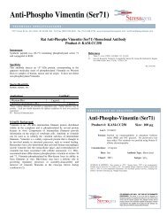
Typical Standard CurveA typical standard curve is shown below. This curve must not be used to calculate PGE 2concentrations; each user must run a standard curve for each assay.10035080280B/Bo (%)6040210140Relative Light Units20B/Bo (%)RLU70<strong>001</strong> 10 100 1,000 10,000PGE 2 conc. (pg/ml)Typical Quality Control ParametersTotal Activity Added = 2804.195 x 10 = 28041.95%Bo/TA = 1.3%Quality of Fit = 0.999 (Calculated from 4 parameter logistic curve fit)20% Intercept = 328.9 pg/mL50% Intercept = 84.0 pg/mL80% Intercept = 12.9 pg/mLPerformance CharacteristicsThe following parameters for this kit were determined using the guidelines listed in the NationalCommittee for Clinical Laboratory Standards (NCCLS) Evaluation Protocols 19 .8
SensitivitySensitivity was calculated in Assay Buffer by determining the average RLU signal bound for sixteen(16) wells run as Bo, and comparing to the average RLU signal for sixteen (16) wells run with Standard#8. The detection limit was determined as the concentration of PGE 2measured at two (2) standarddeviations from the zero along the standard curve.Average RLU for the Bo = 359,060 ± 13,190 (3.7%)Average RLU for Standard #8 = 324,869 ± 18,271 (5.6%)Delta RLU's (0-7.81 pg/mL) = 359,060 - 324,869 = 34,1912 SD's of the Zero Standard = 2 x 13,190 = 26,380Sensitivity = 26,380 x 7.81 pg/mL = 6.03 pg/mL34,191LinearityA sample containing 620.7 pg/mL PGE 2was diluted 4 times 1:2 in the kit Assay Buffer and measuredin the assay. The data was plotted graphically as actual PGE 2concentration versus measured PGE 2concentration.The line obtained had a slope of 0.960 and a correlation coefficient of 0.999.PrecisionIntra-assay precision was determined by taking samples containing low, medium and high concentrationsof PGE 2and running these samples multiple times (n=24) in the same assay. Inter-assayprecision was determined by measuring three samples with low, medium and high concentrations ofPGE 2in multiple assays (n=8).The precision numbers listed below represent the percent coefficient of variation for the concentrationsof PGE 2determined in these assays as calculated by a 4 parameter logistic curve fitting program.PGE 2Intra-assay Inter-assay(pg/mL) %CV %CVLow 7.07 14.7Medium 120 4.1High 330 7.4Low 59.04 9.2Medium 182 7.1High 923 5.69
Cross ReactivitiesThe cross reactivities for a number of related eicosanoid compounds was determined by dissolving thecross reactant (purity checked by N.M.R. and other analytical methods) in Assay Buffer at concentrationsfrom 500,000 to 39 pg/mL. These samples were then measured in the PGE 2Correlate-EIA colorimetric assay, and the measured PGE 2concentration at 50% B/Bo calculated. The % crossreactivity was calculated by comparison with the actual concentration of cross reactant in the sampleand expressed as a percentage.Compound Cross Reactivity Compound Cross ReactivityPGE 2100% PGB 10.1%PGE 170% 13,14-dihydro-15-keto-PGF 2α
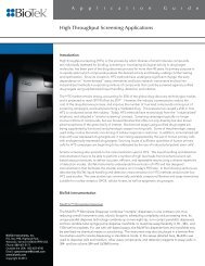
Time Course of Chemiluminescent EmissionThe chemiluminescent signal generated from the reaction of the alkaline phosphatase conjugate andthe <strong>CLIA</strong> substrate is a kinetic reaction that reaches a maximum light output after approximately 4hours. The chemiluminescent emission will last for several hours. The data is presented below.500000400000Relative Light Units3000002000<strong>001</strong>00000References<strong>001</strong>2Time (Hours)1. T. Chard, "An Introduction to Radioimmunoassay & Related Techiques, 4th Ed", (1990)Amsterdam: Elsevier.2. P. Tijssen,"Practice & Theory of Enzyme Immunoassays", (1985) Amsterdam: Elsevier.3. P.W. Ramwell, Biol. Reprod., (1977) 16: 70.4. R.J. Flower & G.J. Blackwell, Biochem. Pharm., (1976) 25: 285.5. S. Moncada & J.R. Vane, Pharm. Rev., (1979) 30: 293.6. B. Sameulsson, et al., Ann. Rev Biochem., (1978) 47: 997.7. P.D.I. Richardson, et al., Brit. J. Pharmacol., (1976) 57: 581.8. J. Raud, et al., Proc. Natl. Acad. Sci., USA, (1988) 85: 2315.9. J.W. Christman, et al., Prostaglandins, (1991) 41: 251.10. O. Hayaishi, J. Biol. Chem., (1988) 263: 14593.11. S. Kuno, et al., Proc. Natl. Acad. Sci., USA, (1986) 83: 3487.12. D.L. Bareis, et al., Proc. Natl. Acad . Sci., USA, (1983) 80: 2514.13. L.G. Raisz, et al., Nature, (1977) 267: 532.14. C.R. Long, et al., Prostaglandins, (1990) 40: 591.15. K. Green, et al., Anal. Biochem, (1973) 54: 434.16. J. Frolich, et al., J. Clin. Invest., (1975) 55: 763.17. J.E. Shaw & P.W. Ramwell, Meth. Biochem. Anal., (1969) 17: 325.18. K. Green, et al., Adv. Prostaglandin & Thromboxane Res., (1978) 5: 15.19. National Committee for Clinical Laboratory Standards Evaluation Protocols, SC1, (1989)Villanova, PA: NCCLS.1134
LIMITED WARRANTYAssay Designs, Inc. warrants that at the time of shipment this product is free from defects in materialsand workmanship. This warranty is in lieu of any other warranty expressed or implied, including butnot limited to, any implied warranty of merchantability or fitness for a particular purpose.Assay Designs must be notified of any breach of this warranty within 48 hours of receipt of the product.No claim shall be honored if Assay Designs is not notified within this time period, or if the producthas been stored in any way other than outlined in this publication. The sole and exclusive remedy ofthe customer for any liability based upon this warranty is limited to the replacement of the product,or refund of the invoice price of the goods.For more details concerning the information within this kit insert, or to orderany of Assay Designs' products, please call (734) 668-6113 between 8:30 a.m. and5:30 p.m. EST. Orders or technical questions can also be transmitted by faxor e-mail 24 hours a day.Material Safety Data Sheet (MSDS) available on our website or by fax.Assay Designs, Inc. Telephone: (734) 668-61135777 Hines Drive (800) 833-8651 (USA & Canada only)Ann Arbor, MI 48108 Fax: (734) 668-2793U.S.A. e-mail: info@assaydesigns.comwebsite: www.assaydesigns.comSimplify Your Science ®Catalog No. 25-0005 December 6, 2006© 20<strong>001</strong>2