DOCTORS NEWSLETTER - Douglass Hanly Moir Pathology
DOCTORS NEWSLETTER - Douglass Hanly Moir Pathology
DOCTORS NEWSLETTER - Douglass Hanly Moir Pathology

You also want an ePaper? Increase the reach of your titles
YUMPU automatically turns print PDFs into web optimized ePapers that Google loves.
GENERAL PRACTICECORPORATISATION, INTEGRATION ANDPROFESSIONAL INDEPENDENCEspecialists associated with the parent primary care company and,in some locations, only lip service has been paid to the importantconcept of free choice and professional independence.Dr Colin GoldschmidtMANAGING DIRECTORThere is also concern that vertical integration might create aconflict of interest whereby a clinician may benefit personally froma referral to a particular specialist or that the integrated structuremight encourage overservicing.Our DecisionSweeping ChangesOver the past year, a significant phenomenon - thecorporatisation of general practice - has taken seed inour health care system. Although a few practices have beenin corporate hands for some time now, the vast majority of generalpractices have continued to be owned and managed in thetraditional manner. The more recent advent of a new crop ofprimary care companies has, however, accelerated the transitionfrom doctor-owned to company-owned practices and, today, asignificant number of practices around Australia are either ownedor managed by corporate entities. This transformation seems to begaining momentum.As pathologists at <strong>Douglass</strong> <strong>Hanly</strong> <strong>Moir</strong>, we do not presume to takea stance in the decisions general practitioners will face inthis area. However, as pathologists, we are taking stock of thechanges happening around us and what they will mean for themedical practice of pathology. It is apparent that, while thereappears to be groundswell support for corporatisation amongstmany general practitioners themselves, there are still many otherdoctors who are opposed to corporatisation on philosophical orother grounds.Potentially far more profound than individual medical practicesbeing managed by an outside company, is the phenomenon ofcross-sector ownership (or management) of general practices,pathology practices and radiology practices by single corporations.In addition to owning/managing general practices, PrimaryHealthcare now owns two pathology practices in Sydney (GCL andSDS) and Revesco owns half of a large Melbourne-basedpathology practice (Gribbles <strong>Pathology</strong>). In New South Wales,Mayne Nickless owns hospitals, medical centres, radiology andpathology practices (Hampsons, Sugermans, Macquarie<strong>Pathology</strong>, Laverty & Associates).These “vertical” holdings by single companies have signalled thepotential “integration” of our healthcare system.Our DilemmaFor <strong>Douglass</strong> <strong>Hanly</strong> <strong>Moir</strong> <strong>Pathology</strong> (and its parent company,Sonic Healthcare), the issue of vertical integration presents adilemma. In Sydney, we have seen referrals from medical centres,now owned by primary care corporations, directed away from us tothe pathology labs owned by those corporations. Doctors in thesemedical centres have been placed under pressure to refer toAs a result of these complexities and others, we have decided thatwe will remain a diagnostic company and will not become directlyinvolved in the corporatisation of general practice. We do not wishto ever compete with our referring doctors, nor to place any doctorin a perceived position of conflict with regard to our pathologyservice provision.We have instead formed a strategic alliance with a primary carecompany, Foundation Healthcare. Sonic Healthcare has taken apassive investment position (ten percent) in Foundation but,importantly, there is no incentive or benefit for Foundationassociateddoctors to refer pathology to Sonic labs. We believethat the Foundation model for general practice is distinctive. It isstructured in such a way that it will find long-term favour withdoctors, by preserving clinical independence, by confining interestto practice management and infrastructure provision (and notinterfering with medical practice), creating value for allstakeholders and generally enhancing the efficiency of Australiangeneral practice into the future.The Need for the Right ModelThe corporatisation of general practice is one of the mostprofound changes facing our health care system and has thepotential to alter the face of general practice forever. In light of this,if any model for corporatisation is to be successful, it needsto be structured appropriately to set in place a system that is fullyembraced by the profession. A successful model must be one thatcan stand alone as a viable business and not depend on coercedreferrals to pathology, radiology, other specialists or hospitals. Itmust provide comfort and benefit to currently practising doctors,in terms of ethics, quality of practice, independence, challenge,finance and lifestyle. It must also provide the right incentive,interest and fulfilment to new and future medical graduates andit must be a model that stands up to scrutiny as being in thegreater interests of Australian medicine.I wish to thank the many doctors who have contacted mewith their views on this subject. Your opinions have contributedtowards the structure of our position on the corporatisation issue.Your understanding of our “dilemma” as a pathology practice, hasalso been most appreciated.I welcome your thoughts and comments on thissubject and invite you to contact me viaFax (02) 9878 5066 orEmail colgold@msn.com.au2
WELCOME TOPATHOLOGISTSWe would like to introduce the pathologists whohave most recently joined our team. We wouldalso like to take this opportunity to farewellDr Esmond Periera who has retired after a long andvalued career.For more complete details about theseand all our other pathologistsvisit our web site atwww.dhm.com.auPathologists who have joined usDr David Blaxland M.B., B.S., F.R.C.P.A., F.A.C.R.R.M.General Pathologist/HistopathologistDavid is the Regional Pathologist for our service in the Riverina area and is based at our Wagga Wagga laboratory.He is a generally trained pathologist with a special interest in histopathology. Prior to coming to Wagga Wagga in early2000, David had been in Warrnambool for 11 years where he was Director of Regional <strong>Pathology</strong> and later worked inprivate pathology practice. He is committed to providing personalised pathology services to practitioners in rural areas.Dr Debbie Clark M.B., Ch.B., F.R.C.P.(U.K.), F.R.C.Path., F.R.C.P.A.HaematologistDebbie has a wealth of experience in both laboratory and clinical haematology and prior to emigrating from the UnitedKingdom, she held the position of Head of Clinical and Laboratory Haematology at a large district hospital in the Peakdistrict of Derbyshire for 17 years. She has been in private laboratory practice in Sydney since 1996 and joined DHM in1998. Her particular interests lie in the fields of flow cytometry, haemoglobinopathies and myelodysplasticsyndromes.Dr Angus Collins M.B., B.S., F.R.C.P.A.Histopathologist/CytopathologistAngus has a broad experience base. He has worked in private pathology practice since 1997 and joined DHM in 1999.He has special interests in dermatopathology, gastrointestinal pathology, haematopathology and fine needleaspiration cytology. He is also interested in the study of molecular biology and the development of its applications tohistopathological diagnosis.Dr Vicki Jordan M.B., B.S., F.R.C.P.A.Histopathologist/CytopathologistPrior to completing her pathology training at the Royal North Shore Hospital, Vicki spent three years in general practiceon Sydney’s Lower North Shore. As well as training in histopathology and cytopathology Vicki also spent a year inhaematology at Westmead Hospital. She joined DHM in early 2000. Vicki has a wide range of interests includinghaematopathology, respiratory pathology and gynaecological pathology.Dr Peter Kench M.B., Ch.B., M.Med.Path. (Clinical), F.R.C.P.A., F.C.A.P., M.I.A.C.HistopathologistPeter trained as a general pathologist but now works primarily in histopathology, where he maintains a broad expertise.Peter has worked in both the public and private arenas of pathology - he has held specialist positions in several majorteaching hospitals, practised as a general pathologist, mainly in country areas of New South Wales and Queensland,and has also conducted his own private cytology practice. Prior to joining DHM in 1998, Peter worked in privatepathology practice in Sydney.Dr Lance Meng M.B., B.S., F.R.C.P.A.General Pathologist/HistopathologistLance is trained in all major disciplines of pathology and has held appointments in both the public and private pathologyarenas, including that of Director of <strong>Pathology</strong> at Wangaratta. Lance and his family live on the Central Coast, where heprovides a comprehensive pathology service for local doctors. He has a particular interest in histopathology andpersonally reports these specimens for Central Coast practitioners. Lance is our Medical Director for the Central Coastand is based full time at our Gosford laboratory.Dr Miriam Paul M.B., B.S., M.M., F.R.A.C.P., F.R.C.P.A.Microbiologist/Infectious Diseases PhysicianMiriam is a physician and a microbiologist. As well as her role at DHM, she has a clinical Infectious Diseases practice onSydney’s North Shore with appointments at the Mater Misericordiae Hospital and the Hills Hospital. Miriam is a memberof a number of infection control committees and also edits a monthly journal for the Australasian HIV Medicine Society.She is particularly interested in assisting doctors with queries regarding patient investigation and management.3
HAEMATOLOGICAL CHANGESIN THE ELDERLYESRDr Debbie ClarkHAEMATOLOGISTThe ESR remains a difficult parameter forinterpretation in old age. Although standard textbooksmay quote a rise of less than 10mm per hour with aging, anumber of studies have shown that the investigation ofmodest elevations in the ESR in the asymptomatic elderlyis likely to be unfruitful. One study, for example, failed todetect any disease process in well elderly women withESRs of up to 69mm per hour.DemographicsWhat disorders are common in the elderly?The changing demographics of the populationhas obvious implications for health care services.Most Western countries anticipate that, in the nexttwenty-five years, the proportion of people over the age of60 will rise. In parts of Europe the rise will be particularlysteep and in Germany, for example, the over 60s areexpected to exceed 35% by the year 2025.Australia and the US are expected to be partially protectedby levels of immigration. Nevertheless, many medicalpractitioners in Australia are likely to encounter anincreasing number of elderly patients in their care.What is ‘normal’ for the elderly?In order to appreciate haematological abnormalities, weneed to first be aware of what is normal.The full blood countIn the past, studies of normal ranges have inadvertentlyincluded people in the early stages of significant disease,hence some early studies suggested a decline inhaematological parameters, such as haemoglobin level,with age.However, more recent research suggests that thereare no significant changes with advancing age (withthe exception of a slight decline in haemoglobin invery elderly males). Indeed, lowering the normal range forolder people could result in underdiagnosis of diseasestates. There are no observed changes in any other redcell parameters (such as MCV) in old age.AnaemiasIn contrast to the younger population, where irondeficiency is the predominant anaemia seen, anaemiain the elderly is more commonly a normochromicanaemia, the ‘anaemia of chronic disorders’. This is alsothe pattern most commonly encountered in hospitalpatients and reflects, in both groups, the influence ofserious underlying disease. Indeed, recently an informalsurvey of moderate anaemias in patients over the age of 80referred to our laboratory by general practitionersconfirmed the dominance of normochromic anaemias inthis group.Iron deficiency anaemia is nevertheless relativelycommon and, as in younger groups, a silent bleedingneoplastic lesion is an important and potentially treatablecause of this condition.Surprisingly perhaps, a macrocytic blood picture came aclose third after iron deficiency. A macrocytic anaemiamay be due to a number of causes including :• B12 or folate deficiency• chronic liver disease• drugs• hypothyroidism (occasionally)However, as life expectancy lengthens, themyelodysplastic syndromes are becoming increasinglyrecognised as a common cause of a macrocytic bloodpicture.The total white cell count, differential, and platelet countsalso do not decline.Leucocyte function, on the other hand, is almost certainlyimpaired in the very old; this correlates well with clinicalobservations of increased susceptibility to infection, butfurther study is needed in this area.Apart from a small decline in Hb levels in elderly males,Hb levels remain constant through old age.4
HAEMATOLOGICAL CHANGESIN THE ELDERLYNeoplastic DisordersLeukaemia and lymphoproliferative diseaseThree groups of haematological neoplastic disorders areespecially important in the elderly because of theirincreased incidence with age. These are :• Myelodysplasia• Chronic lymphocytic leukemia• MyelomaThese diseases have in common the facts that:• Many patients do not necessarily require therapyin the early stages.• They are all essentially incurable in this agegroup.• In contrast to carcinomas, early detection isunlikely to influence survival.It should be noted that all three conditions occasionallyarise in younger patients, and in this group early referraland treatment are important.Myelodysplastic syndromesThis group of related disorders is emerging as animportant cause of cytopenias and otherabnormalities in the blood counts of the very old,although the true incidence of these disorders is not known.Common early abnormalities includemacrocytosis and monocytosis, and these changesmay precede the development of more serious problems bymany years.The incidence of all leukaemias rises sharply with age.The commonest form is chronic lymphocytic leukaemia,which accounts for one third of all leukaemia. One of themost frequent presentations of this disease nowadays isthat of an incidental finding of a lymphocytosis on a fullblood count.In the past, to confirm the diagnosis, it was necessary toperform a bone marrow biopsy, an invasive and sometimesunpleasant procedure. Now, lymphocyte surface markerstudies performed on a specimen of peripheral blood(5mls EDTA) enable us to confirm the diagnosis inmost cases without the immediate necessity formarrow examination. Fortunately, many patients requireno therapy for the disease for a number of years.Myeloma and monoclonal gammopathySome surveys of the very old suggest that up to 10% ofthis population may have an asymptomatic paraprotein.This is commonly detected as a result of biochemicalscreening tests.Many so-called ‘benign’ paraproteins may progress overtime to overt myeloma and, as a result the term‘monoclonal gammopathy of uncertain significance’(MGUS) is replacing the term ‘benign’.Old people with a macrocytosis should initially always beinvestigated to exclude B12 or folate deficiency beforebeing labelled as myelodysplasia. Interestingly, thispresentation of Addisonian pernicious anaemia isbecoming less common as folate supplementation of ourdiet increases and, of course, neurological presentationsare likely to become commoner.Myelodysplastic syndromes may also come to lightfollowing a routine blood count with the finding of anunexplained persisting monocytosis.Other early findings in this varied group of disorders includeneutropenia and thrombocytopenia, and redcell hypochromia with normal or increased ferritin levels.Progressive disease is characterised by the need fortransfusion, troublesome neutropenia andthrombocytopenia and, in some, transformation toacute leukemia.Multiple myeloma: advanced x-ray changes (includingpathological fracture) in an elderly woman presenting with anormochromic anaemia and raised ESR. At the time ofpresentation many patients have advanced skeletaldisease.Investigation of incidental laboratory findings in the elderlyBecause asymptomatic blood disorders are a common incidental finding in this age group, it is sometimes difficult to decide how far to investigate thesepatients. Undoubtedly, individual circumstances will dictate the optimal course of action. The haematologists at <strong>Douglass</strong> <strong>Hanly</strong> <strong>Moir</strong> <strong>Pathology</strong> arealways happy to discuss patients’ findings with practitioners, and bone marrow examinations can readily be arranged if deemed helpful.5
TRAVELLER’S HEALTH PART 2ILLNESS IN THE RETURNING TRAVELLERCauses of Diarrhoea in TravellersDr Ian ChambersDIRECTOR OF MICROBIOLOGYIt is estimated that travellers from developed todeveloping countries have a 15% risk of suffering ahealth problem and that about 8% will be sufficientlyunwell to consult a doctor during travel or on return.A clinician is therefore called upon not only to providepre-travel vaccination advice (as discussed in a previousissue of this Newsletter) but also to assess the travellerwho is unwell on return. This requires an appreciation ofthe important exotic diseases which may occur in travellersand of the basic diagnostic tests available for them. In thisarticle a framework will be presented for the assessment ofthe returning traveller who is unwell.• E. coli (especially enterotoxin producing)• Salmonella species• Campylobacter jejuni• Shigella species• Vibrio species• Giardia intestinalis• Cryptosporidium parvum• Entamoeba histolytica• Viruses (e.g. Norwalk virus, rotavirus)• Cyclospora cayetanesisInvestigations centre on the examinationof a series of 3 stool specimens bymicroscopy and culture. It isimportant to specify thatthe patient has travelledoverseas, as this willinfluence the rangeof pathogens thelaboratorywill lookfor.Travel HistoryA carefully elicited travel history is a vital part of the initialassessment.Details which should be covered in the travel historyinclude:• dates• places and living conditions• pre-travel immunisation and chemoprophylaxis• temporal relationship between potential diseaseexposure and onset of illness(for estimation of incubation periods)• sexual activity while travelling• underlying illnesses• activities such as swimming• illness in companion travellersAny exposure to animals or bites by insects or ticks mayalso be relevant.Categories of Illness in Returning TravellersGastrointestinalDiarrhoea is estimated to affect 20-50% of travellers to thedeveloping world. It is usually bacterial and the commonestcause is enterotoxin-producing E. coli. Chronic diarrhoeamay develop in a significant subset of these patients.FeverThe list of possible causes of fever in a traveller is a longone, but it is possible to formulate a plan of investigation toexclude the commonest and most important.Important Causes of Fever in the Returning TravellerIncubation 21 days• Viral hepatitis• Amoebic liver abscess• Malaria• Typhoid/Paratyphoid fever• Schistosomiasis• Tuberculosis• Filariasis, Leishmaniasis etc• HIV6
TRAVELLER’S HEALTH PART 2ILLNESS IN THE RETURNING TRAVELLERMalariaEosinophilic SyndromesIf a malarious area has been visited, malaria should beconsidered the most important diagnosis to exclude.The four species of malaria that infect humans are:• Plasmodium vivax• P. falciparum• P. malariae• P. ovaleOf these P. falciparum is the most important, both becauseof its potential to be rapidly fatal and because of itsresistance to antimalarial drugsDiagnosis is usually by means of blood films,supplemented by an immunochromatographic test (ICT)to detect antigen and, occasionally, serology.A patient may develop malaria despite taking what wouldappear to be adequate prophylaxis.Many cases result from errorsin assessing the need forprecautions, or fromstopping prophylaxis toosoon after leaving an endemicarea. Malaria can mimic other infectionsand can be seen in association with otherfebrile illnesses, such as pneumonia.A number of infectious and atopic conditions may causepersistent eosinophilia. Parasitic causes are the mostcommon, and a number of investigations are available toexclude the most common.Causes of Significant Eosinophilia (>500/mm 3 )• Atopy• Gastrointestinal parasites (eg. trichuris,hookworm, ascaris)• Strongyloides• Schistosomaiasis• Cutaneous larva migransInvestigations• Direct and serological examination for parasitesIn some cases of persistent eosinophilia with a parasiticcause strongly suspected but unproven, an empiric courseof albendazole may be considered. This agent is effectiveagainst many gut parasites such as trichuris, ascaris etc,but less effective against strongyloides and ineffectiveagainst schistosomiasis.Dengue FeverWorried WellDengue Fever, transmitted by mosquito bite, is an acutefebrile illness, often with rash and thrombocytopenia. It hasa short incubation time (usually 3-7 days and not longerthan 2 weeks) and is readily diagnosed serologically.SalmonellaThe so-called enteric fevers are caused by Salmonellatyphi and S. paratyphi. They have few specific features attheir onset, and typhoid vaccines provide onlyapproximately 70% protection. Therefore this diagnosisshould be considered and excluded (by blood, stool andpossibly urine cultures, not serology) in any febriletraveller.Patients often present having recovered from an illness ornever having been ill. They may be keen for either aretrospective diagnosis, or to exclude the possibility ofinfections such as HIV.Embarking on extensive diagnostic exercises in suchpatients is probably to be discouraged but screening testsfor infections may be indicated in certain circumstances.Travellers to Africa, parts of the Middle East, South EastAsia and Central and South America, for example, whohave been swimming in fresh-water lakes and rivers,commonly request screening for schistosomiasis on theirreturn. A reliable serological test is available(supplemented by examination of stool and urine forschistosomes) but this test may take up to 12 weeks afterexposure to become positive.Laboratory Investigation of Fever• Full blood count and ESR• Biochemistry profile• Blood cultures (x 2-3)• Blood film for malarial parasites (x 3)• Stool microscopy and culture (x 3)• Other cultures as appropriate (urine, wounds etc)• Serology (viral, rickettsial, parasitic etc) – acuteand convalescentApart from traveller’s diarrhoea and acute respiratoryinfections, most travellers are less likely to experienceillness than they are accidents or thefts. However, theoccasionally encountered exotic disease may belife-threatening and therefore an appreciation of therange of infectious hazards, and how to diagnose them,is important. Fever in a returning traveller deservescareful assessment to ensure early diagnosis andeffective treatment.7
AUTOANTIBODIESDIAGNOSTIC TOOLS FOR AUTOIMMUNE DISORDERSDespite these limitations, autoantibodies are a valuable toolfor the diagnosis (when considered with other clinical andlaboratory information) and monitoring of manyautoimmune disorders.What are autoantibodies?Autoantibodies bind non-foreign structureswithin us and have been found in most welldefined autoimmune disorders. They alsooccur in other disorders with an inflammatorycomponent and even in some malignant disordersas paraneoplastic phenomena.With a few important exceptions, autoantibodies haveno direct role in pathogenesis and their main value isas a “marker”.Circulating forms of autoantibodies may be detected byassays on serum. Tissue-bound antibodies may also bedetected by immunofluorescence studies of non-fixedbiopsy specimens.Do autoantibodies ever occur naturally,without clinical associations?Dr Karl BaumgartDIRECTOR OF IMMUNOLOGYLow level autoantibodies occur naturally and morecommonly in persons who are older, female, have chronicdiseases and often a family history of autoimmuneabnormalities. These natural autoantibodies occur in lowconcentrations and have weak binding affinities.Thus low level autoantibodies have limited utility for thediagnosis of autoimmune diseases.How is tissue injury caused in autoimmunedisorders? Are autoantibodies always pathological?Most tissue damage in autoimmune diseases is probablymediated by T cells and their effector mechanisms, ratherthan by B cells and their products, autoantibodies.Normal immune system functions include the recognitionof, and discrimination between, self and non-self targetsand unleashing of effector mechanisms such ascomplement proteins, cytotoxic T cells, cytokines andother phagocytic cells onto non-self targets.Autoantibody production is a consequence of on-goingrecognition of self-targets by both T and B cells.Recognition of self-targets is a necessary part ofsurveillance by the immune system but fortunately isseldom coupled with effector mechanisms that lead totissue damage and autoimmune disease. Breaches oftolerance that may result in autoimmune disease canoccur with the expansion of self-reactive T and B cells byinfectious agents which have cross-reactive antigens,increased expression of HLA-antigens, often followinginfection on certain tissues which present antigens toT cells, and, a number of other factors.Tissue injury in autoimmune disease is often theconsequence of T cell mediated tissue damage. In anumber of disorders, however, the autoantibodies aredirectly pathogenic.Although many of the individual components ofpathological immune responses in autoimmune diseaseshave been characterised in great detail, autoantibodiesremain the markers with greatest clinical utility for theinvestigation of patients with possible autoimmunediseases.How do I use autoantibodies to diagnoseautoimmune disorders?The clinical utility of autoantibodies as markers forautoimmune diseases comes from large sets of empiricalobservations on patients rather than a deep understandingof the biology of these diseases. These observations haveled to the concepts of clusters of autoimmune disordersas well as a broader classification of organ-specific andnon-organ specific disorders.Most autoimmune diseases are diagnosed using acombination of clinical and laboratory features. As ageneral principle, no one clinical or laboratory finding isdiagnostic by itself of an autoimmune disease.Autoantibody tests can provide a diagnosis for aclinician problem like this8
AUTOANTIBODIESDIAGNOSTIC TOOLS FOR AUTOIMMUNE DISORDERSWhich serum autoantibodies have significantclinical utility?• Autoantibodies have been found to add to thediagnosis of many organ-specific diseases.• For some disorders, however, autoantibodieshave little clinical utility and their presencemay be inferred by other tests.• In some disorders, the absence of anautoantibody weighs heavily against thelikelihood of the disease being present. Forexample, smooth muscle antibodies are a markerof autoimmune chronic active hepatitis but canoccur in low levels without the diseasedeveloping. The major clinical value of this test,however, is that the absence of smooth muscleantibodies effectively excludes autoimmunechronic active hepatitis in someone withbiochemical features of hepatitis.Important autoantibody tests, their abbreviations andclinical utility are summarised on the reference sheetenclosed with this Newsletter.Some medications are associated with autoantibodies.What medications should I think of in this regard?More than seventy medications have been associatedwith the induction of autoantibodies, particularly ANAs.Many of the first medications implicated are seldom used,but the major ones to remember include hydralazine,procainamide, quinidine, minocycline, carbamazepine,phenytoin, anti-tuberculous drugs and slow-actinganti-rheumatic drugs.Drug-induced autoantibodies are more common inslow-acetylators, in women, in Caucasians, with higherdoses and with more prolonged therapy.What are some other issues I should think of in respectto interpreting autoantibody results?• Occasionally clinical features of the disordermay precede the development of significantautoantibody titres or levels. This occurs insome patients with connective tissue disorders. Ifthe clinical features are impressive, repeat testingafter a period of time (4-6 weeks) should beconsidered.• Some autoantibodies have predictive value forthe subsequent development of clinicaldisease, particularly when persistently present orwhen present at higher titres. For example, morethan low titres of most of the pathogenicantibodies usually have strong predictive valuefor the subsequent development of the relevantdisease. Similarly, anti-centromere antibodies andanti-mitochondrial antibodies are usually clinicallysignificant, but many years may elapse before thedevelopment of clinical features. By contrast,some autoimmune disorders may have beenpresent for a long time, during which the antibodytitres may have fallen substantially. This is notuncommon with islet cell antibodies in insulindependent diabetes mellitus, adrenal antibodiesin Addison’s disease and ovarian antibodiesin premature ovarian failure.• Antibody-based tests may be unreliable inimmunodeficient patients.For example some patients may be receivingimmunosuppressive therapy, have a protein-losingdisorder or have specific antibody deficiency disorders.Of the antibody deficiency syndromes, IgA deficiency is themost common, and patients with this disorder usually haveno detectable IgA when their total IgA level is measured.What should I do when I have a patient with apositive autoantibody and some clinical featureswithout too much specificity?Certainly the finding of positive autoimmune serologywith relatively high titre antibodies can add weight toa diagnosis as well as identify individuals for futurescrutiny. Low level antibodies clearly do not haveparticular significance unless the disease process is eitherearly or unless the clinical features are very strong.Repeat testing after a period of clinical observation maybe useful.How is the Anti-Nuclear Antibody (ANA) assayperformed?The assay for ANAs is performed by overlaying dilutedpatient sera onto cultured human epithelial cells, knownas “Hep2 cells”. After a period of incubation, the sera arewashed off and then a fluorescein-labelled antibody tohuman IgG is overlaid on the cells. This antibody thereforedetects antibodies in the patient’s serum that have bound tonuclear or cytoplasmic antigens of the Hep2 cell. Theresults are visualised by immunofluorescence microscopy.The ANA results are described as detected or not detectedand any patterns identified are classified according todefined descriptions.Autoantibody detection by indirect immunofluoresenceusing human epithelial cells (Hep2)9
AUTOANTIBODIESDIAGNOSTIC TOOLS FOR AUTOIMMUNE DISORDERSWhat is the significance of the ANA pattern?What is the significance of the ANA titre?Some ANA patterns have clinical associations.For example:• Nucleolar patterns suggest a greater likelihoodof scleroderma.• Centromere patterns are very stronglysuggestive of a more limited form of sclerodermaknown as CREST syndrome (an acronym for thecommon characteristics of Calcinosis, Raynaud’s,Esophageal involvement, Sclerodactyly andTelangiectasia).Centromere• Homogeneous patterns are common in patientswith SLE, drug-induced lupus and autoimmunechronic active hepatitis.When ANAs are detected, the titre(s) of the pattern(s)present are determined.For the initial screening test, the sera have been subjectedto doubling dilutions so that the sera are screened at adilution of one fortieth of the original specimen. This is a“titre” of 40. Positive or borderline results are repeated athigher dilutions and the highest dilution at which theantibody is detected is reported as its “titre”.Thus, ANA results are reported as “detected” or “notdetected”. When detected, they are reported at titres of 40,80, 160, 320, 640, 1280, 2560 or greater than 2560. It ishelpful to explain to patients that these results are not onan “integer scale” and that a result of 1:40 is notnecessarily significantly different to one of 1:80. From oneassay to another, a change in titre to one lower or greaterhas no definite clinical significance. Variation occurs whenresults from laboratories are compared and serial resultsare best done predominantly by the same laboratory.Low level ANAs (titres of 320) are more often clinically significant, althoughnot necessarily so.When I get an ANA result back as “detected”, are thereadditional tests I can do?10• Nuclear membrane patterns may be associatedwith cerebral vasculitis and phospholipidantibodies.• Speckled patterns may occur at low levelswithout pathological significance as well inpatients with lupus and Sjogren’s syndrome.HomogeneousOther patterns that seldom have clinical significance arenot necessarily reported, although different laboratoriesmight have different policies in respect to this.SpeckledOften we will also detect antibodies to cytoplasmicantigens (AMA, SMA, PCA) which may have significancefor other autoimmune disorders but which must becharacterised by specific testing.A not uncommon problem occurs when a patient has beenfound to have a moderate or even high ANA titre withoutany definite clinical features of connective tissue disease.While titres of 1:640 are more often significant than not, thepresence of some kinds of staining patterns can bestrongly suggestive of a particular disorder, as previouslymentioned. To further clarify which antigenicspecificities may be involved in the positive ANA,testing for antibodies to extractable nuclearantigens (ENA) can be helpful.Individuals with only one or two clinical features ofconnective tissue disease and a positive ANA are muchmore likely to develop problems if they are deficient in theC4 component of complement.Similarly, the finding of antibodies to ENA and theircharacterisation, as well as dsDNA antibodies, addto current diagnostic specificity as well as thelikelihood of future clinical problems. Higher levels ofcertain autoantibodies, particularly the ANA, dsDNA, RFand a few others correlate not only with greater diagnosticlikelihood but also disease activity.Repeat estimations of mildly or even moderatelyabnormal results in the presence of mild ornon-specific clinical features after several weeksis often the most useful approach to clarifyingclinical questions.
AUTOANTIBODIESDIAGNOSTIC TOOLS FOR AUTOIMMUNE DISORDERSTell me more about the ENA test. What are theassociations of the different ENA specificities?Originally, when different ANA patterns were described,intense efforts were made to try and identify clinicalcorrelations with the different patterns. Subsequently,other methods were developed to identify antibodies to“extractable nuclear antigens” and a range of clinicalassociations has been described with antibodies todifferent ENA specificities.With ENA testing, a screening assay is performed and, ifpositive, a typing or characterisation assay is thenperformed.• Antibodies to Ribosomal-P occur in only 15% ofpatients with SLE, are highly specific and morecommon in patients with neuropsychiatricmanifestations.• Antibodies to Sm are also highly specific forSLE but occur in only about 10% of patients inAustralia, being more prevalent in differentpatient populations.• Nearly 30% of patients with SLE may haveantibodies to RNP, but otherwise they arequite characteristic as the only antibody inpatients with mixed connective tissuedisease.• Antibodies to Scl-70 are highly specific forscleroderma, occurring in about 70% ofpatients with diffuse scleroderma but alsooccurring in about 15% of patients with morelimited forms of scleroderma including CRESTsyndrome. The anti-centromere antibodypattern, which has very high specificity andpredictive value for CREST syndrome isdetected by the ANA test, not theENA test.• Antibodies to Jo-1 are present in 30% ofpatients with polymyositis-dermatomyositis andmay indicate a greater likelihood of associatedpulmonary fibrosis.• The most common antibodies detected by theENA test are antibodies to SSA and SSB.The finding of antibodies to both SSA and SSBis characteristic of primary Sjogren’ssyndrome. Women with SLE who have boththese antibodies are more likely to developsicca symptoms, cutaneous involvementand if pregnant, their babies may be more atrisk of developing congenital heart block aswell as transient cutaneous lupus. Personswho have have antibodies to SSA alone mayeither have SLE or Sjogren’s syndrome. Asmall number of patients only have antibodiesto SSB and usually have milder disease.Are there any new developments in autoantibodytesting?Increasingly, the target autoantigens of autoantibodies orauto-reactive T cells are being characterised, allowing thedevelopment of tests that may have greater specificityand sensitivity. Important examples include thedevelopment of recombinant or genetically engineeredantigens for testing such as transglutaminase in coeliacdisease.In respect to screening tests for connective tissuedisease, the detection of autoantibodies by the “LE cell”method has been replaced by the use of indirectimmunofluorescence and a human epithelial cell line andthere are now several ELISA assays available. Theclinical validation of these newer assays, however, hasbeen problematic since some assays identify similar andoverlapping, but not the same, patient populations!Consequencly comparison of assays between laboratoriescan be also problematic in selected patients.What tests should I perform to investigate patientswith symptoms of arthritis or possible connectivetissue disease?One approach is summarised here.Formal diagnosis of connective tissue dissordersrequires a combination of clinical and laboratoryfeatures. Negative laboratory tests weigh heavily against,but do not exclude, a particular disorder.Sometimes, when symptoms are of recent onset, repeattesting after a period of time is helpful.ANA / ENA / RFNegativeConsiderInfectious Arthritis(RRV, ASOT, Dnase B, others)Sarcoid (ACE)Reactive (B27)Only RFPositiveANA or ENAPositiveENATypingdsDNA Abs,Cardiolipin Abs,Lupus AnticoagulantPositivedsDNA AbsProbable SLECheck complements,renal functionPossible RheumatoidArthritisLow levelPositiveANA, restNegativeConsiderrepeat testsif relevantsymptomsPositive ENAor High PositveANA ProbableConnectiveTissue Disorder- see comments withENA typing11
OLYMPIC NEWSSONIC BECOMES A SYDNEY 2000 OLYMPIC PROVIDERGreat excitement occurred in our practice earlier this yearwhen Sonic Healthcare was appointed by SOCOG asthe Official Provider of <strong>Pathology</strong> Services to the Sydney2000 Olympic Games. This meant that we had beenchosen to be responsible for providing:• all pathology services (except drug testing) to all athletes andother Olympic Team members during both the Olympic andParalympic Games and• pathology testing in all capital cities in Australia prior to theGames for the members of the Australian Olympic Team.On Site ServicesAlthough all the Sonic Healthcare practices around Australia havebeen involved in the testing required by the Australian OlympicTeam in the months leading up to the Games, the staff of <strong>Douglass</strong><strong>Hanly</strong> <strong>Moir</strong> <strong>Pathology</strong> have been responsible for setting up all thenecessary facilities at the Athletes’ Village and we will beperforming the pathology testing there during the Games.We have fitted out a collection centre and a rapid responselaboratory in the Polyclinic/Medical Centre, which is situated insidethe Athletes’ Village Complex at Homebush Bay. These facilitieswill be open 15 hours a day, 7 days a week and will be staffed byhighly skilled, experienced personnel. We will also be providingan out of hours service for emergencies.The on site laboratory will perform a comprehensive range ofpathology tests and has been accredited by NATA for its role.Testing Off SiteIf any tests not available on site are required, then these will beperformed in conjunction with our main laboratory at North Ryde.A specially designated courier car will transport any specimenswhich need testing off site and the results of these tests will beelectronically transmitted to the Athletes’ Village laboratory usingour PathManager program.Medical DirectorDr Grahame Caldwell has been appointed to the position of MedicalDirector of the laboratory at the Athletes’ Village and his role willinclude consulting with, and advising, clinicians caring for membersof the Olympic teams during the Games.Our appointment by SOCOG is a great honour for the SonicHealthcare practices and all staff who are involved in the processare very proud of this opportunity to be of service to members ofthe Olympic teams. Sonic is providing this service free of charge asour contribution to the Sydney 2000 Olympic Games and theAustralian community.DOUGLASS HANLY MOIR PATHOLOGY PTY LIMITED • ABN 80 003 332 858A subsidiary of SONIC HEALTHCARE LIMITED95 EPPING ROAD • NORTH RYDE • NSW 1670 • AUSTRALIATEL 98 555 222 • TOLL FREE 1800 222 365 • FAX 9878 5077MAIL ADDRESS • LOCKED BAG 145 • NORTH RYDE • NSW 1670 • AUSTRALIA12Official Provider of <strong>Pathology</strong> Services to the Sydney 2000 Olympic Games
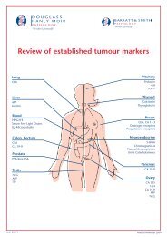
REFERENCE SHEETCLINICAL APPLICATIONS OF AUTOANTIBODY TESTSTEST NAME DISEASE CLINICAL FEATURES SIGNIFICANCE OF LABORATORY RESULTTESTS FOR ORGAN SPECIFIC AUTOIMMUNE DISEASESSKINSkin BM AbBULLOUS PEMPHIGOIDSubepidermal blisters>70% have Abs to dermal-epidermal basement membrane(pathological).Skin ICCS AbGliadin, Endomysial AbPEMPHIGUS VULGARIS Intraepidermal blisters >90% have Abs to intercellular cement substance(pathological).DERMATITIS HERPETIFORMIS Pruritic papules & blisters. Gliadin, endomysial Abs usually positive. Skin biopsyrecommended. Small bowel biopsy useful.HAEMATOLOGICALPlatelet AbIMMUNEVarious forms (acute, drugand chronic)Limited value: Anti-platelet Ab (pathological). Ofteninferred after bone marrow biopsy. Associated Ab tests(ANA) important.Neutrophil AbIMMUNE NEUTROPENIAVarious formsLimited value (pathological). Often inferred after bonemarrow biopsy.Coomb’s testIMMUNE HAEMOLYTICANAEMIAWarm & Cold formsCoomb’s test with modifications. Classification relevant totreatment, prognosis through associated diseases(pathological).RENALGBM AbGOODPASTURE’S SYNDROMEHaemoptysis and HaematuriaAb to glomerular basement membrane (pathological).ANCA(PR3, MPO)WEGENER’S GRANULOMATOSISSystemic vasculitisThe c-ANCA or classic cytoplasmic staining pattern ofAnti-Neutrophil Cytoplasmic Ab, the target antigen beingneutrophil proteinase 3 (PR3) has strong specificity andsensitivity for active Wegener’s granulomatosis.GASTROINTESTINALANCAULCERATIVE COLITISInflammatory bowel diseasep-ANCA, the perinuclear staining cytoplasmic stainingpattern, occurs in 5% and is due to Abs to neutrophilmyeloperoxidase (MPO).– CROHN’S DISEASEInflammatory bowel disease No useful autoAb assays.Gliadin Ab,Endomysial Ab,TGT Ab,IgA levelCOELIAC DISEASEGluten intoleranceGliadin Ab (present in more than 90%, greatest specificity ifboth IgA and IgG Abs are present). Reticulin and endomysialantibodies are less sensitive. Endomysial Abs have greatestspecificity. Tissue transglutaminase antibodies are a newadditional test. IgA deficient patients do not have detectableIgA autoAbs!PCA, IF AbSchilling’s testPERNICIOUS ANAEMIAImpaired B12 absorption90% have parietal cell Abs or intrinsic factor antibodies.Intrinsic factor Abs (block binding of B12 or block absorptionof B12 or block absorption of B12 bound to intrinsic factor)are more specific but more difficult to detect and useful ifPCA are negative (some Abs are pathological)LIVERANCASCLEROSING CHOLANGITISChronic fibrosis of hepatobiliarytree90% have p-ANCA Abs; low titre ANA and Smooth MuscleAbs may be present.SMAAUTOIMMUNE CHRONICChronic hepatitis without evidenceof viral infection80% have Smooth Muscle Abs. High titre, IgG Abs morespecific.LKM AbLKM - Ab ASSOCIATEDChronic hepatitisOther rare variants have liver kidney microsomal antibodiesand antibodies to asialoglycoprotein receptors have beendescribed.AMA(M2 Ab)PRIMARY BILIARY CIRRHOSISProgressive portal fibrosis >95% have Anti-Mitochondrial Antibodies. Many (>9)subtypes of mitochondrial antibodies have been described.Some AMA negative patients have M2 antibodies which canbe detected by a specific ELISA.
REFERENCE SHEETCLINICAL APPLICATIONS OF AUTOANTIBODY TESTSTEST NAME DISEASE CLINICAL FEATURES SIGNIFICANCE OF LABORATORY RESULTENDOCRINEOvarian Ab AUTOIMMUNE OVARIAN FAILUREAmenorrhoea/Early Menopause Majority have antibodies to theca interna cells and oftenother endocrine abnormalities.Sperm AbANTI-SPERM AB Infertility A variety of types and targets have been described in femalesand males as contributing to infertility (pathological).Adrenal AbAUTOIMMUNE ADRENAL DISEASEAddison’s Disease >60% have antibodies to adrenal cortex cells (ACTHreceptor), may be pathological.Islet Cell Ab DIABETES MELLITUS IDDM (Type 1) Islet Cell Antibodies may be detected in almost all patientswith IDDM prior to the development of disease, and thedetection rate falls thereafter.Thyroid Abs(ATG, TMA)HASHIMOTO’S THYROIDITISDiverse thyroid disorders>90% have antibodies to thyroid (TMG) microsomes(thyroid peroxidase): >55% have antibodies to thyroglobulin(ATG).Thyroid Receptor AbGRAVES’ DISEASEDiffuse goitreAlmost all have antibodies to TSH receptor. Clinical utility ofTSH receptor antibodies is for evaluation of diffuse goitre andprediction of both antenatal and post-treatment risk ofdisease.ACHR AbMYASTHENIA GRAVISFatiguability of skeletal muscle>95% have antibodies to acetylcholine receptors. Thepresence of antibodies to striated muscle is suggestive of anunderlying thymoma.NEUROMUSCULARENA (ANA)POLYMYOSITISInflammatory muscleAntibodies of PmScl indicate a high probability of features ofconcomitant scleroderma.GAD AbSTIFF MAN SYNDROMEProgessive rigidity and muscularspasmsAlmost all have antibodies to glutamic acid decarboxylaseand some have features of concomitant scleroderma.Neuronal AbANTI-MYELIN ASSOCIATEDGLYCOPROTEINPeripheral neuropathyAll have antibodies to nerve sheath, of which 35% are tomyelin associated glycoprotein. The majority occur as IgMmonoclonal gammopathies.GM-1 AbGUILLAIN-BARRE SYNDROMEPeripheral neuropathyLimited value as a wide array of antigens have beendescribed of which antibodies to Ganglioside GM-1 havebeen recognised more frequently than others (up to 20%)–MULTIPLE SCLEROSISMultiple sites of CNSdemyelinationSpecific autoAbs have limited value. Oligoclonal bands (ofIgG) by CSF EPG are useful.TESTS FOR CONNECTIVE TISSUE DISEASESRHEUMATOLOGICAL DISORDERSRFRHEUMATOID FACTORRheumatoid ArthritisOther disordersThe level of RF is accurately measured by a number ofmethods with the presence of, and higher titres of, RFhaving prognostic significance.ANAANTI-NUCLEAR ABConnective Tissue DisordersThe majority of persons with connective tissue disordersdevelop antibodies to nuclear antigens.Many patients may have low level antibodies in the initialphase of their illness. In some patients, the titre of ANAs mayvary with the clinical activity of their disease.dsDNADOUBLE-STRANDED DNA AbSLEPresent in patients with renal or cerebral lupus and whenpresent, a useful marker of disease activityENAANTIBODIES TO EXTRACTABLENUCLEAR ANTIGENSConnective Tissue DisordersIt is possible to classify connective tissue disorders on thebasis of clinical and laboratory features into a number ofdifferent subsets with different outcomes and management.The detection and typing or characterisation of antibodies toextractable nuclear antigens is useful in this context.ACL Ab(LAC)ANTI-PHOSPHOLIPID AB(LUPUS ANTICOAGULANT)Excessive clottingAbs to Cardiolipin of IgM, IgG (and less commonly IgA)isotypes have been found in increased frequency inindividuals with excess fetal loss as well as venous andarterial thrombosis. Higher levels, particularly IgG isotypeAbs, are more frequently correlated with disease. Many, butnot necessarily the majority, of such persons have otherconnective tissue disorders.