
You also want an ePaper? Increase the reach of your titles
YUMPU automatically turns print PDFs into web optimized ePapers that Google loves.
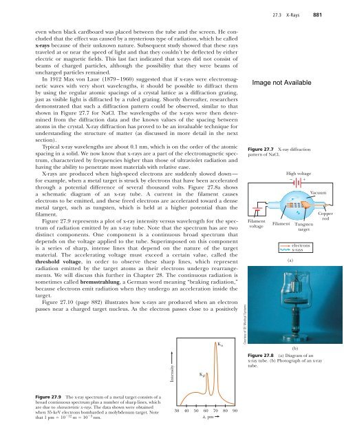
27.3 X-Rays 881even when black cardboard was placed between the tube and the screen. He concludedthat the effect was caused by a mysterious type of radiation, which he calledx-rays because of their unknown nature. Subsequent study showed that these raystraveled at or near the speed of light and that they couldn’t be deflected by eitherelectric or magnetic fields. This last fact indicated that x-rays did not consist ofbeams of charged particles, although the possibility that they were beams ofuncharged particles remained.In 1912 Max von Laue (1879–1960) suggested that if x-rays were electromagneticwaves with very short wavelengths, it should be possible to diffract themby using the regular atomic spacings of a crystal lattice as a diffraction grating,just as visible light is diffracted by a ruled grating. Shortly thereafter, researchersdemonstrated that such a diffraction pattern could be observed, similar to thatshown in Figure 27.7 for NaCl. The wavelengths of the x-rays were then determinedfrom the diffraction data and the known values of the spacing betweenatoms in the crystal. X-ray diffraction has proved to be an invaluable technique forunderstanding the structure of matter (as discussed in more detail in the nextsection).Typical x-ray wavelengths are about 0.1 nm, which is on the order of the atomicspacing in a solid. We now know that x-rays are a part of the electromagnetic spectrum,characterized by frequencies higher than those of ultraviolet radiation andhaving the ability to penetrate most materials with relative ease.X-rays are produced when high-speed electrons are suddenly slowed down—for example, when a metal target is struck by electrons that have been acceleratedthrough a potential difference of several thousand volts. Figure 27.8a showsa schematic diagram of an x-ray tube. A current in the filament causeselectrons to be emitted, and these freed electrons are accelerated toward a densemetal target, such as tungsten, which is held at a higher potential than thefilament.Figure 27.9 represents a plot of x-ray intensity versus wavelength for the spectrumof radiation emitted by an x-ray tube. Note that the spectrum has are twodistinct components. One component is a continuous broad spectrum thatdepends on the voltage applied to the tube. Superimposed on this componentis a series of sharp, intense lines that depend on the nature of the targetmaterial. The accelerating voltage must exceed a certain value, called thethreshold voltage, in order to observe these sharp lines, which representradiation emitted by the target atoms as their electrons undergo rearrangements.We will discuss this further in Chapter 28. The continuous radiation issometimes called bremsstrahlung, a German word meaning “braking radiation,”because electrons emit radiation when they undergo an acceleration inside thetarget.Figure 27.10 (page 882) illustrates how x-rays are produced when an electronpasses near a charged target nucleus. As the electron passes close to a positivelyIntensityK bK aCourtesy of GE Medical SystemsFigure 27.7 X-ray diffractionpattern of NaCl.FilamentvoltageFilamentHigh voltage– +(a)Tungstentargetelectronsx-rays(b)VacuumFigure 27.8 (a) Diagram of anx-ray tube. (b) Photograph of an x-raytube.CopperrodFigure 27.9 The x-ray spectrum of a metal target consists of abroad continuous spectrum plus a number of sharp lines, whichare due to characteristic x-rays. The data shown were obtainedwhen 35-keV electrons bombarded a molybdenum target. Notethat 1 pm 10 12 m 10 3 nm.3040 50 60 70 80 90l, pm